cardiac adaptations to exercise: the role of stem cells and future
TRANSCRIPT
2012/2013
Diogo da Silva Miguel
Cardiac adaptations to exercise: the
role of stem cells and future
therapeutic insights.
março, 2013
Mestrado Integrado em Medicina
Área: Fisiologia
Trabalho efetuado sob a Orientação de:
Doutor Paulo Castro Chaves
Trabalho organizado de acordo com as normas da revis ta:
Cardiovascular Research
Diogo da Silva Miguel
Cardiac adaptations to exercise: the
role of stem cells and future
therapeutic insights.
março, 2013
Projeto de Opção do 6º ano - DECLARAÇÃO DE INTEGRIDADE
Eu, Diogo da Silva Miguel, abaixo assinado, nº mecanográfico 070801255, estudante do 6º ano do
Mestrado Integrado em Medicina, na Faculdade de Medicina da Universidade do Porto, declaro ter
atuado com absoluta integridade na elaboração deste projeto de opção.
Neste sentido, confirmo que NÃO incorri em plágio (ato pelo qual um indivíduo, mesmo por omissão,
assume a autoria de um determinado trabalho intelectual, ou partes dele). Mais declaro que todas as
frases que retirei de trabalhos anteriores pertencentes a outros autores, foram referenciadas, ou
redigidas com novas palavras, tendo colocado, neste caso, a citação da fonte bibliográfica.
Faculdade de Medicina da Universidade do Porto, 20/03/2013
Assinatura:
Projeto de Opção do 6º ano – DECLARAÇÃO DE REPRODUÇÃO
Nome: Diogo da Silva Miguel
Email: [email protected]
Título da Monografia:
Cardiac adaptations to exercise: the role of stem cells and future therapeutic insights.
Orientador:
Paulo Manuel Barreiros de Castro Chaves
Ano de conclusão: 2013
Designação da área do projeto:
Fisiologia
É autorizada a reprodução integral desta Monografia para efeitos de investigação e de divulgação
pedagógica, em programas e projetos coordenados pela FMUP.
Faculdade de Medicina da Universidade do Porto, 20/03/2013
Assinatura:
1
Cardiac adaptations to exercise: the role of stem cells and future
therapeutic insights
Diogo da Silva Miguel1
Faculty of Medicine of University of Porto, Physiology and Cardiothoracic Department
Paulo Castro Chaves MD, PhD
Faculty of Medicine of University of Porto, Physiology and Cardiothoracic Department
Number of words: 5339
1Corresponding author
Faculdade de Medicina da Universidade do Porto, Al. Prof. Hernâni Monteiro, 4200-319 Porto, Portugal
Telephone number: +351 22 551 3600
Fax: +351 22 551 3601
E-mail: [email protected]
2
Diogo da Silva Miguel, Paulo Castro Chaves
Cardiac adaptations to exercise: the role of stem cells and future therapeutic
insights
Abstract
The heart responds to physical exercise through changes in its morphology known as
physiological hypertrophy. Besides cardiomyocyte hypertrophy, there is also an
increased cellular proliferation with formation of new myocytes. Altogether, these
changes lead to an improvement in cardiac function. The molecular mechanism
responsible for the development of physiological hypertrophy is centred in the IGF-
1R/PI3K/Akt signalling pathway, which modulates the activity of transcription factors
involved in protein synthesis and cardiomyocytes proliferation. The endogenous pool of
cardiac stem cells is believed to be crucial for the continuous renewal and regenerative
potential of cardiac tissue, and has also been positively correlated with physiological
hypertrophy. Another system involved in myocardium physiology and pathology is
microRNA. The knowledge of the mechanisms underlying the beneficial effects of
physical exercise over cardiac function may sustain the development of new therapeutic
approaches regarding cardiac pathologies, which represent today an important cause of
morbidity and mortality worldwide.
Keywords: Exercise, Heart, Hypertrophy, microRNA, Stem cells.
3
Introduction
The understanding of heart’s physiology has always been on the top of scientific
community’s priorities. Indeed, there is an enormous amount of work available
regarding this area of knowledge.
The heart responds to increased workload with hypertrophy. According to the nature of
the stimulus, the hypertrophy can be either physiological or pathological1. The main
stimulus that leads to pathological hypertrophy is pressure overload, where the heart
responds with hypertrophy of existing myocytes as an attempt to overwhelm the
increased load2. However, in this setting maladaptive changes, such as apoptosis,
necrosis and a shift in gene expression, also occur, which leads to rapid decline in
cardiac function, with heart failure as possible outcome1, 2. In the opposite side there is
the physiological hypertrophy, with physical exercise appearing as the best example of
this type of cardiac adaptation. Unlike pressure overload, exercise training increases the
blood flow through the heart (volume overload)1, which in turn results in lengthening of
myocytes and consequent eccentric hypertrophy of the whole organ2. The normal
structure of the myocardium is kept, the cardiac function remains unchanged or may
improve3, and the loss of myocytes and increased fibrosis observed in pathological
hypertrophy are actually attenuated with exercise training4.
As noted above, physical exercise is the main trigger of physiological hypertrophy, with
all the resulting benefits. Exercise training is today extensively prescribed and is a cheap
and effective way for both the prevention and management of heart diseases3. Indeed,
current evidence points that an improved fitness level is a strong indicator of freedom
from all-cause mortality5 and is enough to decrease the morbidity and mortality linked
with cardiac disorders6. Besides, physical exercise has been shown to protect the
myocardium from age induced apoptosis, fibrosis and senescence4, 7, 8.
4
Until the past decade, it was believed that the heart was a post-mitotic organ, with no
regenerative potential. However, an increasing body of evidence supports the existence
of a resident population of cardiac stem cells9-12. This endogenous pool not only
reassures the continuous renewal of cardiac tissue13, but also supports some grade of
regeneration after an injury9, 11. The old paradigm of the heart as a terminally
differentiated organ without any renewal potential did not give space for the exploration
of regenerative medicine. However, by the light of the evidence accumulated during the
past decade some work begun to be done in the field of regenerative medicine applied to
the heart.
This review aims to look over the mechanisms known to take part in the control of the
adaptations of myocardium to physical exercise, and open new perspectives about
possible applications of this knowledge in the search of new therapeutic approaches.
Macroscopic and functional changes
In 1975 Morganroth et al. used echocardiography to describe for the first time the
cardiac changes induced by physical exercise, concluding that endurance exercise
induced an increase in left ventricle mass and diastolic cavity size, while the wall
thickness remained normal (eccentric hypertrophy). On the other hand, athletes that
went through a resistance training program showed increased myocardial mass with
ventricular wall thickening, but with normal diastolic cavity size (concentric
hypertrophy)14. This concept has been accepted for decades as the “Morganroth
hypothesis”. Indeed, other studies have found an increase in both left ventricular mass
and left ventricle end diastolic diameter with endurance exercise15-17, though some of
them have failed to show any effect of resistance training in cardiac morphology15, 16.
5
Adaptations at cellular level
Unlike the macroscopic remodelling described in the previous paragraph, due to ethical
limitations there is a paucity of studies describing the changes that occur at the cellular
level in humans. However, a vast amount of studies using mice have shown that an
increase in individual cardiomyocyte dimensions occurs in response to several
swimming training programmes18-20. That increase comprises both the cardiomyocyte’s
long and short axis and mean cell area21, and is remarkably related with the exercise’s
intensity – high intensity exercise induces bigger changes than moderate intensity6. On
the other hand, the pattern of cardiomyocyte growth is also determined by the type of
physical exercise: while endurance training (swimming, running), and the associated
volume overload, prompt myocyte lengthening, resistance training (wrestling, weight
lifting), which is associated with an intermittent pressure overload, drives mainly to
thickening of myocytes2.
Until recently, cardiomyocyte hypertrophy was thought to be the only mean by which
the heart increased its size, which was in line with the paradigm that heart was a post-
mitotic organ without any renewal potential. However, nowadays’ current evidence
indicates that human myocardium is capable of, at least in part, renovate itself by new
cardiomyocyte formation13. Actually, if it was not this self-renewing capacity, the heart
would lose most of its mass in few decades11. Furthermore, increased levels of division
markers, along with a rise in the number of cardiomyocytes, has already been
documented in mice following the application of endurance exercise protocols18, 22.
Interestingly, the increase in the number of cardiomyocytes was positively related with
the intensity of physical exercise the rats have been exposed to22. Therefore, it seems
that the regenerative capacity of myocardium is enhanced by physical activity.
6
Another possible mechanism by which the heart can increase its mass is through a drop
in apoptotic rate. Indeed, several studies point in this direction. Endurance exercise has
been found to attenuate the rise in Bax/Bcl-2 ratio seen with ageing. Consequently there
is a decrease in the activation of caspase-9 and 3 and DNA fragmentation, which is
reflected in the superior number of myocytes per area observed in old exercised rats
compared with sedentary ones4. On the other hand, it was also demonstrated a rise in the
levels of heat shock protein 70 (HSP70), known to inhibit apoptosis, in the ventricles of
exercised mice7. At last but not the least, an improved telomerase activity was observed
in mice exposed to endurance exercise, thereby protecting myocytes from senescence
and apoptosis. This was found already after 21 days of exercise, even before
physiological hypertrophy has been documented8. Furthermore, in studies using obese
rats, endurance exercise was also shown to decrease the number of apoptotic
cardiomyocytes23, 24 and reverse the architectural changes observed to occur in
association with obesity23.
Molecular mechanisms
IGF-1R/PI3K/Akt pathway. The IGF-1R/PI3K/Akt pathway has been extensively
studied and is seen as the hallmark of physiological hypertrophy20, 25, 26. In fact, the
nature of myocardial adaptation to increased workload (physiological vs. pathological)
begins to differentiate already at the growth factor and receptor level – while the
activation of G-protein coupled receptors by adrenergic stimuli results in pathological
adaptation of myocardium27, in the presence of physiological stimuli such as physical
exercise it is the insulin-like growth factor I (IGF-1) acting over its receptor (IGF-1R)
and triggering the beneficial adaptations described above25-27.
7
In one study using rats, it was shown that swimming training increased the IGF-1
mRNA after 2 and 6 weeks of exercise28. These findings are compatible with the work
of Serneri et al. that accessed the concentration of this growth factor in coronary sinus
and concluded that it was significantly bigger in athletes than in controls29. The
importance of the pair IGF-1/IGF-1R was further highlighted by knock-out studies in
which the deletion of IGF-1R in mice attenuated the cardiac hypertrophy seen in
controls after swimming training20. On the other hand, the overexpression of IGF-1R
was associated with a proportional increase in the chamber size and wall thickness of
heart, without any histologic signs of cardiomyopathy, what was also reflected in the
enhanced systolic function of transgenic IGF-1R animals25.
IGF-1R is a tyrosine kinase receptor which once stimulated by its ligand phosphorylates
and activates phosphatidylinositol 3-kinase (PI3K)30. PI3Ks are a family of ubiquitous
expressed lipid kinases, which are essential for a wide range of biological processes in
several different types of tissues27. Class-I PI3K phosphorylates phosphatidylinositol-
4,5-bisphosphate to generate phosphatidylinositol-3,4,5-trisphosphate, which in turn
acts as second messenger to activate Akt. Class-IA PI3K is activated by growth factor
receptors, such as IGF-1R, while class-IB PI3K is activated by G-protein coupled
receptors27. The importance of class-IA PI3K activity for the development of
physiological hypertrophy was well documented by studies in which mice lacking either
the catalytic or regulatory subunit of this kinase failed to show physiological
hypertrophy25-27. However, the same didn’t happen when in the presence of a
pathological stimulus, since even in the absence of the catalytic subunit of class-IA
PI3K, pressure overload successfully induced pathological hypertrophy26.
The downstream effector of the IGF-1R/PI3K/Akt pathway is the serine-threonine
kinase Akt, also known as protein kinase B (PKB). There have been identified 3
8
isoforms of this kinase (Akt1, 2 and 3), and despite their high degree of homology, their
expression varies among different tissues31. Data suggests that the most important
isoform in cardiac physiology is Akt119, 31. As noted above, PI3K plays a key role in the
activation of Akt through generation of phosphatidylinositol-3,4,5-trisphosphate. PTEN
has the opposite effect, and thus decreases the activation of Akt31. Akt phosphorylates
several different targets, which implicates it in a variety of biological processes31. The
importance of Akt for the development of physiological hypertrophy is well supported
by a study in which mice lacking Akt1 failed in achieving the degree of cardiac
adaptation seen in their control littermates after a program of swimming training19. In
addition, this importance is further reinforced by the evidence that overexpression of
Akt causes cardiac hypertrophy both at the molecular and histological level, without any
parallel increase in the collagen content of the heart.
Downstream targets. The mammalian target of rapamycin (mTOR) works as an
important effector that controls protein synthesis, promoting cellular growth. This is one
of the downstream targets of Akt, which phosphorylates and inactivates the product of
tuberous sclerosis gene 2 (TSC2). Doing so, Akt counteracts the inhibition that TSC2
exerts over mTOR, thereby enhancing its growth promoting effect19. This effect is
mediated by the phosphorylation of the ribosomal S6 kinase (S6K), that once activated
phosphorylates and activates the ribosomal protein S632. In addition, the translation
initiation factor-4E binding protein-1 (4E-BP1) sees its activity increased by mTOR6.
The net effect is an increased protein synthesis, with consequent cellular hypertrophy6.
The facts that mice lacking Akt1 showed lower levels of phosphorylated S6K and S6
after exercise training19 and that overexpression of IGF-1R was associated with an
increase in phosphorylated S6K25 highlight the importance of the IGF-1R/PI3K/Akt
pathway for the induction of this system. However, the absence of S6K did not
9
attenuate cardiac physiological hypertrophy when mice were subjected to an exercise
protocol, which suggests that, while important, S6K is not essential for the development
of physiological hypertrophy32.
Another mean by which Akt induces myocyte hypertrophy is by inactivation of
glycogen synthase kinase-3 beta (GSK-3β)19. GSK-3β phosphorylates and inactivates
the transcription factor GATA-4, thereby impairing protein synthesis33. Indeed,
increased levels of the inactive phosphorylated form of GSK-3β were detected in mice
overexpressing Akt, which reflected in the bigger accumulation of GATA-4 in
cardiomyocytes’ nuclei30. Once in the nucleus, GATA-4 regulates the expression of a
variety of cardiac genes related with myocardium hypertrophy33. Moreover, GATA-4
has also been connected to myocyte proliferation in zebrafish34
Besides GATA-4, several other transcription factors have been associated to
cardiomyocytes hypertrophy33. One of the identified transcription factors was C/EBPβ,
whose expression was found to be reduced after exercise training. Interestingly, that fall
was secondary to a higher Akt1 activity, and resulted in a reduction of the repressive
effect that C/EBPβ has over the serum response factor (SRF). SRF is also a transcription
factor that, among others, promotes the expression of GATA-4. Another downstream
target negatively regulated by C/EBPβ is the CBP/p300-interacting transactivator with
ED-rich carboxy-terminal domain 4 (CITED4). In fact, CITED4 was markedly
increased in cardiomyocytes and induced its proliferation, as reflected by an increase in
the number of cells18.
The role of microRNAs
MicroRNAs (miRNA) are a class of recently discovered small molecules, each
composed of a noncoding sequence of nearly 22 nucleotides, which can modulate gene
10
expression by destroying or repressing the translation of a target RNA. Each miRNA
molecule can target more than a different mRNA, and each mRNA can be targeted by
more than a different miRNA, which provides a fine control of gene expression35. Some
miRNAs have already been identified that are known to play important roles in cardiac
development. Namely, miRNA-1 and miRNA-133 are of uttermost importance in
cardiac development by regulating the proliferation and differentiation of
cardiomyocytes36. The role of miRNA-1 and miRNA-133 in adult heart is less certain,
but both pathological and physiological hypertrophic stimuli decrease the expression of
both miRNAs, which suggests an inverse correlation between miRNA-1 and miRNA-
133 and myocardium hypertrophy37.
In an attempt to identify miRNAs regulated by PI3K, Lin et al. found that mice
expressing constitutively active PI3K (caPI3K) expressed lower levels of miRNA-222,
miRNA-34a and miRNA-210 as opposed with controls. Moreover, although the levels
of these miRNAs increased after myocardial infarction, they remained significantly
lower in the group expressing caPI3K, and this was reflected in the improved cardiac
function shown in this group38. Another miRNA that has been shown to be related with
physiological hypertrophy following a swim training programme was miRNA-29c.
However, unlike the former, miRNA-29c levels were upregulated after a programme of
several weeks of physical exercise. The consequence was a downregulation in the
expression of COLIAI and COLIIIAI, with a decrease of total left ventricle collagen
content and improved ventricular compliance39.
Endogenous Cardiac Stem Cells
As described above, the heart harbours a pool of endogenous cardiac stem cells (eCSC).
The fact that exercised animals’ hearts have shown evidence of new cardiomyocytes
11
formation raises a question – what is the relationship between physical exercise and
eCSC?
Some studies exist that try to answer this question. In one of them, the number of c-
Kit pos eCSC was found to be increased in exercised rats in comparison with controls22.
Also the IGF-1R/PI3K/Akt pathway has been connected with eCSC. In this field,
current evidence points to a positive correlation between the activation of this pathway
and the activation and proliferation of eCSC22, 40. Moreover, IGF-1 overexpression
increased the activity of telomerase, which resulted in a delay in eCSC senescence41.
Therefore, if the activation of IGF-1R/PI3K/Akt fosters the activation and proliferation
of eCSC, and if physical exercise promotes the activation of the IGF-1R/PI3K/Akt
signalling pathway, so it is reasonable to hypothesize that physical exercise is an
effective trigger of eCSC activation and consequent cardiac regeneration.
Clinical insights
So far, the only available therapeutic options aim to block the progression on the heart
disease38. However, as noted above, the last decade brought crescent scientific evidence
that supports the possibility of exploring new therapeutic approaches that can improve
the function of the failing heart, thereby improving the quality of life of millions of
people suffering from cardiac pathology.
IGF-1R/PI3K/Akt pathway. As stated above, the IGF-1R/PI3K/Akt pathway is the
central core of physiological hypertrophy. In fact, some studies have already been
performed in the setting of myocardial infarction that showed a reduction in cardiac
fibrosis, cardiomyocytes apoptosis and infarction size, as well as better systolic and
diastolic function, following the administration of IGF-142, 43. Furthermore, in animals
subjected to myocardial infarction, the treatment with an association of IGF-1 and
12
hepatocyte growth factor (HGF) fostered eCSC activation with consequent cardiac
regeneration40, 44, 45. Interestingly, this was observed along with increased expression of
GATA-444 which, as described above, seems to have a role in physiological
hypertrophy induced by physical exercise.
microRNAs. The discovery of miRNAs has brought a new biological target for future
therapies. Actually, today is already possible either to increase the expression, using
vectors46, or antagonize specific miRNAs using ‘antagomirs’, which are cholesterol-
associated RNAs that block the action of miRNAs over their targets47. The expression
of miRNA-34a, aforementioned to be decreased in the cardiomyocytes of mice
expressing caPI3K, has been shown to rise in the ageing heart and following myocardial
infarction. Interestingly, using antigomir against miRNA-34a it was possible to observe
a drop in cell death and cardiac fibrosis, and consequent improvement in contractile
function in mice subjected to myocardial infarction48. Another miRNA described above
as being negatively correlated with PI3K activity was miRNA-210. By those findings, it
would be reasonable to think that a decrease in the expression of this miRNA would be
beneficial. However, other studies found that miRNA-210, whose expression is
upregulated under hypoxic conditions49, is actually beneficial in the setting of
myocardial infarction by significantly inducing angiogenesis and decreasing myocytes’
apoptotic rate, thereby contributing for a more favourable ventricular remodelling and
cardiac function46.
Cell therapy. The first attempts to regenerate myocardial tissue have been done through
transplant of stem cells from other origins than the heart50, 51. Although animal studies
have provided exciting results, several clinical trials in humans where already
performed and showed only modest, if any, improvement in cardiac function50.
13
Therefore, new approaches are needed in order to make this type of therapy more
effective.
One of the first questions to be answered is which type of cells suits better for cell-
based therapy. The discovery of a pool of resident cardiac stem cells has opened a new
window regarding the regenerative potential of this organ, which can be further
explored either as a therapeutic tool or target. Indeed, a phase 1 clinical trial has
documented significant improvements in both the ejection fraction and infarct size in
patients that received autologous cardiac stem cells therapy compared with controls.
Furthermore, the safety of the treatment was absolute, with no adverse effects registered
in any of the treated patients52.
The pool of cardiac stem cells is complex and comprises cells with different capacity to
replicate and form mature myocytes and blood vessels. In an attempt to better
characterize those different cellular populations, D’Amario et al. concluded that cardiac
stem cells expressing IGF-1R were younger and had a better replicative reserve, as
opposed with cells expressing IGF-2R and AT1R. Moreover, cells expressing IGF-1R
were capable of secreting both IGF-1 and IGF-2, the latter being responsible for the
induction of myocyte differentiation. The superiority of IGF-1R expressing cardiac stem
cells was further confirmed by the greater degree of cardiac recovery after the transplant
of these cells into hearts of rats subjected to myocardial infarction53.
Besides the choice of the ideal cell, other strategies can be taken to maximize the
beneficial effects of cell therapy. Interestingly, the combination of cardiac stem cells
administration and nanofibers containing IGF-1 resulted in a higher myocardial
recovery after ischemia when compared with each therapy alone54.
Discussion
14
In the last decade several steps have been taken in order to open new lines of
investigation regarding new therapeutic strategies. Maybe the biggest advances have
been done in the field of cell therapy, with some clinical trials already in progress.
However, as stated above, there is also growing expectation about miRNAs and its
manipulation.
The central role that the IGF-1R/PI3K/Akt has in the development of physiological
hypertrophy has also triggered attempts to modulate this signalling system, namely
through the therapy with IGF-1. In fact, the results obtained with IGF-1 administration
in animal models of myocardial infarction were encouraging42, 43. However, this
signalling pathway is present in a wide range of cell types and because of that systemic
administration of this growth factor can have diffuse effects and result in the
progression of occult neoplasms31.
Concerning cell therapy, some obstacles have been raised regarding the type of cells
that better fulfil the requirements for the success of this therapeutic approach. By one
side, the application of cells from other origins than the heart has shown disappointing
results. Among the causes advanced for this is the source and inadequate preparation of
the cells, as well as the timing and mode of cell delivery. On the other side, it is
believed that embryonic stem cells are the ones that have the greatest potential to be
successful. However, to the risk of teratoma formation and immunogenic
incompatibilities that the use of embryonic stem cells entails, we still have to add the
ethical issues50. Finally, it appears that autologous cardiac stem cells transplantation
may be the best approach, as has been shown by a recent phase 1 clinical trial52. Still,
more research is needed to find new strategies that can increase the feasibility and
effectiveness of cell therapy.
15
The discovery of miRNAs has provided a new overview of cellular regulation
mechanisms. In addition, it represents another process where it is possible to act to
change the fate of diseased heart. Currently, it is already clear that different miRNAs are
important in both the heart development and pathology55. Furthermore, some studies
performed in animals were successful in improving the pattern of cardiac remodelling
after myocardial infarction46, 48. Nevertheless, miRNAs represent a class of molecules
that exerts a very delicate control of specific genes at a post-transcriptional level55.
Consequently, it is critical to better understand the function of each specific miRNA in
the heart’s physiology and pathology, and only after it will be possible to delineate the
best plans to take all the potential from this promising area.
Heart diseases, namely those of ischemic cause, are a major source of morbidity and
mortality worldwide51. Despite the improvement that the development of several classes
of drugs have brought to the prognostic of these patients, advances are need so that it
can be possible to further decrease the burden of disease of cardiac pathologies.
Conflict of interest
None declared.
Bibliography
1. Dorn GW, II. The fuzzy logic of physiological cardiac hypertrophy. Hypertension
2007;49:962-970.
2. Opie LH, Commerford PJ, Gersh BJ, Pfeffer MA. Controversies in Cardiology 4 -
Controversies in ventricular remodelling. Lancet 2006;367:356-367.
3. Ellison GM, Waring CD, Vicinanza C, Torella D. Physiological cardiac remodelling in
response to endurance exercise training: cellular and molecular mechanisms. Heart
(British Cardiac Society) 2012;98:5-10.
16
4. Kwak H-B, Song W, Lawler JM. Exercise training attenuates age-induced elevation in
Bax/Bcl-2 ratio, apoptosis, and remodeling in the rat heart. Faseb Journal 2006;20:791-
+.
5. O'Keefe JH, Vogel R, Lavie CJ, Cordain L. Exercise like a hunter-gatherer: a
prescription for organic physical fitness. Progress in cardiovascular diseases
2011;53:471-479.
6. Kemi OJ, Wisloff U. Mechanisms of exercise-induced improvements in the contractile
apparatus of the mammalian myocardium. Acta physiologica (Oxford, England)
2010;199:425-439.
7. Siu PM, Bryner RW, Martyn JK, Alway SE. Apoptotic adaptations from exercise
training in skeletal and cardiac muscles. Faseb Journal 2004;18:1150-+.
8. Werner C, Hanhoun M, Widmann T, Kazakov A, Semenov A, Poss J, et al. Effects of
physical exercise on myocardial telomere-regulating proteins, survival pathways, and
apoptosis. Journal of the American College of Cardiology 2008;52:470-482.
9. Beltrami AP, Barlucchi L, Torella D, Baker M, Limana F, Chimenti S, et al. Adult
cardiac stem cells are multipotent and support myocardial regeneration. Cell
2003;114:763-776.
10. Torella D, Ellison GM, Karakikes I, Nadal-Ginard B. Resident cardiac stem cells.
Cellular and Molecular Life Sciences 2007;64:661-673.
11. Nadal-Ginard B, Kajstura J, Leri A, Anversa P. Myocyte death, growth, and
regeneration in cardiac hypertrophy and failure. Circulation Research 2003;92:139-150.
12. Saravanakumar M, Devaraj H. Distribution and homing pattern of c-kit(+) Sca-1(+)
CXCR4(+) resident cardiac stem cells in neonatal, postnatal, and adult mouse heart.
Cardiovascular pathology : the official journal of the Society for Cardiovascular
Pathology 2012.
13. Bergmann O, Bhardwaj RD, Bernard S, Zdunek S, Barnabe-Heider F, Walsh S, et al.
Evidence for Cardiomyocyte Renewal in Humans. Science 2009;324:98-102.
17
14. Lewis EJ, McKillop A, Banks L. The Morganroth hypothesis revisited: endurance
exercise elicits eccentric hypertrophy of the heart. The Journal of physiology
2012;590:2833-2834.
15. Venckunas T, Raugaliene R, Mazutaitiene B, Ramoskeviciute S. Endurance rather than
sprint running training increases left ventricular wall thickness in female athletes.
European journal of applied physiology 2008;102:307-311.
16. Spence AL, Naylor LH, Carter HH, Buck CL, Dembo L, Murray CP, et al. A
prospective randomised longitudinal MRI study of left ventricular adaptation to
endurance and resistance exercise training in humans. The Journal of physiology
2011;589:5443-5452.
17. De Luca A, Stefani L, Pedrizzetti G, Pedri S, Galanti G. The effect of exercise training
on left ventricular function in young elite athletes. Cardiovascular ultrasound
2011;9:27.
18. Bostroem P, Mann N, Wu J, Quintero PA, Plovie ER, Panakova D, et al. C/EBP beta
Controls Exercise-Induced Cardiac Growth and Protects against Pathological Cardiac
Remodeling. Cell 2010;143:1072-1083.
19. DeBosch B, Treskov I, Lupu TS, Weinheimer C, Kovacs A, Courtois M, et al. Akt1 is
required for physiological cardiac growth. Circulation 2006;113:2097-2104.
20. Kim J, Wende AR, Sena S, Theobald HA, Soto J, Sloan C, et al. Insulin-Like Growth
Factor I Receptor Signaling Is Required for Exercise-Induced Cardiac Hypertrophy.
Molecular Endocrinology 2008;22:2531-2543.
21. McMullen JR. Role of insulin-like growth factor 1 and phosphoinositide 3-kinase in a
setting of heart disease. Clinical and Experimental Pharmacology and Physiology
2008;35:349-354.
22. Waring CD, Vicinanza C, Papalamprou A, Smith AJ, Purushothaman S, Goldspink DF,
et al. The adult heart responds to increased workload with physiologic hypertrophy,
cardiac stem cell activation, and new myocyte formation. European heart journal 2012.
18
23. Lee SD, Shyu WC, Cheng IS, Kuo CH, Chan YS, Lin YM , et al. Effects of exercise
training on cardiac apoptosis in obese rats. Nutrition, metabolism, and cardiovascular
diseases : NMCD 2012.
24. Peterson JM, Bryner RW, Sindler A, Frisbee JC, Alway SE. Mitochondrial apoptotic
signaling is elevated in cardiac but not skeletal muscle in the obese Zucker rat and is
reduced with aerobic exercise. Journal of applied physiology (Bethesda, Md : 1985)
2008;105:1934-1943.
25. McMullen JR, Shioi T, Huang WY, Zhang L, Tarnavski O, Bisping E, et al. The
insulin-like growth factor 1 receptor induces physiological heart growth via the
phosphoinositide 3-kinase(p110 alpha) pathway. Journal of Biological Chemistry
2004;279:4782-4793.
26. McMullen JR, Shioi T, Zhang L, Tarnavski O, Sherwood MC, Kang PM, et al.
Phosphoinositide 3-kinase(p110 alpha) plays a critical role for the induction of
physiological, but not pathological, cardiac hypertrophy. Proceedings of the National
Academy of Sciences of the United States of America 2003;100:12355-12360.
27. Luo J, McMullen JR, Sobkiw CL, Zhang L, Dorfman AL, Sherwood MC, et al. Class I-
A phosphoinositide 3-kinase regulates heart size and physiological cardiac hypertrophy.
Molecular and Cellular Biology 2005;25:9491-9502.
28. Scheinowitz M, Kessler-Icekson G, Freimann S, Zimmermann R, Schaper W, Golomb
E, et al. Short- and long-term swimming exercise training increases myocardial insulin-
like growth factor-I gene expression. Growth hormone & IGF research : official
journal of the Growth Hormone Research Society and the International IGF Research
Society 2003;13:19-25.
29. Neri Serneri GG, Boddi M, Modesti PA, Cecioni I, Coppo M, Padeletti L, et al.
Increased cardiac sympathetic activity and insulin-like growth factor-I formation are
associated with physiological hypertrophy in athletes. Circ Res 2001;89:977-982.
30. Condorelli G, Drusco A, Stassi G, Bellacosa A, Roncarati R, Iaccarino G, et al. At
induces enhanced myocardial contractility and cell size in vivo in transgenic mice.
19
Proceedings of the National Academy of Sciences of the United States of America
2002;99:12333-12338.
31. Matsui T, Rosenzweig A. Convergent signal transduction pathways controlling
cardiomyocyte survival and function: the role of PI 3-kinase and Akt. Journal of
Molecular and Cellular Cardiology 2005;38:63-71.
32. McMullen JR, Shioi T, Zhang L, Tarnavski O, Sherwood MC, Dorfman AL, et al.
Deletion of ribosomal S6 kinases does not attenuate pathological, physiological, or
insulin-like growth factor 1 receptor-phosphoinositide 3-kinase-induced cardiac
hypertrophy. Mol Cell Biol 2004;24:6231-6240.
33. Akazawa H, Komuro I. Roles of cardiac transcription factors in cardiac hypertrophy.
Circulation Research 2003;92:1079-1088.
34. Kikuchi K, Holdway JE, Werdich AA, Anderson RM, Fang Y, Egnaczyk GF, et al.
Primary contribution to zebrafish heart regeneration by gata4(+) cardiomyocytes.
Nature 2010;464:601-605.
35. Cataluccia D, Latronico MVG, Condorelli G. MicroRNAs control gene expression -
Importance for cardiac development and pathophysiology. In: Sideman S, Beyar R,
Landesberg A, eds. Control and Regulation of Transport Phenomena in the Cardiac
System, 2008:20-29.
36. Cordes KR, Srivastava D. MicroRNA Regulation of Cardiovascular Development.
Circulation Research 2009;104:724-732.
37. Latronico MVG, Catalucci D, Condorelli G. Emerging role of MicroRNAs in
cardiovascular biology. Circulation Research 2007;101:1225-1236.
38. Lin RCY, Weeks KL, Gao X-M, Williams RBH, Bernardo BC, Kiriazis H, et al.
PI3K(p110 alpha) Protects Against Myocardial Infarction-Induced Heart Failure
Identification of PI3K-Regulated miRNA and mRNA. Arteriosclerosis Thrombosis and
Vascular Biology 2010;30:724-732.

20
39. Soci UPR, Fernandes T, Hashimoto NY, Mota GF, Amadeu MA, Rosa KT, et al.
MicroRNAs 29 are involved in the improvement of ventricular compliance promoted by
aerobic exercise training in rats. Physiological Genomics 2011;43:665-673.
40. Ellison GM, Torella D, Dellegrottaglie S, Perez-Martinez C, Perez de Prado A,
Vicinanza C, et al. Endogenous Cardiac Stem Cell Activation by Insulin-Like Growth
Factor-1/Hepatocyte Growth Factor Intracoronary Injection Fosters Survival and
Regeneration of the Infarcted Pig Heart. Journal of the American College of Cardiology
2011;58:977-986.
41. Torella D, Rota M, Nurzynska D, Musso E, Monsen A, Shiraishi I, et al. Cardiac stem
cell and myocyte aging, heart failure, and insulin-like growth factor-1 overexpression.
Circulation Research 2004;94:514-524.
42. O'Sullivan JF, Leblond AL, Kelly G, Kumar AH, Metharom P, Buneker CK, et al.
Potent long-term cardioprotective effects of single low-dose insulin-like growth factor-1
treatment postmyocardial infarction. Circulation Cardiovascular interventions
2011;4:327-335.
43. Lai NC, Tang T, Gao MH, Saito M, Miyanohara A, Hammond HK. Improved function
of the failing rat heart by regulated expression of insulin-like growth factor I via
intramuscular gene transfer. Human gene therapy 2012;23:255-261.
44. Ruvinov E, Leor J, Cohen S. The promotion of myocardial repair by the sequential
delivery of IGF-1 and HGF from an injectable alginate biomaterial in a model of acute
myocardial infarction. Biomaterials 2011;32:565-578.
45. Bocchi L, Savi M, Graiani G, Rossi S, Agnetti A, Stillitano F, et al. Growth factor-
induced mobilization of cardiac progenitor cells reduces the risk of arrhythmias, in a rat
model of chronic myocardial infarction. PloS one 2011;6:e17750.
46. Hu S, Huang M, Li Z, Jia F, Ghosh Z, Lijkwan MA, et al. MicroRNA-210 as a novel
therapy for treatment of ischemic heart disease. Circulation 2010;122:S124-131.
21
47. Krutzfeldt J, Kuwajima S, Braich R, Rajeev KG, Pena J, Tuschl T, et al. Specificity,
duplex degradation and subcellular localization of antagomirs. Nucleic acids research
2007;35:2885-2892.
48. Boon RA, Iekushi K, Lechner S, Seeger T, Fischer A, Heydt S, et al. MicroRNA-34a
regulates cardiac ageing and function. Nature 2013.
49. Chan YC, Banerjee J, Choi SY, Sen CK. miR-210: the master hypoxamir.
Microcirculation 2012;19:215-223.
50. Fraccarollo D, Galuppo P, Bauersachs J. Novel therapeutic approaches to post-
infarction remodelling. Cardiovascular research 2012;94:293-303.
51. Dimmeler S, Burchfield J, Zeiher AM. Cell-based therapy of myocardial infarction.
Arteriosclerosis Thrombosis and Vascular Biology 2008;28:208-216.
52. Bolli R, Chugh AR, D'Amario D, Loughran JH, Stoddard MF, Ikram S, et al. Cardiac
stem cells in patients with ischaemic cardiomyopathy (SCIPIO): initial results of a
randomised phase 1 trial. Lancet 2011;378:1847-1857.
53. D'Amario D, Cabral-Da-Silva MC, Zheng H, Fiorini C, Goichberg P, Steadman E, et al.
Insulin-like growth factor-1 receptor identifies a pool of human cardiac stem cells with
superior therapeutic potential for myocardial regeneration. Circ Res 2011;108:1467-
1481.
54. Padin-Iruegas ME, Misao Y, Davis ME, Segers VF, Esposito G, Tokunou T, et al.
Cardiac progenitor cells and biotinylated insulin-like growth factor-1 nanofibers
improve endogenous and exogenous myocardial regeneration after infarction.
Circulation 2009;120:876-887.
55. Small EM, Olson EN. Pervasive roles of microRNAs in cardiovascular biology. Nature
2011;469:336-342.
22
Figure legends
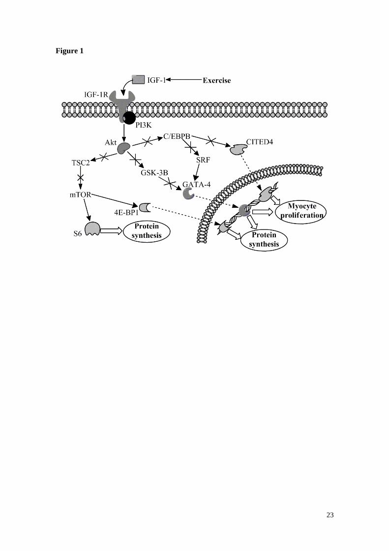
Figure 1 – IGF-1R/PI3K/Akt signalling pathway and downstream targets. Arrows mean
activation; arrows with a cross mean inhibition; C/EBPB – C/EBPβ; GSK-3B – GSK-
3β. For more details consult the text.
23
Figure 1

ANEXO
Instruções aos Autores
INSTRUCTIONS TO AUTHORS
Policy Cardiovascular Research is the international basic science journal of the European Society of
Cardiology. The Journal is concerned with both basic and translational research, across different
disciplines and areas, enhancing insight in cardiovascular disease mechanisms and the perspective
for innovation. The Journal welcomes submission of papers both at the molecular, subcellular,
cellular, organ, and organism level, and of clinical proof-of-concept and translational studies.
Manuscripts may be submitted as Original Articles, Rapid Communications, or Reviews. Moreover,
the Journal publishes Letters to the Editor and Editorials (the latter are usually invited).
An author should indicate whether his/her manuscript should be considered for one of the
Spotlight Issues that address particular themes. Manuscripts are normally evaluated by three
members from an international panel of reviewers, and an editorial decision is made on average
within 22 days of receipt of a manuscript.
Preparation of manuscripts (for regular papers) The manuscript should be typed double-spaced and pages should be numbered. Original articles
should not exceed 5750 words, including the abstract, manuscript text, references, and figure
legends. Abbreviations should be kept to a minimum and should not appear in the Abstract unless
they may be understood by non-expert readership. Manuscripts should be submitted electronically
(see below, under Submission). Authors presently unable to take advantage of online submission
should fax the Editorial Office for further instructions (+49 641 99 47 209). Manuscripts submitted
to the journal may be checked for originality using anti-plagiarism software.
(1) Title page. This is the first page of the manuscript submission file. Title length should be no
longer than 120 characters, including spaces. Provide the names of all authors including first
name, department where the work was performed, all authors' affiliations, name of corresponding
author with address, telephone number, fax and e-mail. Also give current addresses of any
authors who have moved since the work was finished. If there are more than 10 authors, a
statement of the contribution of each to the study should be provided in your cover letter. The
number of words should be mentioned on the title page.
(2) Abstract. The abstract should be submitted as a separate file. Repeat in normal sized, but
bold font, names of the authors and the title of the manuscript at the top of the page. The
abstract should not exceed one page of the manuscript and should be 250 words or less. It should
be structured into the subsections "Aims," "Methods and Results" and "Conclusion(s)."Give the
name of the animal species, if applicable, in the subsection "Methods".
(3) Keywords. These will be published with your article. During online submission, they are
typed into a window. A maximum of 5 keywords is allowed. Keywords can be selected from the
linked alphabetically formatted list or can be of your own choice.
(4) Classifications. These are used for administration purposes and selection of reviewers.
During online submission they are chosen by ticking boxes in a formatted list. Authors should first
choose classifications concerning Discipline, Object of Study, Level,and Expertise from the
linked list and then specific classifications, listed here alphabetically. Please tick as many keywords
as you feel necessary to characterize your manuscript.
(5) Introduction. This section should position the study with regard to objective, rationale, and
preceding work of other authors.
(6) Methods. This section should be divided into headed subsections. To reduce a lengthy
methods section, experimental details (buffer compositions, primer sequences, etc.) may be
included in a separate supplementary file for online publication. However, each method must be
briefly described and thoroughly referenced in the main article.
(6a) NEW: For investigations involving procedures with animals or animal tissues, the main
Methods section should provide the generic name of the anaesthetic and analgesic agent(s) used,
the dose, and the route and frequency of administration. Note also that neuromuscular blocking or
paralytic agents should never be used without general anaesthesia. Methods used for monitoring
of the adequacy of anaesthesia must be described. Methods used for euthanasia should likewise
be explicitly described. For experiments involving isolated tissues or primary cell cultures, the
procedures used for their isolation should be described, including methods of anaesthesia and/or
euthanasia. Finally, it should be stated whether the investigation conforms to either the Guide for
the Care and Use of Laboratory Animals published by the United States National Institutes of
Health or the Directive 2010/63/EU of the European Parliament. Please see the editorial statement
of this journal for more specific information on anesthetics.
(6b) If human subjects or tissues are used, you should state whether the investigation conforms
with the principles outlined in the Declaration of Helsinki.
(6c) In addition, for both animal and human research, you should declare whether approval was
granted by a local or university ethics review board (approval reference number to be given, if
available). All manuscripts will be sent to an ethics subeditor for approval, if applicable, before the
peer-review process is initiated.
(7) Results. If pertinent, the section may be divided into headed subsections. For presentation of
data, figures are preferred to tables. Also, extensive numerical data should appear in legends to
the figures rather than in the main body of text. SI units should be used.
(8) Discussion. This section should not contain paragraphs dealing with topics that are beyond
the scope of the study. Four manuscript pages should in general be enough to compare and
interpret the data with regard to previous work by yourself and others.
(9) Funding. Details of all funding sources for the work in question should be given in a separate
section entitled 'Funding'. This should appear before the 'Acknowledgements' section.
The following rules should be followed:
• The sentence should begin: ‘This work was supported by …’
• The full official funding agency name should be given, i.e. ‘the National Cancer Institute at the
National Institutes of Health’ or simply 'National Institutes of Health' not ‘NCI' (one of the 27
subinstitutions) or 'NCI at NIH’ (full RIN-approved list of UK funding agencies) Grant numbers
should be complete and accurate and provided in brackets as follows: ‘[grant number ABX
CDXXXXXX]'
• Multiple grant numbers should be separated by a comma as follows: ‘[grant numbers ABX
CDXXXXXX, EFX GHXXXXXX]’
• Agencies should be separated by a semi-colon (plus ‘and’ before the last funding agency)
• Where individuals need to be specified for certain sources of funding the following text should
be added after the relevant agency or grant number 'to [author initials]'.
An example is given here: ‘This work was supported by the National Institutes of Health
[AA123456 to C.S., BB765432 to M.H.]; and the Alcohol & Education Research Council [P50
CA098252 and CA118790 to R.B.S.R.].’
Oxford Journals will deposit all NIH-funded articles in PubMed Central.
Seehttp://www.oxfordjournals.org/for_authors/repositories.html for details. Authors must ensure
that manuscripts are clearly indicated as NIH-funded using the guidelines above.
(10) Acknowledgements.
(11) Conflict of Interest. All authors must make a formal statement indicating any potential
conflict of interest that might constitute an embarrassment to any of the authors if it were not to
be declared and were to emerge after publication. Such conflicts might include, but are not limited
to, shareholding in or receipt of a grant or consultancy fee from a company whose product
features in the submitted manuscript or which manufactures a competing product. If none of the
authors has a conflict of interest, then type: ‘Conflict of Interest: none declared.’
(12) References. Note: This format has been recently changed – journal names should be in
italics, volume numbers in bold, and page numbers should be fully written out. In-text citations
should be numerical and superscripted.
Regular papers:
Coronel R, Opthof T, Taggart P, Tytgat J, Veldkamp M. Differential electrophysiology of
repolarisation from clone to clinic. Cardiovasc Res 1997;33:503-517.
Books:
Wit AL, Janse MJ. The Ventricular Arrhythmias of Ischemia and Infarction. Electrophysiological
Mechanisms. Mount Kisco, NY: Futura Publishing Company, Inc, 1992.
Chapter in book:
Weber KT. Cardiac Interstitium: Extracellular Space of the Myocardium. In: Fozzard HA, Haber E,
Jennings RB, Katz AM, Morgan HE, eds. The Heart and Cardiovascular System. Scientific
Foundations, 2nd ed. New York: Raven Press, 1991:1465-1480.

Thesis:
Dekker L.R.C. Role of intracellular calcium in ischemic damage and preconditioning in cardiac
muscle. Amsterdam: University of Amsterdam. 1996 (Thesis).
Abstract:
Like regular paper, but add (Abstract) at end.
Please note: If the bibliography contains more than six authors, et al. should be added following
the sixth author.
(13) Figure Legends. Figure legends should start on a new page of the manuscript, but one
page may contain legends to more than one figure.
(14) Figures/Tables. A maximum of 6 figures on 6 pages is allowed. These may have multiple
panels, but they must be able to withstand reduction by up to 50%. Additional figures may be
uploaded as a supplement. Tables can be included in the manuscript file. Figures should be
attached as a separate file(s) during the submission process and labelled (entitled "Figure 1", for
example, in the box marked "Description" visible during submission). Electronically submitted
figures should be of high resolution (300 dpi or greater) and in one of the following formats: tiff
(.tif), bitmap (.bmp), jpeg (.jpg), portable data format (.pdf), or postscript (.ps or .eps). Any
lettering in the figures should be large enough to stand photographic reduction. You should
prepare your figures for either one column width (84 mm) or the entire page width (175 mm). The
maximum height is 240 mm. Photomicrographs should contain a scale bar that represents a given
length in the figure (e.g. 5 µm). The Publisher will determine the degree of any reduction or
enlargement required and in general, line drawings will be reduced to one column width if
possible.
(15) Colour Figures. For colour reproduction in print, you will receive information regarding the
costs from Oxford Journals after receipt of your accepted article. Each colour page in print costs
approx. £350/$600/€525. For further information on the preparation of electronic artwork, please
see http://cpc.cadmus.com/da.
Please note: Because of the high cost of colour, authors are advised to submit figures where the
colour is not essential in black and white or greyscale. In line graphs, different lines can be indicated with dots, dashes or symbols (♦ ◊ V X + and so on) or with labels and arrows. Bars in
bar charts can be black, white, and grey, or include cross hatching.
Where colour is necessary for proper interpretation, figures should be submitted in colour.
Manuscripts submitted in colour will be published in colour, both online and in print. Note also that
you are required to make a statement in your covering letter whether you agree to pay the cost of
printing your colour figures (see below under "Submission").
Language editing Particularly if English is not your first language, before submitting your manuscript you may wish
to have it edited for language. This is not a mandatory step, but may help to ensure that the
academic content of your paper is fully understood by journal editors and reviewers. Language
editing does not guarantee that your manuscript will be accepted for publication. If you would like
information about such services please clickhere. There are other specialist language editing
companies that offer similar services and you can also use any of these. Authors are liable for all
costs associated with such services.
Supplementary Data Supplementary material can be submitted to support and enhance your scientific research.
Supplementary files supplied will be published online alongside the electronic version of your
article. Authors should submit the material in electronic format together with the article online and
supply a concise and descriptive caption for each file. Regarding supplementary methods, please
note that a reader should be able to understand what techniques were used, with at least a simple
description or adequate reference to another source in the literature. Buffer components, SDS gel
composition, primer sequences, etc., may be placed in supplementary methods.
Please note: supplementary data cannot be altered or replaced after the paper has been
accepted for publication. This will not undergo typesetting or copyediting.
Preparation of Review Articles Review articles should be divided into the following sections: a short abstract (unstructured)
followed by various subsections that may include an introduction and may also be further
subdivided, and a summary or similar concluding section. The maximum number of words is 7500,
including references.

Rapid Communications These are high priority manuscripts that report major advances or provide important, novel insight
into the field of cardiovascular medicine and basic science. They are organized like regular
manuscripts (above) but are relatively short and concise (no longer than 4000 words, including
references, and 5 display items, preferably no colour figures). An accompanying covering letter
should justify why it belongs in this category. The decision to admit a manuscript to this track
rests with the Editor.
Online copyright licence form Upon receipt of accepted manuscripts at Oxford Journals authors will be invited to complete an
online copyright licence to publish form.
Please note that by submitting an article for publication you confirm that you are the
corresponding/submitting author and that Oxford University Press ("OUP") may retain your email
address for the purpose of communicating with you about the article. Please notify OUP
immediately if your details change. If your article is accepted for publication OUP will contact you
using the email address you have used in the registration process. Please note that OUP does not
retain copies of rejected articles.
Open access option for authors Cardiovascular Research authors have the option to publish their paper under the Oxford
Open initiative; whereby, for a charge, their paper will be made freely available online immediately
upon publication. After your manuscript is accepted the corresponding author will be required to
accept a mandatory licence to publish agreement. As part of the licensing process you will be
asked to indicate whether or not you wish to pay for open access. If you do not select the open
access option, your paper will be published with standard subscription-based access and you will
not be charged.
You can pay Open Access charges using our Author Services site. This will enable you to pay
online with a credit/debit card, or request an invoice by email or post.
Open access charges can be viewed here in detail; discounted rates are available for authors
based in some developing countries (click here for a list of qualifying countries). Please note that
these charges are in addition to any colour/page charges that may apply.
Orders from the UK will be subject to the current UK VAT charge. For orders from the rest of the
European Union, OUP will assume that the service is provided for business purposes. Please
provide a VAT number for yourself or your institution and ensure you account for your own local
VAT correctly.
Self-archiving and post-print policy Authors may deposit the post-print of their article into PubMedCentral, other subject repositories
or institutional repositories, but must stipulate that public availability be delayed until 12 months
after the first online publication. For further details of this policy please visit: Author Self-archiving
Policy
Submission Manuscripts should be submitted electronically by the corresponding author at the
URLhttp://www.editorialmanager.com/cardiovascres/default.asp. Three files are required to be
uploaded for the submission process: (1) the abstract; (2) the manuscript (with title
page, not as a PDF file); and (3) the covering letter including the following declarations: (i) That
"the manuscript, or part of it, has neither been published (except in form of abstract or thesis) nor
is currently under consideration for publication by any other journal"; (ii) The submitting author
should declare that the co-author(s) has (have) read the manuscript and approved its submission
to Cardiovascular Research; (iii) In the case of colour figures, the authors should declare that they
agree to pay for the cost of printing. A specification of costs will be sent by publisher after final
acceptance of the manuscript.
Checklist Covering letter?
Length of Title?
Addresses and affiliations?
Number of words?
Title and authors repeated at top of Abstract?
Abstract structured?
Abstract length one page?

Species mentioned in Abstract?
Ethics statement?
Figures OK?
Reference format?
Double spacing?