cardiac arrest and cardiopulmonary resuscitation
TRANSCRIPT

Cardiac arrest and cardiopulmonary resuscitationdysregulates the hypothalamic–pituitary–adrenalaxisGretchen Neigh, Emory UniversityKate Karelina, Ohio State UniversityNing Zhang, Ohio State UniversityErica R Glasper, Ohio State UniversityMichael J Owens, Emory UniversityPaul M Plotsky, Emory UniversityCharles B Nemeroff, Emory UniversityA Courtney DeVries, Ohio State University
Journal Title: Journal of Cerebral Blood Flow and MetabolismVolume: Volume 29, Number 10Publisher: Nature Publishing Group: Open Access Hybrid Model Option B |2009-10Type of Work: Article | Post-print: After Peer ReviewPublisher DOI: 10.1038/jcbfm.2009.85Permanent URL: http://pid.emory.edu/ark:/25593/fjzp6
Final published version:http://www.nature.com/jcbfm/journal/v29/n10/full/jcbfm200985a.html
Copyright information:© 2009 ISCBFM All rights reserved
Accessed December 4, 2021 4:23 AM EST

Cardiac arrest and cardiopulmonary resuscitation dysregulatesthe hypothalamic–pituitary–adrenal axis
Gretchen N Neigh1,2, Kate Karelina3, Ning Zhang3, Erica R Glasper3, Michael J Owens1,Paul M Plotsky1, Charles B Nemeroff1, and A Courtney DeVries3,4
1Department of Psychiatry and Behavioral Sciences, Emory University, Atlanta, Georgia, USA2Center for Behavioral Neuroscience, Atlanta, Georgia, USA3Department of Psychology and Neuroscience, The Ohio State University, Columbus, Ohio, USA4Institute for Behavioral Medicine, Columbus, Ohio, USA
AbstractCardiac arrest and cardiopulmonary resuscitation (CA/CPR) increase the risk for affectivedisorders in human survivors. Postischemic anxiety- and depressive-like behaviors have beendocumented in animal models of CA/CPR; however, the stability of post-CA/CPR anxiety-likebehavior over time and the underlying physiologic mechanisms remain unknown. Thehypothalamic–pituitary–adrenal (HPA) axis and the corticotropin releasing factor (CRF) systemmay mediate the pathophysiology of anxiety and depression; therefore, this study measured CA/CPR-induced changes in CRF receptor binding and HPA axis negative feedback. Mice wereexposed to CA/CPR or SHAM surgery and assessed 7 or 21 days later. Consistent with earlierdemonstrations of anxiety-like behavior 7 days after CA/CPR, increased anxiety-like behavior inthe open field was also present 21 days after CA/CPR. On postoperative day 7, CA/CPR wasassociated with an increase in basal serum corticosterone concentration relative to SHAM, but thisdifference resolved by postoperative day 21. The Dexamethasone Suppression Test showed thatthe CA/CPR group had enhanced negative feedback compared with SHAM controls atpostoperative day 21. Furthermore, there was a gradual increase in CRF1 receptor binding in theparaventricular nucleus of the hypothalamus and bed nucleus of the stria terminalis, as well as atransient decrease of both CRF1, and CRF2A receptors in the dorsal hippocampus. Therefore,sustained changes in activity of the HPA axis and the CRF system after CA/CPR may contributeto the postischemic increase in affective disorders.
Keywordscardiac arrest; corticosterone; CRF; dexamethasone; HPA; ischemia
IntroductionIncreased survival from cardiac arrest has created a growing patient population with uniquemental and physical challenges (Bunch et al, 2003; Paradis et al, 2002; The Hypothermiaafter Cardiac Arrest Study Group, 2002). Several investigators have reported that cardiacarrest and cardiopulmonary resuscitation (CA/CPR) survivors experience apathy (Reich et
© 2009 ISCBFM All rights reserved
Correspondence: Dr GN Neigh, Department of Psychiatry and Behavioral Sciences, Emory University, 101 Woodruff Circle, Suite4000, Atlanta, GA 30322, USA. [email protected].
NIH Public AccessAuthor ManuscriptJ Cereb Blood Flow Metab. Author manuscript; available in PMC 2013 November 03.
Published in final edited form as:J Cereb Blood Flow Metab. 2009 October ; 29(10): . doi:10.1038/jcbfm.2009.85.
NIH
-PA Author Manuscript
NIH
-PA Author Manuscript
NIH
-PA Author Manuscript
al, 1983), decreased quality of life (de Vos et al, 1999; Wachelder et al, 2009), dysfunctionalpsychosocial behavior (Miranda, 1994; Sunnerhagen et al, 1996), and increased risk forposttraumatic stress disorder (Ladwig et al, 1999). The underlying pathophysiology of thesepostischemia anxiety-related behaviors is not yet fully understood but the phenomenon hasbeen reproduced in rodent models (Dhooper et al, 1997; Neigh et al, 2004) and is sustainedafter the resolution of neurologic deficits (Albertsmeier et al, 2007), suggesting that there isa robust and conserved neurobiologic mechanism. Aberrations in hypothalamic–pituitary–adrenal (HPA) axis function may underlie a subset of both anxiety and depressive disorders(Muller et al, 2004; Tsigos and Chrousos, 2002), and there is some evidence to suggest HPAaxis dysfunction after CA/CPR in mice (Neigh et al, 2005).
Although the effects of CA/CPR on HPA axis physiology are largely uncharacterized,elevated postresuscitation cortisol has been documented in Patients who regained effectivecirculation after CA/CPR (Hekimian et al, 2004) Cerebral damage after CA/CPR ispredominantly located in ‘watershed’ regions of the brain, including the hippocampus(Bottiger et al, 1999; Kofler et al, 2004; Neigh et al, 2004; Sadowski et al, 1999; Weil et al,2008), because of intensification of neuronal injury during the hyporeperfusion experienceof cardiovascular postresuscitation syndrome (Bottiger et al, 1997; Cerchiari et al, 1993;Neumar et al, 2008). The hippocampus is a key regulatory region for HPA axis negativefeedback; therefore, damage from CA/CPR may disrupt negative feedback (Brown et al,1999). As CA/CPR has been shown to induce neuronal damage in the hippocampus, theeffects of CA/CPR on HPA axis function were assessed in this study.
HPA axis activity also can be disrupted by changes in the corticotropin releasing factor(CRF) circuits, the primary regulator in the central nervous system of the HPA axis. Inaddition, CRF has been implicated in ischemia-induced changes in cerebral blood flow (DeMichele et al, 2005), blood–brain barrier permeability (Esposito et al, 2003), and cell death(Stevens et al, 2003). Furthermore, CRF levels rise in the amygdala, hypothalamus,hippocampus, and bed nucleus of the stria terminalis within hours of ischemia (Wong et al,1995). Although CRF is known to change in response to ischemia, the effects of ischemia onCRF receptor binding have not been characterized. Given the connection between CRF andischemia, the central role of CRF in the regulation of the HPA axis, and the contribution ofCRF to the pathophysiology of depression and anxiety, the experiments described heresought to characterize the short- (7d) and long-term (21d) effects of CA/CPR on CRFreceptor binding.
The overall hypotheses tested are that (1) post-CA/CPR anxiety-like behavior is still present21 days after insult and (2) CA/CPR alters HPA axis function. HPA axis assessments wereconducted at both acute (7d) and chronic (21d) timepoints to determine if post-CA/CPRaberrations were transient or stable across time. A better understanding of theneurophysiologic mechanisms of ischemia-induced affective disorders will guide thedevelopment of more efficacious treatment strategies for this growing patient population.
Materials and methodsAnimals
Adult male C57BL/6 mice (12 weeks) were housed individually in polycarbonate cages (28cm × 17 cm × 12 cm) in rooms maintained on a 14:10 light:dark cycle (lights on at 0100 heastern standard time) at 20°C ± 4°C and relative humidity of 50% ± 5%. Tap water andfood (LabDiet 5001; PMI Nutrition; Brentwood, MO, USA) were available ad libitumthroughout the study. At the beginning of the experiment, mice were randomly assigned toone of the following experimental groups: (1) SHAM surgery with 7d survival, (2) SHAMsurgery with 21d survival, (3) CA/CPR with 7d survival, or (4) CA/CPR with 21d survival.
Neigh et al. Page 2
J Cereb Blood Flow Metab. Author manuscript; available in PMC 2013 November 03.
NIH
-PA Author Manuscript
NIH
-PA Author Manuscript
NIH
-PA Author Manuscript
A total of 187 mice were used in the described studies. The sensitivity of the HPA axis tobehavioral testing, dexamethasone administration, and blood samples required that separatecohorts of mice be used for the assessment of anxiety-like behavior at 21d survival,dexamethasone suppression, and CRF binding. Specific samples sizes per group for eachstudy are detailed below.
Cardiac Arrest ProcedureThe CA/CPR procedure used in this study has been described earlier in detail (Kofler et al,2004; Neigh et al, 2004). Briefly, mice were anesthetized with 3% halothane in air,intubated, and maintained on 1.5% halothane. A sterile PE10 catheter was inserted into theright jugular vein for drug administration. Continuous monitoring of arterial blood pressurewas achieved through a blood pressure transducer (Columbus Instruments, Columbus, OH,USA) connected to a right femoral artery cannula (Fine Science, Foster City, CA, USA).Mice were ventilated with a tidal volume of 120 µL and a respiratory rate of 160 breaths perminute (Columbus Instruments). Blood pressure (Figure 1) and temperature measurements(rectal and temporal) were taken throughout the procedure. As established earlier, peripheralhypothermia (27°C) was used to prevent organ damage (Kofler et al, 2004; Neigh et al,2004). Head temperature was manipulated independently of body temperature through theuse of a double lumen coil that was placed around the head and filled with circulating waterto maintain a brain temperature of 37°C. To induce cardiac arrest, cold KC1 (50.0 µL, 0.5M, 4°C) was injected through the jugular catheter and the mouse was detached from theventilator. Consistent with earlier work, at 7 mins 45 sees into the arrest period, the mousewas reattached to the ventilator and ventilated on 100% oxygen, and at 8 mins after injectionof KC1, 8 µg of epinephrine (EPI) in 0.5 mL saline, warmed to 37°C, was injected throughthe jugular vein catheter and chest compressions (approximately 300 per minute) wereinitiated. Mice were maintained on 100% oxygen for 15 mins after initiation of spontaneouscirculation and then extubated, followed by the removal of catheters and suturing of wounds(Kofler et al, 2004; Neigh et al, 2004). Rewarming was established with a combination ofheat lamp, heating pad, and circulation of warm water through a double lumen coil. Therectal temperature probe and temporal probe provided a continuous readout of body andhead temperature. An injection of 0.75 mL of prewarmed lactated Ringers solution (37°C)was administered subcutaneously immediately after the conclusion of the procedure. Micewere placed in a clean cage on a thermal barrier for an additional hour before return to thecolony.
The surgical preparations, anesthetic exposure, and temperature modulation described abovewere similar for CA/CPR and SHAM operated mice, except that SHAM operated micereceived a 50.0 µL injection of isotonic saline instead of KC1 and a 0.5 mL injection ofisotonic saline instead of EPI. The SHAM operated mice did not experience CA/CPR andwere not exposed to anoxia, EPI, or chest compressions.
Behavioral TestingBehavioral testing and scoring were performed during the dark phase by an experimenterwho was not aware of experimental assignment. The mice were tested once, 21 days afterCA/CPR (n = 7) or SHAM (n = 10) surgery. Behavioral testing 7 days after CA/CPR andSHAM surgery has been reported earlier (Neigh et al, 2004).
Activity in an open field—Locomotor activity was assessed in Flex Field photobeamactivity systems (San Diego Instruments, San Diego, CA, USA). The apparatus wasenclosed in individual sound attenuating chambers equipped with a 15 W fluorescent whitelight and ventilating fan that also provided masking noise. A clear Plexiglas insert (40 cm ×40 cm × 37.5 cm) was fitted inside a metal frame consisting of 16 equally spaced infrared
Neigh et al. Page 3
J Cereb Blood Flow Metab. Author manuscript; available in PMC 2013 November 03.
NIH
-PA Author Manuscript
NIH
-PA Author Manuscript
NIH
-PA Author Manuscript

photocell detectors. The photocells were located 2 cm from the floor, along two adjacentwalls of the chamber. Interruptions in the infrared light sources by the experimental animalwere recorded. Beam breaks were converted to distance traveled (in cm). Data were alsoanalyzed to determine peripheral versus central activity (a 90 cm2 zone in the middle of theapparatus). Rearing behavior was also tabulated. Locomotor activity was assessed during60mins sessions once during the dark cycle.
Elevated plus maze—The elevated plus maze was used as a measure of anxiety-likebehavior (Pellow et al, 1985). The apparatus consisted of two open arms and two closedarms arranged in a ‘ +’ orientation. The arms were 65 cm long and 5 cm wide. The wallsenclosing the closed arms were 15 cm high. The mouse was placed in the center of theapparatus facing an open arm and the following measures were recorded: latency to enterarms, duration of time spent in closed and open arms, and frequency of arm entries. The5mins test was administered during the dark cycle.
Dexamethasone Suppression TestThe Dexamethasone Suppression Test was performed in separate cohorts of mice onpostsurgical day 7 or 21. After SHAM or CA/CPR surgery, mice were undisturbed. A bloodsample was collected from separate cohorts of mice at baseline (no injection), afterintraperitoneal injection of the vehicle (0.25 mL sterile isotonic saline; SHAM n = 6 for each7d and 21d survival, CA/CPR n = 6 for each 7d and 21d survival), or after injection one ofthree doses of dexamethasone (DEX; SHAM n = 6, CA/CPR n = 6 for each concentrationand for each 7d and 21d survival; low: 50 µg/kg; med: 100 µg/kg; or high: 200 µg/kg).Blood samples were collected through the retro-orbital sinus 6h after injection. The sampleswere centrifuged at 6,000 r.p.m. for 30mins at 4°C; sera were collected and stored at −80°Cuntil corticosterone concentrations were assessed with a radioimmunoassay. We conductedthe radioimmune assay following the [125I] double-antibody kit instructions (MPBiomedical, Solon, OH, USA). The radioimmune assay is highly specific, crossreacting atless than 1% with other hormones and a detection limit of 5ng/mL. The coefficients ofvariation were <10%, and the intraassay variation was <4%. The standard curve was run intriplicate and samples were run in duplicate. All samples within an experiment were run in asingle assay.
CRF Receptor AutoradiographyCRF receptor binding was determined in a separate cohort of mice. Serial coronal sections(20 µm) of the mouse brains were prepared on a cryostat at −18°C, thaw-mounted ontoSuperFrost Plus slides (Fisher Scientific, Pittsburgh, PA, USA), and stored with Humi-Capdesiccant capsules (United Desicants, Pennsauken, NJ, USA) at 80°C until the assays.Receptor binding autoradiography for two CRF receptor subtypes, CRF1 and CRF2A, wasperformed as detailed earlier (Skelton et al, 2000). Brain tissue was collected at twotimepoints, 7 days after either SHAM or CA/CPR procedure (SHAM n = 9, CA/CPR n = 10)or 21 days after either SHAM or CA/CPR (SHAM n = 9, CA/CPR n = 10).
Image AnalysisImages on film from receptor autoradiography assays were digitized with a CCD-72(DAGE-MTI, Michigan City, IN, USA) image analysis system equipped with a camera(Nikon, Tokyo, Japan) using MCID software (Imaging Research, Inc., St Catherine’s, ON,Canada). Optical densities were calibrated against [I125] standards for receptorautoradiography. Density of CRF receptor binding was calculated for distinct anatomicregions as defined by Paxinos and Watson (1986) in each brain slice by subtracting theneutral background density from the specific signal. For each animal, brain region, and
Neigh et al. Page 4
J Cereb Blood Flow Metab. Author manuscript; available in PMC 2013 November 03.
NIH
-PA Author Manuscript
NIH
-PA Author Manuscript
NIH
-PA Author Manuscript
assay four to eight individual measurements were averaged to produce a single value for thatanimal. Measurements made by two independent ‘blinded’ observers for each animal wereindistinguishable in the final results.
Data Analysis and StatisticsBlood pressure, head temperature, and body temperature were compared using two-wayrepeated measures ANOVA assessing the effects of time and surgery. Post hoc analysis wasused to further distinguish among groups, and all differences were considered statisticallysignificant if P < 0.05, unless the assumptions of normality or equal variance were violated.If these assumptions were violated, then a was adjusted to P < 0.025 for initial analyses andP < 0.01 for post hoc analyses to correct for the increased likelihood of type I error (Keppel,1991). This conservative adjustment in P-value was necessary for the analysis of bloodpressure and temporalis temperature; the data for the SHAM and CA/CPR groups met theassumption of equal variance at baseline and reperfusion, but during the 8 mins arrestperiod, cessation of blood flow substantially decreased variation in the CA/CPR group(approaching 0 variance for the blood pressure measure), in turn necessitating acompensatory adjustment of P-value as indicated by Keppel (1991) and described in earlierstudies (Kofler et al, 2004; Neigh et al, 2004). Serum corticosterone concentrations, CRFreceptor binding, total surgery time, behavioral parameters, and neuronal damage wereassessed using one-way ANOVA. Data are presented as the mean ± s.e.m.
ResultsThe CA/CPR Procedure Suspended Blood Pressure for 8 mins
As established earlier (Neigh et al, 2004), the described procedure produced a mean femoralblood pressure of less than 5 mm Hg (limit of detection, see Figure 1A) and respirationceased during the cardiac arrest period. EPI and CPR restored blood pressure andrespiration. Mean blood pressure was lower in the CA/CPR groups as compared with theSHAM groups during the arrest period and higher than the SHAM groups during thereperfusion period (F51,765 = 273.42, P < 0.05). There were no differences in peri-surgicalblood pressure between the 7d and 21d survival SHAM groups or between the 7d and 21dsurvival CA/CPR groups (P > 0.05). Although not shown in Figure 1, earlier work hasestablished that blood pressure returns to baseline levels among CA/CPR groups within 30mins of resuscitation (Neigh et al, 2004). Although CA/CPR groups had higher temporalistemperatures and lower body temperatures during arrest and reperfusion than SHAMcontrols (Figure 1B, temporalis temperature F51,765 = 29.89, P < 0.05; Figure 1C, rectaltemperature F51,765 = 9.962, P<0.05), regional body temperatures did not differ by morethan 2°C between SHAM and CA/CPR groups at any one timepoint and there were nodifferences between the 7d and 21d CA/CPR mice or the 7d and 21d SHAM mice (Figures1B and 1C). There were also no significant differences in surgical time between SHAM andCA/CPR procedures (P > 0.05). Body mass did not differ between mice in the SHAM (25.03± 0.20 g) and CA/CPR groups (25.18 ± 0.15 g; P > 0.05). The SHAM groups had a 100%survival rate. The survival rate was 64% for 7d survival CA/CPR group and 63% for the 21dsurvival CA/CPR group.
Post-CA/CPR Anxiety-Like Behavior Is PersistentLocomotor activity was similar for CA/CPR and SHAM operated mice that were tested 21dafter surgery (Figure 2; 5,801 ± 305 counts and 5,380 ± 286 counts, respectively, P > 0.05).Increased anxiety-like behavior, as demonstrated earlier Id and 7d post-CA/CPR (Neigh etal, 2004) was still evident at the 21d timepoint. When observed in an open field, mice thatunderwent CA/CPR spent less time in the center of the arena than SHAM operated mice(10.4% ± 0.6% and 13.3% ± 0.9%, respectively, t15 = 2.29, P < 0.05). In addition, mice that
Neigh et al. Page 5
J Cereb Blood Flow Metab. Author manuscript; available in PMC 2013 November 03.
NIH
-PA Author Manuscript
NIH
-PA Author Manuscript
NIH
-PA Author Manuscript

underwent CA/CPR spent less time in the open arms of the elevated plus maze than SHAMoperated mice (48.2% ± 6.5% and 64.2% ± 3.9%, respectively, t15 = 2.21, P < 0.05), despitea similar number of total arm entries (SHAM = 8.7 ± 0.7 entries and CA/CPR = 8.0 ± 0.9entries, P > 0.05).
CA/CPR Alters Basal HPA Axis Activity and Enhances Negative FeedbackBasal serum corticosterone concentrations increased in mice that underwent CA/CPR (84.2± 8.0 ng/mL), as compared with SHAM operated mice (43.9 ± 7.0 ng/mL) 7d after theprocedure (see Figure 3; F4,51 = 3.34, P < 0.05); however, these differences resolved by 21dpost-CA/CPR (P > 0.05, compared with 21d SHAM). At the 7d timepoint, mice in the CA/CPR group failed to exhibit the typical corticosterone response to handling and injection,which could be considered a mild stressor. Specifically, injection of isotonic saline resultedin a significant increase in corticosterone concentration relative to basal concentrationsamong mice in the SHAM (91.2 ± 8.2 ng/mL versus 43.9 ± 6.9 ng/mL; q = 6.9, P < 0.05)but not CA/CPR groups (79.1 ± 8.9ng/mL versus 84.2 ± 8.2 ng/mL q = 0.60, P > 0.05). Atthe 21d timepoint, both the CA/CPR and SHAM group responded to saline injection with asignificant increase in serum corticosterone concentration relative to basal concentrations(SHAM: 39.5 ± 7.0 ng/mL versus 104.1 ± 7.0ng/mL; q = 9.2, P < 0.05; CA/CPR: 38.8 ± 8.4ng/mL versus 70.8 ± 7.4 ng/mL q = 4.0, P < 0.05); although among the saline treated mice,those that experienced SHAM surgery had significantly higher corticosterone concentrationsthan those that underwent CA/CPR (SHAM = 104.0 ± 7.0 ng/mL; CA/CPR = 71.0 ± 7.0 ng/mL, q = 33.30, P <0.05).
Dexamethasone-induced suppression of the HPA axis is also altered after CA/CPR (Figure3). On day 7, dexamethasone suppression of endogenous corticosterone is apparent in bothSHAM and CA/CPR mice and appears to be similar in magnitude (P > 0.05). However, by2Id after surgery, mice that underwent CA/CPR exhibit an enhanced suppression ofcorticosterone after dexamethasone administration as compared with SHAM operated mice.Enhanced negative feedback is evident at the low (50 µg/kg; SHAM = 68.1 ±7.0 ng/mL;CA/CPR = 6.2 ± 7.8 ng/mL; F4,88 = 6.5, q = 8.3, P < 0.05) and middle dose (100 µg/kg;SHAM = 37.0 ± 6.4ng/mL; CA/CPR = 7.5 ± 7.0 ng/mL; q = 4.4, P < 0.05), but not at thehighest dose of dexamethasone tested (200 µg/kg; SHAM = 4.4 ± 6.7 ng/mL; CA/CPR = 6.2± 6.7ng/mL; P > 0.05).
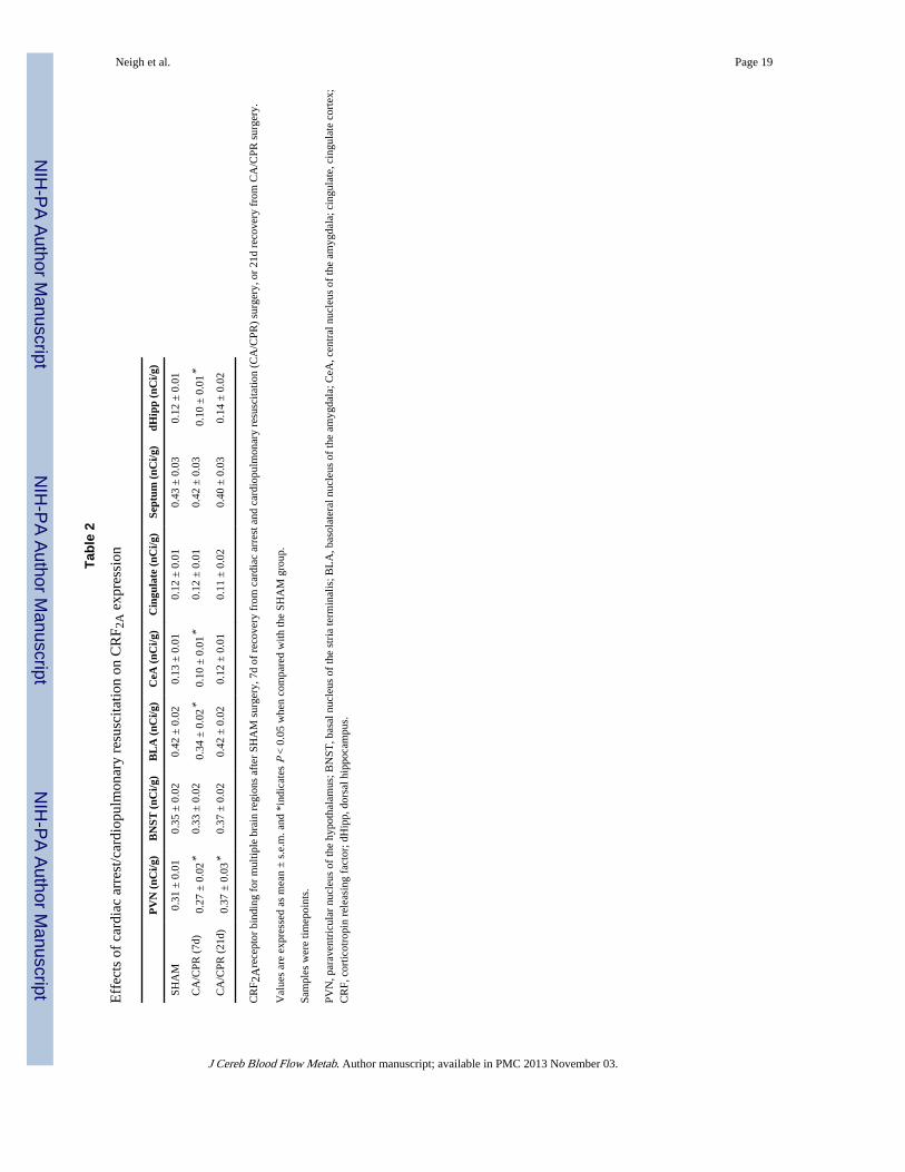
CA/CPR Alters CRF Receptor Binding in a Region-Specific and Time-Specific MannerA complete summary of the observed changes in CRF receptor binding after either 7d or 21dof recovery from CA/CPR or SHAM surgery are depicted in Tables 1 and 2. As all bindingassays were run simultaneously, the data from the 7d and 21d SHAM groups were collapsedinto one SHAM control group for the CRF receptor binding, after establishing that therewere no statistical differences in expression between these two timepoints. CA/CPR causes agradual increase in CRF1 receptor binding in the paraventricular nucleus of thehypothalamus (PVN), as compared with a similar timepoint in the SHAM group, which wasstatistically significant by 2Id after surgery (F2,29 = 4.70, P < 0.05). CRF2A receptor bindingin the PVN initially decreased after CA/CPR (7d after surgery), but was elevated by 21dafter surgery (F2,29 = 6.80, P < 0.05) as compared with SHAM operated mice. CRF1 bindingin the bed nucleus of the stria terminalis showed a pattern similar to the PVN with a slightincrease 7d after CA/CPR, which was significant by 21d following the procedure ascompared with SHAM operated mice (t23 = 2.50, P < 0.05). The cingulate cortex showed theopposite pattern, a trend toward decreased CRF1 binding 7d after CA/CPR, that becamesignificant by 21d, as compared with SHAM operated mice (F2,18 = 4.70, P < 0.05). Therewere no CA/CPR-induced changes in CRF1 binding in either the basolateral or centralamygdala (P > 0.05). CRF2A binding was transiently decreased in the basolateral amygdala
Neigh et al. Page 6
J Cereb Blood Flow Metab. Author manuscript; available in PMC 2013 November 03.
NIH
-PA Author Manuscript
NIH
-PA Author Manuscript
NIH
-PA Author Manuscript

at 7d (F2,35 = 4.82, P < 0.05), but not 21d (P > 0.05) after the CA/CPR procedure, andbinding in the central nucleus was decreased, as compared with SHAM operated mice, atboth 7d and 21d after CA/CPR (F2,22 = 4.51, P < 0.05). A transient decrease in the dorsalhippocampus of both CRF1 and CRF2A binding occurred in the CA/CPR mice as comparedwith SHAM controls (F2,35 = 4.44, P < 0.05; F2,33 = 5.21, P < 0.05). A transient decrease inCRF1 binding in the septum was documented 7d (t13 = 2.53, P < 0.05), but not 21d, after theCA/CPR procedure (P > 0.05).
DiscussionPeople who survive cardiac arrest are plagued by persistent affective disorders and havedifficulty with social integration. Indeed, as many as 3 years after cardiac arrest, 30% ofsurvivors report anxiety and nearly 75% report low social participation (Wachelder et al,2009). In addition, high levels of anxiety are associated with increased risk of suddencardiac death (Albert et al, 2005). Compared with depression, relatively few clinical studieshave addressed the topic of anxiety after recovery from cardiac arrest or cardiopulmonarybypass (Ladwig et al, 1999; Murphy et al, 2008; Vingerhoets et al, 1995; Wachelder et al,2009). The underlying pathophysiologic causes remain unknown but may involvedysfunction in the HPA axis (Hekimian et al, 2004; Yin et al, 2007). This study shows thatCA/CPR in mice is associated with persistent alterations in anxiety-like behavior andchanges in HPA axis regulation that may contribute to the development of anxiety.
An important step in establishing a physiologic mechanism involves developing an animalmodel that produces long-term anxiety-like behavior after cardiac arrest. Anxiety-likebehavior after CA/CPR has been reported in mice (Kofler et al, 2004; Neigh et al, 2004,2005) and rats (Dhooper et al, 1997) within days of resuscitation. Although some othermodels of global cerebral ischemia have produced increased anxiety-like behavior (Ishibashiet al, 2006; Yan et al, 2007), others have failed to do so (Bantsiele et al, 2004; Plamondonand Khan, 2005). Most of these studies suffer the limitation of examining anxiety soon afterischemia. This study extends our earlier work reporting the presence of anxiety-likebehavior after a short-term recovery, within 7d of the procedure (Neigh et al, 2004), andprovides evidence that anxiety-like behavior is still evident 21d after CA/CPR (Figure 2).Thus, CA/CPR-induced increases in anxiety-like behavior persist well beyond the ischemicevent.
CA/CPR also alters several aspects of HPA axis function, including (1) a transient increasein basal corticosterone concentrations (Figure 3), (2) sensitization of negative feedback onthe HPA axis (Figure 3), and (3) a dynamic change in CRF receptor binding (Tables 1 and2).
Mean basal corticosterone concentration was significantly higher among CA/CPR thanSHAM operated mice on postsurgical day 7, but not on postsurgical day 21 (Figure 3).These data confirm and extend earlier work showing that CA/CPR elevates basalcorticosterone concentration and attenuates the corticosterone response to a restraint stressor(Neigh et al, 2005). Data from this study suggest that the rise in basal corticosteroneconcentrations after CA/CPR is transient, because by 21 days, basal corticosteroneconcentrations are similar for SHAM and CA/CPR groups. Furthermore, over time CA/CPRincreases the sensitivity of the HPA axis to dexamethasone-induced suppression ofcirculating corticosterone concentrations (Figure 3), a measure of HPA axis negative-feedback regulation. On postsurgical day 7, both the CA/CPR and SHAM groups exhibit theexpected dose-dependent decrease in serum corticosterone after treatment withdexamethasone, and values at each concentration of dexamethasone are similar for the twogroups. In contrast, on postsurgical day 21, the SHAM group exhibits the expected dose-
Neigh et al. Page 7
J Cereb Blood Flow Metab. Author manuscript; available in PMC 2013 November 03.
NIH
-PA Author Manuscript
NIH
-PA Author Manuscript
NIH
-PA Author Manuscript

dependent decrease in corticosterone concentration after treatment with dexamethasone,whereas the CA/CPR group exhibits maximal suppression at even the lowest dose;corticosterone concentrations are significantly lower for the CA/CPR than SHAM groupafter treatment with 50 and 100 µg of dexamethasone. At the highest dose of dexamethasone(200 µg), the corticosterone concentrations of the CA/CPR and SHAM groups are similar.The delayed onset of enhanced HPA axis negative feedback, and resolution of the elevatedbasal corticosterone, may indicate that negative feedback is enhanced to compensate for theelevated basal concentrations of corticosterone that manifest soon after CA/CPR. In fact,adrenal insufficiency shortly after CA/CPR in humans is associated with a higher rate ofdeath because of septic shock (Hekimian et al, 2004), suggesting that an optimal amount ofcortisol may be beneficial after CA/CPR. The long-term implications of prolonged, robustincreases in glucocorticoids after ischemia are unknown but our data suggest that thenegative feedback on the HPA axis may change in response to the ischemia-inducedelevations in glucocorticoids. To our knowledge, dexamethasone suppression has not beenassessed after CA/CPR in the human population.
The CA/CPR-induced changes in basal corticosterone concentrations, which precedealterations in negative feedback, may be a manifestation of ischemia-induced changes inCRF neurotransmission. CRF mRNA is rapidly upregulated in the ischemic cortex andamygdala (Wong et al, 1995), and extracellular CRF is elevated in the paraventricularnucleus of the hypothalamus, amygdala, hippocampus, and cortex after cerebral ischemia(Khan et al, 2004). In turn, CRF influences cerebral blood flow, vascular permeability, andcell death (De Michele et al, 2005; Khan et al, 2004; Stevens et al, 2003). Indeed, ischemia-induced neuronal loss is attenuated when CRF antagonists are administered (Charron et al,2008; Wong et al, 1995). Studies of ischemia in CRF1 and CRF2A knockout mice indicatethat CRF1 is an important mediator of ischemic injury (Stevens et al, 2003). The reductionin CRF1 and CRF2A receptor binding in the hippocampus observed 7d after CA/CPR in thisstudy (Tables 1 and 2) may be a compensatory response to the increase in CRF that othershave reported (Khan et al, 2004; Wong et al, 1995). At postsurgical day 21, the CA/CPRgroup had more CRF1 binding in the paraventricular nucleus of the hypothalamus and thebed nucleus of the stria terminalis than the SHAM group. These changes in CRF physiologyare consistent with changes documented in other anxiety models (Dunn and Berridge, 1990),and may contribute to the anxiety-like behavior documented 21d after CA/CPR (Figure 2).
Neurons, microglia, and astroglia express both CRF1 and CRF2A receptors, and activation ofthe CRF1 receptor in particular influences the inflammatory response and the extent ofneuronal damage that results from focal ischemia (Stevens et al, 2003). Modulation of CRFneurotransmission after cerebral ischemia can also influence behavior; administration of theCRF1 receptor antagonist CP154, 526 either shortly before or after global ischemiaattenuates the increase in locomotor activity that typically appears 4 to 7d after globalischemia, without altering neurodegeneration (Plamondon and Khan, 2006). Whether thechanges in CRF1 and CRF2A binding observed in this study reflect alterations in neurons,microglia, astroglia or a combination of these cell types, and how these changes affectneuronal survival and inflammation after CA/CPR requires further study.
Given the established role of HPA axis pathology in depressive and anxiety disorders(Tsigos and Chrousos, 2002), and the increased incidence of affective disorders in CA/CPRsurvivors (Ladwig et al, 1999; Wachelder et al, 2009), we sought to establish whether CA/CPR caused long-term changes in the HPA axis. The data presented here show that there is atransient increase in basal corticosterone concentrations after CA/CPR, which resolvesbetween 7d and 21d after injury and may reflect changes in CRF neurotransmission, whichhave been documented earlier to occur after ischemia (De Michele et al, 2007; Wong et al,1995). Furthermore, augmented negative feedback regulation of the HPA axis manifests by
Neigh et al. Page 8
J Cereb Blood Flow Metab. Author manuscript; available in PMC 2013 November 03.
NIH
-PA Author Manuscript
NIH
-PA Author Manuscript
NIH
-PA Author Manuscript
21d after injury. The enhanced sensitivity to negative feedback after CA/CPR is intriguingand warrants additional work to determine whether persistent anxiety after resuscitation iscausally related to the observed changes in the HPA axis. Together, the data presented hereshow dynamic changes in the HPA axis after CA/CPR and a concomitant increase inanxiety. Better understanding of the pathology that underlies post-CA/CPR behavioralchanges will ultimately lead to better treatments of the unique mental and physical disordersof CA/CPR survivors. In addition, the continued remodeling of the HPA axis showed in thisstudy highlights the importance of studying long-term physiologic and behavioral changesafter ischemia beyond the initial recovery period.
AcknowledgmentsThe authors thank Lorraine Smith, Faketa Zejnelovic, Greg Norman, and Susan Plott for technical assistance. Wethank Dr Kerry Ressler and the journal referees for editorial comments on this manuscript. This work wassupported in part by NIH Institutional Research and Academic Career Development grant #K12 GM00680-05(GNN), R01HL080249 (ACD), and P30 NS0457558 (ACD).
Disclosure/conflict of interest
Gretchen N Neigh, PhD
Research Grants
NARSAD, AHA, NIH, GlaxoSmithKline
Kate Karelina
Research Grants
AHA
Ning Zhang, MD
None
Erica R Glasper, PhD
Research Grants
United Negro College Fund · Merck Postdoctoral Science Research Fellowship
Michael J Owens, PhD
Research Grants
NIH, Eli Lilly, Pfizer, GlaxoSmithKline, Lundbeck, Cyberonics, Ortho-McNeil Janssen, AstraZeneca
Consultant
H Lundbeck A/S
Honorarium
Eli Lilly
Patents
‘A method to estimate transporter occupancy’
Paul Plotsky, PhD
Research Grants
Neigh et al. Page 9
J Cereb Blood Flow Metab. Author manuscript; available in PMC 2013 November 03.
NIH
-PA Author Manuscript
NIH
-PA Author Manuscript
NIH
-PA Author Manuscript

Lundbeck, Cyberonics, NIH
Charles B Nemeroff, MD, PhD
Research Grants
NIH
Scientific Advisory Board
AFSP; AstraZeneca; Forest Laboratories; NARSAD; Quintiles; Janssen/Ortho-McNeil, PharmaNeuroboost, Mt.Cook Pharma, Inc
Stockholder or Equity
Corcept; Revaax; NovaDel Pharma; CeNeRx, PharmaNeuroboost
Board of Directors
American Foundation for Suicide Prevention (AFSP); George West Mental Health Foundation; NovaDel Pharma,Mt. Cook Pharma, Inc
Patents
Method and devices for transdermal delivery of lithium (US 6,375,990 Bl)
‘A method to estimate transporter occupancy’
A Courtney DeVries, PhD
Research Grants
AHA, NIH
ReferencesAlbert CM, Chae CU, Rexrode KM, Manson JE, Kawachi I. Phobic anxiety and risk of coronary heart
disease and sudden cardiac death among women. Circulation. 2005; 111:480–487. [PubMed:15687137]
Albertsmeier M, Teschendorf P, Popp E, Galmbacher R, Vogel P, Bottiger BW. Evaluation of a taperemoval test to assess neurological deficit after cardiac arrest in rats. Resuscitation. 2007; 74:552–558. [PubMed: 17449165]
Bantsiele GB, Bentue-Ferrer D, Amiot N, Allain H, Bourin M, Reymann JM. Does rat global transientcerebral ischemia serve as an appropriate model to study emotional disturbances? Fundam ClinPharmacol. 2004; 18:685–692. [PubMed: 15548240]
Bottiger BW, Krumnikl JJ, Gass P, Schmitz B, Motsch J, Martin E. The cerebral “no-reflow”phenomenon after cardiac arrest in rats—influence of low-flow reperfusion. Resuscitation. 1997;34:79–87. [PubMed: 9051828]
Bottiger BW, Teschendorf P, Krumnikl JJ, Vogel P, Galmbacher R, Schmitz B, Motsch J, Martin E,Gass P. Global cerebral ischemia due to cardiocirculatory arrest in mice causes neuronaldegeneration and early induction of transcription factor genes in the hippocampus. Brain Res MolBrain Res. 1999; 65:135–142. [PubMed: 10064884]
Brown ES, Rush AJ, McEwen BS. Hippocampal remodeling and damage by corticosteroids:implications for mood disorders. Neuropsychopharmacology. 1999; 21:474–484. [PubMed:10481830]
Bunch TJ, White RD, Gersh BJ, Meverden RA, Hodge DO, Ballman KV, Hammill SC, Shen WK,Packer DL. outcomes of out-of-hospital cardiac arrest after successful early defibrillation. N Engl JMed. 2003; 348:2626–2633. [PubMed: 12826637]
Neigh et al. Page 10
J Cereb Blood Flow Metab. Author manuscript; available in PMC 2013 November 03.
NIH
-PA Author Manuscript
NIH
-PA Author Manuscript
NIH
-PA Author Manuscript

Cerchiari EL, Safar P, Klein E, Cantadore R, Pinsky M. Cardiovascular function and neurologicoutcome after cardiac arrest in dogs. The cardiovascular post-resuscitation syndrome. Resuscitation.1993; 25:9–33. [PubMed: 8446790]
Charron C, Frechette S, Proulx G, Plamondon H. In vivo administration of corticotropin-releasinghormone at remote intervals following ischemia enhances CA1 neuronal survival and recovery ofspatial memory impairments: a role for opioid receptors. Behav Brain Res. 2008; 188:125–135.[PubMed: 18055027]
De Michele M, Sette G, Chalmers DT, Dewar D, Toni D, Sancesario G, McCulloch J. Focal cerebralischaemia induces corticotropin releasing factor (CRF) vascular immunoreactivity in rat occludedhemisphere. Regul Pept. 2007; 143:69–75. [PubMed: 17477982]
De Michele M, Touzani O, Foster AC, Fieschi C, Sette G, McCulloch J. Corticotropin-releasing factor:effect on cerebral blood flow in physiologic and ischaemic conditions. Exp Brain Res. 2005;165:375–382. [PubMed: 15864562]
de Vos R, de Haes HC, Koster RW, de Haan RJ. Quality of survival after cardiopulmonaryresuscitation. Arch Intern Med. 1999; 159:249–254. [PubMed: 9989536]
Dhooper A, Young C, Reid KH. Ischemia-induced anxiety following cardiac arrest in the rat. BehavBrain Res. 1997; 84:57–62. [PubMed: 9079772]
Dunn AJ, Berridge CW. Physiological and behavioral responses to corticotropin-releasing factoradministration: is CRF a mediator of anxiety or stress responses? Brain Res Brain Res Rev. 1990;15:71–100. [PubMed: 1980834]
Esposito P, Basu S, Letourneau R, Jacobson S, Theoharides TC. Corticotropin-releasing factor (CRF)can directly affect brain microvessel endothelial cells. Brain Res. 2003; 968:192–198. [PubMed:12663088]
Hekimian G, Baugnon T, Thuong M, Monchi M, Dabbane H, Jaby D, Rhaoui A, Laurent I, Moret G,Fraisse F, Adrie C. Cortisol levels and adrenal reserve after successful cardiac arrest resuscitation.Shock. 2004; 22:116–119. [PubMed: 15257083]
Ishibashi S, Kuroiwa T, LiYuan S, Katsumata N, Li S, Endo S, Mizusawa H. cognitive andneuropsychological symptoms after global cerebral ischemia in Mongolian gerbils. Acta NeurochirSuppl. 2006; 96:299–302. [PubMed: 16671475]
Keppel, G. Design and analysis. Upper Saddle River, NJ: Prentice Hall; 1991.
Khan S, Milot M, Lecompte-Collin J, Plamondon H. Time-dependent changes in CRH concentrationsand release in discrete brain regions following global ischemia: effects of MK-801 pretreatment.Brain Res. 2004; 1016:48–57. [PubMed: 15234251]
Kofler J, Hattori K, Sawada M, DeVries AC, Martin LJ, Hurn PD, Traystman RJ. Histopathologicaland behavioral characterization of a novel model of cardiac arrest and cardiopulmonaryresuscitation in mice. J Neurosci Methods. 2004; 136:33–44. [PubMed: 15126043]
Ladwig KH, Schoefinius A, Dammann G, Danner R, Gurtler R, Herrmann R. Long-actingpsychotraumatic properties of a cardiac arrest experience. Am J Psychiatry. 1999; 156:912–919.[PubMed: 10360132]
Miranda DR. Quality of life after cardiopulmonary resuscitation. Chest. 1994; 106:524–530. [PubMed:7774331]
Muller MB, Uhr M, Holsboer F, Keck ME. Hypothalamic-pituitary-adrenocortical system and mooddisorders: highlights from mutant mice. Neuroendocrinology. 2004; 79:1–12. [PubMed:14755129]
Murphy BM, Elliott PC, Worcester MU, Higgins RO, Le Grande MR, Roberts SB, Goble AJ.Trajectories and predictors of anxiety and depression in women during the 12 months following anacute cardiac event. Br J Health Psychol. 2008; 13:135–153. [PubMed: 17535492]
Neigh GN, Glasper ER, Bilbo SD, Traystman RJ, Courtney DeVries A. Cardiac arrest/cardiopulmonary resuscitation augments cell-mediated immune function and transientlysuppresses humoral immune function. J Cereb Blood Flow Metab. 2005; 25:1424–1432. [PubMed:15874972]
Neigh GN, Kofler J, Meyers JL, Bergdall V, La Perle KM, Traystman RJ, DeVries AC. Cardiac arrest/cardiopulmonary resuscitation increases anxiety-like behavior and decreases social interaction. JCereb Blood Flow Metab. 2004; 24:372–382. [PubMed: 15087706]
Neigh et al. Page 11
J Cereb Blood Flow Metab. Author manuscript; available in PMC 2013 November 03.
NIH
-PA Author Manuscript
NIH
-PA Author Manuscript
NIH
-PA Author Manuscript
Neumar RW, Nolan JP, Adrie C, Aibiki M, Berg RA, Bottiger BW, Callaway C, Clark RS, GeocadinRG, Jauch EC, Kern KB, Laurent I, Longstreth WT Jr, Merchant RM, Morley P, Morrison LJ,Nadkarni V, Peberdy MA, Rivers EP, Rodriguez-Nunez A, Sellke FW, Spaulding C, Sunde K,Vanden Hoek T. -cardiac arrest syndrome: epidemiology, pathophysiology, treatment, andprognostication. A consensus statement from the International Liaison Committee onResuscitation (American Heart Association, Australian and New Zealand Council onResuscitation, European Resuscitation Council, Heart and Stroke Foundation of Canada, InterAmerican Heart Foundation, Resuscitation Council of Asia, and the Resuscitation Council ofSouthern Africa); the American Heart Association Emergency Cardiovascular Care Committee;the Council on Cardiovascular Surgery and Anesthesia; the Council on Cardiopulmonary,Perioperative, and Critical Care; the Council on Clinical Cardiology; and the Stroke Council.Circulation. 2008; 118:2452–2483. [PubMed: 18948368]
Paradis NA, Wenzel V, Southall J. Pressor drugs in the treatment of cardiac arrest. Cardiol Clin. 2002;20:61–78. viii. [PubMed: 11845545]
Paxinos, G.; Watson, C. The rat brain in stereotaxic coordinates. San Diego: Academic Press; 1986.
Pellow S, Chopin P, File SE, Briley M. Validation of open:closed arm entries in an elevated plus-mazeas a measure of anxiety in the rat. J Neurosci Methods. 1985; 14:149–167. [PubMed: 2864480]
Plamondon H, Khan S. Characterization of anxiety and habituation profile following global ischemiain rats. Physiol Behav. 2005; 84:543–552. [PubMed: 15811389]
Plamondon H, Khan S. The CRH1 antagonist CP154,526 failed to alter ischemia-inducedneurodegeneration and spatial memory deficits in rats but inhibited behavioral activity in the novelopen field. Behav Brain Bes. 2006; 166:85–92.
Reich P, Regestein QR, Murawski BJ, DeSilva RA, Lown B. Unrecognized organic mental disordersin survivors of cardiac arrest. Am J Psychiatry. 1983; 140:1194–1197. [PubMed: 6614228]
Sadowski M, Wisniewski HM, Jakubowska-Sadowska K, Tarnawski M, Lazarewicz JW,Mossakowski MJ. Pattern of neuronal loss in the rat hippocampus following experimental cardiacarrest-induced ischemia. J Neurol Sci. 1999; 168:13–20. [PubMed: 10500268]
Skelton KH, Nemeroff CB, Knight DL, Owens MJ. administration of the triazolobenzodiazepinealprazolam produces opposite effects on corticotropin-releasing factor and urocortin neuronalsystems. J Neurosci. 2000; 20:1240–1248. [PubMed: 10648728]
Stevens SL, Shaw TE, Dykhuizen E, Lessov NS, Hill JK, Wurst W, Stenzel-Poore MP. Reducedcerebral injury in CRH-R1 deficient mice after focal ischemia: a potential link to microglia andatrocytes that express CRH-R1. J Cereb Blood Flow Metab. 2003; 23:1151–1159. [PubMed:14526225]
Sunnerhagen KS, Johansson O, Herlitz J, Grimby G. Life after cardiac arrest; a retrospective study.Resuscitation. 1996; 31:135–140. [PubMed: 8733020]
The Hypothermia after Cardiac Arrest Study Group. Mild therapeutic hypothermia to improve theneurologic outcome after cardiac arrest. N Engl J Med. 2002; 346:549–556. [PubMed: 11856793]
Tsigos C, Chrousos GP. Hypothalamic-pituitary-adrenal axis, neuroendocrine factors and stress. JPsychosom Res. 2002; 53:865–871. [PubMed: 12377295]
Vingerhoets G, de Soete G, Jannes C. Subjective complaints versus neuropsychological testperformance after cardiopulmonary bypass. J Psychosom Res. 1995; 39:843–853. [PubMed:8636916]
Wachelder EM, Moulaert VR, van Heugten C, Verbunt JA, Bekkers SC, Wade DT. Life after survival:long-term daily functioning and quality of life after an out-of-hospital cardiac arrest. Resuscitation.2009; 80:517–522. [PubMed: 19282084]
Weil ZM, Norman GJ, Barker JM, Su AJ, Nelson RJ, Devries AC. Social isolation potentiates celldeath and inflammatory responses after global ischemia. Mol Psychiatry. 2008; 13:913–915.[PubMed: 18800053]
Wong ML, Loddick SA, Bongiorno PB, Gold PW, Rothwell NJ, Licinio J. Focal cerebral ischemiainduces CRH mRNA in rat cerebral cortex and amygdala. Neuroreport. 1995; 6:1785–1788.[PubMed: 8541482]
Neigh et al. Page 12
J Cereb Blood Flow Metab. Author manuscript; available in PMC 2013 November 03.
NIH
-PA Author Manuscript
NIH
-PA Author Manuscript
NIH
-PA Author Manuscript

Yan B, He J, Xu H, Zhang Y, Bi X, Thakur S, Gendron A, Kong J, Li XM. Quetiapine attenuates thedepressive and anxiolytic-like behavioural changes induced by global cerebral ischemia in mice.Behav Brain Res. 2007; 182:36–41. [PubMed: 17568696]
Yin YQ, Luo AL, Guo XY, Li LH, Huang YG. Postoperative neuropsychological change and itsunderlying mechanism in patients undergoing coronary artery bypass grafting. Chin Med J (Engl).2007; 120:1951–1957. [PubMed: 18067777]
Neigh et al. Page 13
J Cereb Blood Flow Metab. Author manuscript; available in PMC 2013 November 03.
NIH
-PA Author Manuscript
NIH
-PA Author Manuscript
NIH
-PA Author Manuscript
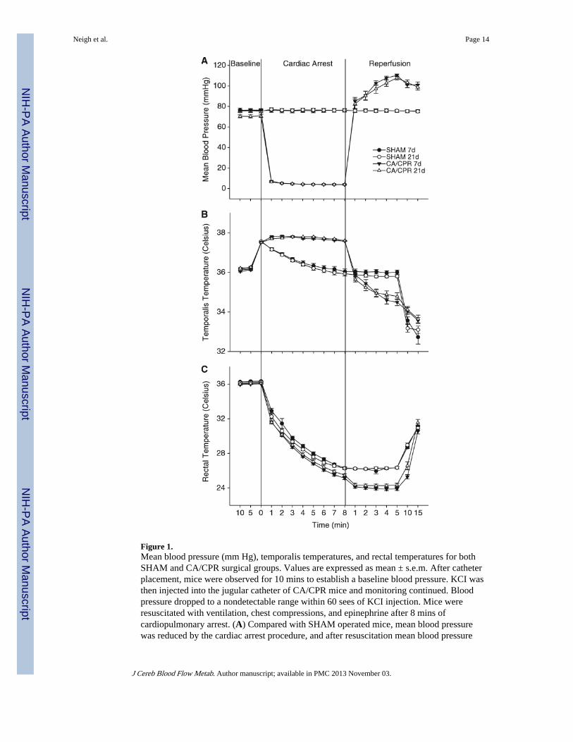
Figure 1.Mean blood pressure (mm Hg), temporalis temperatures, and rectal temperatures for bothSHAM and CA/CPR surgical groups. Values are expressed as mean ± s.e.m. After catheterplacement, mice were observed for 10 mins to establish a baseline blood pressure. KCI wasthen injected into the jugular catheter of CA/CPR mice and monitoring continued. Bloodpressure dropped to a nondetectable range within 60 sees of KCI injection. Mice wereresuscitated with ventilation, chest compressions, and epinephrine after 8 mins ofcardiopulmonary arrest. (A) Compared with SHAM operated mice, mean blood pressurewas reduced by the cardiac arrest procedure, and after resuscitation mean blood pressure
Neigh et al. Page 14
J Cereb Blood Flow Metab. Author manuscript; available in PMC 2013 November 03.
NIH
-PA Author Manuscript
NIH
-PA Author Manuscript
NIH
-PA Author Manuscript
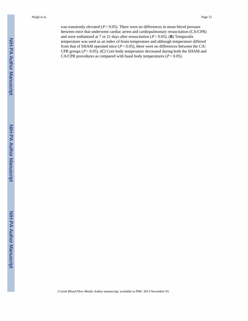
was transiently elevated (P < 0.05). There were no differences in mean blood pressurebetween mice that underwent cardiac arrest and cardiopulmonary resuscitation (CA/CPR)and were euthanized at 7 or 21 days after resuscitation (P > 0.05). (B) Temporalistemperature was used as an index of brain temperature and although temperature differedfrom that of SHAM operated mice (P < 0.05), there were no differences between the CA/CPR groups (P > 0.05). (C) Core body temperature decreased during both the SHAM andCA/CPR procedures as compared with basal body temperatures (P < 0.05).
Neigh et al. Page 15
J Cereb Blood Flow Metab. Author manuscript; available in PMC 2013 November 03.
NIH
-PA Author Manuscript
NIH
-PA Author Manuscript
NIH
-PA Author Manuscript

Figure 2.After a 21d recovery period, mice were tested for anxiety-like behavior in the elevated plusmaze (A–C) and open field (D–F) during their dark cycle. Locomotor activity did not differbetween SHAM and CA/CPR operated mice in the elevated plus maze (A) or the open field(D). (B) Anxiety-like behavior was shown by a decrease in time in the open arms entries inthe CA/CPR group when compared with SHAM operated mice (mean ±s.e.m., *indicates P< 0.05). (C) However, there was no group difference in percent open arm entries. (E)Central tendency in the open field was also decreased in CA/CPR mice when compared withSHAM operated mice (mean ± s.e.m., *indicates P < 0.05). (F) There was no groupdifference in rearing frequency in the open field.
Neigh et al. Page 16
J Cereb Blood Flow Metab. Author manuscript; available in PMC 2013 November 03.
NIH
-PA Author Manuscript
NIH
-PA Author Manuscript
NIH
-PA Author Manuscript
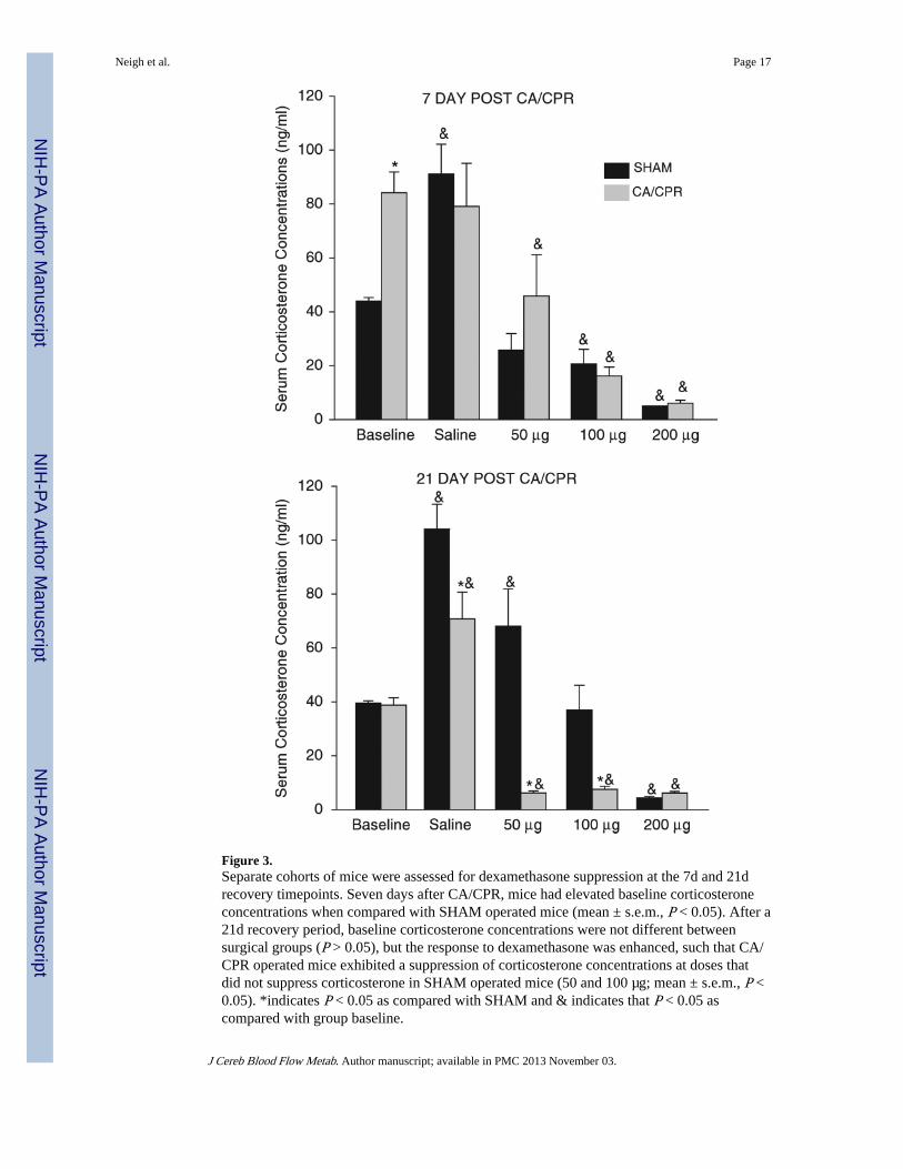
Figure 3.Separate cohorts of mice were assessed for dexamethasone suppression at the 7d and 21drecovery timepoints. Seven days after CA/CPR, mice had elevated baseline corticosteroneconcentrations when compared with SHAM operated mice (mean ± s.e.m., P < 0.05). After a21d recovery period, baseline corticosterone concentrations were not different betweensurgical groups (P > 0.05), but the response to dexamethasone was enhanced, such that CA/CPR operated mice exhibited a suppression of corticosterone concentrations at doses thatdid not suppress corticosterone in SHAM operated mice (50 and 100 µg; mean ± s.e.m., P <0.05). *indicates P < 0.05 as compared with SHAM and & indicates that P < 0.05 ascompared with group baseline.
Neigh et al. Page 17
J Cereb Blood Flow Metab. Author manuscript; available in PMC 2013 November 03.
NIH
-PA Author Manuscript
NIH
-PA Author Manuscript
NIH
-PA Author Manuscript

NIH
-PA Author Manuscript
NIH
-PA Author Manuscript
NIH
-PA Author Manuscript
Neigh et al. Page 18
Tabl
e 1
Eff
ects
of
card
iac
arre
st/c
ardi
opul
mon
ary
resu
scita
tion
on C
RF 1
exp
ress
ion
PV
N (
nCi/g
)B
NST
(nC
i/g)
BL
A (
nCi/g
)C
eA (
nCi/g
)C
ingu
late
(nC
i/g)
Sept
um (
nCi/g
)dH
ipp
(nC
i/g)
SHA
M0.
33 ±
0.0
20.
52 ±
0.0
20.
58 ±
0.0
20.
18 ±
0.0
10.
53 ±
0.0
10.
61 ±
0.0
40.
26 ±
0.0
1
CA
/CPR
(7d
)0.
37 ±
0.0
30.
57 ±
0.0
40.
56 ±
0.0
50.
15 ±
0.0
10.
47 ±
0.0
20.
50 ±
0.0
2*0.
21 ±
0.0
1*
CA
/CPR
(21
d)0.
43 ±
0.0
3*0.
60 ±
0.0
3*0.
53 ±
0.0
3*0.
18 ±
0.0
20.
46 ±
0.0
3*0.
57 ±
0.0
30.
27 ±
0.0
1
CR
F 1 r
ecep
tor
bind
ing
for
mul
tiple
bra
in r
egio
ns a
fter
SH
AM
sur
gery
, 7d
of r
ecov
ery
from
car
diac
arr
est a
nd c
ardi
opul
mon
ary
resu
scita
tion
(CA
/CPR
) su
rger
y, o
r 21
d of
rec
over
y fr
om C
A/C
PR s
urge
ry.
Val
ues
are
expr
esse
d as
mea
n ±
s.e
.m. a
nd *
indi
cate
s P
< 0
.05
whe
n co
mpa
red
with
the
SHA
M g
roup
.
Sam
ples
wer
e tim
epoi
nts.
PVN
, par
aven
tric
ular
nuc
leus
of
the
hypo
thal
amus
; BN
ST, b
asal
nuc
leus
of
the
stri
a te
rmin
alis
; BL
A, b
asol
ater
al n
ucle
us o
f th
e am
ygda
la; C
eA, c
entr
al n
ucle
us o
f th
e am
ygda
la; c
ingu
late
, cin
gula
te c
orte
x;C
RF,
cor
ticot
ropi
n re
leas
ing
fact
or; d
Hip
p, d
orsa
l hip
poca
mpu
s.
J Cereb Blood Flow Metab. Author manuscript; available in PMC 2013 November 03.
NIH
-PA Author Manuscript
NIH
-PA Author Manuscript
NIH
-PA Author Manuscript
Neigh et al. Page 19
Tabl
e 2
Eff
ects
of
card
iac
arre
st/c
ardi
opul
mon
ary
resu
scita
tion
on C
RF 2
A e
xpre
ssio
n
PV
N (
nCi/g
)B
NST
(nC
i/g)
BL
A (
nCi/g
)C
eA (
nCi/g
)C
ingu
late
(nC
i/g)
Sept
um (
nCi/g
)dH
ipp
(nC
i/g)
SHA
M0.
31 ±
0.0
10.
35 ±
0.0
20.
42 ±
0.0
20.
13 ±
0.0
10.
12 ±
0.0
10.
43 ±
0.0
30.
12 ±
0.0
1
CA
/CPR
(7d
)0.
27 ±
0.0
2*0.
33 ±
0.0
20.
34 ±
0.0
2*0.
10 ±
0.0
1*0.
12 ±
0.0
10.
42 ±
0.0
30.
10 ±
0.0
1*
CA
/CPR
(21
d)0.
37 ±
0.0
3*0.
37 ±
0.0
20.
42 ±
0.0
20.
12 ±
0.0
10.
11 ±
0.0
20.
40 ±
0.0
30.
14 ±
0.0
2
CR
F 2A
rece
ptor
bin
ding
for
mul
tiple
bra
in r
egio
ns a
fter
SH
AM
sur
gery
, 7d
of r
ecov
ery
from
car
diac
arr
est a
nd c
ardi
opul
mon
ary
resu
scita
tion
(CA
/CPR
) su
rger
y, o
r 21
d re
cove
ry f
rom
CA
/CPR
sur
gery
.
Val
ues
are
expr
esse
d as
mea
n ±
s.e
.m. a
nd *
indi
cate
s P
< 0
.05
whe
n co
mpa
red
with
the
SHA
M g
roup
.
Sam
ples
wer
e tim
epoi
nts.
PVN
, par
aven
tric
ular
nuc
leus
of
the
hypo
thal
amus
; BN
ST, b
asal
nuc
leus
of
the
stri
a te
rmin
alis
; BL
A, b
asol
ater
al n
ucle
us o
f th
e am
ygda
la; C
eA, c
entr
al n
ucle
us o
f th
e am
ygda
la; c
ingu
late
, cin
gula
te c
orte
x;C
RF,
cor
ticot
ropi
n re
leas
ing
fact
or; d
Hip
p, d
orsa
l hip
poca
mpu
s.
J Cereb Blood Flow Metab. Author manuscript; available in PMC 2013 November 03.