cardiac trauma chapter 13 daniel eiferman, r. nathan ... · advanced trauma life support (atls)...
TRANSCRIPT

Chapter 13
Cardiac Trauma
Daniel Eiferman,R. Nathan Cotterman andMichael Firstenberg
Additional information is available at the end of the chapter
http://dx.doi.org/10.5772/55723
1. Introduction
1.1. History of cardiac trauma
The treatment of trauma to the heart has been written about since 3000 BC and had aninauspicious beginning. Until the late 19th century, the commonly held belief agreed withBoerhaave’s sentiments that, “all penetrating cardiac trauma is fatal.” Theodore Billrothwarned, “The surgeon who should attempt to suture a wound of the heart would lose therespect of his colleagues.” Paget believed that “surgery of the heart has probably reached thelimits set by nature to all surgery: no new method of discovery can overcome the naturaldifficulties that attend a wound of the heart.” However, reports of successful treatment ofcardiac injuries began to surface toward the beginning of the 20th century. Like many surgicaladvances, times of war brought about new innovations and techniques for treating injuries.
Around the time of World War II, it was recognized that cardiac tamponade could be suc‐cessfully managed by pericardiocentesis. With the advent of cardiopulmonary bypass byGibbon in 1953, repair of more complex injuries became possible. This ushered in the modernera of treating injuries to the heart. Diagnosis of cardiac injury and tamponade has also beenfacilitated by portable ultrasound becoming the standard of care in the evaluation of traumapatients. The focused assessment with sonography for trauma (FAST) scan allows for simple,quick, and non-invasive assessment and recognition of cardiac trauma [1].
Cardiac trauma, especially penetrating injuries to the heart, still carries a very high mortality,but certainly is no longer considered uniformly fatal and attempt at repair is now the standardof care in patients presenting with signs of life upon arrival to the hospital[2, 3].
© 2013 Eiferman et al.; licensee InTech. This is an open access article distributed under the terms of theCreative Commons Attribution License (http://creativecommons.org/licenses/by/3.0), which permitsunrestricted use, distribution, and reproduction in any medium, provided the original work is properly cited.

2. Initial assessment and general assessment
The initial care of the trauma patient with cardiac injuries does not vary from standardAdvanced Trauma Life Support (ATLS) protocols. The primary priority is ensuring the patencyof the airway and establishing adequate oxygenation and ventilation. This may include tubethoracostomy for drainage of hemothorax from the pleural space to allow re-expansion of thelung. Subsequently, the circulatory system is assessed. Priority is given to establishingintravenous access for the administration of crystalloid and/or blood products. If cardiactamponade is suspected, this should be confirmed with sonographic confirmation of hemo‐pericardium and/or right ventricular collapse during diastole[4]. If tamponade physiology ispresent, treatment for immediate drainage of the pericardial space should be initiated. Thiscan be accomplished percutaneously by pericardiocentesis or via open pericardial window.
The treatment algorithm for cardiac injured patients branches at this point depending on themechanism of injury and hemodynamic status. As is the standard in all trauma care, cardiacinjuries are categorized as either blunt or penetrating and we will explore their assessmentand treatment separately.
3. Penetrating trauma
Penetrating trauma to the heart most frequently occur with trauma to the anterior chest, butshould also be suspected with wounds to the upper abdomen, chest, back, and neck [5]. Of thepatients that do present to the hospital, the majority of the injuries are to the low pressure,anteriorly located right side of the heart (Table 1) [6]. Survival following penetrating traumais often dependent on the state of the pericardial wound.[7] When the pericardial wound isopen and blood is able to flow freely into the pleural space, the patient can often be supportedwith fluid resuscitation and chest tube thoracostomy. Persistent drainage from the thoracos‐tomy tube should warn of possible cardiac injury and surgical exploration is indicated.Conversely, if the blood is retained in the pericardial space, cardiac tamponade and physiologywill ensue if not drained immediately.
Right Atrium 14% Left Atrium 5%
Right Ventricle 43% Left Ventricle 33%
Coronary Arteries Involved 3.1-4.4%
Table 1. Anatomic Location of Penetrating Cardiac Injuries
The protocol for treatment of patients with penetrating cardiac trauma can be further subdi‐vided based upon the patient's vital signs upon presentation to the hospital (Figure 1).Management of the stable patient (systolic blood pressure greater than 90 mm Hg) allows fora more complete evaluation including chest x-ray and echocardiography. Unstable patients
Principles and Practice of Cardiothoracic Surgery340
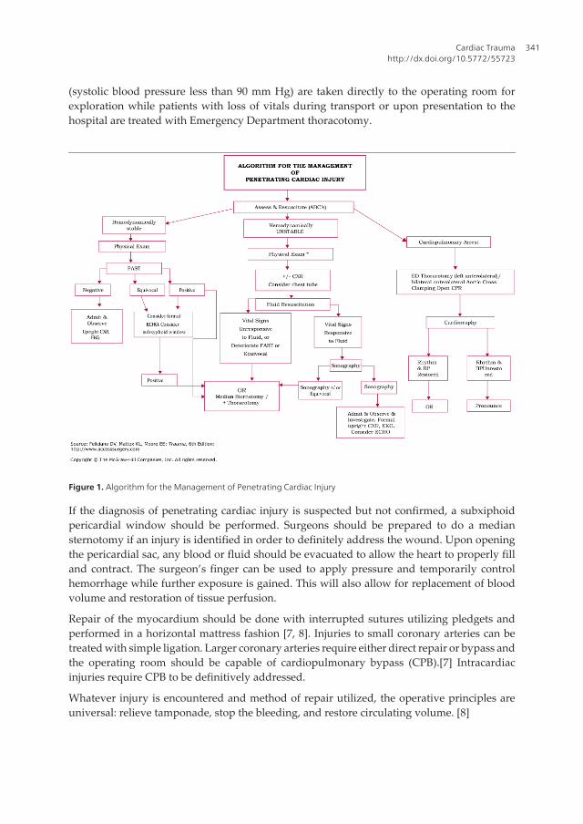
(systolic blood pressure less than 90 mm Hg) are taken directly to the operating room forexploration while patients with loss of vitals during transport or upon presentation to thehospital are treated with Emergency Department thoracotomy.
Figure 1. Algorithm for the Management of Penetrating Cardiac Injury
If the diagnosis of penetrating cardiac injury is suspected but not confirmed, a subxiphoidpericardial window should be performed. Surgeons should be prepared to do a mediansternotomy if an injury is identified in order to definitely address the wound. Upon openingthe pericardial sac, any blood or fluid should be evacuated to allow the heart to properly filland contract. The surgeon’s finger can be used to apply pressure and temporarily controlhemorrhage while further exposure is gained. This will also allow for replacement of bloodvolume and restoration of tissue perfusion.
Repair of the myocardium should be done with interrupted sutures utilizing pledgets andperformed in a horizontal mattress fashion [7, 8]. Injuries to small coronary arteries can betreated with simple ligation. Larger coronary arteries require either direct repair or bypass andthe operating room should be capable of cardiopulmonary bypass (CPB).[7] Intracardiacinjuries require CPB to be definitively addressed.
Whatever injury is encountered and method of repair utilized, the operative principles areuniversal: relieve tamponade, stop the bleeding, and restore circulating volume. [8]
Cardiac Traumahttp://dx.doi.org/10.5772/55723
341

Picture 1. Cardiac Laceration from anterior stab woundPicture
Picture 2. Successful pledgeted repair
Principles and Practice of Cardiothoracic Surgery342

Figure 2. Schematic Depiction of Right Ventricular Repair
3.1. Iatrogenic injuries
Another form of penetrating cardiac injury that has increased in the modern era is iatrogenicinjuries. As the fields of interventional and electrophysiology cardiology continue to increasethe number of percutaneous procedures performed, there is a concomitant increase iniatrogenic injuries to the heart. Pacemaker and ICD placement, ASD occlusion devices,coronary catheterization, pericardiocentesis, and even central line placement can cause cardiactrauma. Usually the treatment is observational, but sometimes intervention is necessary.Fortunately these are rare complications but the incidence of iatrogenic injury has beenreported as high as 6% for certain radiofrequency ablation procedures.[9] Awareness andprompt recognition of an injury are essential to successful treatment.
Cardiac Traumahttp://dx.doi.org/10.5772/55723
343

3.2. Cardiac fistulas
Although hemorrhage and tamponade are the most common injuries seen in penetratingcardiac trauma, cardiac fistulas are another uncommon yet dramatic complication from cardiactrauma (including iatrogenic injuries). Fistulous connections can occur between coronaryarteries, aorta, and directly with the cardiac chambers. Patients, if symptomatic, usuallypresent with congestive heart failure and surgical repair is usually required.[10, 11]. Presen‐tation is variable from acutely after the injury to decades post-injury. Echocardiography andcoronary angiography are the cornerstones of diagnosis and necessary to plan surgical repair.
Figure 3. Pledgets are used to reinforce the suture line
Principles and Practice of Cardiothoracic Surgery344
4. Blunt injury
4.1. Background (mechanism, incidence, and pathophysiology)
Blunt cardiac injury (BCI) is a spectrum of traumatic heart diseases with severity that can rangefrom myocardial contusion and EKG changes to septal rupture and death. Earlier in thecentury, cardiac contusion or concussion were terms used to diagnose cardiac changes fromblunt thoracic trauma. More recently, BCI is the term used to better incorporate and classify
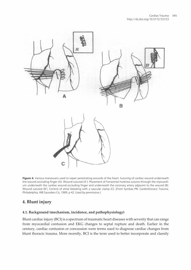
Figure 4. Various maneuvers used to repair penetrating wounds of the heart. Suturing of cardiac wound underneaththe wound-occluding finger (A). Wound sutured (A’). Placement of horizontal mattress sutures through the myocardi‐um underneath the cardiac wound-occluding finger and underneath the coronary artery adjacent to the wound (B).Wound sutured (B’). Control of atrial bleeding with a vascular clamp (C). (From Symbas PN: Cardiothoracic Trauma.Philadelphia, WB Saunders Co, 1989, p 42. Used by permission.)
Cardiac Traumahttp://dx.doi.org/10.5772/55723
345

the myriad of cardiac injuries that result from blunt trauma. BCI is estimated to occur in 20%of motor vehicle collisions and in greater than 75% of thoracic blunt injuries independent ofthe mechanism. The primary mechanism of injury to the heart is from high-speed motor vehiclecollision, but any injury that applies force in the form of kinetic energy to the chest wall andheart can result in a form of BCI. The following mechanisms of injury may result in BCI: directprecordial impact, a crush injury between the sternum and spine, a deceleration injury causinginjury from the fixation points of the aorta and vena cava, a hydraulic effect from an intraab‐dominal injury that sends force to the great vessels and heart, or a crush injury [12].
Figure 5.
Principles and Practice of Cardiothoracic Surgery346

Since blunt cardiac injury is a spectrum of injuries to the heart, a classification scheme wasdeveloped to allow clinicians to categorize the types of injury based on outcomes and treatmentoptions. These categories are as follows: 1) BCI with free wall rupture, 2) BCI with septalrupture, 3) BCI with coronary artery rupture, 4) BCI with cardiac failure, 5) BCI with complexarrhythmias, and 6) BCI with minor ECG or cardiac enzyme abnormalities. The AmericanAssociation for the Surgery of Trauma (AAST) has also published a cardiac injury scale (Table2) that may help to codify injury for diagnosis and research. Injuries sustained with bluntcardiac injury (BCI) include contusion, ruptures, septal defects, valvular injuries, and coronaryartery injuries. Table 3 lists each of these types and the incidence seen from both autopsy andclinical series. Contusion is the most common type of injury with left atrial chamber rupturebeing least common. Injuries can often occur concomitantly; approximately 20% of injurieswith chamber rupture will have another chamber involved. The right heart is the mostcommonly injured as it is closest to the sternum which is impacted anteriorly by the steeringwheel in motor vehicle collisions. Besides having concomitant cardiac injuries, the forceneeded to cause a BCI will often cause associated injuries such as chest pain, rib fractures,pulmonary contusions, and solid organ injuries; the most common associated injuries thatoccur with BCI are listed in Table 5.
4.2. Diagnosis
The best test for diagnosing blunt cardiac injury has been debated for many years. Cardiacenzymes, radionuclide scans, EKG, cardiac ultrasound and continuous monitoring are someof the major methods that have been investigated. Although cardiac enzymes and radionuclidescans have had many supporters these have not shown reliable predictability in diagnosingblunt cardiac injury and have therefore been left out of the Eastern Association for the Surgeryof Trauma (EAST) guidelines (figure 6). Cardiac enzymes, specifically serial troponin meas‐urements are mentioned in the suggested BCI algorithm by Schultz and Trunkey 2004 (figure7) as an adjunct to increase the negative predictive value of the normal EKG when you have apatient who has either a history of cardiac disease or increased age. EKG has emerged as theprimary screening tool for blunt cardiac injury. There are no pathognomonic findings;however, the presence of a new arrhythmia is a sign that workup needs to be escalated. If theEKG is negative in a young hemodynamically stable patient without a history of cardiacdisease there is no further need for workup [12]. If the EKG is abnormal, and the patient hasa history of cardiac disease, increased age or hemodynamic instability then continuoustelemetry monitoring for 24-48 hours is recommended. Those with hemodynamic instabilityrequire continuous monitoring in a surgical ICU. Any arrhythmia may be detected after BCIincluding sinus tachycardia, supraventricular arrhythmias, ventricular arrhythmias, any typeof heart block, ST-T changes or Q waves [13].
Although, these patients are likely to have had a FAST exam in the emergency room, it isimportant to figure out who needs a formal echocardiogram. The key indication is hemody‐namic instability and a possible diagnosis of blunt cardiac injuiry. Anyone meeting thesecriteria requires a formal echocardiogram. There has been debate over whether to use trans‐thoracic or transesophageal echocardiography. The recommendations are that the patient
Cardiac Traumahttp://dx.doi.org/10.5772/55723
347

receive the first available study method. If transthoracic echocardiography is used andadequate imaging cannot be obtained, then a transesophageal echocardiogram should beinitiated immediately.
*Advance one grade for multiple wounds to a single chamber or multiple chamber involvement.
Table 2. Cardiac Injury Scale
4.3. Management
Since blunt cardiac injury describes a spectrum of disease states, the treatment depends on theactual problem. Arrhythmia can be managed medically with the caveat that anticoagulationneeds to be used cautiously in trauma patients. Hemopericardium can be seen with or without
Principles and Practice of Cardiothoracic Surgery348

hypotension or tamponade. If hemopericardium is suspected and the patient is stable asubxiphoid pericardial window can be used to verify the hemopericardium. Once a pericardialwindow is performed, the surgeon must be prepared to proceed with a median sternotomy.If the patient is hypotensive and tamponade is expected then either a subxiphoid pericardialwindow or a thoracotomy can be performed. As a rule free wall rupture is more common inthe atria than the ventricles and more common on the right than the left. This is thought to be
Cardiac Injury Incidence of injury in autopsy series
of patients with BCI
Incidence of injury in clinical series
of patients with BCI
Myocardial contusion 60% to 100% 60% to 100%
Chamber Rupture
Right Ventricle 19% to 32% 17% to 32%
Right Atrium 10% to 15% 8% to 65%
Left Ventricle 5% to 44% 8% to 15%
Left Atrium 1% to 7% 0% to 31%
Atrial Septal Defect 7% Case reports
Valve Injury in BCI 5% Case reports
Ventricular Septal Defect 4% Case reports
Coronary Artery Injury 3% Case reports
Table 3. BCI Patterns of Injury
Associated Injuries Incidence of finding in patients with BCI
Chest Pain 18% to 92%
Rib Fracture 18% to 69%
Aortic or great vessel injury 20% to 40%
Hemothorax 7% to 64%
Pulmonary Contusion 6% to 58%
Pneumothorax 7% to 40%
Flail Chest 4% to 38%
Sternal Fracture 0% to 60%
Traumatic Brain Injury 20% to 73%
Extremity Injury 20% to 66%
Abdominal Solid Organ Injury 5% to 43%
Spinal Injury 10% to 20%
Table 4. Injuries Associated with BCI
Cardiac Traumahttp://dx.doi.org/10.5772/55723
349

due in part to the position of the heart in the chest. The method of repairing the atria is to graspeach side of the atrial wound, place a vascular clamp across the defect, and sew it closed. Themethod of repair of the ventricle is to place a finger of the non-dominant hand over the injuryoccluding the wound and stopping the blood loss. Then pledgeted mattress sutures are placedunder the finger in order to approximate the wound without tearing through the injuredmyocardium. Septal rupture requires the patient to be placed on bypass. Coronary arteryinjury, valve injury and papillary muscle rupture are all very rare. These entities generally
EAST guidelines
A. Level I
1. An admission EKG should be performed on all patients in who there is suspected
BCI.
B. Level II
1. If the admission EKG is abnormal (arrhythmia, ST changes, ischemia, heart block,
unexplained ST), the patient should be admitted for continuous EKG monitoring
for 24 to 48 hours. Conversely, if the admission EKG is normal, the risk of having a
BCI that requires treatment is insignificant, and the pursuit of diagnosis should be
terminated.
2. If the patient is hemodynamically unstable, an imaging study (echocardiogram)
should be obtained. If an optimal transthoracic echocardiogram cannot be
performed, then the patient should have a transesophageal echocardiogram.
3. Nuclear medicine studies add little when compared to echocardiography and, thus,
are not useful if an echocardiogram has been performed.
C. Level III
1. Elderly patients with known cardiac disease, unstable patients, and those with an
abnormal admission EKG can be safely operated on provided they are
appropriately monitored. Consideration should be given to placement of a
pulmonary artery catheter in such cases.
2. The presence of a sternal fracture does not predict the presence of BCI and, thus,
does not necessarily indicate that monitoring should be performed.
3. Neither creatinine phosphokinase with isoenzyme analysis nor measurement of
circulating cardiac troponin T are useful in predicting which patients have or will
have complications related to BCI.
Screening of Blunt Cardiac Injury. Pasquale, N K and Clark, J. s.l. : The Eastern
Association for the Surgry of Trauma, 1998.
Figure 6. EAST guidelines for Blunt Cardiac Injury
Principles and Practice of Cardiothoracic Surgery350

present with clinically significant acute congestive heart failure. Another rare entity ispericardial rupture with cardiac herniation. This requires opening the chest with replacementof the heart in the normal anatomic position and repair of any injured vasculature. Whetheryou utilize a thoracotomy or sternotomy will depend on the details of the cardiac herniation.
Outcomes of emergency department thoracotomy for blunt trauma are universally poor. Thesalvage rate of patients with or without vital signs on arrival to the emergency department is1%-2% [14]. This low survival rate mandates that before an emergency department thoracot‐
Blunt Cardiac Injury. Schultz JM, Trunkey DD. Critical Care Clin. 20 (2004) 57-70[12]
Figure 7. Algorithm for treatment of suspected BCI
Cardiac Traumahttp://dx.doi.org/10.5772/55723
351

omy is undertaken both the mechanism of injury and the length or presence of CPR be takeninto consideration.
Author details
Daniel Eiferman*, R. Nathan Cotterman and Michael Firstenberg
Ohio State University Medical Center, Department of Surgery, Division of Trauma, CriticalCare, and Burn, Columbus, OH, USA
Figure 8. Schematic Representation of right atrial repair
Principles and Practice of Cardiothoracic Surgery352

References
[1] Rozycki, G.S., et al., The role of ultrasound in patients with possible penetrating car‐diac wounds: a prospective multicenter study. J Trauma, 1999. 46(4): p. 543-51; dis‐cussion 551-2.
[2] Thourani, V.H., et al., Penetrating cardiac trauma at an urban trauma center: a 22-year perspective. Am Surg, 1999. 65(9): p. 811-6; discussion 817-8.
[3] Velmahos, G.C., et al., Penetrating trauma to the heart: a relatively innocent injury.Surgery, 1994. 115(6): p. 694-7.
[4] Plummer, D., et al., Emergency department echocardiography improves outcome inpenetrating cardiac injury. Ann Emerg Med, 1992. 21(6): p. 709-12.
[5] Karrel, R., M.A. Shaffer, and J.B. Franaszek, Emergency diagnosis, resuscitation, andtreatment of acute penetrating cardiac trauma. Ann Emerg Med, 1982. 11(9): p.504-17.
[6] Asensio, J.A., et al., Penetrating cardiac injuries: a prospective study of variables pre‐dicting outcomes. J Am Coll Surg, 1998. 186(1): p. 24-34.
[7] Symbas, P.N., Cardiothoracic trauma. Curr Probl Surg, 1991. 28(11): p. 741-97.
[8] Evans, J., et al., Principles for the management of penetrating cardiac wounds. An‐nals of Surgery, 1979. 189(6): p. 777-84.
[9] Kang, N., et al., Penetrating cardiac injury: overcoming the limits set by Nature. In‐jury, 2009. 40(9): p. 919-27.
[10] Hancock Friesen, C., J.G. Howlett, and D.B. Ross, Traumatic coronary artery fistulamanagement. Ann Thorac Surg, 2000. 69(6): p. 1973-82.
[11] Lowe, J.E., et al., The natural history and recommended management of patientswith traumatic coronary artery fistulas. Ann Thorac Surg, 1983. 36(3): p. 295-305.
[12] Schultz, J.M. and D.D. Trunkey, Blunt cardiac injury. Crit Care Clin, 2004. 20(1): p.57-70.
[13] Sutherland, G.R., et al., Anatomic and cardiopulmonary responses to trauma with as‐sociated blunt chest injury. J Trauma, 1981. 21(1): p. 1-12.
[14] Cothren, C.C. and E.E. Moore, Emergency department thoracotomy for the criticallyinjured patient: Objectives, indications, and outcomes. World J Emerg Surg, 2006. 1:p. 4.
Cardiac Traumahttp://dx.doi.org/10.5772/55723
353
