cardiophys - sinoe medical association tmsinoemedicalassociation.org/ap2/cardio/cardiophys.pdf ·...
TRANSCRIPT

1/9/2009
1
Danil Hammoudi.MD

1/9/2009
2
The heart is composed of three major types of cardiac muscle:• atrial muscle, •Ventricular muscle, •specialized excitatory and conductive muscle fibers.
The atrial and ventricular types of muscle contract in much the same way as skeletal muscle, except that the duration of contraction is much longer.
Conversely, the specialized excitatory and conductive fibers contract only feebly because they contain few contractile fibrils; instead, they exhibit either automatic rhythmical electrical discharge in the form of y gaction potentials or conduction of the action potentials through the heart, providing an excitatory system that controls the rhythmical beating of the heart.
HEART(PUMP) R AUTOREGULATION
CARDIOVASCULAR SYSTEM
VESSELS
RE
GU
LAT
ION
NEURAL
HORMONAL
VESSELS(DISTRIBUTION SYSTEM) RENAL-BODY FLUID
CONTROL SYSTEM

1/9/2009
3
Heart HORMONESNatriuretic Peptides
In response to a rise in blood pressure, the heart releases two peptides: •A-type Natriuretic Peptide (ANP) This hormone of 28 amino acids is released from stretched atria (hence the "A"). •B-type Natriuretic Peptide (BNP)
This hormone is released from the ventricles. (It was first discovered in brain tissue; hence the "B".)
Both hormones lower blood pressure by •relaxing arterioles •inhibiting the secretion of renin and aldosterone•inhibiting the reabsorption of sodium ions by the kidneys.
The latter two effects reduce the reabsorption of water by the kidneys So the volume of urineThe latter two effects reduce the reabsorption of water by the kidneys. So the volume of urine increases as does the amount of sodium excreted in it. The net effect of these actions is to reduce blood pressure by reducing the volume of blood in the circulatory system.
These effects give ANP and BNP their name (natrium = sodium; uresis = urinate).

1/9/2009
4
Heart muscle:pacemaker can funciton for many years without interruption
Is stimulated by nerves and is self‐excitable (automaticity)Contracts as a unitHas a long (250 ms) absolute refractory period
Ach (from ParaSym terminals of vagus nerve Xth cranial nerve) = slows HR by increasing K+ conductance & reducing Ca2+ conductance of pacemaker cells
norepinephrine (Sym NS)y pCardiac muscle contraction is similar to skeletal muscle contraction
norepinephrine (Sym NS) accelerates pacemake potential = increasing HR

1/9/2009
5
Autorhythmic cells:Initiate action potentials Initiate action potentials Have unstable resting potentials called pacemaker potentialsUse calcium influx (rather than sodium) for rising phase of the action potential
Action potentials (electrical impulses) in the heart originate in specialized cardiac muscle cells called autorhythmic cells.
These cells are self-excitable, able to generate an action potential without external stimulation by nerve cells.
The autorhythmic cells serve as a pacemaker to initiate the cardiac cycle (pumping cycle of the heart) and provide a conduction system to coordinate the contraction of muscle cells throughout the heart.

1/9/2009
6
The autorhythmic cells are concentrated inthe following areas.
•The sinoatrial (SA) node, located in the upperwall of the right atrium, initiates the cardiac cycleby generating an action potential that spreadsthrough both atria through the gap junctions ofthe cardiac muscle fibers.
Th t i t i l (AV) d l t d•The atrioventricular (AV) node, located nearthe lower region of the interatrial septum,receives the action potential generated by theSA node. A slight delay of the electricaltransmission occurs here, allowing the atria tofully contract before the action potential ispassed on to the ventricles.
•The atrioventricular (AV) bundle (bundle ofHis) receives the action potential from the AVnode and transmits the impulse to the ventriclesby way of the right and left bundle branches.Except for the AV bundle, which provides theonly electrical connection, the atria areelectrically insulated from the ventricles.
•The Purkinje fibers are large-diameter fibersthat conduct the action potential from theinterventricular septum, down to the apex, andthen upward through the ventricles.
Sino‐atrial (SA) nodeAtrio‐ventricular (AV) nodeBundle of HisBundle branchesPurkinje fibersPurkinje fibers

1/9/2009
7
Sequence of excitationi t i l (SA) d d t b th t i1. sinoatrial (SA) node ‐ spreads to both atria
▪ 90 ‐ 100 action potentials per minute
2. atrioventricular (AV) node ▪ 40 ‐50 action potentials per minute
3. atrioventricular (AV) bundle (bundle of His)▪ 20‐40 action potentials per minute
4. right & left bundle branches ▪ in the interventricular septum
5. Purkinje fibers▪ conduction myofibers

1/9/2009
8

1/9/2009
9
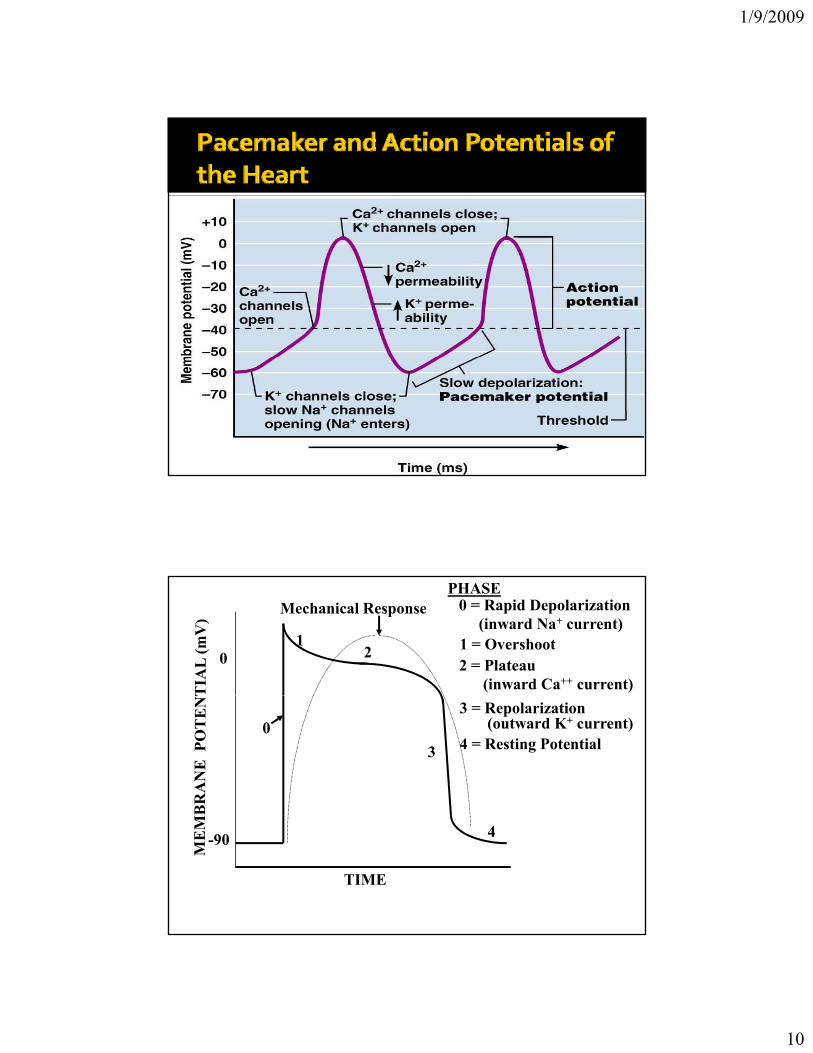
SA node ‐ no stable resting membrane potentialPacemaker potential Pacemaker potential
gradual depolarization from ‐60 mV, slow influx of Na+
Action potential occurs at threshold of ‐40 mVdepolarizing phase to 0 mV▪ fast Ca2+ channels open, (Ca2+ in)
repolarizing phase▪ K+ channels open, (K+ out)▪ at ‐60 mV K+ channels close, pacemaker potential starts over
Each depolarization creates one heartbeatSA node at rest fires at 0.8 sec, about 75 bpm
1/9/2009
10
Figure 18.13
01
2
PHASE0 = Rapid Depolarization
(inward Na+ current) 1 = Overshoot2 = Plateau
(inward Ca++ current)
Mechanical Response
03
3 = Repolarization(outward K+ current)
4 = Resting Potential
-90 4
TIME

1/9/2009
11
Figure 18.12
SINGLE VENTRICULAR ACTION POTENTIAL
ENDOCARDIAL FIBER
EPICARDIAL FIBER
ATRIALFIBER
R
1 mV
EPICARDIAL FIBER
ECG PQ S
TRepolarization of ventricles
Depolarization of ventriclesDepolarization of atria

1/9/2009
12
Sinoatrial (SA) node generates impulses ( ) g pabout 75 times/minute
Atrioventricular (AV) node delays the impulse approximately 0.1 second
Impulse passes from atria to ventricles via the atrioventricular bundle (bundle of His)
AV bundle splits into two pathways in the p p yinterventricular septum (bundle branches)Bundle branches carry the impulse toward the apex of the heartPurkinje fibers carry the impulse to the heart apex and ventricular wallsand ventricular walls

1/9/2009
13
Figure 18.14a

1/9/2009
14
Figure 18.12

1/9/2009
15
SA node generates impulse;atrial excitation begins
Impulse delayedat AV node
Impulse passes toheart apex; ventricular
excitation begins
Ventricular excitationcomplete
excitation begins
SA node AV node Purkinjefibers
Bundlebranches
Figure 18.17
SA node generates impulse;atrial excitation begins
Impulse delayedat AV node
Impulse passes toheart apex; ventricular
excitation begins
Ventricular excitationcomplete
excitation begins
SA node AV node Purkinjefibers
Bundlebranches
Figure 18.17

1/9/2009
16
Heart is stimulated by the sympathetic cardioacceleratory center Heart is inhibited by the parasympathetic p y pcardioinhibitory center
Figure 18.15
Figure 18.19

1/9/2009
17
Heart sounds (lub‐dup) are associated with ( p)closing of heart valvesFirst sound occurs as AV valves close and signifies beginning of systoleSecond sound occurs when SL valves close at the beginning of ventricular diastolebeginning of ventricular diastole
Cardiac cycle refers to all events associated ywith blood flow through the heartSystole – contraction of heart muscleDiastole – relaxation of heart muscle

1/9/2009
18
Ventricular filling –mid‐to‐late diastolegHeart blood pressure is low as blood enters atria and flows into ventriclesAV valves are open, then atrial systole occurs
Ventricular systoleyAtria relax Rising ventricular pressure results in closing of AV valvesIsovolumetric contraction phaseVentricular ejection phase opens semilunar valvesVentricular ejection phase opens semilunar valves

1/9/2009
19
Isovolumetric relaxation – early diastoleyVentricles relaxBackflow of blood in aorta and pulmonary trunk closes semilunar valves
Dicrotic notch – brief rise in aortic pressure caused by backflow of blood rebounding off caused by backflow of blood rebounding off semilunar valves
Phase 1 ‐Atrial ContractionPhase 2 ‐ IsovolumetricContractionPhase 3 ‐ Rapid EjectionPhase 4 ‐ Reduced EjectionPhase 5 ‐ IsovolumetricRelaxationPhase 6 ‐ Rapid FillingPhase 7 ‐ Reduced Filling

1/9/2009
20
Figure 18.20
Pressure and Volume Relationships in the Cardiac Cycle

1/9/2009
21

1/9/2009
22
CO is the amount of blood pumped by each p p yventricle in one minuteCO is the product of heart rate (HR) and stroke volume (SV)HR is the number of heart beats per minuteSV is the amount of blood pumped out by a
i l i h h bventricle with each beatCardiac reserve is the difference between resting and maximal CO
Cardiac Output CO is the amount of blood pumped by each ventricle in one minute CO is the product of heart rate (HR) and stroke volume (SV) HR is the number of heart beats per minute SV is the amount of blood pumped out by a ventricle with each beat
SV = EDV ‐ ESV EDV = amount of blood collected in a ventricle during diastole ESV = amount of blood remaining in a ventricle after contraction
Ejection Fraction (EF) = Stroke Volume / End Diastolic Volume Example of Cardiac Output
CO (ml/min) = HR (75 beats/min) x SV (70 ml/beat) CO CO = ______________________
Factors Affecting Stroke Output Preload ‐ amount ventricles are stretched by contained blood Contractility ‐ cardiac cell contractile force due to factors other than EDV ▪ Increase in contractility comes from:
▪ Increased sympathetic stimuli ▪ Certain hormones ▪ Ca2+ and some drugs
▪ Agents/factors that decrease contractility: ▪ Acidosis ▪ Increased extracellular K+ ▪ Calcium channel blockers
Afterload ‐back pressure exerted by blood in the large arteries leaving the heart Frank‐Starling Law of the Heart ▪ Preload, or degree of stretch, of cardiac muscle cells before they contract is the critical factor controlling stroke volume ▪ Slow heartbeat and exercise increase venous return to the heart, increasing SV

1/9/2009
23

1/9/2009
24
CO (ml/min) = HR (75 beats/min) x SV (70 ml/beat)CO = 5250 ml/min (5.25 L/min)
SV = end diastolic volume (EDV) minus end ( )systolic volume (ESV)EDV = amount of blood collected in a ventricle during diastoleESV = amount of blood remaining in a ventricle after contractionventricle after contraction

1/9/2009
25
Figure 20.19a-d

1/9/2009
26
Preload – amount ventricles are stretched by ycontained blood Contractility – cardiac cell contractile force due to factors other than EDVAfterload – back pressure exerted by blood in the large arteries leaving the heartthe large arteries leaving the heart
Preload, or degree of stretch, of cardiac , g ,muscle cells before they contract is the critical factor controlling stroke volumeSlow heartbeat and exercise increase venous return to the heart, increasing SVBlood loss and extremely rapid heartbeat Blood loss and extremely rapid heartbeat decrease SV

1/9/2009
27
Figure 18.21

1/9/2009
28
Contractility is the increase in contractile ystrength, independent of stretch and EDVIncrease in contractility comes from: Increased sympathetic stimuliCertain hormonesC 2+ d dCa2+ and some drugs
Agents/factors that decrease contractility g / yinclude:AcidosisIncreased extracellular K+
Calcium channel blockers

1/9/2009
29
Extracellular fluid
Adenylate cyclaseCa2+
channel
Ca2+β1-Adrenergicreceptor
Norepinephrine
GTP GDP
Inactive protein kinase A
Active protein kinase A
ATP cAMP
GTP
Ca2+
Cytoplasm
Ca2+
uptakepump
1
3
2
Sympathetic stimulation releases norepinephrine and initiates a cyclic AMP second-messenger
SR Ca2+
channel
Ca2+
bindsto
TroponinEnhancedactin-myosininteraction
Sarcoplasmicreticulum (SR)
Cardiac muscleforce and velocity
system
Figure 18.22

1/9/2009
30
Positive chronotropic factors increase heart prateNegative chronotropic factors decrease heart rate
Sympathetic nervous system (SNS) stimulation is activated by stress anxiety stimulation is activated by stress, anxiety, excitement, or exercise Parasympathetic nervous system (PNS) stimulation is mediated by acetylcholine and opposes the SNS PNS d i t th t i ti l ti PNS dominates the autonomic stimulation, slowing heart rate and causing vagal tone

1/9/2009
31
Atrial (Bainbridge) reflex – a sympathetic ( g ) y preflex initiated by increased blood in the atriaCauses stimulation of the SA nodeStimulates baroreceptors in the atria, causing increased SNS stimulation
The hormones epinephrine and thyroxine increase heart rateIntra‐ and extracellular ion concentrations must be maintained for normal heart function

1/9/2009
32
Figure 18.23
1/9/2009
33
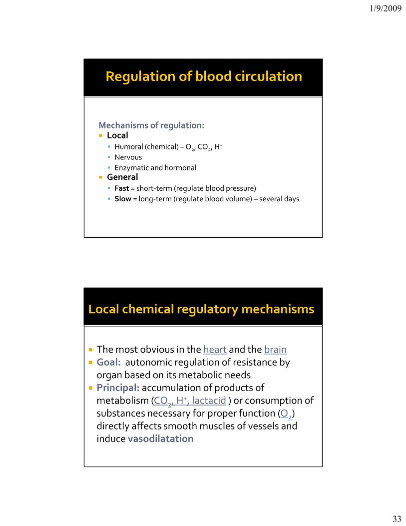
Mechanisms of regulation:Local
Humoral (chemical) –O2, CO2, H+
NervousEnzymatic and hormonal
GeneralGeneralFast = short‐term (regulate blood pressure)Slow = long‐term (regulate blood volume) – several days
Th t b i i th h t d th b iThe most obvious in the heart and the brainGoal: autonomic regulation of resistance by organ based on its metabolic needsPrincipal: accumulation of products of metabolism (CO2, H+, lactacid ) or consumption of substances necessary for proper function (O2) directly affects smooth muscles of vessels and induce vasodilatation

1/9/2009
34
The most obvious in the skin and mucousThe most obvious in the skin and mucousGoal: central regulation of blood distributionPrincipal:Autonomic nervous system
Sympaticus▪ Vasoconstriction – activation of α receptors in vessels‐ noradrenalin(glands, GIT, skin, mucous, kidneys, other inner organs)
▪ Vasodilatation – activation of β receptors in vessels – adrenalin (heart, brain, skeletal muscles)
Parasympaticus ‐Acetylcholin▪ Vasoconstriction – heart▪ Vasodilatation – salivatory glands, GIT, external genitals
Kinin ↑ vasodilatationKinin ↑ = vasodilatationCells of GIT glands contain kallikrein – changes kininogen to kinin → kallidin → bradykinin (vasodilatation)Kinins are any of various structurally related polypeptides, such as bradykinin and kallikrein, that act locally to induce vasodilationand contraction of smooth muscle. A role in inflammation, blood pressure control, coagulation and pain.
Hormones of adrenal medula: adrenalin (vasodilatation), noradrenalin (vasoconstriction)

1/9/2009
35
N t i flNervous autonomic reflexesBaroreflex▪ glomus caroticum, glomus aorticum▪ Afferentation: IX and X spinal nerve▪ Centre: medulla oblongata, nucleus tractus solitarii
ff l f b▪ Efferentation: X spinal nerve, sympatetic fibres▪ Effector: heart (atriums), vessels▪ Effect: After acute increase of blood pressure – activation of receptors – decrease of blood pressure (vasodilatation, decrease of effect of sympaticus)
Receptors in the heartReceptors in the heartReflex of atrial receptors – mechano- and volumoreceptors – activated by increased blood flow through the heart▪ A receptors – sensitive to ↑ of wall tension after systole of
atriums▪ B receptors – sensitive to ↑ of wall tension after systole ofB receptors sensitive to ↑ of wall tension after systole of
ventriclesVentricular receptors – mechano- and chemical receptors -activated in pathological cases▪ Hypoxia of myocardium → decrease of heart rate (Bezold-
Jarisch reflex) → protection of myocardium of larger damage

1/9/2009
36
Humoral mechanismsHumoral mechanismsAdrenalin – β receptors → vasodilatation → ↓ peripheral resistance → blood from skin and GIT to skeletal muscles, heart and brain → ↑ minute heart volumeNoradrenalin – α receptors → vasoconstriction → ↑ blood pressureRenin‐angiotensin – activated by ↓ pressure in vas afferensRenin angiotensin activated by ↓ pressure in vas afferens
Regulatory mechanisms of water and electrolytes exchangesRegulatory mechanisms of water and electrolytes exchangesRegulation of total blood volume by kidneysWhen ↑ blood pressure →↑ of filtration pressure in glomeruli →↑ production of urine →↓ volume of circulating blood →↓ blood pressure
Increase of ADH (vasopressin)↑ADH →↑ of the permeability of collecting ductus for the water →water is reabsorbed →↑ volume of circulating blood →↑ blood pressure
Increase of Aldosterone↑ aldosterone →↑ reabsorbtion Na+ and water →↓ volume of urine →↑volume of circulating blood →↑ blood pressure

1/9/2009
37
F k St li ’ l i iti l l th f th fib i d t i d Frank‐Starling’s law = initial length of the fibers is determined by the degree of diastolic filling of the heart, and the pressure developed in the ventricle is proportionate to the total tension developed.The developed tension increases as the diastolic volume increases until it reaches a maximum, then tends to decrease.
Ionotropic effect of heart rhythm↑ heart frequency→↑ amount of Ca2+ that goes into heart cells →↑Ca2+ available for tubules of sarkoplasmatic reticulum →↑Ca2+ that is freed by each contraction → ↑ strength of contraction

1/9/2009
38
Cardiomotoric centersCardiomotoric centersInhibition – ncl. Ambiguus (beginning of n. vagus in medulla oblongata)Excitation ‐Th1‐3 beginning of sympathetic fibres
Vasomotoric centersIn brain stem (medulla oblongata, Pons Varoli)( g , )In the hypothalamus (controls activity of vasomotoric centers in brain stem)Brain cortex – control both the hypothalamus and the brain stem

1/9/2009
39

1/9/2009
40
♥ ↑ Peripheral resistance ↑mean arterial pressure↑ ↑♥ ↑Cardiac output ↑mean arterial pressure
♥ ↑Stroke volume ↑ pulse pressure♥ ↓Arterial compliance ↑pulse pressure♥ ↑Heart Rate ↓pulse pressure♥ ↑Blood Volume ↑ arterial & venous♥ ↑Blood Volume ↑ arterial & venous

1/9/2009
41
Changes in central arterial pressure d t t d b b tare detected by baroreceptors
(pressure receptors) in the carotid and aortic arteries. These receptors provide information to the cardiovascular centres in the hind brain.
Carotid baroreceptors are located in the carotid sinus at the branch of the carotid artery. Aortic baroreceptors are less sensitive than carotid pressure receptors.
♥ Reduced arterial blood pressure decreased baroreceptor activity.
♥ Decreased parasympathetic tone to the heart.♥ Increased heart rate.♥ Increased cardiac output and blood pressure.
♥ Baroreceptor activity reduced.♥ Increased sympathetic tone to the heart.y p♥ Increased heart rate and contractility, increased stroke
volume.♥ Increased cardiac output and blood pressure.

1/9/2009
42
♥ Reduced arterial blood pressure decreased baroreceptor activityactivity.
♥ Increased sympathetic tone to blood vessels.
♥ Elevated total peripheral resistance and blood pressure.♥ (Coronary and cerebral circulation are largely unaffected.)
♥ Elevated venous tone.Red ced eno s capacitance red ced eno s ol me♥ Reduced venous capacitance, reduced venous volume.
♥ Increased circulating volume, increased venous return.♥ Increased stroke volume, cardiac output and blood pressure.
Three mechanisms have been suggested to explain acute autoregulation.
1) Myogenic mechanisms
2) Tissue pressure
3) local metabolites3) local metabolites

1/9/2009
43
♥ Increased pressure increases arteriolar wall tension.
♥ Vascular smooth muscle contracts when stretched and relaxed when passively shortened.
♥ Action is purely myogenic, no mediators required.
♥ Involves stretch sensitive ion channels on the cell bmembrane.
O V t i t ( t l )(i t b i )♥ O2 Vasoconstrictor (not pulmonary)(import. brain)♥ Glucose vasoconstrictor (at least coronary vessels)
♥ K+ Vasodilator (skeletal muscle)♥ CO2 vasodilator (not pulmonary)(import. brain)♥ adenosine vasodilator (coronary)♥ H+ vasodilator (import brain)♥ H vasodilator (import. brain)♥ PO4
3‐ vasodilator♥ osmolarity vasodilator

1/9/2009
44
Sympathetic activityy p yParasympathetic activityChemical secretionKidney
Noradrenaline (NA) from sympathetic nerves and circulating adrenaline, increase the heart rate and enhances conduction of the AP. Acetylcholine (ACh) released from parasympathetic nerves reduces the heart rate and conduction across the AV node.

1/9/2009
45
At rest heart rate is under both sympathetic andsympathetic and parasympathetic tone.
Normally the parasympathetic inhibition of rate is larger than the sympathetic stimulation.
It regulates the action potential frequency of g p q ythe SA node.Regulates vasoconstriction.Regulates venomotor tone.Stimulate the secretion of epinephrine and reninrenin.

1/9/2009
46
Through the release of Ach, it controls the g ,action potential frequency of the SA node.
Epinephrine secretion regulates Venous p p gPressure, Stroke Volume, and Heart Rate.An increase in either venous pressure, stroke volume or heart rate leads to an increase in blood pressure.

1/9/2009
47
Kidney regulates the secretion of:y gReninAngiotensin IIAldosterone
Renin and Angiotensin II controls Total Peripheral Resistance.Ald l h i Aldosterone controls the urine output.
Changes in extracellular fluid volume alter blood volume.
Altered blood volume changes the end diastolic volume and filling pressure of the heart.
Changes in cardiac pre-load alter stroke volume and cardiac output.
Al d di hAltered cardiac output changes blood pressure.
AND by autoregulation changes total peripheral resistance. Further changing blood pressure.

1/9/2009
48
Urine production is dependent on arterial blood
Increased arterial pressure increases fil i d i
ppressure. filtration and urine
production.
Increased urine production reduces extracellular fluid (ECF) and blood
A renal output curve (ROC) shows the relationship between pressure and urine volume.
(ECF) and blood volume.
♥ ECF volume is continually lost as urine.♥ ECF volume is maintained only if intake is sufficient to balance
loss.♥ Loss of ECF volume is dependent on blood pressure♥ Loss of ECF volume is dependent on blood pressure.♥ Increased blood pressure increases ECF volume loss and blood
pressure falls.♥ Net loss of ECF stops when blood pressure is sufficient for ECF
loss from urine to just balances fluid intake.
♥ Imbalance in osmolarity is controlled by the osmoreceptor tsystem.
♥ Salt load is generaly more important than water as the osmoreceptors regulate water to the salt load.

1/9/2009
49
Drugs trying to cure high blood pressure are g y g g pcurrently available.High blood pressure drugs try to dilate the arteries, so the peripheral resistance would increase and thus the blood pressure would decreasedecrease.
Other types of drugs areyp g1. Diuretics‐which cause the body to excrete
water and salt.2. ACE inhibitors‐ reduce the production of
angiotensin, a chemical that causes arteries to constrictto constrict.

1/9/2009
50
(3) Beta‐Blockers‐ block the effects of adrenaline, thus easing the heart’s pumping action and widening blood vessels.
(4) Vasodilators‐ expand blood vessels.(5) Calcium‐channel blockers‐which help decrease heart contractions.
Sympathetic, Parasympathetic, chemical activities and kidney control blood pressure.Baroreceptors action potential frequency is the input for sympathetic, and parasympathetic activity.The output for sympathetic activity involve (1) venomotor tone (2) vasoconstriction (3) ventricular contraction (4) heart rate

1/9/2009
51
Mean Arterial Pressure
Arterial baroreceptors
Parasympathetic Sympathetic
SA Node VentricularMyocardium
Veins Arteries
Action Potential
Contractility Venomotor tone
Vasoconstriction
Heart Rate Stroke Volume
Total Peripheral Resistance
Feedback
Mean Arterial Pressure