cardiopulmonary health effects: toxicity of semi-volatile ...cardiopulmonary health effects:...
TRANSCRIPT

Cardiopulmonary Health Effects: Toxicity of Semi-Volatile
and Non-Volatile Components of PM
Final Report
Agreement No. 07-307
Michael Kleinman, Principal Investigator Department of Medicine
University of California, Irvine Irvine, CA 92697-1825
12 April 2013
Prepared for the California Air Resources Board and the California Environmental Protection Agency

ii
Disclaimer
The statements and conclusions in this Report are those of the contractor and not
necessarily those of the California Air Resources Board. The mention of commercial
products, their source, or their use in connection with material reported herein is not to be
construed as actual or implied endorsement of such products.
Acknowlegements
This project was funded under the ARB’s Dr. William F. Friedman Health Research
Program. During Dr. Friedman’s tenure on the Board, he played a major role in guiding
ARB’s health research program. His commitment to the citizens of California was
evident through his personal and professional interest in the Board’s health research,
especially in studies related to children’s health. The Board is sincerely grateful for all of
Dr. Friedman’s personal and professional contributions to the State of California. The
research was a joint effort of Air Pollution Health Effects Laboratory at the Department
of Medicine, University of California, Irvine (UCI) and the Department of Civil
Engineering at the University of Southern California (USC). At UCI I want to
acknowledge the technical efforts of Dr. Loyda Mendez, Andrew Keebaugh, Glenn
Gookin, Paul Willett, Michael MacKinnon and Karina Salazar for the exposure, the
chemical and biochemical assays and the data analyses on which this project depended.
At USC I want to thank Co-Principal Investigator Dr. Constantinas Sioutas who was
instrumental in the overall study design, data analysis and for the extraordinary support of
this project by supplying the particle concentrator and thermal denuder used in this study
and the important contributions of Drs. Vishal Verma, Payam Pakbin (USC) and James
Schauer (U. Wisc.). This Report was submitted in fulfillment of ARB Agreement No. 07-
307, “Cardiopulmonary Health Effects: Toxicity of Semi-Volatile and Non-Volatile
Components of PM” under the partial sponsorship of the California Air Resources Board.
Work was completed as of December 2012.

iii
Table of Contents
Cardiopulmonary Health Effects: Toxicity of Semi-Volatile and Non-Volatile
Components of PM .............................................................................................................. i
Disclaimer ................................................................................................................. ii
Acknowlegements ..................................................................................................... ii
Table of Contents ..................................................................................................... iii
List of Figures ........................................................................................................... v
List of Tables .......................................................................................................... vii
List of Inventions ................................................................................................... viii
List of Copyrighted Materials ................................................................................ viii
List of Terms, Abbreviations and Symbols ............................................................. ix
Abstract ........................................................................................................................ xi
Executive Summary ................................................................................................... xiii
Background ............................................................................................................ xiii
Methods.................................................................................................................. xiv
Results ..................................................................................................................... xv
Conclusions ............................................................................................................. xv
Cardiopulmonary Health Effects: Toxicity of Semi-Volatile and Non-Volatile
Components of PM ............................................................................................................. 1
Introduction ................................................................................................................... 1

iv
Methods and Materials .................................................................................................. 3
Experimental Techniques.......................................................................................... 3
Selection and Characteristics of the Animal Model ............................................. 3
Animal Husbandry ................................................................................................ 3
Ambient Particle Concentrator ............................................................................. 4
Exposure Chambers .............................................................................................. 6
Exposures .............................................................................................................. 7
Exposure Procedure .............................................................................................. 9
Bioassay and Data Analysis Methods ................................................................... 9
Vascular and Cardiac Histology ......................................................................... 10
Inflammatory and anti-inflammatory cytokines and markers of vascular
inflammation ............................................................................................................. 10
Electrocardiographic (ECG) telemetry ............................................................... 11
Results ..................................................................................................................... 12
Concentration and Composition of Undenuded and Denuded Ultrafine PM ..... 12
Atherosclerosis Development after Exposure to Denuded and Undenuded
Particles ..................................................................................................................... 19
ECG Assessments ............................................................................................... 21
Biomarkers of Systemic and Vascular Inflammation ......................................... 26
Conclusions ................................................................................................................. 29
v
List of Figures
Figure 1. Exposure System for Comparing the Effects of Denuded and Undenuded
Ambient Particles. ............................................................................................................... 5
Figure 2. Schematic Diagram of VACES and Modified Dekati Thermodenuder for
Comparing Particle Free Organic Vapors and Semivolatiles with Undenuded Ultrafine
Particles. .............................................................................................................................. 6
Figure 3. Drawing of the mobile exposure cage showing the rectangular stainless-
steel pan (50 cm length × 27 cm width × 15 cm height), perforated stainless-steel floor,
partitions, copper aerosol inlet and distribution pipe, and copper aerosol outlet. The
aerosol return line below the floor is not shown. The aerosol inlet is designed to connect
to the outlet of the VACES aerosol concentrator. ............................................................... 7
Figure 4. High Resolution Time-of-Flight Aerosol Mass Spectrometer ...................... 8
Figure 5. Particle Size Dependent Composition of Concentrated Ambient Particles. 18
Figure 6. Progressive Losses of Organic Constituents and Oxidant Capacity with
Increased Denuder Temperature ....................................................................................... 19
Figure 7. Arterial Plaque Development is Accelerated in Arteries from Mice Exposed
to Concentrated UF Particles Compared to Plaque Development in Mice Exposed to
Purified Air or the Non-volatile Particles Remaining After Thermal Denuding. ............. 20
Figure 8. Lipid content of aortic arch samples (Air = Purified Air, Total =
Undenuded PM and NV = Denuded PM. These can be compared with stained histology
sections of the aortic arch and the C1 coronary artery from mice exposed to purified air,
denuded and undenuded CAPs. ........................................................................................ 21
Figure 9. Effects of Undenuded Particles and Particle Free Organic Compounds on
Total Cholesterol and LDL-Cholesterol (Upper and Lower Left n = 13; Upper and Lower
Right n = 8) ....................................................................................................................... 27

vi
Figure 10. Serum Lipid Peroxidation Levels in Mice Exposed to Denuded PM,
Undenuded PM and Particle Free Organics (PFO) ........................................................... 29

vii
List of Tables
Table 1. Chemical Composition of Exposure Atmospheres (Undenuded s. Denuded)
........................................................................................................................................... 14
Table 2. Effects of Denuded and Undenuded PM on Heart Rate Variability (Indexed
as % Change From Baseline) ............................................................................................ 23
Table 3. Effects of Particle Free Organic (PFO) compounds and Undenuded PM on
Heart Rate Variability (Indexed as % Change From Baseline) ........................................ 25
Table 4. Serum Levels of Glutathione and C-Reactive Protein (ng/mL) .................. 28

viii
List of Inventions
None
List of Copyrighted Materials
None

ix
List of Terms, Abbreviations and Symbols
Symbol or
Abbreviation
Explanation
AAALAC Association for the Assessment and Accreditation of
Laboratory Animal Care
APHEL Air Pollution Health Effects Laboratory, University of
California, Irvine
apoE-/- Mice in which the gene regulating apoE is knocked out
AQMD Southern California Air Quality Management District
Arteriosclerosis A chronic disease in which deposits of cholesterol and/or
calcium cause abnormal thickening and hardening of the arterial
walls with resulting loss of elasticity
Atherosclerosis A stage of arteriosclerosis in which fatty deposits
(atheromas) form inside the arterial walls, thus narrowing the
arteries
Atherothrombosis Disruption of an atherosclerotic plaque or lesion resulting in
release of fragments that can block an artery
BAL Bronchoalveolar Lavage
CAPs Concentrated Ambient Particles
CO Carbon Monoxide
CRP C-reactive protein (acute phase protein which is a marker of
systemic inflammation)
CVD Cardiovascular disease
EC Elemental Carbon
ECG Electrocardiogram
GSH Glutathione
HEPA High Efficiency Particle Air filter

x
LDL Low density lipoprotein
NYU New York University
OC Organic Carbon
PFO Particle Free Organics
PM Particulate matter
PM2.5 Particulate matter less than 2.5 μm in aerodynamic diameter
PTFE Fluorocarbon-based filter media
SS Stainless Steel
UFP Ultrafine Particles (dp ≤ 0.18 µm)
VACES Versatile Aerosol Concentration Enrichment System
VCAM Vascular Cell Adhesion Molecule

xi
Abstract
The goal of this project was to determine whether or not the toxicity of ultrafine
(UFP; particles ≤ 0.18 µm aerodynamic diameter) particles depends on the concentration
and composition of semi-volatile and non-volatile fractions of the PM. We tested the
hypothesis that adverse effects of exposure to these UFP, which are primarily emitted by
combustion sources and are highly enriched in semi-volatile components, will be
significantly attenuated by removal of those components from the aerosol. We used a
unique mobile in vivo rodent exposure system in combination with a particle concentrator
and thermal denuder to study the cardiopulmonary effects of UFP, before and after the
removal of the semi-volatile components. The study used genetically modified (apoE-/-)
mice that had impaired lipid metabolism and were therefore predisposed to the
development of atherosclerotic-like plaques. Exposures were 6 hr/day, 4 days per week
for 8 weeks and were conducted near the University of Southern California campus in
central Los Angeles. Detailed chemical and physical characterization examinations of the
concentrated ambient UFP (CAPs) and thermally denuded CAPs were conducted. The
thermal denuder removed more than 60% of the particle-associated organic compounds
(OC) but did not remove the non-volatile components such as elemental carbon (EC) or
trace metals. Exposure to undenuded CAPs accelerated the development of
atherosclerotic plaque in the apoE-/- mice, characterized by decreased arterial lumen
diameters and increased incorporation of lipids in arterial walls. The lumen diameters
and arterial wall lipid contents in apoE-/- mice exposed to thermally denuded CAPs
suggested significantly less plaque development than in the mice exposed to undenuded
CAPs and were not different from plaque levels in apoE-/- exposed to purified air, as
controls. In addition, heart rate variability was decreased in the mice exposed to
undenuded CAPs but not in the mice exposed to either purified air or denuded CAPs. In
a separate experiment apoE-/- mice were exposed to air, undenuded CAPs and the
particle free organic compounds (PFO) that were stripped from the CAPs in the
thermodenuder and delivered to the exposure system. This study demonstrated that the
xii
organic compounds, independent of the presence of particles, played an active role in the
acceleration of plaque development. Cholesterol and low density lipoprotein-cholesterol
(LDL) levels were relatively high in the apoE-/- mice, as would be expected. Exposure
to undenuded CAPs, denuded CAPs and PFO all induced increased levels of both
cholesterol and LDL in the serum of these mice, but only the undenuded CAPs and the
PFO caused significant serum lipid peroxidation, which is a known contributor to plaque
formation. We therefore conclude that the organic constituents of UFP contribute to the
accelerated development of atherosclerotic plaque in arteries, lipid oxidation is an
important mechanism of action in PM-induced coronary artery disease, and that removal
of the organic compounds from PM greatly ameliorates plaque development associated
with air pollutant exposure. These findings suggest that emission control measures that
remove and sequester or destroy organic constituents of combustion generated aerosols
could benefit public health because coronary artery disease is a leading contributor to
heart-related deaths, which represents about 50% of deaths, annually, in California and
other states as well.

xiii
Executive Summary
Background
Heart disease is the leading cause of death in the U.S. Recent data have indicated that
exposure to air pollutants is a risk factor for cardiopulmonary diseases and may represent
an important preventable contributor to both morbidity and mortality among populations
living in polluted environments. The strong and relatively consistent epidemiological
associations between cardiovascular morbidity and mortality may be related to PM-
induced oxidative stress and/or inflammation. Epidemiological and in vivo exposure
studies demonstrate that exposure to particles (fine and ultrafine) in close proximity to
mobile source emissions may be more deleterious to health than are exposures to airborne
particles more distant from these mobile source emissions. Our earlier studies [1, 2] of
mice exposed to fine and ultrafine PM 50 m downwind of a freeway showed that these
exposures had significant biological activity which was associated with elemental and
organic carbon fractions of the aerosol.
Ultrafine particles are capable of inducing the greatest amount of pulmonary
inflammation per unit of PM mass. This has been attributed to the physical and chemical
characteristics of ultrafine particles, including, high particle number, high pulmonary
deposition efficiency, and a surface chemistry involving a high surface area that can carry
adsorbed or condensed toxic air pollutants (oxidant gases, organic compounds and
transition metals). The specific mechanisms by which particulate matter (PM) exposure
disrupts cardiac function and worsens cardiovascular disease (CVD) are not well
understood. There is a growing body of knowledge that suggests that PM exposure can
induce inflammatory changes in blood vessels and exacerbate atherogenesis leading to
the development of atherosclerotic plaques and lesions. We initially hypothesized that
PM exposure would increase free radical production, contribute to the induction of
oxidative stress that could abnormally activate endothelial cells and induce vascular
inflammation. This process could then lead to the accelerated formation of arterial
plaques, which are a hallmark of atherosclerosis. However, preliminary studies [3]

xiv
demonstrated that if the particles were stripped of most of their organic constituents they
also lost a substantial amount of their ability to elicit free radicals and they lost their
oxidant potential. We therefore designed this study to test a new hypothesis that removal
of organic constituents of PM would reduce the particle’s ability to induce or accelerate
atherosclerosis.
Specific Aims: The objective of this 5-year project was to determine how the
atherogenicity of ultrafine particles depends on the concentration and characteristics of
semi-volatile and non-volatile fractions of PM emitted from vehicles and other sources.
We tested the hypothesis that the atherogenicity of near-source PM was due to
nanoparticles that are composed largely of semi-volatile components and that biological
activity will be attenuated by removal of those components from the aerosol. We used
our in vivo rodent exposure system in combination with particle concentrator-thermal
denuder technology [3-6] to separately study the cardiopulmonary effects of PM, before
and after the thermal device was used to denude (i.e. remove the semi-volatile
components from) the particles. Detailed chemical and physical characterizations of
concentrated ambient PM (CAPs) and thermally denuded CAPs were conducted by Dr.
Sioutas and colleagues.
Methods
The study was performed using mice that were genetically susceptible to the
development of atherosclerosis (apoE-/- mice). Groups of 18 mice were each exposed to:
(a) undenuded concentrated ambient ultrafine particles (CAPs; PM0.18); (b) denuded
CAPs; or (c) purified air for 4 days per week, 6 hours per day for 8 weeks. The average
exposure concentration was about 58 μg/m3, over the total of 192 exposure hours. We
examined several factors relevant to mechanisms of atherogenesis and the development
of cardiovascular heart disease. Plasma was assayed for total cholesterol and low density
lipoprotein cholesterol concentrations and C-reactive protein, which is produced in the
liver and is an acute phase protein that increases during systemic inflammation. Assays
were also conducted for protein carbonyl content and lipid peroxidation as markers of

xv
oxidative stress and glutathione (GSH), as a measure of antioxidant capacity. We
measured the effects of exposure on cardiac function in a subset of the mice that were
implanted with cardiographic transponders. Heart rate and heart rate variability (HRV)
were determined and HRV was shown to decrease after exposure to undenuded PM. The
initial study suggested an important role of the organic constituents of PM, notably
significant reductions in atherogenicity and serum lipid peroxidation in animals exposed
to denuded PM as opposed to undenuded PM. We therefore conducted a follow-on study
to directly examine the effects of the organic compounds stripped from the denuded
particles. To accomplish this, the thermal denuder was modified; the activated carbon
adsorber was removed, a pre-fired quartz filter was added and apoE-/- mice were exposed
to the resulting particle-free organic vapor (PFO), purified air and undenuded PM.
Results
The main findings of this study are that: (1) the VACES and the Dekati
Thermodenuder can be used in tandem to deliver undenuded ultrafine ambient PM (UFP,
dp ≤ 0.18 µm), denuded UFP and PFO (consisting of organic compounds stripped from
the PM by the denuder) to genetically modified, apoE-/-, mice in a mobile rodent
exposure system; (2) exposures to undenuded PM or to PFO accelerated the development
of atherosclerotic plaques and induced decreases in heart rate variability (3) the organic
constituents of UFP are important contributors to atherosclerotic plaque development and
significantly accelerate the growth of arterial plaques after an 8 week exposure; (4)
exposure to both organic and inorganic constituents of UFP raise serum concentrations of
cholesterol and low density lipoprotein-cholesterol (LDL), but (5) exposures to denuded
UFP (PM denuded of most organic constituents) did not promote serum lipid
peroxidation while exposures to undenuded UFP or to PFO did promote serum lipid
peroxidation.
Conclusions
This study has demonstrated that the semi-volatile PM fraction of ambient ultrafine
particulate matter could be an important contributor to the development of atherosclerosis

xvi
and heart disease. PM exposure was also shown to increase serum levels of cholesterol
and LDL-cholesterol, both of which are known risk factors for atherosclerosis and heart
disease. In this study, exposure to undenuded UFP and to PFO also promoted the
peroxidation of serum lipids while exposure to denuded UFP did not. Peroxidation of
serum lipids, especially LDL, has been specifically implicated in atherosclerotic plaque
formation. Removal of the semivolatile organics by a process of thermal denuding
significantly mitigated the adverse effects of acceleration of atherosclerosis and reduced
HRV induced by exposures to undenuded PM in a genetically modified animal model of
atherosclerosis. We did not determine if these effects would be observed in unmodified,
or wild type mice.
Chemical characterization of the ambient PM using a high resolution aerosol mass
spectrometer showed that the UFP fraction, particles with diameters ≤ 0.18 µm, was more
enriched in organic compounds that were less oxygenated, hence less polar, than particles
with diameters ≥ 0.18 µm. Studies of mobile source emissions have shown that
emissions from heavy duty diesel vehicles are enriched in UFP and that those UFP are
characterized by low oxygenation levels which are less lipophilic and more nonpolar.
This is important because organics that are nonpolar are better able to cross cell
membranes than are more polar molecules and once inside the cell have the potential for
toxic interactions with critical cell components.
An important corollary to these results is that they suggest that removal of UFP-
associated semivolatile compounds could significantly mitigate adverse cardiovascular
effects. Modern diesel engines are often provided with emission control technologies
that remove effectively the non-volatile fraction, but not necessarily the volatile fraction.
Some research has shown that removal of the non-volatile PM fraction can increase the
concentration of the volatile fraction by enhancing nucleation of condensing organic
vapors. This project has provided findings that improve our understanding of the
mechanism of toxic action of freshly-emitted combustion aerosols and has identified
organic constituents of ambient aerosols as being causally related to potential health
effects. This information will also aid regulators and planners in developing air quality

xvii
mitigation strategies and land use guidance to better protect the health of California
residents.

1
Cardiopulmonary Health Effects: Toxicity of Semi-Volatile
and Non-Volatile Components of PM
Introduction
Heart disease is the leading cause of death in the U.S and is responsible for nearly
50% of mortality for all non-accidental causes. Recent data have indicated that exposure
to air pollutants is a risk factor for cardiovascular disease and may represent an important
preventable contributor to heart-related morbidity and mortality among populations living
in polluted environments [7-9]. There are strong and relatively consistent mechanistic
associations between cardiovascular morbidity or mortality and oxidative stress
associated with inflammation [7, 10]. Ultrafine particles are capable of inducing
pulmonary inflammation due to their physical and chemical characteristics[11],
including, high particle number, high pulmonary deposition efficiency, and a surface
chemistry involving a high surface area that can carry adsorbed or condensed toxic air
pollutants (oxidant gases, organic compounds and transition metals). These PM
components have been identified as having pro-inflammatory effects[12]. A seminal
series of studies performed by Chen, Lippmann and colleagues at New York University
(NYU) demonstrated that exposure to concentrated ambient fine (PM2.5) particles in
Tuxedo NY could exacerbate the development of atherosclerotic lesions in mice that
were genetically predisposed to abnormal lipid metabolism [13, 14]. Studies conducted
in Los Angeles demonstrated that UFP were more potent than fine (PM2.5) particles with
respect to accelerating development of atherosclerotic plaques in apoE-/- mice [15, 16].
The specific mechanisms by which particulate matter (PM) exposure disrupts cardiac
function and worsens cardiovascular disease (CVD) are still under investigation, however
there is a growing body of knowledge that suggests that PM exposure can oxidize
circulating lipoproteins and induce inflammatory changes in blood vessels leading to the
development of atherosclerotic plaques and lesions [17]. Epidemiological and in vivo

2
exposure studies demonstrate that particles (fine and ultrafine) in close proximity to
mobile source emissions can affect cardiopulmonary health to a greater degree than
particles in the air more distant from the source [2, 18, 19]. Our earlier studies of mice
exposed to fine and ultrafine PM 50 m downwind of a freeway showed that these
exposures had significant biological activity which was associated with elemental and
organic carbon fractions of the aerosol. When exposures were performed 150 m
downwind of the freeway the biological activity was greatly diminished and there were
no measurable exposure-related effects [1, 2]. Careful measurements in a roadway tunnel
[20] and near a major freeway with heavy-duty diesel traffic [21-23] demonstrated that
there were rapid shifts in aerosol size and composition within minutes of emission and
that the volatility of the PM increases with decreasing particle size. We initially
hypothesized that PM exposure would increase free radical production, contribute to the
induction of oxidative stress thus abnormally activating endothelial cells thereby inducing
vascular inflammation leading to the accelerated formation of arterial plaques which are a
hallmark of atherosclerosis. Sioutas and colleagues developed the capacity to couple a
thermal denuder to a Versatile Aerosol Concentration Enrichment System (VACES) [3].
The thermal denuder heats the aerosol to a specified temperature to evaporate and remove
semi-volatile components, and then returns the aerosol to the original temperature. The
VACES can increase the concentration of the processed aerosol by factors of 20 to 30 to
provide adequate concentrations for performing acute in vivo toxicology exposure
studies. Preliminary studies demonstrated that if the particles were stripped of most of
their organic constituents they also lost a substantial amount of their ability to elicit free
radicals and they lost their oxidant potential [3]. Using this VACES/denuder system,
combined with our mobile rodent field exposure/cardiac monitoring unit, we were able to
systematically examine the role of the semivolatile components of PM on heart function,
plaque development and cardiac physiology. We therefore designed this study to test the
hypothesis that removal of organic constituents of PM would reduce the particle’s ability
to induce or accelerate atherosclerosis. This research examined the link between particle-
induced inflammation and the development of atherosclerosis in atherosclerosis-prone
mice and used a thermal denuder coupled with a particle concentrator and mobile
3
exposure system to specifically address the question “Do semivolatile organic
constituents of UFP play an important role in the PM-induced acceleration of
atherosclerotic plaque development?” In addition, we examined oxidative stress and
inflammation-associated biomarkers to determine the relative importance of these modes
of action in the development or exacerbation of cardiovascular disease. Improved
understanding of the roles of these specific modes of action could lead to improved
techniques for preventing or treating heart diseases caused by environmental
contaminants.
Methods and Materials
Experimental Techniques
Selection and Characteristics of the Animal Model
The transgenic mouse model of cardiovascular disease that we proposed to use in this
study was developed from the C57BL/6 mouse. The C57BL/6 strain lacking the
apolipoprotein E receptor (apoE-/-) has been shown to be particularly susceptible to
cardiovascular effect but is also subject to adverse pulmonary effects from a variety of
inhaled substances including O3 [24], acid-coated carbon particles [25], ovalbumin [26]
and concentrated pseudo-ultrafine particulate matter (PM0.18) [15].
Animal Husbandry
Male apoE-/- 6 week old mice were purchased from a commercial supplier and
housed two to a cage in AAALAC accredited animal housing facility at the Air Pollution
Health Effects Laboratory (APHEL). All animals used in this study were apoE-/- mice.
The mice that were implanted with telemetry devices were housed singly so that ECG
parameters could be monitored while the mice were in the vivarium. Animals were
provided with food and water ad libitum. Animals were transported to the exposure site
[2] near the campus of USC in Los Angeles using a van and customized

4
transport/exposure modules. During transport the animals breathed filtered, purified air.
Telemeter-equipped mice were monitored during exposures and while they were in the
vivarium, but were not monitored during transport. On the average, mice were monitored
about 20 hr per day. When ECG data were evaluated the data from the same time periods
were evaluated on exposure and non-exposure days so the unmonitored transport time did
not impact our analyses.
Ambient Particle Concentrator
Ambient particles with particle diameters smaller than 0.18 μm were concentrated
using the Versatile Aerosol Concentration Enrichment System (VACES) which has been
described in detail by Kim et al. [4, 5]. VACES consists of a size selective inlet, a
saturator/chiller module that supersaturates the aerosol with water vapor causing fine and
ultrafine particles to grow to a size that can be inertially separated using a virtual
impactor, and a diffusion drier module that removes the excess water vapor and returns
the aerosol to a size distribution that is very close to that in the unconcentrated ambient
air. The system is mobile and capable of enriching the concentration of particles in the
range of 0.03- 2.0 m by up to a factor of 30 x ambient, depending on the output flow

5
rate [27]. The concentration efficiency falls off above 2.0 μm or below 0.03 μm.
Figure 1. Exposure System for Comparing the Effects of Denuded and
Undenuded Ambient Particles.
To determine the role of semivolatile components of ultrafine particles on
cardiovascular disease, the VACES was coupled to a Dekati thermodenuder which is
designed to remove volatile and semivolatile organic compounds from sample ambient
particles [3]. This allowed us to compare the effects of denuded and undenuded particles
on cardiac physiology and the development of atherosclerotic plaque. In a subsequent
exposure, we modified the thermodenuder, by removing the activated carbon annular
denuder and replacing it with a pre-fired quartz filter, to allow mice to be exposed to the
vapor and semivolatile organic components in the absence of particles (PFO) and to
allow us to compare the effects of undenuded particles on mice to effects of mice
exposed to PFO.
Ambient Air
Ultrafine
Impactor(Dp 50 =180 nm
Heater
Thermodenuder
CPC
Electrostatic
Classifier
CPC
Tef lon f ilter
Adsorption /Cooling
Pump
Quartz f ilter
Saturation Tank
Virtual Impactor
Diffusion Dryer
315 lpm
300 lpm
5 lpm
15 lpm
Heater
Pump
Tef lon f ilter
Quartz f ilter
Electrostatic
Classifier
10 lpm
5 lpm
4.7 lpm
4.7 lpm
2.5 lpm
2.5 lpm
VACES
Condenser
0.3 lpm

6
Figure 2. Schematic Diagram of VACES and Modified Dekati Thermodenuder
for Comparing Particle Free Organic Vapors and Semivolatiles with Undenuded
Ultrafine Particles.
Exposure Chambers
A whole-body exposure mouse chamber (Figure 3) was designed specifically for use
with the VACES. Each stainless steel (SS) chamber (20 inches X 12 inches X 6 inches)
was segmented into 18 cubicles (1 mouse per cubicle) separated by perforated SS sheets
(0.078" hole diameter, 36% open and staggered, (McMaster-Carr, New Brunswick, NJ).
Concentrated ambient particles (CAPs) were delivered through SS particle delivery tubes
that distributed CAPs uniformly throughout the exposure chamber [28]. A raised sub-
floor constructed from perforated SS sheet (0.25” hole diameter, 50% open, staggered)
was used, which permitted urine and feces to fall to the bottom of the vat and kept the
mice relatively clean. The exposure atmosphere was exhausted from below the sub-floor
through 2 SS tubes, each 40 cm in length with 28 0.5 mm downward-facing holes.
Absorbent sheets impregnated with an antibiotic to prevent fecal bacteria from generating
Pump
Water
Chiller
(-4˚C)
Virtual
impactor
Temperature
controller
Saturating bath
(~30˚C)
Condenser
Heater (120 C)
Temperature
controller
Chiller
(10˚C)
To exposure
chambers
1.5 LPM each X2
Teflon+
Vapor sampler
(4LPM)
T = 25˚C
VACES
Pump
7.5 L.min-1 7.5 L.min-1
To CPC for
concentration
check (0.3 LPM)
Air flow
Filter Holder
Quartz filter
Teflon+
Vapor sampler
(4LPM)
To exposure
chambers
1.5 LPM each X2 Quartz filter
holder
37 mm Quartz
filter 0.5 LPM

7
ammonia from urine were placed under the exhaust lines to absorb urine and to collect
feces.
Figure 3. Drawing of the mobile exposure cage showing the rectangular
stainless-steel pan (50 cm length × 27 cm width × 15 cm height), perforated
stainless-steel floor, partitions, copper aerosol inlet and distribution pipe, and
copper aerosol outlet. The aerosol return line below the floor is not shown. The
aerosol inlet is designed to connect to the outlet of the VACES aerosol concentrator.
Exposures
During exposures, we monitored concentrations of ambient and CAPs particles.
Samples were also collected on quartz filters that were pre-treated at 400°C to remove
adsorbed organic compounds. These filters were composited on a weekly basis and
analyzed for elemental carbon (EC) and organic carbon concentrations (OC). EC is a
reasonable tracer for particles originating from diesels [29] and represents between 5 -
20% of the UFP in ambient samples. EC and OC were measured by a thermal
photometric method on a fraction of the filter; the remaining fractions were stored (-80º
C) and subsequently analyzed for organic and inorganic constituents. Particles were also
collected for mass concentration and chemical analyses on pre-weighed fluorocarbon
filters. Following collection, the filters were equilibrated overnight at constant humidity
and weighed. These filters were submitted for ICP/MS analysis of elemental
constituents, including Fe, V, Zn, Cr, Ni, Cu, Pb and Mn, among many others (the entire
8
list is seen in Table 1). These specific metals were identified because previous inhalation
or in vitro studies have shown them to be potentially toxic [30-36]. We plan to examine
possible links between these metals and health-related outcomes in the future. Some of
the other metal and non-metal constituents that were measured may also be evaluated
(e.g. As, Se).
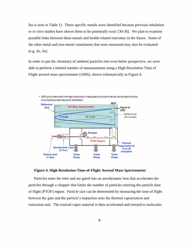
In order to put the chemistry of ambient particles into even better perspective, we were
able to perform a limited number of measurements using a High Resolution Time of
Flight aerosol mass spectrometer (AMS), shown schematically in Figure 4.
Figure 4. High Resolution Time-of-Flight Aerosol Mass Spectrometer
Particles enter the inlet and are gated into an aerodynamic lens that accelerates the
particles through a chopper that limits the number of particles entering the particle time
of flight (PTOF) region. Particle size can be determined by measuring the time of flight
between the gate and the particle’s impaction onto the thermal vaporization and
ionization unit. The ionized vapor material is then accelerated and ionized to molecules

9
in the TOF Mass Spectrometer and mass to charge ratios are compared with an internal
library to provide molecular identities.
Exposure Procedure
Animals were housed at UC Irvine (UCI), transported daily to Los Angeles while
breathing purified air and exposed. At UCI the mice were housed in ventilated caging
attached to an air purification system. The air purifier delivered filtered air at flows
adequate to provide 15-20 air exchanges per hour in each ventilated cage unit. The
vivarium was supplied with Class 100 filtered air using a laminar flow air purifier that
consisted of a 1000 CFM blower, an oxidizing adsorbent canister containing
permanganate-impregnated alumina spheres, and a high efficiency particle air (HEPA)
filter system. Between exposures the animals were supplied with purified air, clean water
and food, ad lib. During the first set of exposures, groups of 18 animals were placed into
sealed, compartmentalized exposure chambers and were each exposed to undenuded
concentrated ultrafine particles (CAPs) or to CAPs from which semivolatile components
were removed using the Dekati thermal denuder (denuded CAPs) for 6 hours per day, 4
days per week for 8 weeks. During the second set of exposures, groups of 18 animals
were each exposed to particle free vapor removed from the CAPs using the Dekati
thermal denuder or to undenuded concentrated ultrafine particles for 6 hours per day, 4
days per week for 8 weeks. Control animals in each set of exposures received purified air
under conditions identical to those of the animals exposed to CAPs. Temperature was
monitored continuously during the exposures and held to 75 ± 5 °C. Animals were
observed throughout the experiment for signs of distress (e.g. changes in grooming, food
and water uptake, shaking). The Versatile Aerosol Concentrator for Exposure Studies
(VACES) designed by Sioutas and colleagues at USC [4, 5, 27, 37, 38] was used for the
PM exposures. This device has been used by UCI for exposures of mice near freeways in
Los Angeles [39]. The animals were euthanized 24 hr after the last exposure on the
eighth week of the study.
Bioassay and Data Analysis Methods

10
Blood: serum samples were collected from each animal from the descending aorta for
cytokine and circulating biomarker levels.
Heart: A sample of the heart was collected and frozen for subsequent gene expression
analyses. The remaining tissue was fixed for subsequent morphological study.
Arteries: arterial tissue was coronary arteries and the aorta for histological evaluations of
plaque and biochemical assays for oxidative stress and plaque lipid analyses.
At sacrifice, the mice were euthanized with an overdose of pentobarbital and the heart
and aorta perfused with 4% paraformaldehyde. The heart and thoracic and abdominal
aorta were removed en bloc. Sections of the aorta were obtained from the proximal,
central and distal areas, snap frozen and stored in liquid nitrogen. These samples were
homogenized and used for assays of gene expression, and for analyses of biomarkers of
vascular inflammation and oxidative stress. The remaining proximal and distal aorta
segments were cut longitudinally. One section was frozen in O.C.T. embedding medium
(Sakura Finetek, Torrence, CA) and reserved for subsequent laser capture
microdissection and genomic and proteomic analysis. The remaining aorta sections were
fixed in buffered formaldehyde and examined for total atherosclerotic lesion areas, lipid
contents, and cellularity [40]. A section of the heart was removed, snap-frozen in liquid
nitrogen and reserved for gene expression analysis. The remaining tissues were fixed in
4% paraformaldehyde. The fixed cardiac tissue was embedded in paraffin, sectioned and
stained with hematoxylin and eosin for subsequent histological analysis.
Vascular and Cardiac Histology
All morphological assessments were done blind (i.e. without knowledge of the
treatment group). Fixed tissues were embedded in paraffin and sectioned at 5 µm.
Samples were stained (H&E) and assessed using an optical microscope.
Inflammatory and anti-inflammatory cytokines and markers of vascular
inflammation

11
Sera were analyzed for inflammatory and oxidative stress cytokines and acute phase
proteins using a state of the art multiplexed bead assay system (Luminex) as well as
adaptations of more conventional enzyme linked immunosorbant assay systems (ELISA).
The Luminex system uses fluorescently tagged beads to which antibodies to specific
proteins are attached. After incubation with the sample, the beads are incubated with
detection antibodies and then analyzed using a fluorescent flow sorting system (similar in
working principle to flow cytometry) and the data are recorded and quantified using an
online computer. Assays also included arterial disease biomarkers (total cholesterol, low
density lipoprotein cholesterol (LDL) and C-reactive protein (CRP)) and arterial wall
oxidative stress indicators (protein carbonyl content (PCC), glutathione (GSH), and lipid
peroxidation). Total glutathione (GSH) was measured as an indicator of antioxidant
capacity using an enzymatic recycling method [41]. Malondialdehyde (MDA) was
measured as an indicator of lipid peroxidation using a colorimetric assay [42, 43].
Protein carbonyl content was measured with a fluorometric assay [44] as an indicator of
protein oxidation.
Electrocardiographic (ECG) telemetry
The protocol for surgical implantation of telemetry devices (PhysioTel Telemetry
system, Data Sciences International, St. Paul, MN) to measure biopotential (ECG
tracings), temperature, and physical activity in mice has been previously described [45,
46]. Aseptic techniques were used throughout the implantation procedure.
ECG Data Analysis: The DataQuest A.R.T. system was used to detect, collect and
analyze biopotential, body core temperature and activity telemetry signals from each
animal. The acquisition program interfaced with a receiver that was tuned to each
animal’s implanted ECG telemetry device. At the start of our field study, data was
sampled each day of exposure for 15 min. before, for 5 min every 30 min during the 6
hour exposure period and 15 minutes post-exposure. As the project progressed, we were
able to expand the monitoring to include monitoring overnight while the animals were
housed in the vivarium. The acquisition program automatically cycled through the

12
animals, and acquired data for 5 min out of every 30 min in groups of 4 mice at a time.
ECG waveforms were stored on a dedicated computer for subsequent analysis and
analyzed to determine heart rate variability (HRV). Changes in HRV may represent
alterations in autonomic control of cardiac function [47]. Reduced HRV in humans can
represent an adverse effect. Analysis of the ECG waveform was used to extract measures
of HRV (the magnitude of variance explained (power) in the heart’s rhythm across
different frequency bands (spectra) of periodic oscillations in heart rate). Portions of
these spectra reflect different autonomic influences on heart rate and blood pressure (BP)
[48]. The high frequency (HF) band (1.5 – 5.0 Hz) of the heart period power spectrum
has been used to estimate cardiac vagal control [49]. HRV in this band is linked to
respiratory influences and has been referred to as “respiratory sinus arrhythmia” [50].
Heart period oscillations at lower frequencies (LF, 0.1 to 1.5 Hz) are less well
understood. They may represent mixed sympathetic-parasympathetic and
thermoregulatory influences [51-53].
Results
Concentration and Composition of Undenuded and Denuded Ultrafine PM
The results of compositional analyses of the denuded and undenuded particles are
detailed in Table 1. The total undenuded particle concentration was 58 µg/m3, however
about 49% of the mass was lost after thermally denuding the aerosol at 120°C, i.e. the
denuded particle mass concentration was 28.6 µg/m3. Organic carbon represented about
44% of the total mass of the undenuded particles (25.4 µg/m3) and approximately 35% of
the organic carbon mass was lost during the denuding process. The residual organic
carbon however still represented about 31% of the mass of the denuded particles. These
data are consistent with our results which showed that the mass of organic compounds
stripped from the particles during the denuder process was increased as the temperature
of the denuder was increased. Why did we select 120°C for the denuder temperature for
this study? This was done because at that temperature we were able to demonstrate that
there was no nucleation of new particles downstream of the heated zone of the denuder.
13
The concentrations of chemical components in the particulate phase, before and after
denuding, were contrasted using a 2-sample t-test. As expected, elemental carbon was
conserved during the denuder process, i.e., EC concentrations before and after denuding
were not significantly different (p ≥ 0.05). Water soluble organic carbon was
significantly reduced after denuding (p ≤ 0.01). Ions (Cl-, SO4
= and NH4
+) were
conserved during the denuding process, however there were significant (40%; p ≤ 0.05)
losses of NO3-, which was not unexpected. The particles contained small amounts of
polycyclic aromatic hydrocarbons (PAHs). While concentrations of phenanthrene,
anthracene and fluoranthene were not significantly changed by the denuding process (p ≥
0.05), pyrene was reduced by 29% (p ≤ 0.01) and 87% or more of the benzo-addition
compounds of fluoranthene, pyrene and anthracene were lost (p ≤ 0.01). There were
small, mostly non-significant (p ≥ 0.05) changes in the concentrations of alkali metals
(Li, Na and K) after denuding. Concentrations of some of the common metals (Al,
Cr,Fe, Co, Cu, Sn, and Pb) were slightly, but significantly reduced, but on the average,
55% was retained in the particle phase after denuding.

14
Table 1. Chemical Composition of Exposure Atmospheres (Undenuded s. Denuded)
Particle Composition
Total UFP (Undenuded) Thermal denuded (Denuded)
Ratio of Denuded/Undenuded
Concentration
Mean Uncertainty
S.E. Concentration
Mean Uncertainty
S.E.
Tota
l U
FP,
µg/
m3
58.20
28.65
49%
ECO
C,
µg/
m3 OC 25.382 1.522 8.826 0.716
35% **1
EC 1.472 0.371 2.008 0.392
136% ns
TC 26.855 1.724 10.834 0.953
40% **
WSO
C
µg/
m3
5.701 0.219 2.322 0.142
41% **
IC, µ
g/m
3
Chloride 0.027 0.039 0.100 0.041
368% ns
Nitrate 0.359 0.049 0.214 0.037
60% *
Sulfate 1.959 0.206 2.401 0.250
123% ns
Ammonium 1.372 0.145 1.171 0.126
85% ns
Potassium µg/m3
Sodium µg/m3
Phosphate µg/m3 0.119 0.086
PA H,
ng
/m 3 Naphthalene
1 ns = non significant (p ≥ 0.05); * p ≤ 0.05; ** p ≤ 0.01

15
Acenaphthylene
Fluorene
Phenanthrene 0.250 0.071 0.216 0.064
86% ns
Anthracene 0.023 0.020 0.075 0.026
320% ns
Fluoranthene 0.142 0.035 0.108 0.030
76% ns
Pyrene 0.326 0.071 0.095 0.029
29% **
Methylfluoranthene 0.018 0.018
9-Methylanthracene 0.018 0.018
Benzo(ghi)fluoranthene 0.018 0.018
Cyclopenta(cd)pyrene 0.018 0.018
Benzo(a)anthracene 0.018 0.018
Chrysene 0.018 0.018
1-Methyl chrysene 0.018 0.018
Retene 0.018 0.018
Benzo (b)fluoranthene 0.912 0.183 0.028 0.019
3% **
Benzo (k)fluoranthene
Benzo(e)pyrene 0.765 0.154 0.018
0% **
Benzo(a)pyrene 0.788 0.159 0.018
0% **
Perylene 0.018 0.018
Indeno(123-cd)pyrene 0.096 0.027 0.018
0% ns
Benzo(ghi)perylene 0.216 0.047 0.028 0.019
13% **
Dibenzo(ah)anthracene 0.023 0.019 0.018
0% ns
Picene 0.018 0.018
Coronene 0.204 0.060 0.022 0.031
11% **
Dibenzo(ae)Pyrene
M eta
ls
An d
Tra
ce
Ele
me
nts , ng
/m 3 Li7 0.162 0.020 0.289 0.045
179% *

16
B11 3.681 0.096 2.137 0.094
58% **
Na23 55.537 6.811 82.356 10.017
148% ns
Mg25 20.678 2.057 14.998 1.904
73% ns
Al27 256.878 17.390 106.785 9.682
42% **
P31 12.968 1.024 9.416 1.354
73% ns
S34 890.648 80.678 298.446 54.494
34% **
K39 35.409 7.619 54.840 11.405
155% ns
Ca44 39.716 4.899 85.710 14.913
216% *
Sc45 0.009 0.005 0.011 0.005
125% ns
Ti49 22.059 1.607 13.202 1.814
60% **
V51 5.009 0.302 16.306 1.323
326% **
Cr52 6.315 0.336 4.299 0.342
68% **
Mn55 6.716 0.295 6.864 0.463
102% ns
Fe57 320.185 14.725 175.638 14.212
55% **
Co59 2.902 0.127 1.985 0.184
68% **
Ni60 6.528 0.517 9.276 1.079
142% ns
Cu63 17.661 0.695 11.119 0.809
63% **
Zn66 16.620 1.413 24.472 2.867
147% ns
As75 6.258 0.594 6.541 0.765
105% ns
Se82 0.795 0.185 0.356 0.213
45% ns
Rb85 0.119 0.012 0.180 0.035
151% ns
Sr88 0.632 0.052 0.825 0.085
131% ns
Y89 0.064 0.005 0.023 0.004
35% **
Nb93 0.043 0.005 0.033 0.008
76% ns
Mo95 3.304 0.255 2.643 0.448
80% ns
Rh103 0.001 0.001 0.001 0.001
90% ns

17
Pd105 0.017 0.008 0.013 0.010
78% ns
Ag109 10.785 2.531 3.665 1.219
34% ns
Cd111 0.094 0.011 0.135 0.028
144% ns
Sn118 20.364 1.178 10.916 1.440
54% **
Sb121 1.460 0.113 1.381 0.175
95% ns
Cs133 0.007 0.002 0.010 0.004
150% ns
Ba138 7.668 0.399 7.866 0.611
103% ns
La139 0.402 0.023 0.252 0.018
63% **
Ce140 0.392 0.017 0.242 0.020
62% **
Pr141 0.019 0.001 0.010 0.002
52% **
Nd143 0.059 0.005 0.028 0.004
46% **
Sm147 0.013 0.003 0.008 0.001
61% ns
Eu151 0.006 0.001 0.007 0.001
127% ns
Dy163 0.006 0.001 0.003 0.001
54% ns
Ho165 0.001 0.001 0.001 0.000
88% ns
Yb173 0.003 0.001 0.001 0.001
41% ns
Lu175 0.001 0.000 0.001 0.000
96% ns
W184 0.209 0.009 0.200 0.009
95% ns
Pt195 0.007 0.002 0.004 0.002
59% ns
Tl205 0.005 0.001 0.008 0.001
173% ns
Pb 7.264 0.401 4.856 0.384
67% **
Th232 0.016 0.003 0.010 0.002
62% ns
U238 0.010 0.001 0.006 0.001
59% *

18
High Resolution Aerosol Mass Spectrometer Results
Some results are shown in Figure 5. While these data were obtained for concentrated PM in
Irvine, CA rather than Los Angeles, CA the observed results are quite consistent with the
characterizations conducted on the LA aerosol [3].
Figure 5. Particle Size Dependent Composition of Concentrated Ambient Particles.
The particles in the “accumulation” mode (≥ 0.18 µm diameter) are comprised of organic
compounds, sulfates, nitrates and ammonium ions. The particles smaller than 0.18 µm diameter
(the particle size range used in our comparison of denuded vs. undenuded particles) are more
highly concentrated in organic compounds. Another interesting contrast is that the organic
constituents of the accumulation mode particles are nearly 10 times more oxygenated (based on
the mass/charge ratios for marker oxygenated and non-oxygenated ions) than are the organics
from the ultrafine particles. This is consistent with studies of emissions from diesel engines
which are reported to be major sources of ambient ultrafine particles.

19
As shown in Table 1, the denuder process significantly alters particle mass and composition.
These changes are more apparent when one changes the denuder temperature. In Figure 6
changes that are observed when particles are denuded at 50°C, 100°C and 200°C, both in
composition and oxidation potential, as measured using the dithiothreitol (DTT) assay method.
The data are adapted from Verma et al. [54]
Figure 6. Progressive Losses of Organic Constituents and Oxidant Capacity with
Increased Denuder Temperature
Atherosclerosis Development after Exposure to Denuded and Undenuded
Particles
Figure 7 summarizes the total plaque areas in coronary arteries after exposure to air, undenuded
concentrated ultrafine particles (total UF) and the non-volatile particles from denuded UF
(NVUF). The upper panels show the average wall thickness of the aortic arch and A1 measured
from histological cross-sections of each. The middle and lower panels show aortic plaque areas
and lipid content also measured from histological sections with lipids stained by Oil Red-O.
Aortic arch wall thickness was significantly greater in mice exposed to undenuded PM
compared to air exposed mice. There was no difference at the p ≤ 0.05 level in aortic arch wall
thickness between mice exposed to denuded PM and air. A1 wall thickness was not significantly
different between the three exposure groups.

20
Plaque development, by plaque area and lipid content measures, was accelerated in the
mice exposed to undenuded PM compared to either that in mice exposed to air or denuded PM.
The difference was significant for the difference between denuded and undenuded PM
exposures, but the differences between air and undenuded PM exposures were not significant at
the p ≤ 0.05 level.
Figure 7. Arterial Plaque Development is Accelerated in Arteries from Mice Exposed
to Concentrated UF Particles Compared to Plaque Development in Mice Exposed to
Purified Air or the Non-volatile Particles Remaining After Thermal Denuding.
The panel on the left in Figure 8 are a measured lipid content of homogenized samples of the
aortic arch from mice exposed to purified air, total (undenuded) PM and the non-volatile (denuded or
NV) particles. There is a significant increase in lipid content in the mice exposed to undenuded PM
Undenuded vs. Air (P ≤ 0.05)
Undenuded vs. Air (P ≤ 0.05)
Undenuded vs. Denuded (P ≤ 0.05)

21
compared to that in mice exposed to denuded PM. The panels on the right are a series of stained
histology sections of the aortic arch and the C1 coronary artery from mice exposed to air, denuded
and undenuded CAPs. Nearly all the mice had some occlusion of the C1 artery, even those exposed
to purified air. However the occlusion in mice exposed to undenuded CAPs was considerably more
serious than for mice exposed to either air or undenuded CAPs.
Figure 8. Lipid content of aortic arch samples (Air = Purified Air, Total = Undenuded
PM and NV = Denuded PM. These can be compared with stained histology sections of the
aortic arch and the C1 coronary artery from mice exposed to purified air, denuded and
undenuded CAPs.
ECG Assessments
As mentioned earlier, the analysis of the power spectrum of changes in heart rate frequency
is a useful measure of heart rate variability (HRV) which reflects an influence of the autonomic
nervous system on heart work. In humans, the ratio between low and high frequency components
(LF/HF ratio) of HRV spectra may represent a measure of sympatho-vagal balance. As shown in
Table 2 the LF/HF ratio in mice exposed to concentrated, undenuded PM was significantly
greater than the ratios in mice exposed to either purified air or denuded PM. The results were
normalized with respect to baseline measurements in these mice measured during a 1 week “run-

22
in” period to compensate for individual group mean differences between the mice. These results
suggest that, in addition to accelerating the development of arterial plaque, exposure to
undenuded ambient particles induce changes in autonomic control of cardiac physiology.

23
Table 2. Effects of Denuded and Undenuded PM on Heart Rate Variability (Indexed as
% Change From Baseline)
LF/HF Ratio (mean ± SE)
Week Purified air Denuded Undenuded
1 22.5±5 6.3±5 22.5±5
2 39.4±3 10.4±6 39.4±3
3 16.4±5 16.4±5 57.5±3
4 39.0±2 20.7±6 57.8±9
5 14.7±5 14.7±8 57.4±3
6 13.3±5 31.0±3 45.3±6
7 35.8±7 7.4±6 55.6±5
8 44.9±12 -8.4±40 44.9±12
Average ± S.E. 28.3 ± 4.5 12.3 ±4.1 47.6 ±4.4
One-Way Anova
Results
I----------------------------I
Air vs Denuded P ≥ 0.10
I------------------------------I
Denuded vs. undenuded P ≤ 0.01
During the course of the study Dr. Sioutas and his team developed a unique modification of
the thermodenuder which allowed us to directly test the hypothesis that the semivolatile
components of PM were active toxins that promoted atherosclerotic plaque development. In the
modified thermodenuder the activated carbon annular trap was replaced with a quartz filter

24
which removed all of the particles but permitted free passage to the volatilized organic
compounds on a dynamic basis. Accordingly, apoE-/-mice were exposed to purified air, particle
free organic (PFO) compounds that were stripped from the denuded particles and to undenuded
particles. The HRV results are summarized in Table 3. Exposure to PFO produced a significant
increase in the LF/HF ratio compared to both purified air-exposed and denuded PM-exposed
mice. The undenuded PM also increased the LF/HF ratio compared to purified air, but the
increase was not statistically significant at the p ≤ 0.05 level.

25
Table 3. Effects of Particle Free Organic (PFO) compounds and Undenuded PM on
Heart Rate Variability (Indexed as % Change From Baseline ± SE)
LF/HF Ratio
Week Purified air PFO Undenuded
1 -6.1±5 12.6±17.8 4.2±17.8
2 6.9±2 -0.2±16.6 6.9±10.5
3 14.5±10 26.75±19.0 -5.8±7.6
4 3.6±2 49.5±24.8 16.8±6.3
5 7.6±1 24.0±15.3 7.6±22.6
6 -0.5±5 33.7±19.7 18.8±12.3
7 5.4±1 51.1±27.2 18.2±18.3
8 -7.6±8 62.3±33.6 21.7±24.3
Average ± S.E. 3.0 ± 2.6 32.4 ± 7.4 11.1 ± 3.2
One-Way Anova
Results
I-----------------------I I-----------------------I
p ≤ 0.005 p ≤ 0.05
I-------------------------------------------------------I
p ≥ 0.10

26
Biomarkers of Systemic and Vascular Inflammation
We examined several biomarkers that could cast light on potential mechanisms for the
observed effects of PM exposures on atherosclerotic plaque development. ApoE-/- mice have
aberrant lipid metabolism giving rise to increased levels of cholesterol and LDL-cholesterol. As
shown in Figure 9, most of the serum cholesterol in these mice is associated with LDL-
cholesterol.

27
Figure 9. Effects of Undenuded Particles and Particle Free Organic Compounds on
Total Cholesterol and LDL-Cholesterol (Upper and Lower Left n = 13; Upper and Lower
Right n = 8)
It can however be see that there is a clear pattern of increased LDL and cholesterol in the sera
of mice that were exposed to either undenuded PM or to the particle free organics that were
removed from the PM by thermal denuding compared to the concentrations observed in mice
exposed to purified air.
8
10
12
14
Air Undenuded Denuded
Seru
m L
DL
(mM
)
Serum LDL
4
6
8
10
Air Undenuded PFOSe
rum
LD
L (m
M)
Serum LDL
4
6
8
10
Air Undenuded Denuded
Seru
m C
ho
lest
ero
l (m
M)
Serum Cholesterol
4
6
8
10
12
Air Undenuded PFO
Seru
m C
ho
lest
ero
l (m
M)
Serum Cholesterol
28
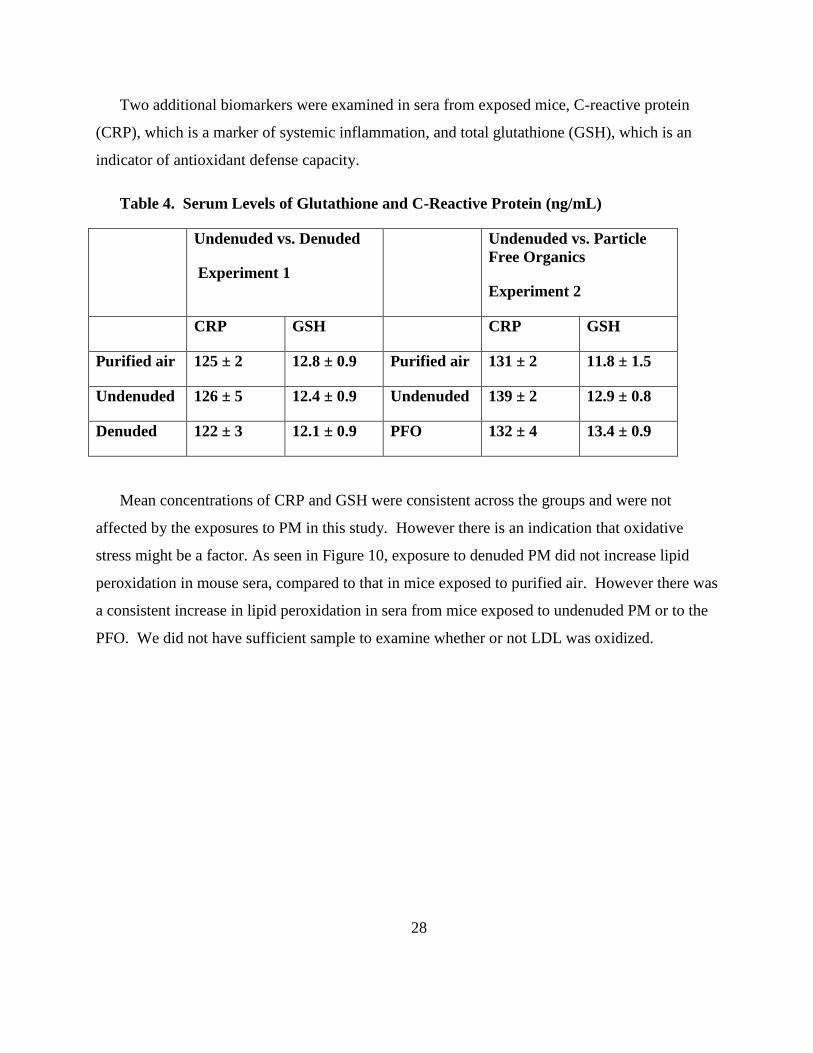
Two additional biomarkers were examined in sera from exposed mice, C-reactive protein
(CRP), which is a marker of systemic inflammation, and total glutathione (GSH), which is an
indicator of antioxidant defense capacity.
Table 4. Serum Levels of Glutathione and C-Reactive Protein (ng/mL)
Undenuded vs. Denuded
Experiment 1
Undenuded vs. Particle
Free Organics
Experiment 2
CRP GSH CRP GSH
Purified air 125 ± 2 12.8 ± 0.9 Purified air 131 ± 2 11.8 ± 1.5
Undenuded 126 ± 5 12.4 ± 0.9 Undenuded 139 ± 2 12.9 ± 0.8
Denuded 122 ± 3 12.1 ± 0.9 PFO 132 ± 4 13.4 ± 0.9
Mean concentrations of CRP and GSH were consistent across the groups and were not
affected by the exposures to PM in this study. However there is an indication that oxidative
stress might be a factor. As seen in Figure 10, exposure to denuded PM did not increase lipid
peroxidation in mouse sera, compared to that in mice exposed to purified air. However there was
a consistent increase in lipid peroxidation in sera from mice exposed to undenuded PM or to the
PFO. We did not have sufficient sample to examine whether or not LDL was oxidized.

29
Figure 10. Serum Lipid Peroxidation Levels in Mice Exposed to Either Denuded PM,
Undenuded PM and Particle Free Organics (PFO)
Conclusions
The findings of this study are that: (1) the VACES and the Dekati Thermodenuder can be
used in tandem to deliver undenuded ultrafine ambient PM (UFP, dp ≤ 0.18 µm), denuded UFP
and particle free organic PM constituents (PFO) to genetically modified, apoE-/-, mice in a
mobile rodent exposure system; (2) the organic constituents of UFP are important contributors to
atherosclerotic plaque development and significantly accelerate the growth of arterial plaques
after an 8 week exposure; and (3) exposure to both organic and inorganic constituents of UFP
raise serum concentrations of cholesterol and low density lipoprotein-cholesterol (LDL), but
exposures to UFP that were denuded of most organic constituents did not promote serum lipid
peroxidation while exposures to undenuded UFP or to PFO did promote serum lipid
peroxidation.
This study has demonstrated that the semi-volatile PM fraction of ambient ultrafine
particulate matter is an important contributor to the development of atherosclerosis and heart
disease. PM exposure was also shown to increase serum levels of cholesterol and LDL-
cholesterol, both of which are known risk factors for atherosclerosis and heart disease. In this
study exposure to undenuded UFP and to PFO also promoted the peroxidation of serum lipids
0
50
100
150
200
250
Air Undenuded Denuded
Lip
id P
ero
xid
atio
n (
nm
ol M
DA
/mg
pro
tein
)
Lipid Peroxidation
0
50
100
150
200
250
300
Air Undenuded PFOLip
id P
ero
xid
atio
n (
nm
ol M
DA
/mg
pro
tein
)
Lipid Peroxidation

30
while exposure to denuded UFP did not. Peroxidation of serum lipids, especially LDL, has been
specifically implicated in atherosclerotic plaque formation. In fact, serum levels of oxidized
LDL are increased in individuals with exposures to traffic-related air pollution [55] and oxidized
LDL contributes to formation of lipid-laden macrophages (foam cells) and promote the induction
of foam cell formation, inflammatory cytokines secretion [56]. Removal of the semivolatile
organics by a process of thermal denuding significantly mitigated the adverse effects of PM
exposures in a laboratory animal model of atherosclerosis.
Chemical characterization of the ambient PM using a high resolution aerosol mass
spectrometer showed that the UFP fraction, particles with diameters ≤ 0.18 µm, was more
enriched in organic compounds that were less oxygenated, hence less polar, than particles with
diameters ≥ 0.18 µm. This is important because organics that are nonpolar are better able to
cross cell membranes than are more polar molecules. Additionally, studies of mobile source
emissions have shown that emissions from heavy duty diesel vehicles are enriched in UFP and
that those UFP are characterized by low oxygenation levels.
An important corollary to these results is that they suggest that removal of UFP-associated
semivolatile compounds can significantly mitigate adverse cardiovascular effects. Modern diesel
engines are often provided with emission control technologies that remove effectively the non-
volatile fraction, but not necessarily the volatile fraction. Some research has shown that removal
of the non-volatile PM fraction can increase the concentration of the volatile fraction by
enhancing nucleation of condensing organic vapors. This project has provided findings that
improve our understanding of the mode of toxic action of freshly-emitted combustion aerosols
and has identified organic constituents of ambient aerosols as influencing potential health effects.
This information will also aid regulators and planners in developing air quality mitigation
strategies and land use guidance to better protect the health of California residents.

31
References:
1. Kleinman MT, Hamade A, Meacher D, Oldham M, Sioutas C, Chakrabarti B, Stram D,
Froines JR, Cho AK: Inhalation of concentrated ambient particulate matter near a
heavily trafficked road stimulates antigen-induced airway responses in mice. J Air
Waste Manage 2005, 55(9):1277-1288.
2. Kleinman MT, Sioutas C, Froines JR, Fanning E, Hamade A, Mendez L, Meacher D,
Oldham M: Inhalation of concentrated ambient particulate matter near a heavily
trafficked road stimulates antigen-induced airway responses in mice. Inhalation
Toxicology 2007, 19 Suppl 1:117-126.
3. Verma V, Pakbin P, Cheung KL, Cho AK, Schauer JJ, Shafer MM, Kleinman MT,
Sioutas C: Physicochemical and oxidative characteristics of semi-volatile components
of quasi-ultrafine particles in an urban atmosphere. Atmospheric Environment 2011,
45(4):1025-1033.
4. Kim S, Jaques PA, Chang MC, Barone T, Xiong C, Friedlander SK, Sioutas C: Versatile
aerosol concentration enrichment system (VACES) for simultaneous in vivo and in
vitro evaluation of toxic effects of ultrafine, fine and coarse ambient particles - Part
II: Field evaluation. J Aerosol Sci 2001, 32(11):1299-1314.
5. Kim S, Jaques PA, Chang MC, Froines JR, Sioutas C: Versatile aerosol concentration
enrichment system (VACES) for simultaneous in vivo and in vitro evaluation of
toxic effects of ultrafine, fine and coarse ambient particles - Part I: Development
and laboratory characterization. J Aerosol Sci 2001, 32(11):1281-1297.
6. Wehner B, Philippin S, Wiedensohler A: Design and calibration of a thermodenuder
with an improved heating unit to measure the size-dependent volatile fraction of
aerosol particles. J Aerosol Sci 2002, 33(7):1087-1093.
7. Dhalla NS, Temsah RM, Netticadan T: Role of oxidative stress in cardiovascular
diseases. J Hypertens 2000, 18(6):655-673.
8. Dominici F, Peng RD, Bell ML, Pham L, McDermott A, Zeger SL, Samet JM: Fine
particulate air pollution and hospital admission for cardiovascular and respiratory
diseases. JAMA 2006, 295(10):1127-1134.
9. Fang SC, Mehta AJ, Alexeeff SE, Gryparis A, Coull B, Vokonas P, Christiani DC,
Schwartz J: Residential Black Carbon Exposure and Circulating Markers of
Systemic Inflammation in Elderly Males: The Normative Aging Study.
Environmental Health Perspectives 2012, 120(5):674-680.
10. Kodavanti UP, Schladweiler MC, Ledbetter AD, Watkinson WP, Campen MJ, Winsett
DW, Richards JR, Crissman KM, Hatch GE, Costa DL: The spontaneously
hypertensive rat as a model of human cardiovascular disease: evidence of
exacerbated cardiopulmonary injury and oxidative stress from inhaled emission
particulate matter. Toxicol Appl Pharmacol 2000, 164(3):250-263.
11. Warheit DB, Reed KL, Sayes CM: A role for nanoparticle surface reactivity in
facilitating pulmonary toxicity and development of a base set of hazard assays as a

32
component of nanoparticle risk management. Inhalation Toxicology 2009, 21 Suppl
1:61-67.
12. Schulz H, Harder V, Ibald-Mulli A, Khandoga A, Koenig W, Krombach F, Radykewicz
R, Stampfl A, Thorand B, Peters A: Cardiovascular effects of fine and ultrafine
particles. Journal of Aerosol Medicine-Deposition Clearance and Effects in the Lung
2005, 18(1):1-22.
13. Sun Q, Wang A, Jin X, Natanzon A, Duquaine D, Brook RD, Aguinaldo JG, Fayad ZA,
Fuster V, Lippmann M et al: Long-term air pollution exposure and acceleration of
atherosclerosis and vascular inflammation in an animal model. Jama 2005,
294(23):3003-3010.
14. Sun Q, Yue P, Deiuliis JA, Lumeng CN, Kampfrath T, Mikolaj MB, Cai Y, Ostrowski
MC, Lu B, Parthasarathy S et al: Ambient air pollution exaggerates adipose
inflammation and insulin resistance in a mouse model of diet-induced obesity.
Circulation 2009, 119(4):538-546.
15. Araujo JA, Barajas B, Kleinman M, Wang X, Bennett BJ, Gong KW, Navab M, Harkema
J, Sioutas C, Lusis AJ et al: Ambient particulate pollutants in the ultrafine range
promote early atherosclerosis and systemic oxidative stress. Circulation Research
2008, 102(5):589-596.
16. Gong KW, Zhao W, Li N, Barajas B, Kleinman M, Sioutas C, Horvath S, Lusis AJ, Nel
A, Araujo JA: Air pollutant chemicals and oxidized lipids exhibit genome-wide
synergistic effects on endothelial cells. Arteriosclerosis Thrombosis and Vascular
Biology 2007, 27(6):E78-E78.
17. Hofnagel O, Luechtenborg B, Weissen-Plenz G, Robenek H: Statins and foam cell
formation: impact on LDL oxidation and uptake of oxidized lipoproteins via
scavenger receptors. Biochimica et Biophysica Acta 2007, 1771(9):1117-1124.
18. Tonne C, Melly S, Mittleman M, Coull B, Goldberg R, Schwartz J: A case-control
analysis of exposure to traffic and acute myocardial infarction. Environ Health
Perspect 2007, 115(1):53-57.
19. Garshick E, Laden F, Hart JE, Caron A: Residence near a major road and respiratory
symptoms in U.S. Veterans. Epidemiology 2003, 14(6):728-736.
20. Phuleria HC, Geller MD, Fine PM, Sioutas C: Size-resolved emissions of organic
tracers from light- and heavy-duty vehicles measured in a California roadway
tunnel. Environmental Science & Technology 2006, 40(13):4109-4118.
21. Zhu Y, Kuhn T, Mayo P, Hinds WC: Comparison of daytime and nighttime
concentration profiles and size distributions of ultrafine particles near a major
highway. Environmental Science & Technology 2006, 40(8):2531-2536.
22. Sahu M, Hu S, Ryan PH, Le Masters G, Grinshpun SA, Chow JC, Biswas P: Chemical
compositions and source identification of PM(2).(5) aerosols for estimation of a
diesel source surrogate. The Science of the total environment 2011, 409(13):2642-2651.
23. Biswas S, Verma V, Schauer JJ, Cassee FR, Cho AK, Sioutas C: Oxidative potential of
semi-volatile and non volatile particulate matter (PM) from heavy-duty vehicles
retrofitted with emission control technologies. Environmental Science & Technology
2009, 43(10):3905-3912.

33
24. Kleeberger SR, Reddy S, Zhang LY, Jedlicka AE: Genetic susceptibility to ozone-
induced lung hyperpermeability: role of toll-like receptor 4. Am J Respir Cell Mol
Biol 2000, 22(5):620-627.
25. Ohtsuka Y, Clarke RW, Mitzner W, Brunson K, Jakab GJ, Kleeberger SR: Interstrain
variation in murine susceptibility to inhaled acid-coated particles. American Journal
of Physiology-Lung Cellular and Molecular Physiology 2000, 278(3):L469-L476.
26. Morokata T, Ishikawa J, Ida K, Yamada T: C57BL/6 mice are more susceptible to
antigen-induced pulmonary eosinophilia than BALB/c mice, irrespective of systemic
T helper 1/T helper 2 responses. Immunology 1999, 98(3):345-351.
27. Kim S, Chang MC, Kim D, Sioutas C: A new generation of portable coarse, fine, and
ultrafine particle concentrators for use in inhalation toxicology. Inhalation
Toxicology 2000, 12:121-137.
28. Oldham MJ, Phalen RF, Robinson RJ, Kleinman MT: Performance of a portable
whole-body mouse exposure system. Inhal Toxicol 2004, 16(9):657-662.
29. Geller VD, Sardar SB, Phuleria H, Fine PN, Sioutas C: Measurements of particle
number and mass concentrations and size distributions in a tunnel environment.
Environmental Science & Technology 2005, 39(22):8653-8663.
30. Ghio AJ, Stonehuerner J, Dailey LA, Carter JD: Metals associated with both the water-
soluble and insoluble fractions of an ambient air pollution particle catalyze an
oxidative stress. Inhal Toxicol 1999, 11(1):37-49.
31. Hlavay J, Polyak K, Weisz M: Monitoring of the natural environment by chemical
speciation of elements in aerosol and sediment samples. J Environ Monit 2001,
3(1):74-80.
32. Campen MJ, Nolan JP, Schladweiler MC, Kodavanti UP, Evansky PA, Costa DL,
Watkinson WP: Cardiovascular and thermoregulatory effects of inhaled PM-
associated transition metals: a potential interaction between nickel and vanadium
sulfate. Toxicol Sci 2001, 64(2):243-252.
33. Lippmann M, Ito K, Hwang JS, Maciejczyk P, Chen LC: Cardiovascular effects of
nickel in ambient air. Environmental Health Perspectives 2006, 114(11):1662-1669.
34. Magari SR, Schwartz J, Williams PL, Hauser R, Smith TJ, Christiani DC: The
association of particulate air metal concentrations with heart rate variability.
Environ Health Perspect 2002, 110(9):875-880.
35. Natusch DFS, Wallace JR, Evans CA: Toxic Trace-Elements - Preferential
Concentration in Respirable Particles. Science 1974, 183(4121):202-204.
36. Carter JD, Ghio AJ, Samet JM, Devlin RB: Cytokine production by human airway
epithelial cells after exposure to an air pollution particle is metal-dependent. Toxicol
Appl Pharmacol 1997, 146(2):180-188.
37. Sioutas C, Kim S, Chang M: Development and evaluation of a prototype ultrafine
particle concentrator. J Aerosol Sci 1999, 30(8):1001-1017.
38. Kim S, Sioutas C, Chang MC, Gong H: Factors affecting the stability of the
performance of ambient fine-particle concentrators. Inhalation Toxicology 2000,
12:281-298.
39. Kleinman MT, Hamade A, Meacher D, Oldham M, Sioutas C, Chakrabarti L, Stram D,
Froines JR, Cho AK: Inhalation of concentrated ambient particulate matter near a

34
heavily trafficked road stimulates antigen-induced airway responses in mice. Journal
of the Air & Waste Management Association 2005, 55(9):1277-1288.
40. Wadsworth MP, Sobel BE, Schneider DJ, Taatjes DJ: Delineation of the evolution of
compositional changes in atheroma. Histochemistry and Cell Biology 2002, 118(1):59-
68.
41. Griffith OW: Determination of glutathione and glutathione disulfide using
glutathione reductase and 2-vinylpyridine. Analytical biochemistry 1980, 106(1):207-
212.
42. Erdelmeier I, Gerard-Monnier D, Yadan JC, Chaudiere J: Reactions of N-methyl-2-
phenylindole with malondialdehyde and 4-hydroxyalkenals. Mechanistic aspects of
the colorimetric assay of lipid peroxidation. Chemical Research in Toxicology 1998,
11(10):1184-1194.
43. Gerard-Monnier D, Erdelmeier I, Regnard K, Moze-Henry N, Yadan JC, Chaudiere J:
Reactions of 1-methyl-2-phenylindole with malondialdehyde and 4-hydroxyalkenals.
Analytical applications to a colorimetric assay of lipid peroxidation. Chemical
Research in Toxicology 1998, 11(10):1176-1183.
44. Mohanty JG, Bhamidipaty S, Evans MK, Rifkind JM: A fluorimetric semi-microplate
format assay of protein carbonyls in blood plasma. Analytical biochemistry 2010,
400(2):289-294.
45. Simkhovich BZ, Kleinman MT, Kloner RA: Particulate air pollution and coronary
heart disease. Current Opinion in Cardiology 2009, 24(6):604-609.
46. Simkhovich BZ, Kleinman MT, Kloner RA: Air pollution and cardiovascular injury
epidemiology, toxicology, and mechanisms. Journal of the American College of
Cardiology 2008, 52(9):719-726.
47. Schwartz J, Litonjua A, Suh H, Verrier M, Zanobetti A, Syring M, Nearing B, Verrier R,
Stone P, MacCallum G et al: Traffic related pollution and heart rate variability in a
panel of elderly subjects. Thorax 2005, 60(6):455-461.
48. Rowan WH, 3rd, Campen MJ, Wichers LB, Watkinson WP: Heart rate variability in
rodents: uses and caveats in toxicological studies. Cardiovascular Toxicology 2007,
7(1):28-51.
49. Pyetan E, Akselrod S: Do the high-frequency indexes of HRV provide a faithful
assessment of cardiac vagal tone? A critical theoretical evaluation. IEEE Trans
Biomed Eng 2003, 50(6):777-783.
50. Pyetan E, Akselrod S: A theoretical appraisal of the dependence of respiratory sinus
arrhythmia on gradual vagal blockade. Methods of information in medicine 2004,
43(1):52-55.
51. Shinar Z, Akselrod S, Dagan Y, Baharav A: Autonomic changes during wake-sleep
transition: a heart rate variability based approach. Auton Neurosci 2006, 130(1-
2):17-27.
52. Peles E, Goldstein DS, Akselrod S, Nitzan H, Azaria M, Almog S, Dolphin D, Halkin H,
Modan M: Interrelationships among measures of autonomic activity and
cardiovascular risk factors during orthostasis and the oral glucose tolerance test.
Clin Auton Res 1995, 5(5):271-278.

35
53. Baharav A, Kotagal S, Gibbons V, Rubin BK, Pratt G, Karin J, Akselrod S: Fluctuations
in autonomic nervous activity during sleep displayed by power spectrum analysis of
heart rate variability. Neurology 1995, 45(6):1183-1187.
54. Verma V, Larsen BD, Coombs W, Lin XM, Sarrou E, Taffet SM, Delmar M: Design and
characterization of the first peptidomimetic molecule that prevents acidification-
induced closure of cardiac gap junctions. Heart Rhythm 2010, 7(10):1491-1498.
55. Jacobs L, Emmerechts J, Hoylaerts MF, Mathieu C, Hoet PH, Nemery B, Nawrot TS:
Traffic air pollution and oxidized LDL. PLoS One 2011, 6(1):e16200.
56. Al Gadban MM, Smith KJ, Soodavar F, Piansay C, Chassereau C, Twal WO, Klein RL,
Virella G, Lopes-Virella MF, Hammad SM: Differential trafficking of oxidized LDL
and oxidized LDL immune complexes in macrophages: impact on oxidative stress.
PLoS One 2010, 5(9).