cardiovascular block arterial blood pressure & its regulation
TRANSCRIPT
Dr. Ahmad Al-Shafei, MBChB, PhD, MHPEAssociate Professor in Physiology
KSU
Dr. Ahmad Al-Shafei, MBChB, PhD, MHPEAssociate Professor in Physiology
KSU
Cardiovascular Block
Arterial Blood Pressure & Its Regulation

Learning outcomes
Describe systemic and pulmonary vascular resistances.Define cardiac function in modern physiological terms.Compare and contrast activity, pressure and volume of the left and right ventricles of the heart.Discuss structure related to functions of arterial system.Define arterial pulse and describe its characteristics and clinical significance. State normative data of arterial blood pressure for pulmonary and systemic circulations.Compare and contrast measurement of arterial blood pressure in systemic and pulmonary circulations.Discuss factors affecting systolic and diastolic blood pressures.
After reviewing the PowerPoint presentation, lecture notes and associated material, the student should be able to:
Discuss regulation of blood pressure: The arterial baroreceptors.The arterial baroreflex.Limits of effectiveness of the arterial baroreceptors.Baroreceptors and gravity
Atrial volume receptors (Bainbridge reflex).
Arterial chemorecptors
Discuss hormonal and renal regulation of blood pressure.Define systemic hypertension and state its complications.
Learning outcomes, continued

Learning Resources
Textbooks :
Guyton and Hall, Textbook of Medical Physiology; 12th Edition.Mohrman and Heller, Cardiovascular Physiology; 7th Edition.Ganong’s Review of Medical Physiology; 24th Edition.
Websites:
http://accessmedicine.mhmedical.com/

Systemic and pulmonary vascular resistances
As blood flows through the systemic and pulmonary circulations friction develops between the moving fluid and the stationary walls of the blood vessels. Thus, the vessels tend to resist fluid movement through them. Such resistance is known as vascular resistance.The resistance to the flow of the blood through the systemic circulation is known as systemic vascular resistance and the resistance to the flow of the blood through the pulmonary circulation is known as pulmonary vascular resistance. The ratio of pulmonary to systemic vascular resistance is approximately 1:6.It is because the vascular resistance to flow that we need the heart in the cardiovascular system.

Arterial blood pressure (ABP)
Blood pressure is defined as the force exerted by the blood against a vessel wall.
Blood pressure is an important characteristic of our body since it is the force that drives blood along blood vessels after it has left the heart.
Without blood pressure, nutrients, oxygen, and proteins could not travel from the arterial side of the body to the venous side.
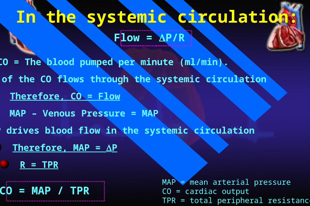
Flow = P/R
MAP – Venous Pressure = MAP
MAP = mean arterial pressureCO = cardiac outputTPR = total peripheral resistance
In the systemic circulation:
CO = The blood pumped per minute (ml/min).
Therefore, CO = Flow
MAP drives blood flow in the systemic circulation
Therefore, MAP = P
R = TPR
CO = MAP / TPR
All of the CO flows through the systemic circulation

In the systemic circulation:
CO = MAP / TPR
MAP = CO X TPR
= SV X HR X TPR
What are the factors affecting vascular resistance?- Resistance is inversely proportional to r4.- Resistance is directly proportional to viscosity. The viscosity of - blood is dependent on the haematocrit and plasma protein concentration.

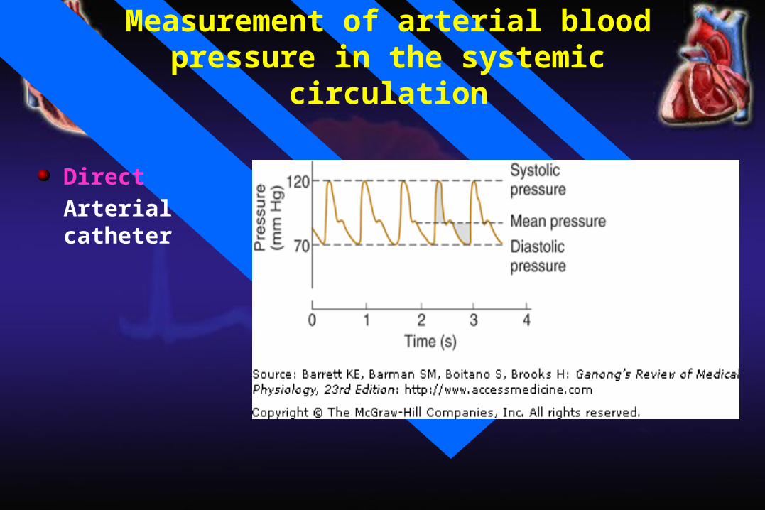
Measurement of arterial blood pressure in the systemic circulation
DirectArterial catheter
IndirectStethoscope and blood pressure cuff
Measurement of arterial blood pressure in the systemic circulation
DirectArterial catheter

Indirect measurementof arterial blood pressurein the systemic circulation
Systolic blood pressure: Is the maximum level of arterial blood pressure. It is reached during the rapid ejection phase of ventricular systole. For a normal adult, it is about 120 mmHg (range of normal: 90-140).
Diastolic blood pressure: Is the minimum level of arterial blood pressure. It is reached during the isometric contraction phase of ventricular systole. For a normal adult, it is about 80 mmHg (range of normal: 60-90).
Arterial blood pressure (ABP)
Mean arterial pressure (MAP)
MAP is the average pressure responsible for driving blood into the tissues throughout the cardiac cycle.
MAP is not halfway between systolic and diastolic pressures because arterial pressure remains closer to diastolic than to systolic pressure for a longer period of each cardiac cycle.
A good approximation of the MAP can be determined using the following formula:
MAP = Diastolic pressure + 1/3 pulse pressure
Mean arterial pressure tends to increase with age because of an age-dependent increase in total peripheral resistance which is controlled primarily by arterioles.

Right Heart PressuresSwan-Ganz / PA Catheter
Measurement of arterial blood pressure in the pulmonary circulation

Pressures in in the systemic and pulmonary circulations
Pressures in thepulmonary circulation
Pressures in thesystemic circulation
Right ventricle 25/0 mm Hg
Left ventricle 120/0 mm Hg
Pulmonary artery 25/10 mm Hg
Aorta 120/80 mm Hg
Mean pulmonary artery 15 mm Hg
Mean arterial BP 93 mm Hg
Capillary 7-9 mm Hg
Capillary: skeletal 30 mm Hg
renal glomerular 45-50 mm Hg
Pulmonary veins 5 mm Hg
Peripheral veins 7-15 mm Hg
Left atrium 5-10 mm Hg
Right atrium (CVP) 0 mm Hg
Pressure gradient 15-5 = 10 mm Hg
Pressure gradient 93-0 = 93 mm Hg
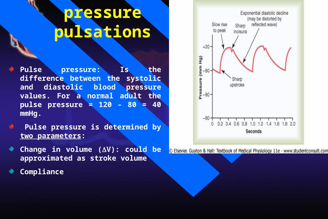
Arterial pressure pulsations
Pulse pressure: Is the difference between the systolic and diastolic blood pressure values. For a normal adult the pulse pressure = 120 – 80 = 40 mmHg.
Pulse pressure is determined by two parameters:
Change in volume (V): could be approximated as stroke volume
Compliance

Abnormalities in pulse pressure
Atherosclerosis: Pulse pressure tends to increase with ageing because of a decrease in arterial compliance ("hardening of the arteries").
Aortic regurgitation: in early diastole, blood leaks back into the ventricles. As a results, diastolic pressure falls to very low levels.

Regulation of blood pressure
BP varies over 24 hours:
lower during sleep
higher during waking, especially in stress
BP is well regulated – why?
to provide organs (especially brain) with adequate perfusion pressure
to optimize cardiovascular work
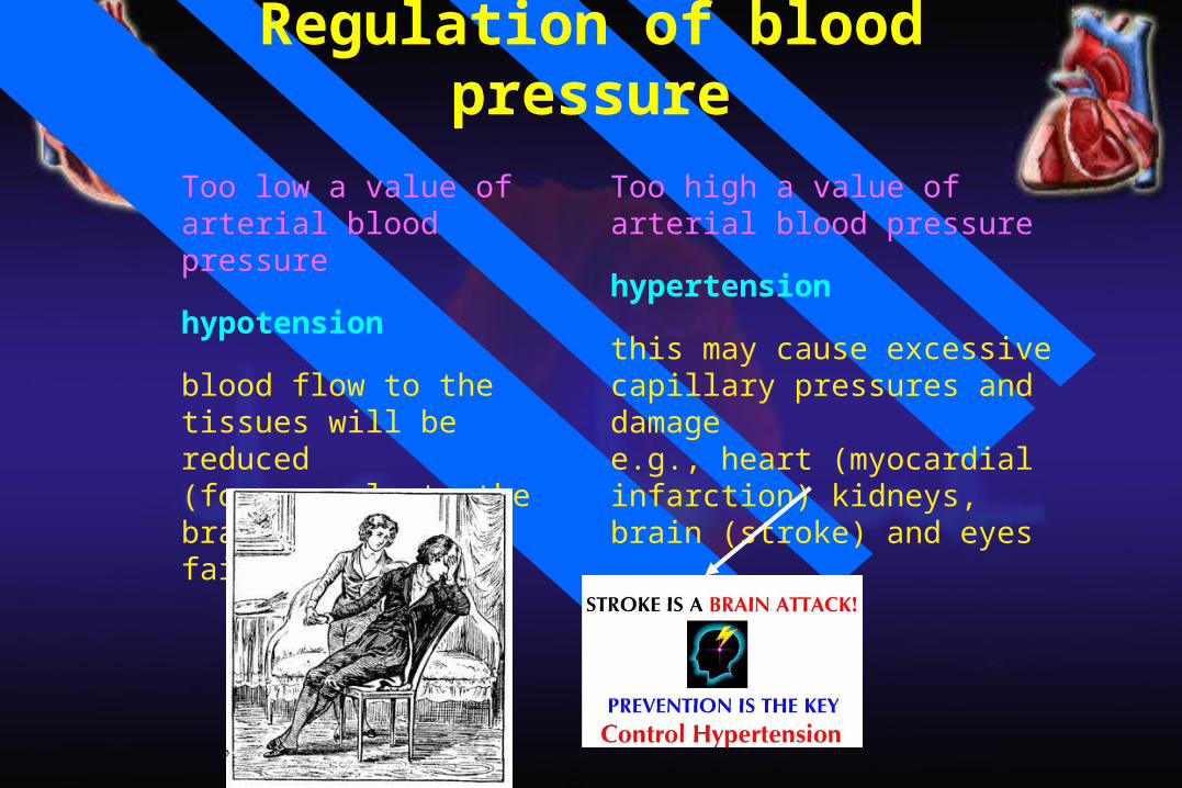
Regulation of blood pressureToo low a value of arterial blood pressure
hypotension
blood flow to the tissues will be reduced(for example to the brain and induce a faint)
Too high a value of arterial blood pressure
hypertension
this may cause excessive capillary pressures and damagee.g., heart (myocardial infarction) kidneys, brain (stroke) and eyes
Short term mechanisms (seconds to minutes): are largely neural. They regulate cardiac function and arteriolar diameter.
Long term mechanisms (minutes to days): are largely renal and hormonal. They regulate blood volume.
Regulation of blood pressure
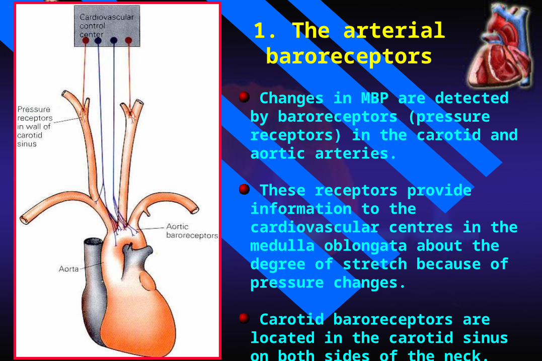
1. The arterial baroreceptors
Changes in MBP are detected by baroreceptors (pressure receptors) in the carotid and aortic arteries.
These receptors provide information to the cardiovascular centres in the medulla oblongata about the degree of stretch because of pressure changes.
Carotid baroreceptors are located in the carotid sinus on both sides of the neck. Aortic baroreceptors are located in the aortic arch.

1. The arterial baroreceptors

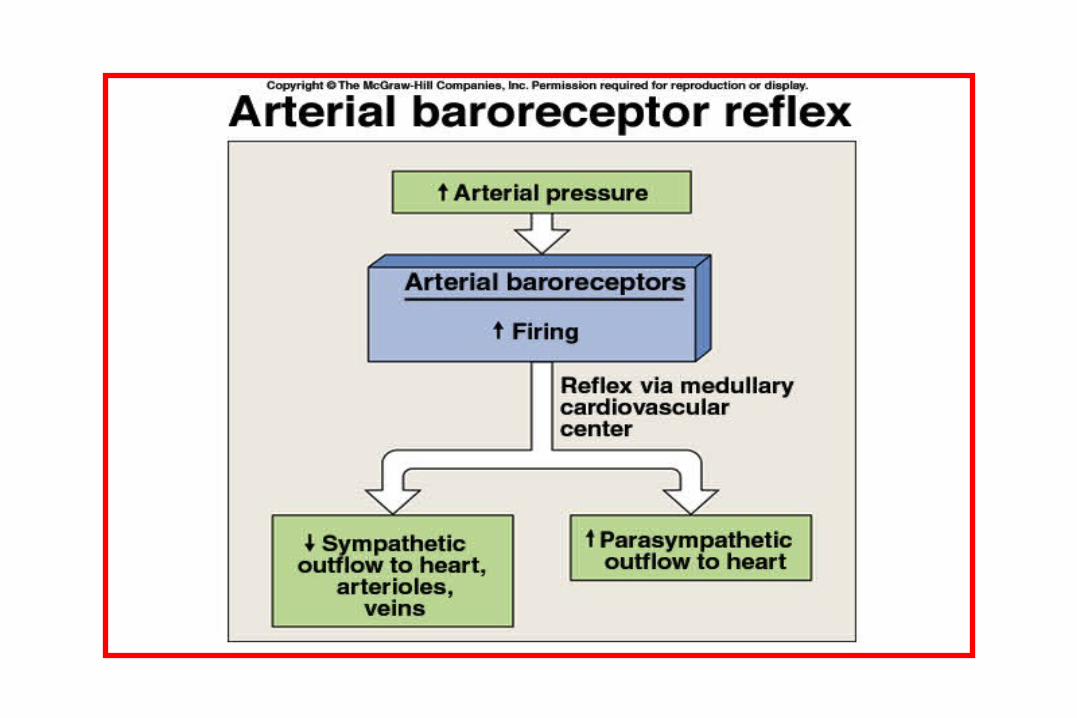
Increased arterial pressure increases baroreceptor activity
- At normal arterial pressure the baroreceptors are active.
- Increased pressure increases their rate of fire, while decreased pressure decreases the rate of fire.
- They play an important role in maintaining relatively constant blood flow to vital organs (such as brain) during rapid changes in pressure, such as standing up after lying down. That is why they are called “pressure buffers”.


↑ MAP
Baroreceptors
↑ Parasympathetic(vagal) activity
↓ Sympathetic activity
↓ HR ↓ SV ↓ TPR ↓ MAP
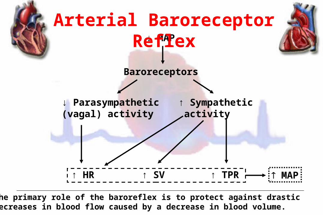
Arterial Baroreceptor Reflex
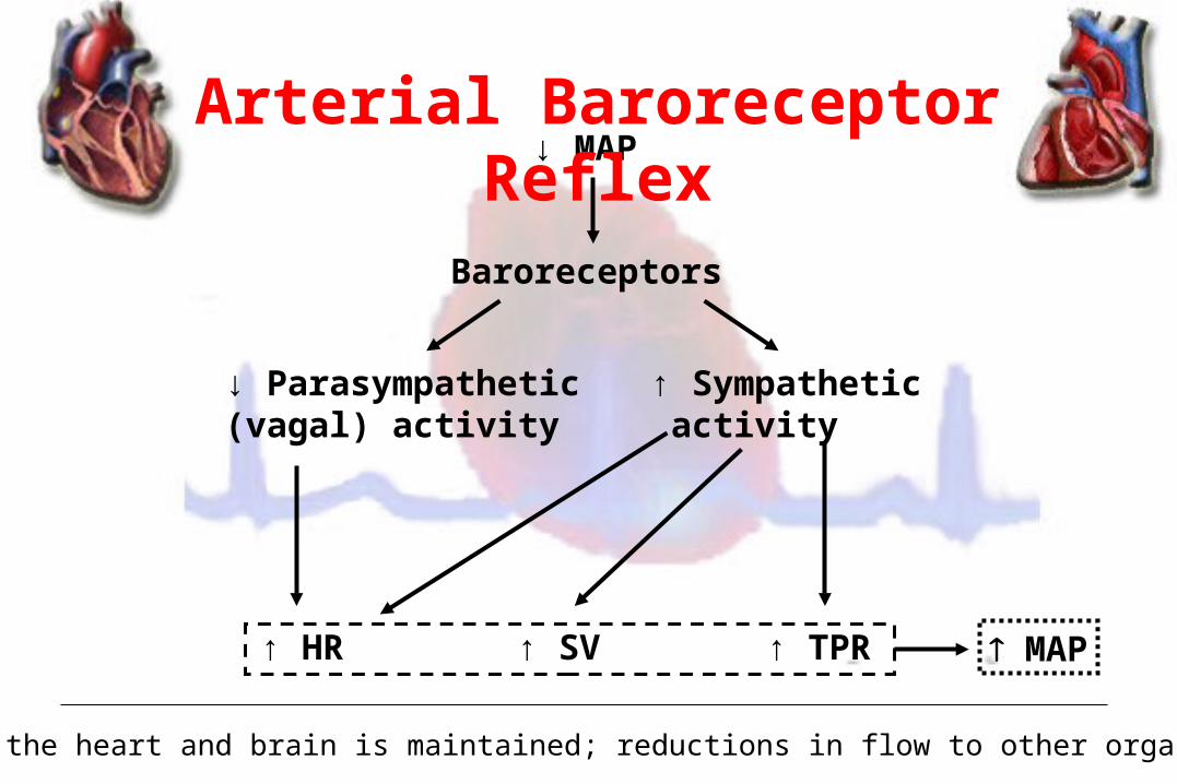
↓ MAP
Baroreceptors
↓ Parasympathetic(vagal) activity
↑ Sympathetic activity
↑ HR ↑ SV ↑ TPR
• Flow to the heart and brain is maintained; reductions in flow to other organs.
MAP
Arterial Baroreceptor Reflex
↓ MAP
Baroreceptors
↓ Parasympathetic(vagal) activity
↑ Sympathetic activity
↑ HR ↑ SV ↑ TPR MAP
The primary role of the baroreflex is to protect against drasticdecreases in blood flow caused by a decrease in blood volume.
Arterial Baroreceptor Reflex
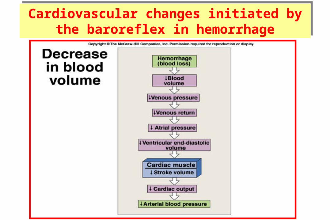
Cardiovascular changes initiated by the baroreflex in hemorrhage
Cardiovascular changes initiated by the baroreflex in hemorrhage
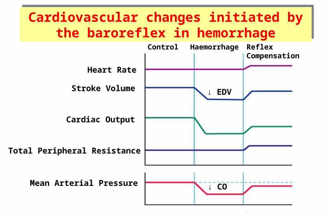
Heart Rate
Stroke Volume
Cardiac Output
Total Peripheral Resistance
Mean Arterial Pressure
Control Haemorrhage ReflexCompensation
↓ EDV
↓ CO
Cardiovascular changes initiated by the baroreflex in hemorrhage
Cardiovascular changes initiated by the baroreflex in hemorrhage
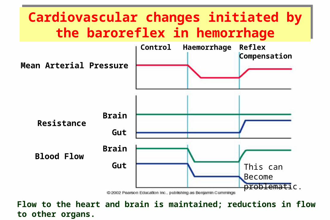
Flow to the heart and brain is maintained; reductions in flow to other organs.
Control Haemorrhage ReflexCompensation
Mean Arterial Pressure
ResistanceBrain
Gut
Blood FlowBrain
Gut This canBecome problematic.
Cardiovascular changes initiated by the baroreflex in hemorrhage
Cardiovascular changes initiated by the baroreflex in hemorrhage
Unimportance of arterial baroreceptors in long-term pressure control
The baroreceptor reflex mechanism functions primarily as a short-term regulator of ABP. It is activated at once by any blood pressure change and attempt to restore blood pressure rapidly toward normal.
Yet, if ABP deviates from its normal operating point for more than a few days, the arterial baroreceptors adapt to this new pressure.
This adaptation of the baroreceptors obviously prevents the baroreflex from functioning as a long-term blood pressure control system.
2. Atrial volume receptors
There are receptors in the very large veins close to the heart and in the walls of the atria.
They are sensitive to blood volume. The following mechanisms occurs to prevent excess elevation in blood pressure upon response to an increase of body fluid volume
An increased blood volume stretch of atria activate atrial volume receptors sensory afferent nerves to medulla inhibiting the cardiovascular centre This results into decreased blood volume through:
(a) sympathetic drive to kidney:
- dilate afferent arterioles glomerular capillary hydrostatic pressure GFR blood volume (towards normal).
- renin secretion (Renin is an enzyme which activates angiotensinogen in blood). Hence inhibition of renin secretion inhibit RAAS inhibit aldosterone production Blood volume (towards normal)
(b) ADH secretion blood volume (towards normal).
(c) Atrial Natriuretic Peptide (ANP) causes loss of blood volume.
3. Arterial chemoreceptors
• Supply of O2 to tissues depends on both respiratory and cardiovascular factors.
• Reduced blood flow (due to reduced ABP) stimulates the chemoreceptors through oxygen lack, increased hydrogen ions or carbon dioxide.
• Response is excitatory, NOT inhibitory; mainly through activation of sympathetic nervous system.
• They reduce blood flow to unessential areas and protect vital tissues like brain and heart.

↓ MAP
Sympathetic Activity
Splanchnic nerve
Adrenal Medulla
Adrenaline Secretion
HR (SA node) and SV (ventricular myocardium)
A. Catecholamines (Adrenaline and Noradrenaline)
Hormonal regulation of blood pressure

-adrenoceptor
-adrenoceptor
Adrenoceptor = Adrenergic Receptor(binds adrenaline and noradrenaline)
A. Catecholamines (Adrenaline and Noradrenaline)
Hormonal regulation of blood pressure

-adrenoceptor
-adrenoceptor
-adrenoceptor stimulation promotes vasoconstriction -adrenoceptor stimulation promotes vasodilation
A. Catecholamines (Adrenaline and Noradrenaline)
Hormonal regulation of blood pressure

-adrenoceptor
-adrenoceptor
NoradrenalineNoradrenalineNoradrenaline
Noradrenaline released from the sympathetic nerves bindsprimarily to adrenoceptors.
A. Catecholamines (Adrenaline and Noradrenaline)
Hormonal regulation of blood pressure
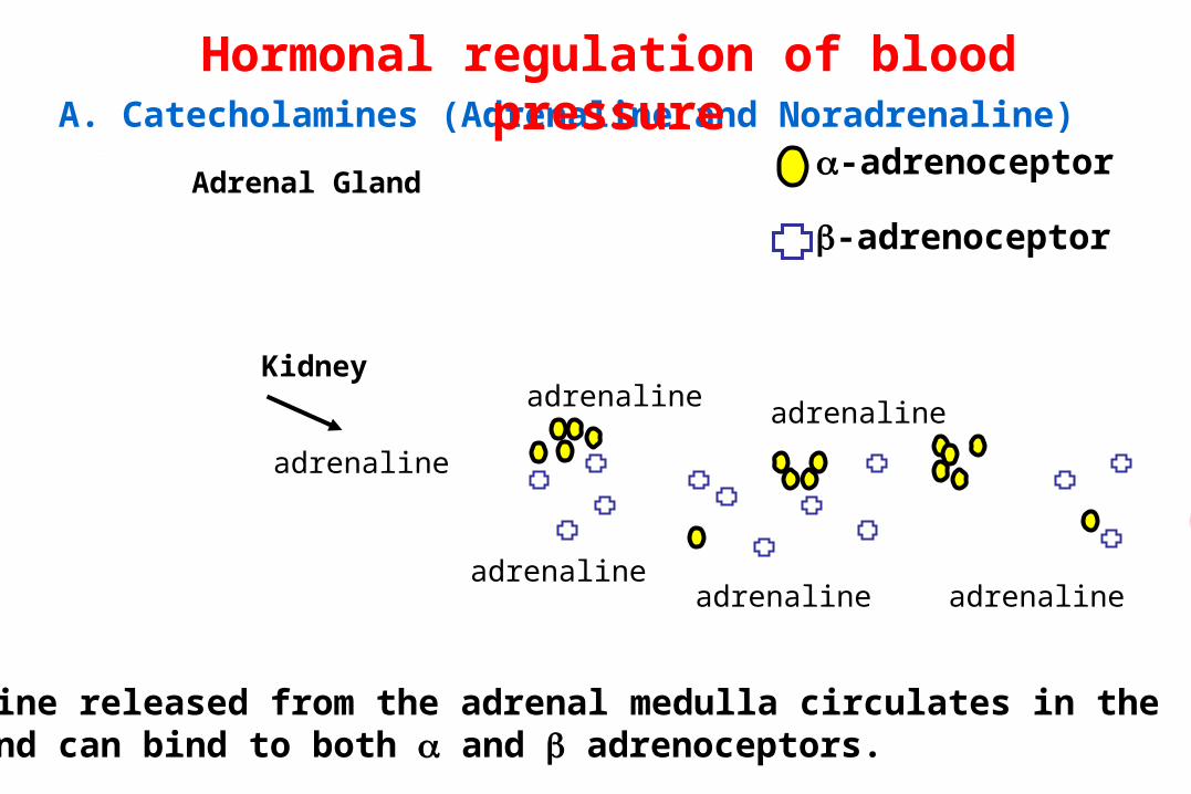
-adrenoceptor
-adrenoceptor
adrenaline
adrenalineadrenaline
adrenaline adrenaline
adrenaline
Adrenaline released from the adrenal medulla circulates in theblood and can bind to both and adrenoceptors.
Adrenal Gland
Kidney
A. Catecholamines (Adrenaline and Noradrenaline)
Hormonal regulation of blood pressure
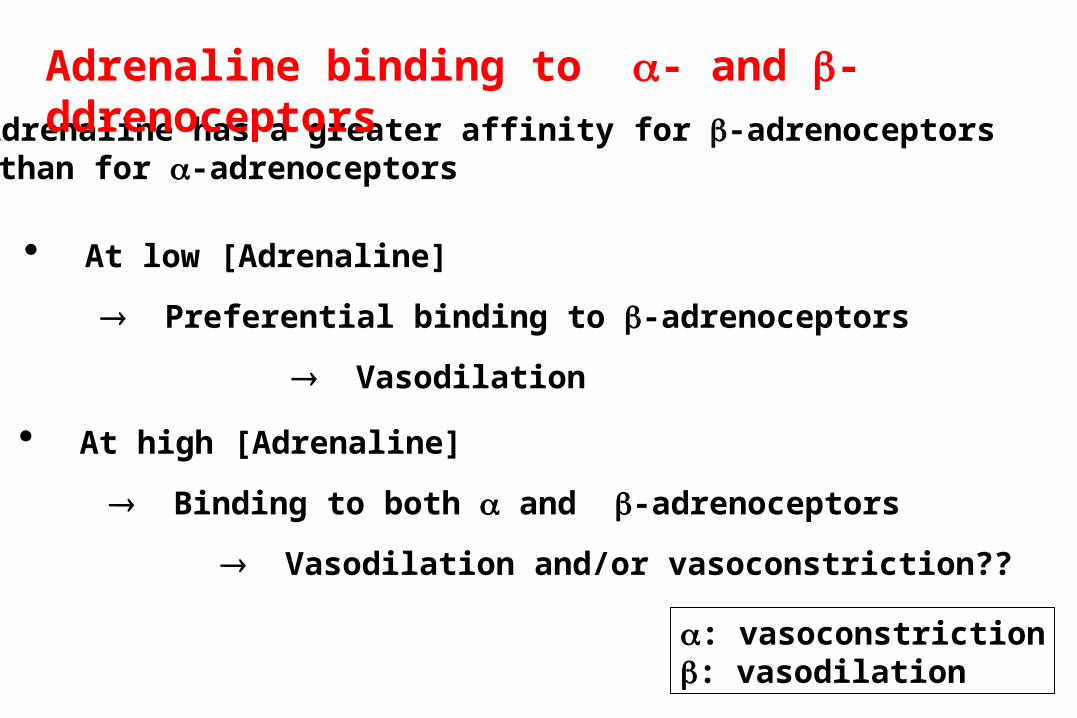
• Adrenaline has a greater affinity for -adrenoceptors than for -adrenoceptors
• At low [Adrenaline]
Preferential binding to -adrenoceptors
Vasodilation
• At high [Adrenaline]
Binding to both and -adrenoceptors
Vasodilation and/or vasoconstriction??
Adrenaline binding to - and -ddrenoceptors
: vasoconstriction: vasodilation
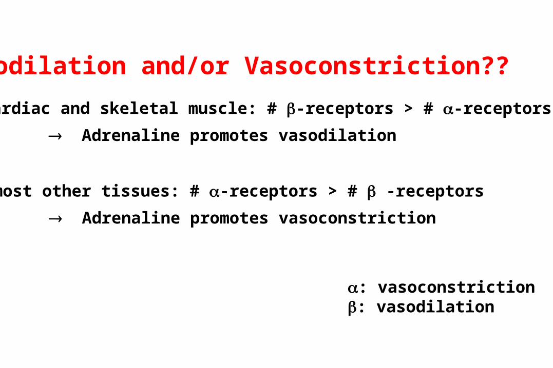
Vasodilation and/or Vasoconstriction??
: vasoconstriction: vasodilation
• In cardiac and skeletal muscle: # -receptors > # -receptors
Adrenaline promotes vasodilation
• In most other tissues: # -receptors > # -receptors
Adrenaline promotes vasoconstriction

hypothalamus
pituitary
• ADH (vasopressin) is synthesized within the
Paraventricular Nucleus of the hypothalamus.
• ADH is stored in the posterior pituitary.
Paraventricular nucleus
• Vasopressin exerts a pressor effect (i.e., ↑BP)
B. Role of vasopressin (Antidiuretic hormone; ADH )
Hormonal regulation of blood pressure

hypothalamus
pituitary
1. Dehydration or salt ingestion
4. Triggers ADH release from the pituitary
5a. Causes vasoconstriction
5b. Promotes water retention by the kidney
2. ↑ Blood osmolarity
3. Stimulates osmoreceptors in the hypothalamus
↑ TPR ↑ BP
↑ Blood Volume
B. Vasopressin (Antidiuretic hormone; ADH )
Hormonal regulation of blood pressure

Renal regulation of blood pressure
The baroreceptor reflex and other reflex mechanisms are important for the short term control of blood pressure.
Long term control of blood pressure requires control of blood volume.
Blood volume is controlled by the kidney.
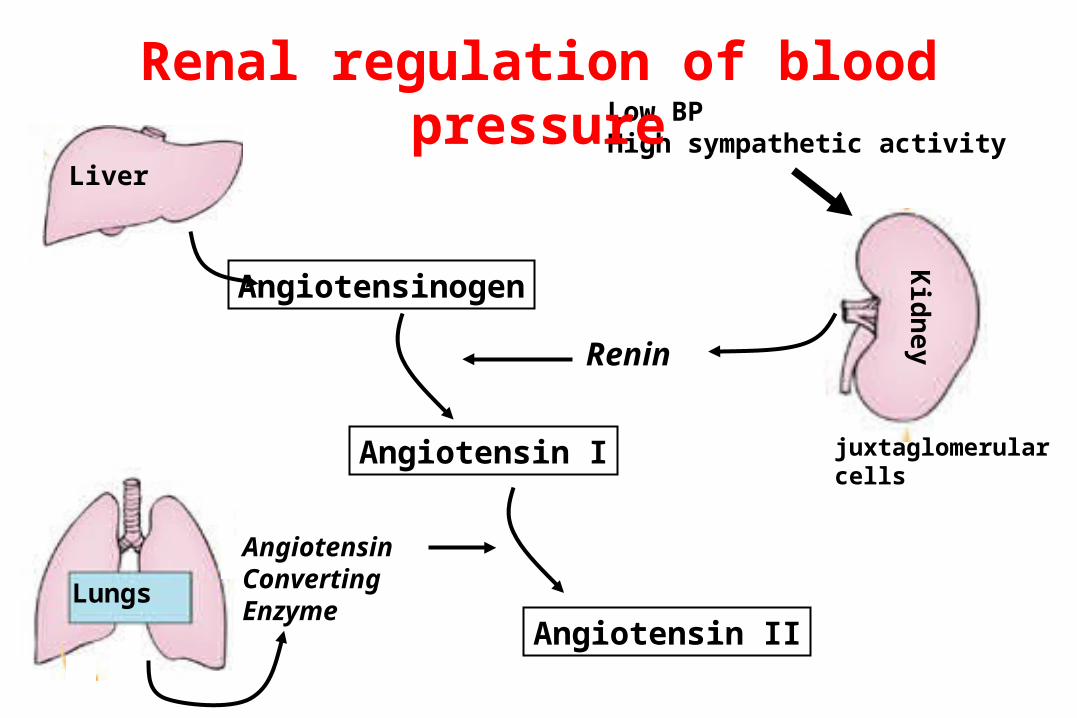
LiverKidney
Angiotensinogen
Renin
Angiotensin I
Lungs
AngiotensinConvertingEnzyme
Angiotensin II
Low BPHigh sympathetic activity
juxtaglomerularcells
Renal regulation of blood pressure
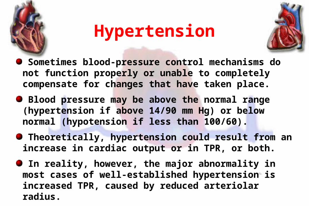
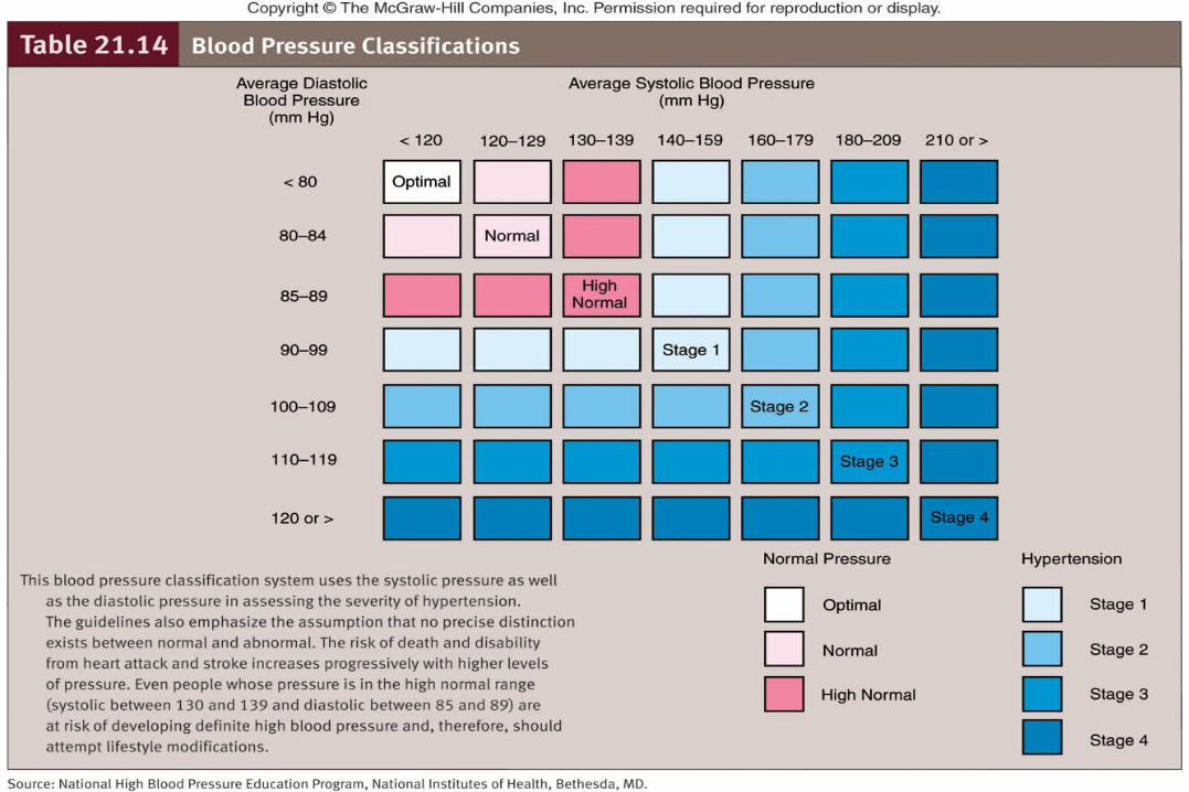
Hypertension
Sometimes blood-pressure control mechanisms do not function properly or unable to completely compensate for changes that have taken place.
Blood pressure may be above the normal range (hypertension if above 14/90 mm Hg) or below normal (hypotension if less than 100/60).
Theoretically, hypertension could result from an increase in cardiac output or in TPR, or both.
In reality, however, the major abnormality in most cases of well-established hypertension is increased TPR, caused by reduced arteriolar radius.

In only a small fraction of cases (only 10% of cases) the cause of arteriolar constriction is known.
Hypertension that occurs secondary to another primary problem is called secondary hypertension.
The underlying cause is unknown in the remaining 90% of hypertension cases. Such hypertension is known as primary (essential or idiopathic) hypertension.
Hypertension

Secondary hypertensionCauses:
1- renal diseases account for over 80% of the case of secondary hypertension.
2- Endocrine causes:
- Conn’s syndrome.
- Adrenal hyperplasia.
- Phaeochromocytoma.
- Cushing’s syndrome.
- Acromegaly.
3. Coarctation of the aorta.
4. Drugs.
5. Pregnancy.
What are the complications of hypertension?