care of the critically ill pregnant patient - critical care canada · care of the critically ill...
TRANSCRIPT

Care of the Critically Ill
Pregnant Patient
Stephen E. Lapinsky
Mount Sinai HospitalToronto

No conflict of interest related to this topic
May describe “off-label” use considering limited data in pregnancy

Case Presentation
26 year old woman, 27 weeks gestation
Admitted with:
- hypoxic respiratory failure
- bilateral pulmonary infiltrates
FiO2 0.60
pH 7.29
pCO2 46
pO2 52
HCO3 22
Sat 79%
Overview
• Physiological changes in pregnancy
• Risks to the fetus of maternal ICU stay
• Causes of critical illness during pregnancy
• Management

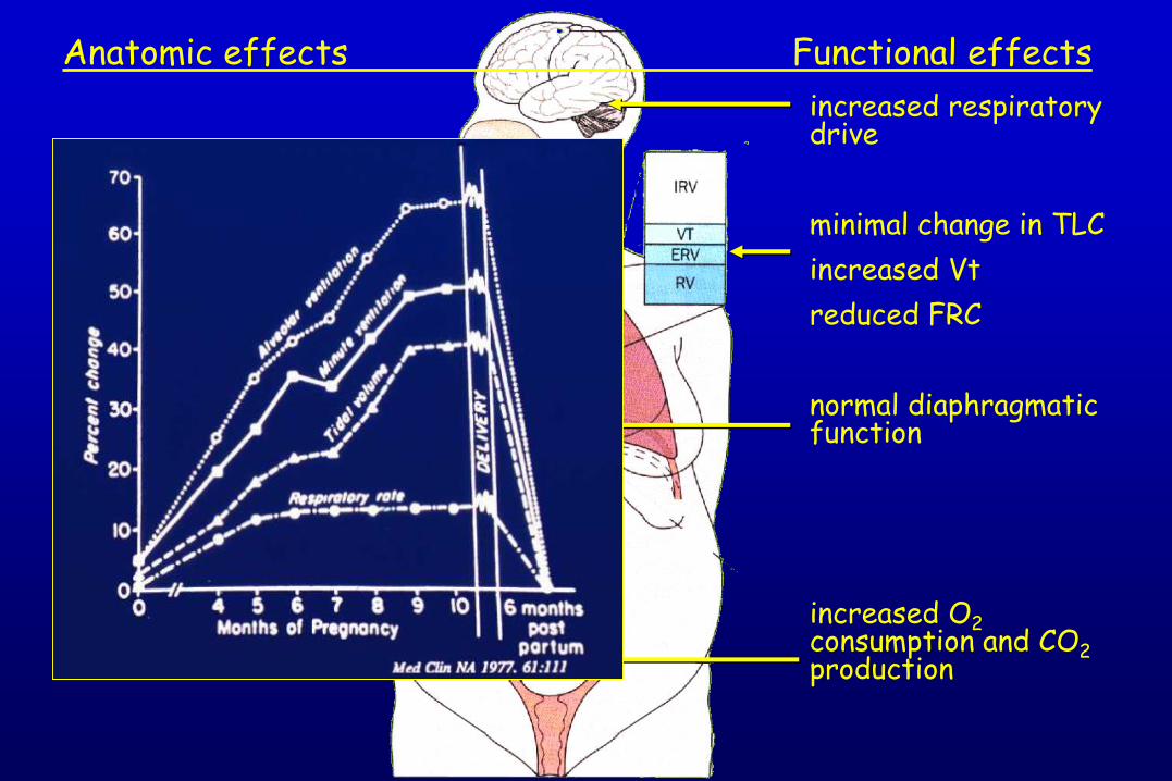
airway edema, friability
widened AP and transverse diam.
elevated diaphragm
widened subcostal angle
enlarging uterus
Anatomic effects Functional effects
airway edema, friability
widened AP and transverse diam.
elevated diaphragm
widened subcostal angle
enlarging uterus
Anatomic effects Functional effects
increased respiratory drive
minimal change in TLC
increased Vt
reduced FRC
normal diaphragmatic function
increased O2consumption and CO2production

airway edema, friability
widened AP and transverse diam.
elevated diaphragm
widened subcostal angle
enlarging uterus
Anatomic effects Functional effects
increased respiratory drive
minimal change in TLC
increased Vt
reduced FRC
normal diaphragmatic function
increased O2consumption and CO2production
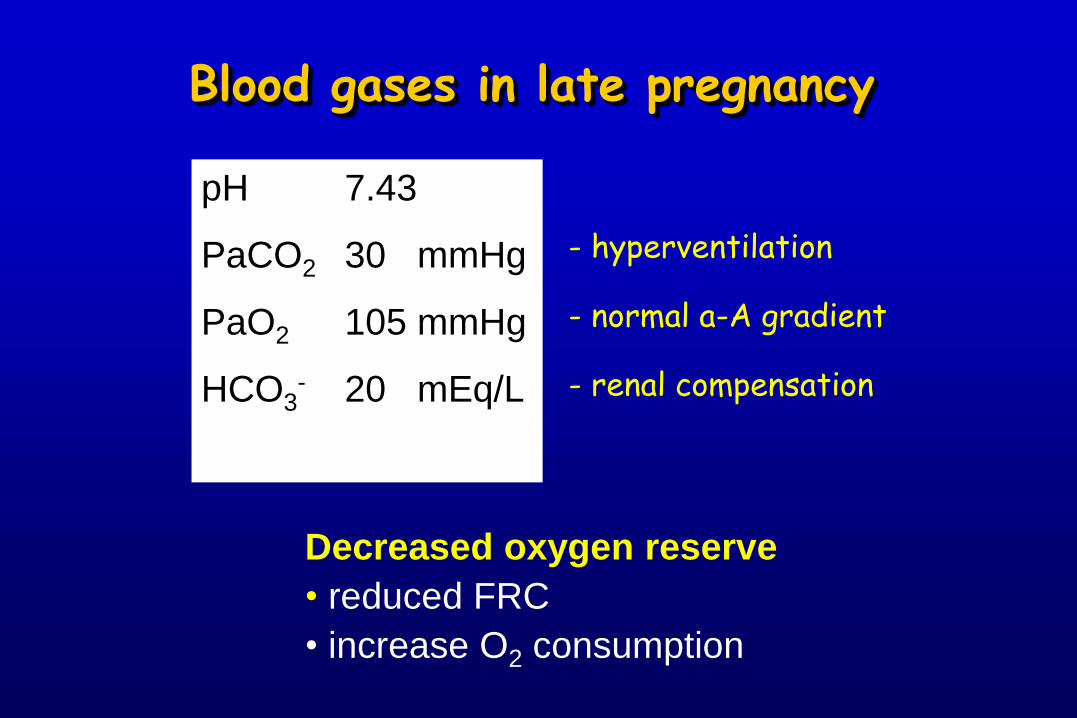
Blood gases in late pregnancy
pH 7.43
PaCO2 30 mmHg
PaO2 105 mmHg
HCO3- 20 mEq/L
- hyperventilation
- normal a-A gradient
- renal compensation
Decreased oxygen reserve
• reduced FRC
• increase O2 consumption

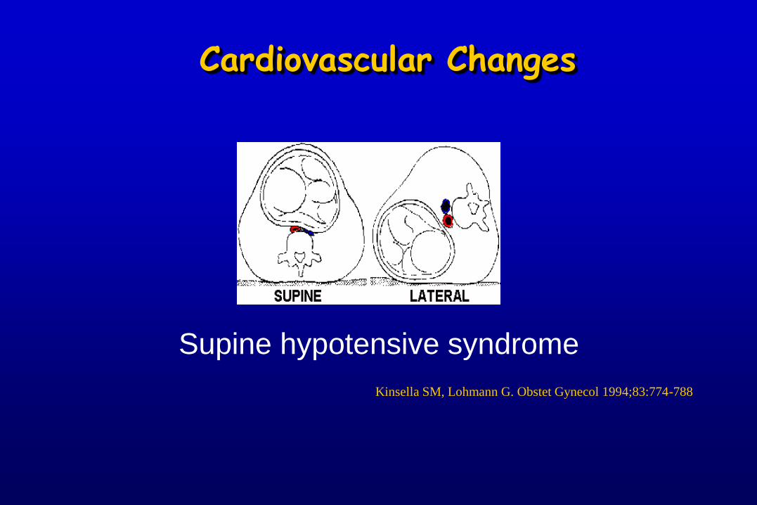
Cardiovascular Changes
increased blood volume (up 40% by third trimester)
increased cardiac output 30 – 50% by 25 – 32 weeks
decrease in blood pressure 10 – 20%, nadir 28 weeks
decreased SVR
increased LV mass and LV ED dimension
Cardiovascular Changes
Supine hypotensive syndrome
Kinsella SM, Lohmann G. Obstet Gynecol 1994;83:774-788
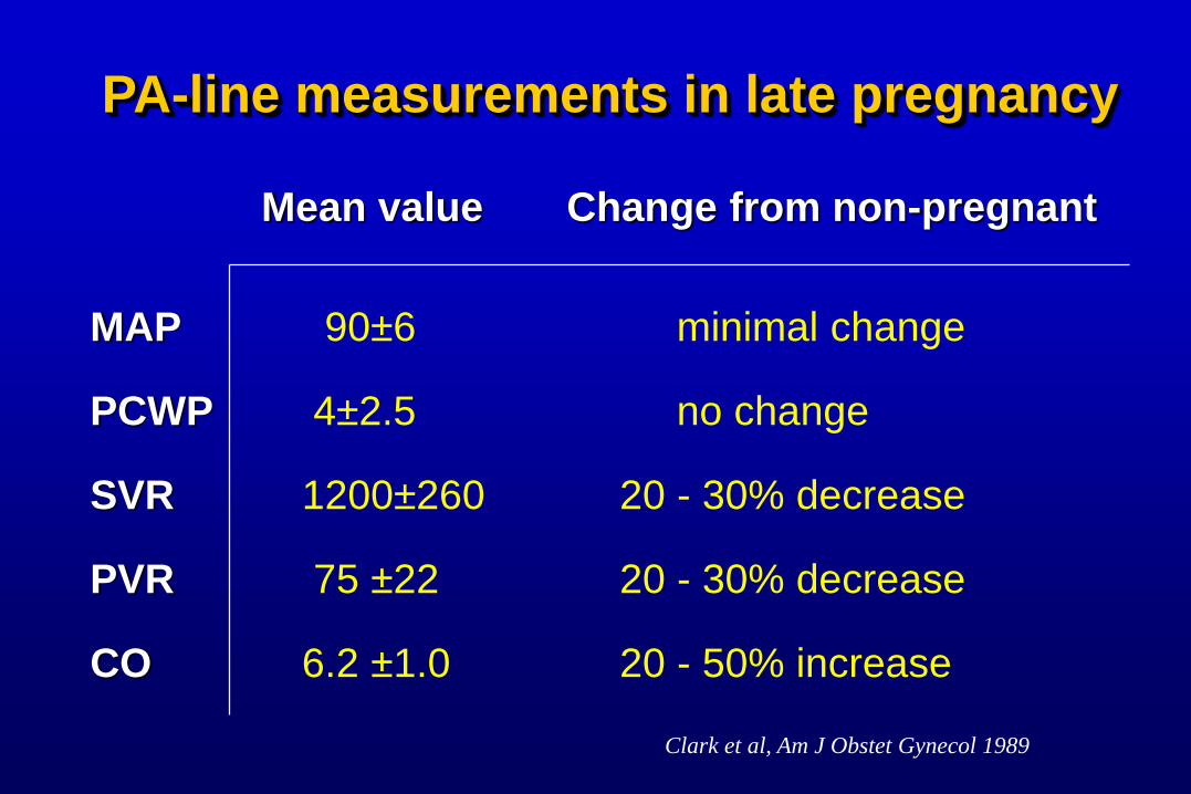
PA-line measurements in late pregnancy
Mean value Change from non-pregnant
MAP 90±6 minimal change
PCWP 4±2.5 no change
SVR 1200±260 20 - 30% decrease
PVR 75 ±22 20 - 30% decrease
CO 6.2 ±1.0 20 - 50% increase
Clark et al, Am J Obstet Gynecol 1989
Risks to the fetus
Fetal hypoxia
Radiological investigations
Drug therapy
Risks to the fetus
Fetal hypoxia
Radiological investigations
Drug therapy
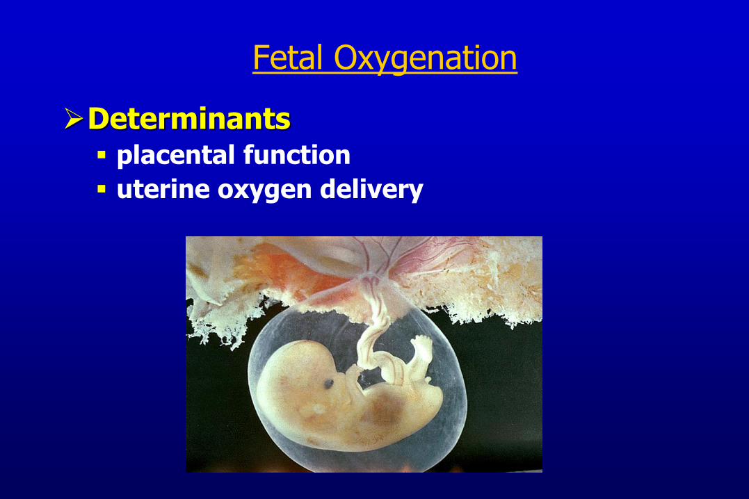
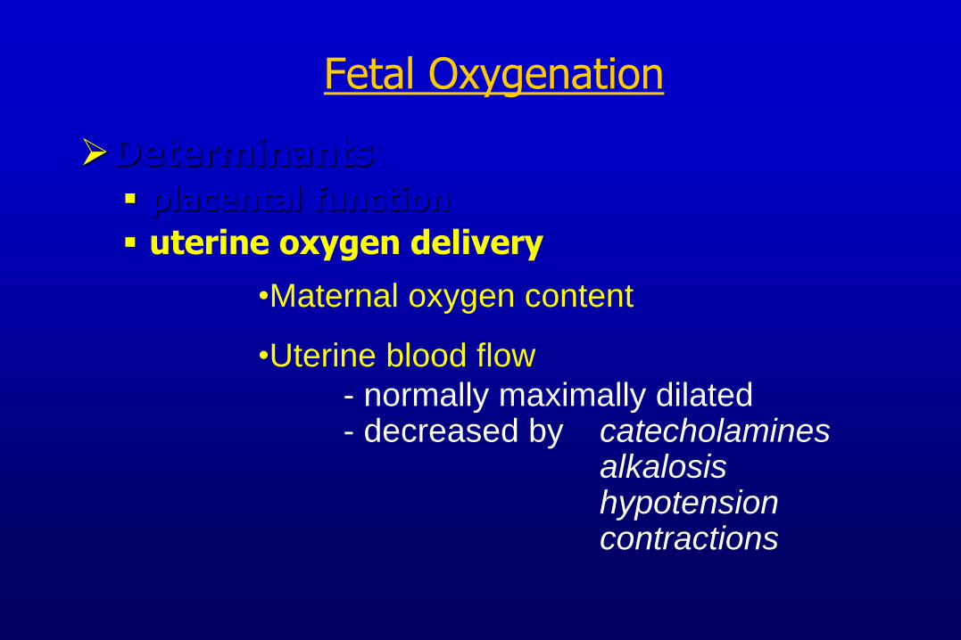
Fetal Oxygenation
Determinants placental function
uterine oxygen delivery

Fetal Oxygenation
Determinants placental function
uterine oxygen delivery
•Maternal oxygen content
•Uterine blood flow
Fetal Oxygenation
Determinants placental function
uterine oxygen delivery
•Maternal oxygen content
•Uterine blood flow
- normally maximally dilated- decreased by catecholamines
alkalosishypotensioncontractions

Fetal Oxygenation
Risks to the fetus
Fetal hypoxia
Radiological investigations
Drug therapy
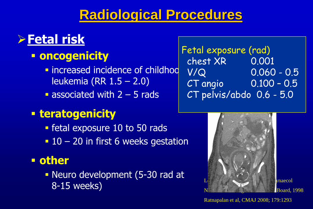
Radiological Procedures
Fetal risk
oncogenicity
increased incidence of childhood leukemia (RR 1.5 – 2.0)
associated with 2 – 5 rads
teratogenicity
fetal exposure 10 to 50 rads
10 – 20 in first 6 weeks gestation
other
Neuro development (5-30 rad at 8-15 weeks)
Lowe 2004, Austr NZ J Obstet Gynaecol
National Radiological Protection Board, 1998
Ratnapalan et al, CMAJ 2008; 179:1293
Fetal exposure (rad)chest XR 0.001V/Q 0.060 - 0.5CT angio 0.100 – 0.5CT pelvis/abdo 0.6 - 5.0
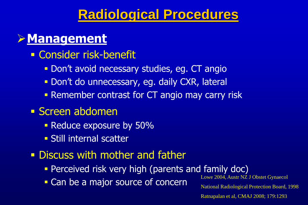
Radiological Procedures
Management
Consider risk-benefit
Don’t avoid necessary studies, eg. CT angio
Don’t do unnecessary, eg. daily CXR, lateral
Remember contrast for CT angio may carry risk
Screen abdomen
Reduce exposure by 50%
Still internal scatter
Discuss with mother and father
Perceived risk very high (parents and family doc)
Can be a major source of concernLowe 2004, Austr NZ J Obstet Gynaecol
National Radiological Protection Board, 1998
Ratnapalan et al, CMAJ 2008; 179:1293
Risks to the fetus
Fetal hypoxia
Radiological investigations
Drug therapy
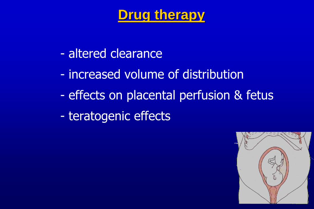
Drug therapy
- altered clearance
- increased volume of distribution
- effects on placental perfusion & fetus
- teratogenic effects
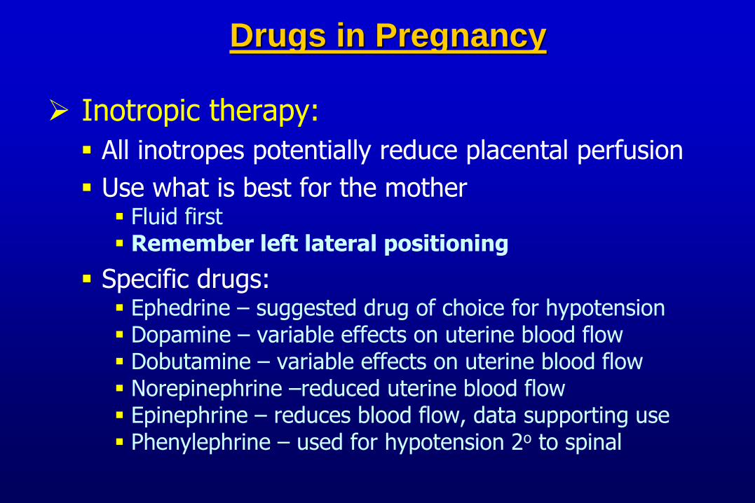
Drugs in Pregnancy
Inotropic therapy:
All inotropes potentially reduce placental perfusion
Use what is best for the mother Fluid first Remember left lateral positioning
Specific drugs: Ephedrine – suggested drug of choice for hypotension Dopamine – variable effects on uterine blood flow Dobutamine – variable effects on uterine blood flow Norepinephrine –reduced uterine blood flow Epinephrine – reduces blood flow, data supporting use Phenylephrine – used for hypotension 2o to spinal
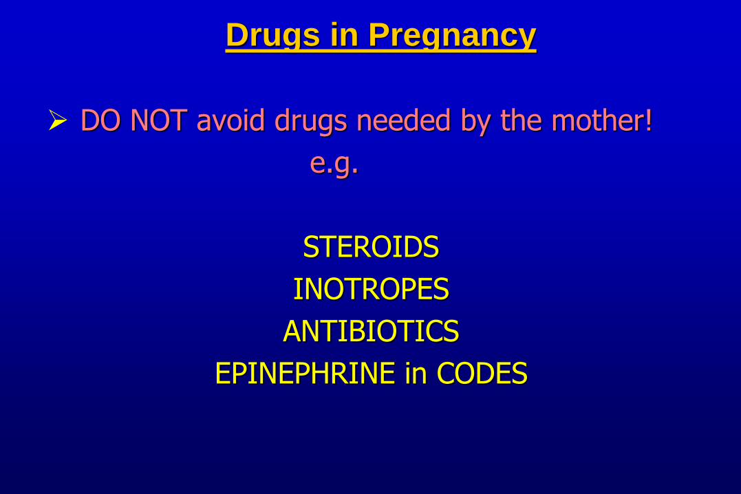
Drugs in Pregnancy
DO NOT avoid drugs needed by the mother!
e.g.
STEROIDS
INOTROPES
ANTIBIOTICS
EPINEPHRINE in CODES
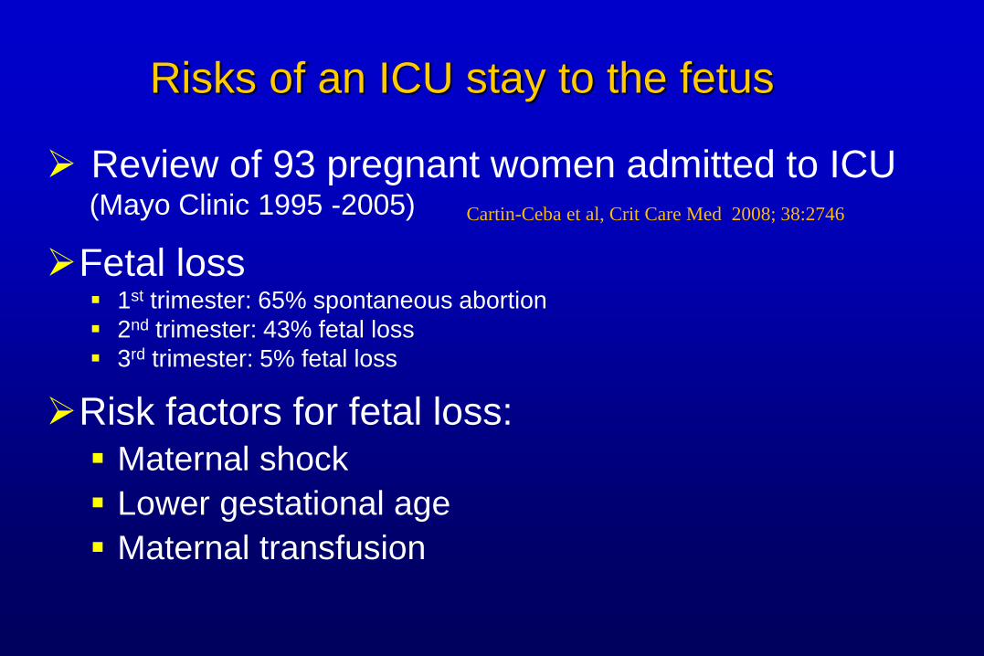
Review of 93 pregnant women admitted to ICU(Mayo Clinic 1995 -2005)
Fetal loss 1st trimester: 65% spontaneous abortion
2nd trimester: 43% fetal loss
3rd trimester: 5% fetal loss
Risk factors for fetal loss: Maternal shock
Lower gestational age
Maternal transfusion
Risks of an ICU stay to the fetus
Cartin-Ceba et al, Crit Care Med 2008; 38:2746
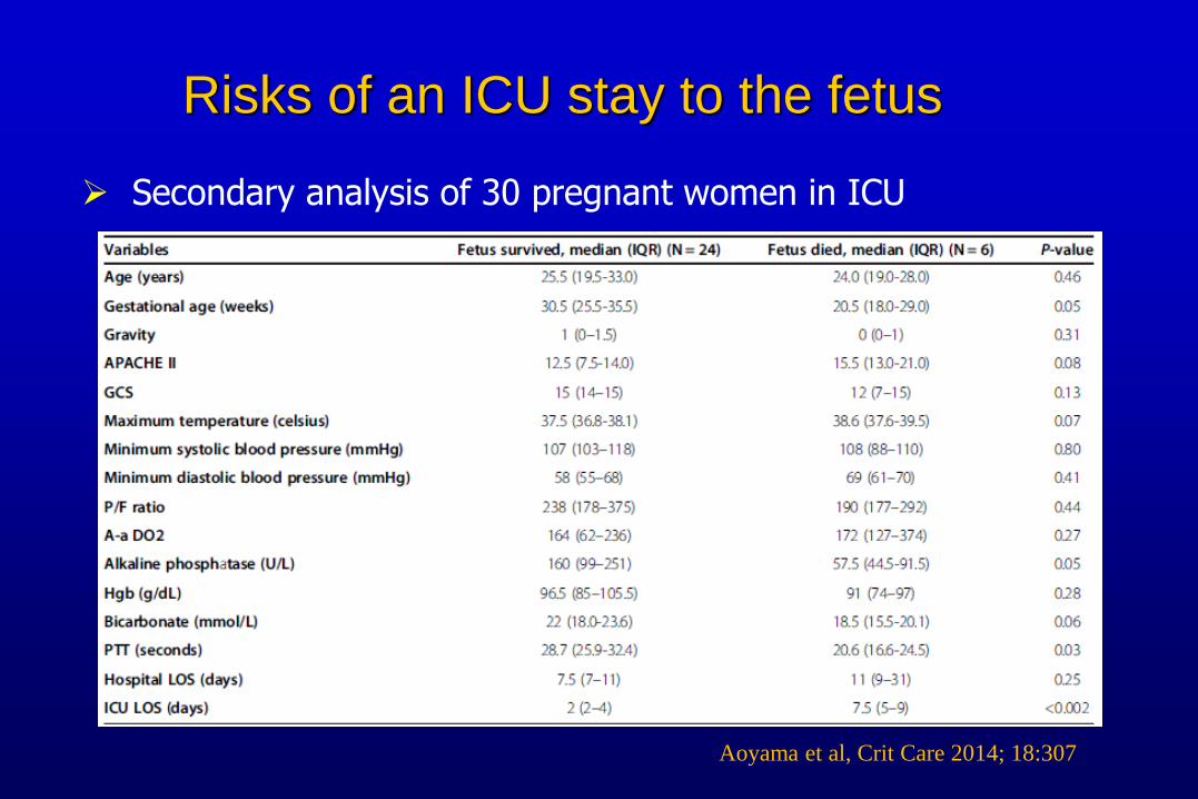
Risks of an ICU stay to the fetus
Aoyama et al, Crit Care 2014; 18:307
Secondary analysis of 30 pregnant women in ICU

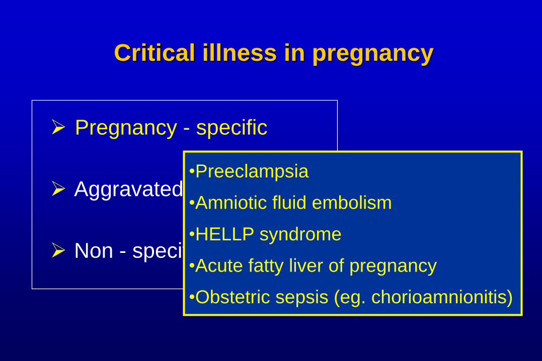
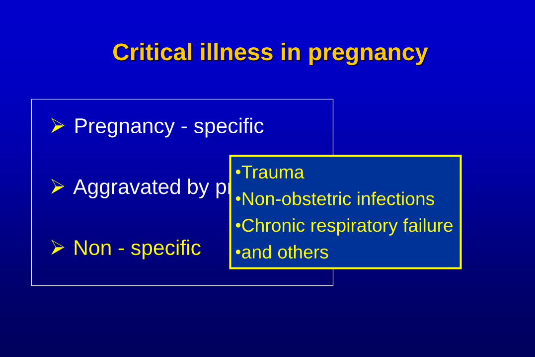
Critical illness in pregnancy
Pregnancy - specific
Aggravated by pregnancy
Non - specific
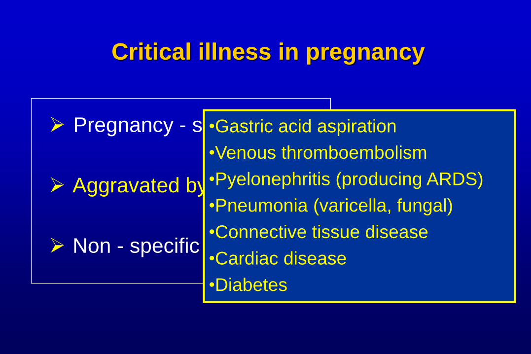
Critical illness in pregnancy
Pregnancy - specific
Aggravated by pregnancy
Non - specific
•Preeclampsia
•Amniotic fluid embolism
•HELLP syndrome
•Acute fatty liver of pregnancy
•Obstetric sepsis (eg. chorioamnionitis)
Critical illness in pregnancy
Pregnancy - specific
Aggravated by pregnancy
Non - specific
•Gastric acid aspiration
•Venous thromboembolism
•Pyelonephritis (producing ARDS)
•Pneumonia (varicella, fungal)
•Connective tissue disease
•Cardiac disease
•Diabetes
Critical illness in pregnancy
Pregnancy - specific
Aggravated by pregnancy
Non - specific
•Trauma
•Non-obstetric infections
•Chronic respiratory failure
•and others

Preeclampsia
Syndrome of
hypertension
proteinuria
after 20 weeks gestation
Complications:
pulmonary edema
cerebral edema
hypertensive crises
renal failure
eclampsia (seizures)
hepatic: HELLP
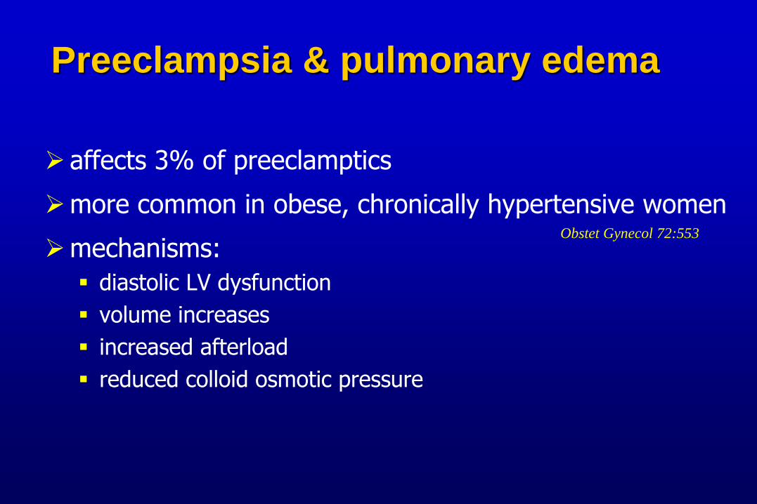
Preeclampsia & pulmonary edema
affects 3% of preeclamptics
more common in obese, chronically hypertensive women
mechanisms:
diastolic LV dysfunction
volume increases
increased afterload
reduced colloid osmotic pressure
Obstet Gynecol 72:553
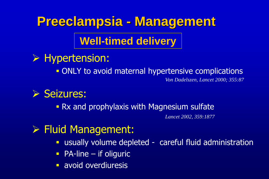
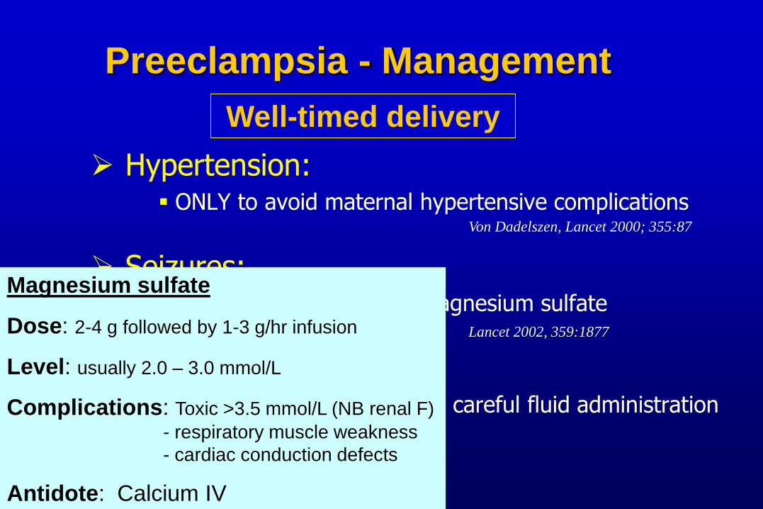
Preeclampsia - Management
Hypertension: ONLY to avoid maternal hypertensive complications
Seizures: Rx and prophylaxis with Magnesium sulfate
Fluid Management: usually volume depleted - careful fluid administration
PA-line – if oliguric
avoid overdiuresis
Well-timed delivery
Lancet 2002, 359:1877
Von Dadelszen, Lancet 2000; 355:87
Magnesium sulfate
Dose: 2-4 g followed by 1-3 g/hr infusion
Level: usually 2.0 – 3.0 mmol/L
Complications: Toxic >3.5 mmol/L (NB renal F)
- respiratory muscle weakness
- cardiac conduction defects
Antidote: Calcium IV

Preeclampsia - Management
Hypertension: ONLY to avoid maternal hypertensive complications
Seizures: Rx and prophylaxis with Magnesium sulfate
Fluid Management: usually volume depleted - careful fluid administration
PA-line – if oliguric
avoid overdiuresis
Well-timed delivery
Lancet 2002, 359:1877
Von Dadelszen, Lancet 2000; 355:87
HELLP syndrome
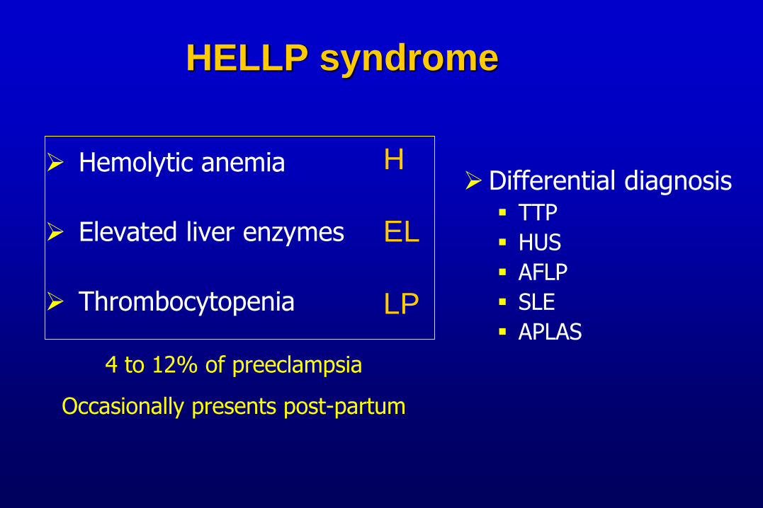
Hemolytic anemia
Elevated liver enzymes
Thrombocytopenia
4 to 12% of preeclampsia
Occasionally presents post-partum
Differential diagnosis TTP
HUS
AFLP
SLE
APLAS
HELLP syndrome
H
EL
LP
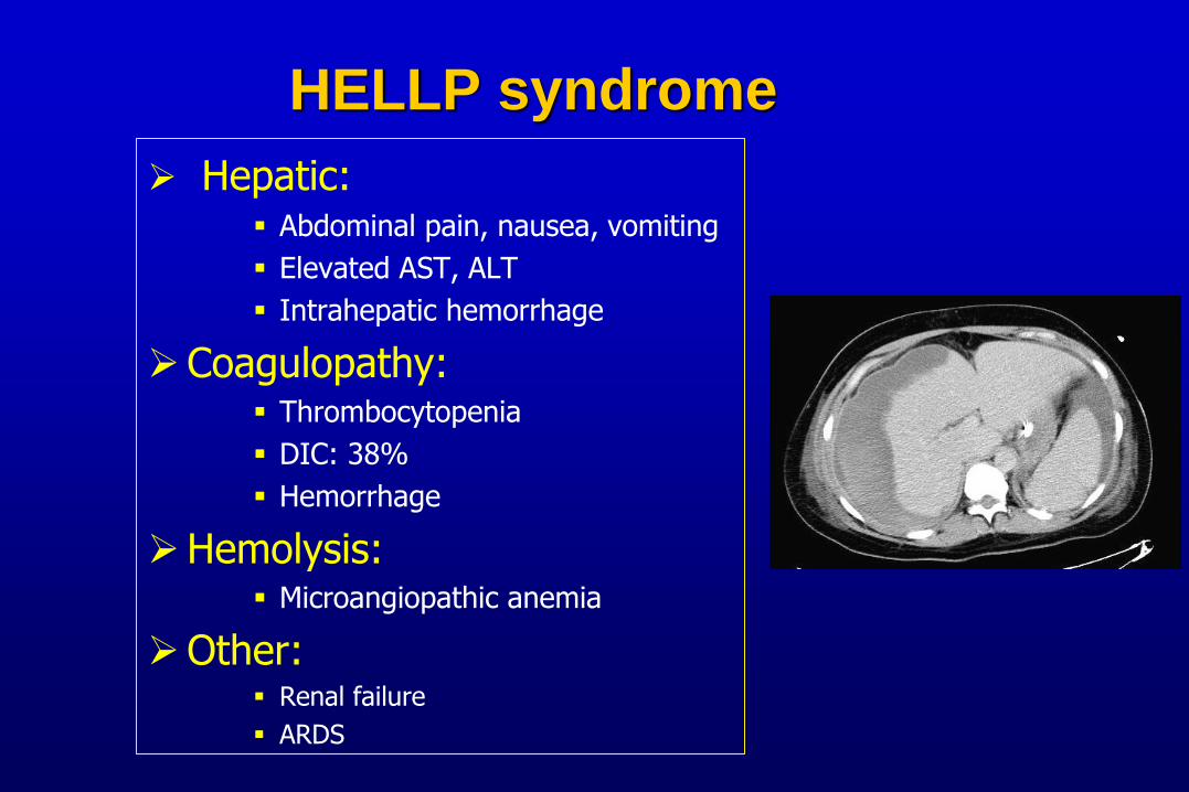
Hepatic: Abdominal pain, nausea, vomiting
Elevated AST, ALT
Intrahepatic hemorrhage
Coagulopathy: Thrombocytopenia
DIC: 38%
Hemorrhage
Hemolysis: Microangiopathic anemia
Other: Renal failure
ARDS
HELLP syndrome
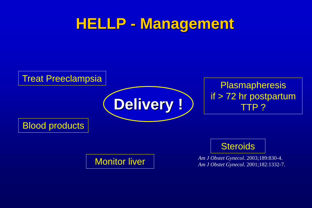
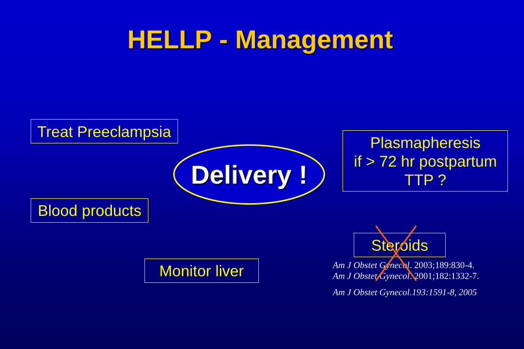
HELLP - Management
Delivery !
Treat Preeclampsia
Blood products
Monitor liver
Plasmapheresis
if > 72 hr postpartum
TTP ?
SteroidsAm J Obstet Gynecol. 2003;189:830-4.
Am J Obstet Gynecol. 2001;182:1332-7.
HELLP - Management
Delivery !
Treat Preeclampsia
Blood products
Monitor liver
Plasmapheresis
if > 72 hr postpartum
TTP ?
SteroidsAm J Obstet Gynecol. 2003;189:830-4.
Am J Obstet Gynecol. 2001;182:1332-7.
Am J Obstet Gynecol.193:1591-8, 2005

Acute Fatty Liver of Pregnancy

Clinical Features
onset usually late third trimester
anorexia, vomiting, jaundice
abdominal pain
coagulopathy, encephalopathy, renal failure
uncommon 1 in 15,000 pregnancies
Early reports: fulminant hepatic failure, high mortality
More recently: early recognition, improved outcome
Acute Fatty Liver of Pregnancy
Acute Fatty Liver of Pregnancy
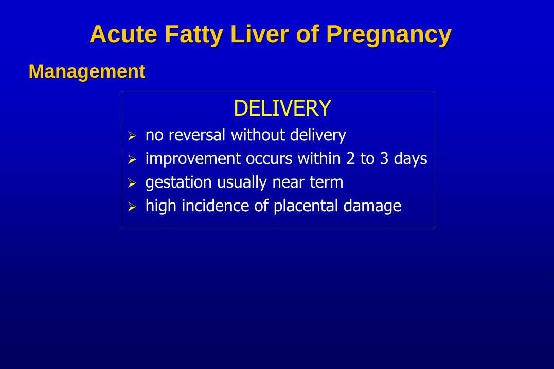
DELIVERY no reversal without delivery
improvement occurs within 2 to 3 days
gestation usually near term
high incidence of placental damage
Management
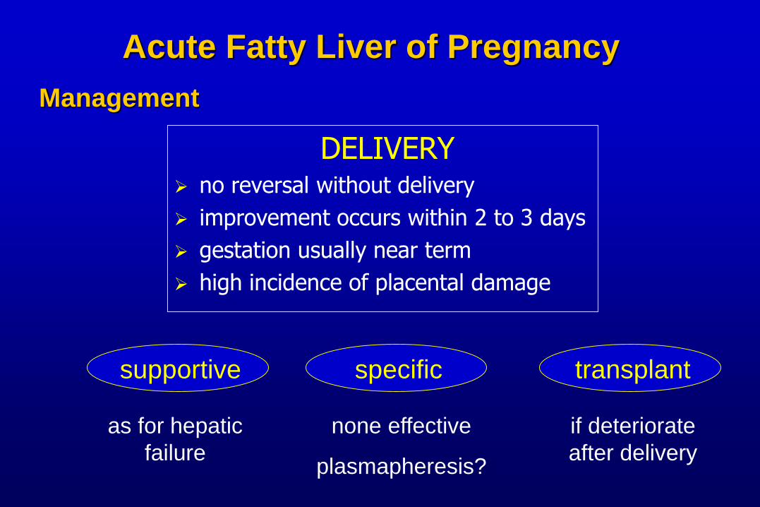
Acute Fatty Liver of Pregnancy
Management
supportive specific transplant
as for hepatic
failure
none effective
plasmapheresis?
if deteriorate
after delivery
DELIVERY no reversal without delivery
improvement occurs within 2 to 3 days
gestation usually near term
high incidence of placental damage

Liver disease in pregnancy - Etiology
79% of mothers carrying a fetus homozygous for a specific mutation of long chain 3-hydroxyacyl-CoA dehydrogenase had AFLP
N Engl J Med 1999; 340:1723

Amniotic Fluid Embolism

Rare: 1/8,000 to 1/80,000
Catastrophic: mortality 10 - 86%
Presentation: cardiorespiratory collapse
fetal distress
cardiac arrest, seizures
Late effects: ARDS & DIC
Amniotic Fluid Embolism

Pathophysiology: amniotic fluid enters venous circulation
cellular contents and humoral factors
Abnormal maternal immune response
pulmonary hypertension & myocardial dysfunction
Management: Supportive - ventilation, inotropes
Steroids ?
Anticipate ARDS & DIC
Amniotic Fluid Embolism
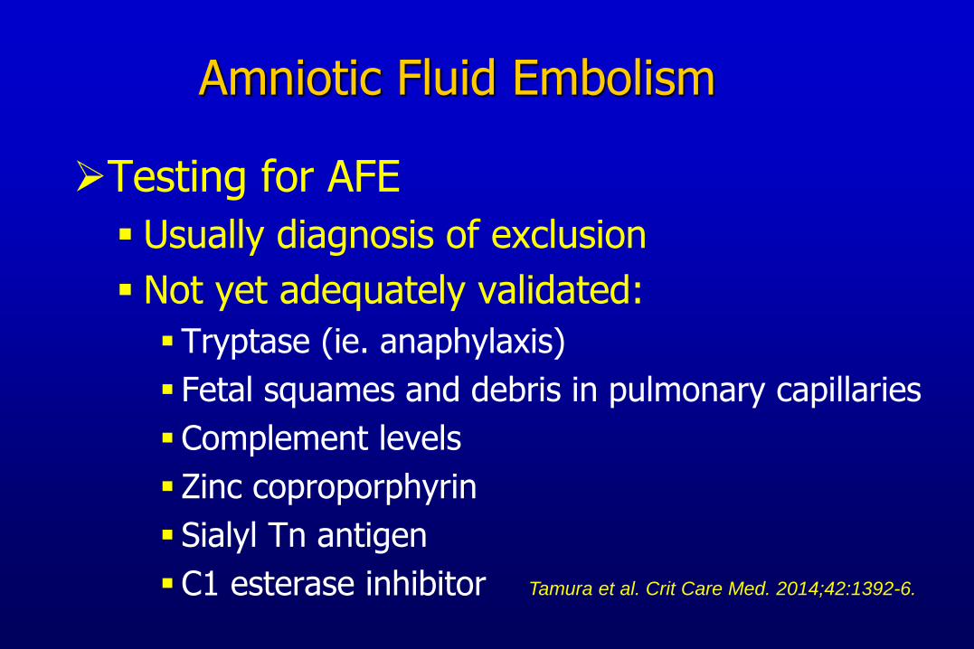
Testing for AFE
Usually diagnosis of exclusion
Not yet adequately validated:
Tryptase (ie. anaphylaxis)
Fetal squames and debris in pulmonary capillaries
Complement levels
Zinc coproporphyrin
Sialyl Tn antigen
C1 esterase inhibitor
Amniotic Fluid Embolism
Tamura et al. Crit Care Med. 2014;42:1392-6.
ICU Management
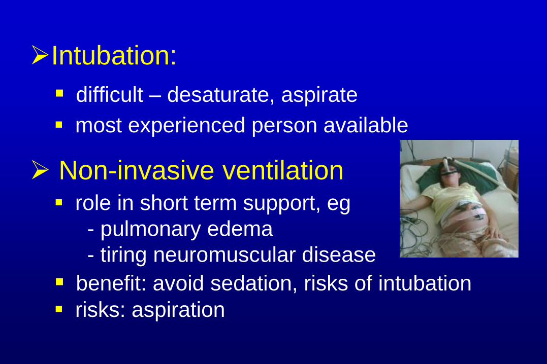
Intubation:
difficult – desaturate, aspirate
most experienced person available
Non-invasive ventilation role in short term support, eg
- pulmonary edema
- tiring neuromuscular disease
benefit: avoid sedation, risks of intubation
risks: aspiration
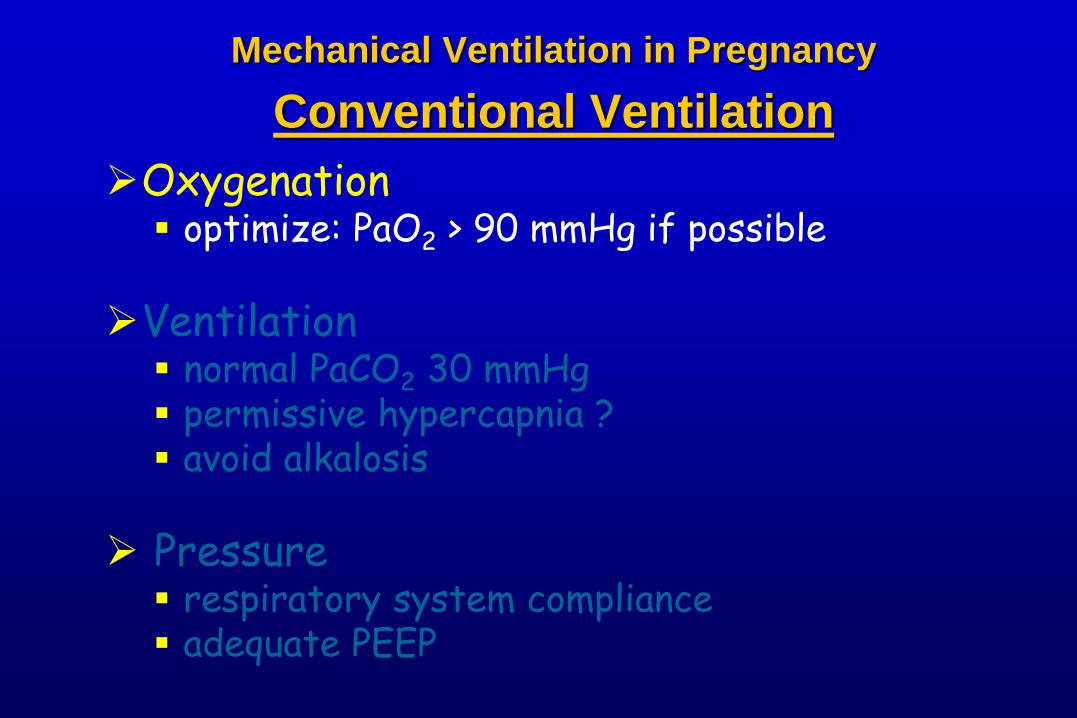
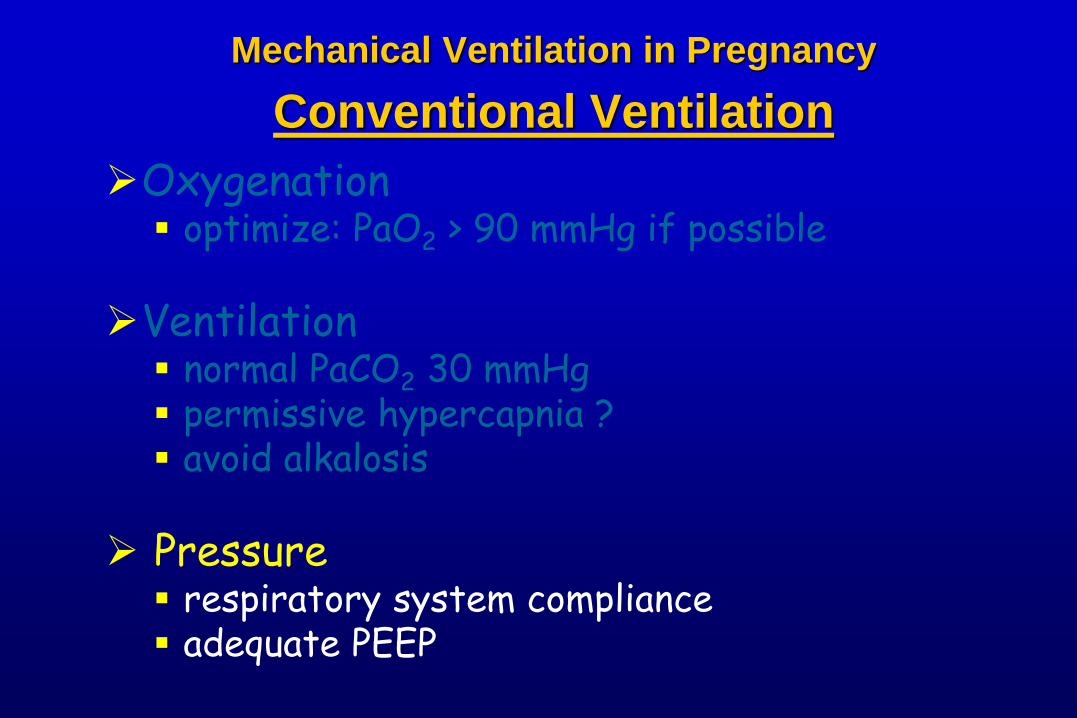
Mechanical Ventilation in Pregnancy
Conventional Ventilation
Oxygenation optimize: PaO2 > 90 mmHg if possible
Ventilation normal PaCO2 30 mmHg permissive hypercapnia ? avoid alkalosis
Pressure respiratory system compliance adequate PEEP
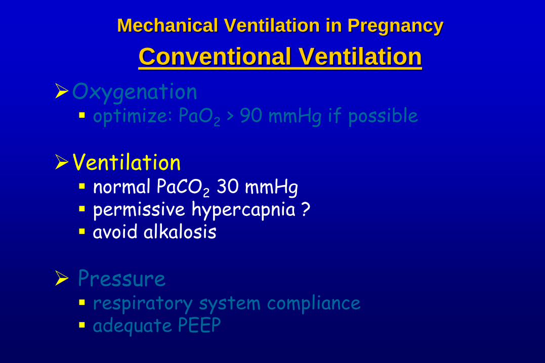
Mechanical Ventilation in Pregnancy
Conventional Ventilation
Oxygenation optimize: PaO2 > 90 mmHg if possible
Ventilation normal PaCO2 30 mmHg permissive hypercapnia ? avoid alkalosis
Pressure respiratory system compliance adequate PEEP

Hypocapnia Maternal PaCO2 < 25 mmHg:
- decreased UBF ->fetal hypoxia and acidosis
Hypercapnia Fetal acidemia 2o to maternal acidemia: not 2o to fetal hypoxemia
Maternal PaCO2 of 50-60 mmHg seems well tolerated
Small studies: mild hypoventilation (pH 7.36) better tolerated by fetus than mild hyperventilation (pH7.5)
Peng et al, Br J Anasth 1972, 44:1173
Buss Am J Physiol 1975; 228:1497
Clark Anesth Analg 1971; 50:713
Conventional Ventilation
Hollemen, Acta Anaesth Scan 1972, 221
Ivankovic et al, Am J Obstet Gynecol 1970
Mechanical Ventilation in Pregnancy
Conventional Ventilation
Oxygenation optimize: PaO2 > 90 mmHg if possible
Ventilation normal PaCO2 30 mmHg permissive hypercapnia ? avoid alkalosis
Pressure respiratory system compliance adequate PEEP
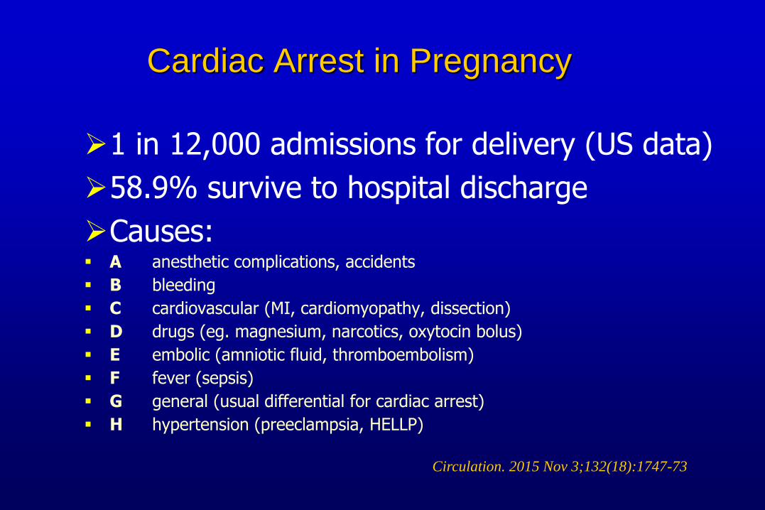
Cardiac Arrest in Pregnancy
1 in 12,000 admissions for delivery (US data)
58.9% survive to hospital discharge
Causes: A anesthetic complications, accidents
B bleeding
C cardiovascular (MI, cardiomyopathy, dissection)
D drugs (eg. magnesium, narcotics, oxytocin bolus)
E embolic (amniotic fluid, thromboembolism)
F fever (sepsis)
G general (usual differential for cardiac arrest)
H hypertension (preeclampsia, HELLP)
Circulation. 2015 Nov 3;132(18):1747-73
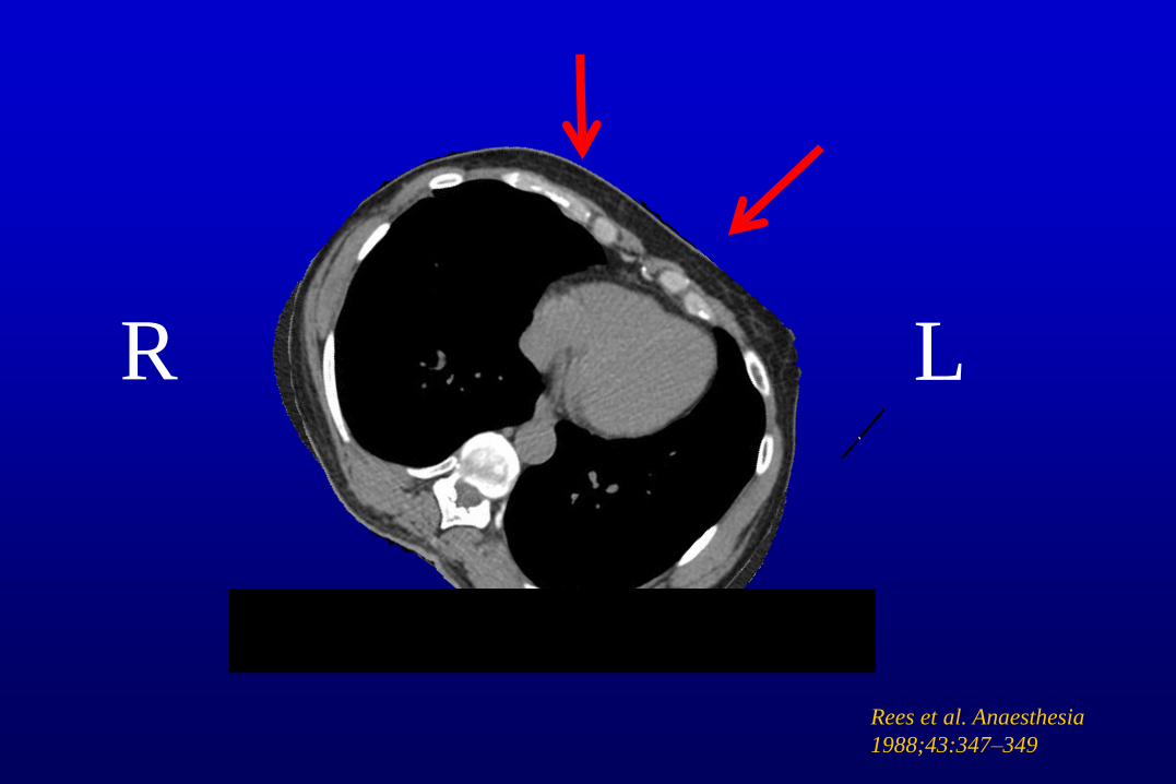
R L
Rees et al. Anaesthesia
1988;43:347–349

Cardiac Arrest in PregnancyManagement differences:

Cardiac Arrest in PregnancyManagement differences:
Manually displace uterus to left
No change in defibrillation
No change in drug therapy
Attention to oxygenation
Place IV access above the diaphragm
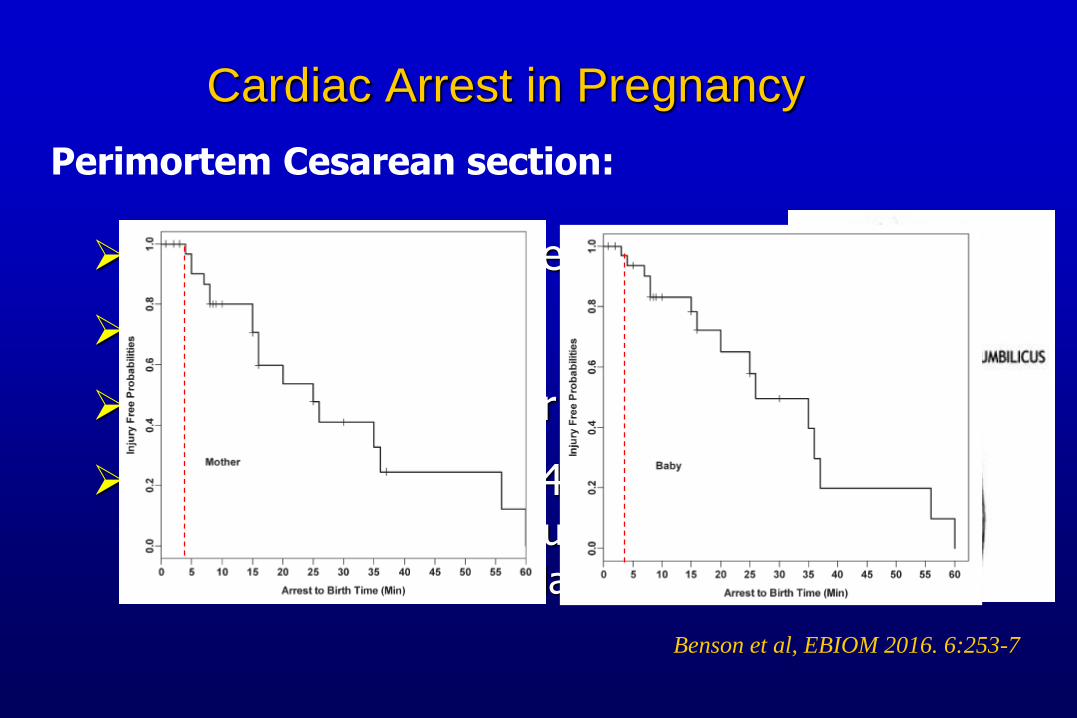
Perimortem Cesarean section

Cardiac Arrest in Pregnancy
Perimortem Cesarean section:
Who? Potentially viable fetus
Don’t move patient
Benefit to both mother and fetus
Initiate if no ROSC at 4 minutes Immediately, if timing unclear
Still beneficial if much later
Cardiac Arrest in Pregnancy
Perimortem Cesarean section:
Who? Potentially viable fetus
Don’t move patient
Benefit to both mother and fetus
Initiate if no ROSC at 4 minutes Immediately, if timing unclear
Still beneficial if much later
Benson et al, EBIOM 2016. 6:253-7
Sedation & NM blockade
Fetal monitoring
Delivery
Other Management Issues
Other Management Issues
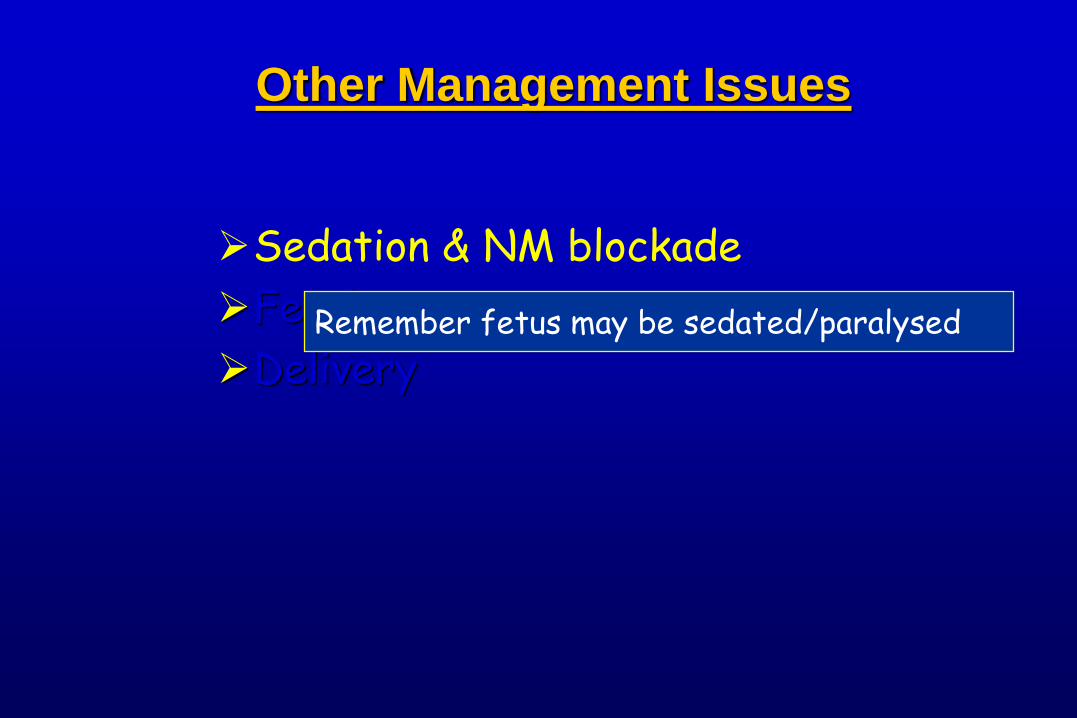
Sedation & NM blockade
Fetal monitoring
DeliveryRemember fetus may be sedated/paralysed
Other Management Issues
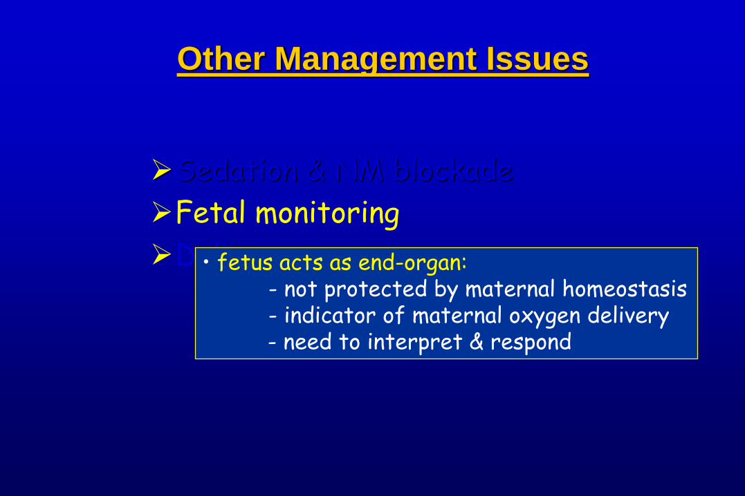
Sedation & NM blockade
Fetal monitoring
Delivery • fetus acts as end-organ:- not protected by maternal homeostasis- indicator of maternal oxygen delivery- need to interpret & respond

Other Management Issues
Sedation & NM blockade
Fetal monitoring
Delivery

Delivery of the fetus
Given the physiological changes, it may be considered that delivery of the pregnant women with respiratory failure is beneficial to the mother
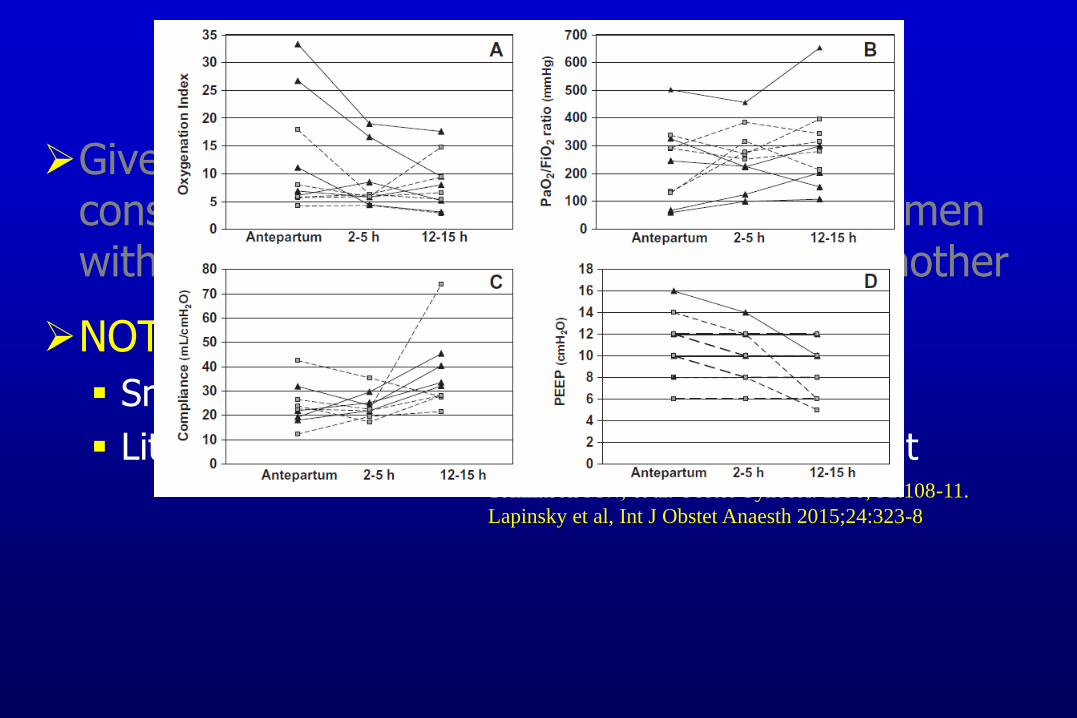
Delivery of the fetus
Given the physiological changes, it may be considered that delivery of the pregnant women with respiratory failure is beneficial to the mother
NOT always the case:
Small oxygenation improvement
Little change in compliance or PEEP requirementTomlinson MW, et al. Obstet Gynecol. 1998; 91:108-11.
Lapinsky et al, Int J Obstet Anaesth 2015;24:323-8
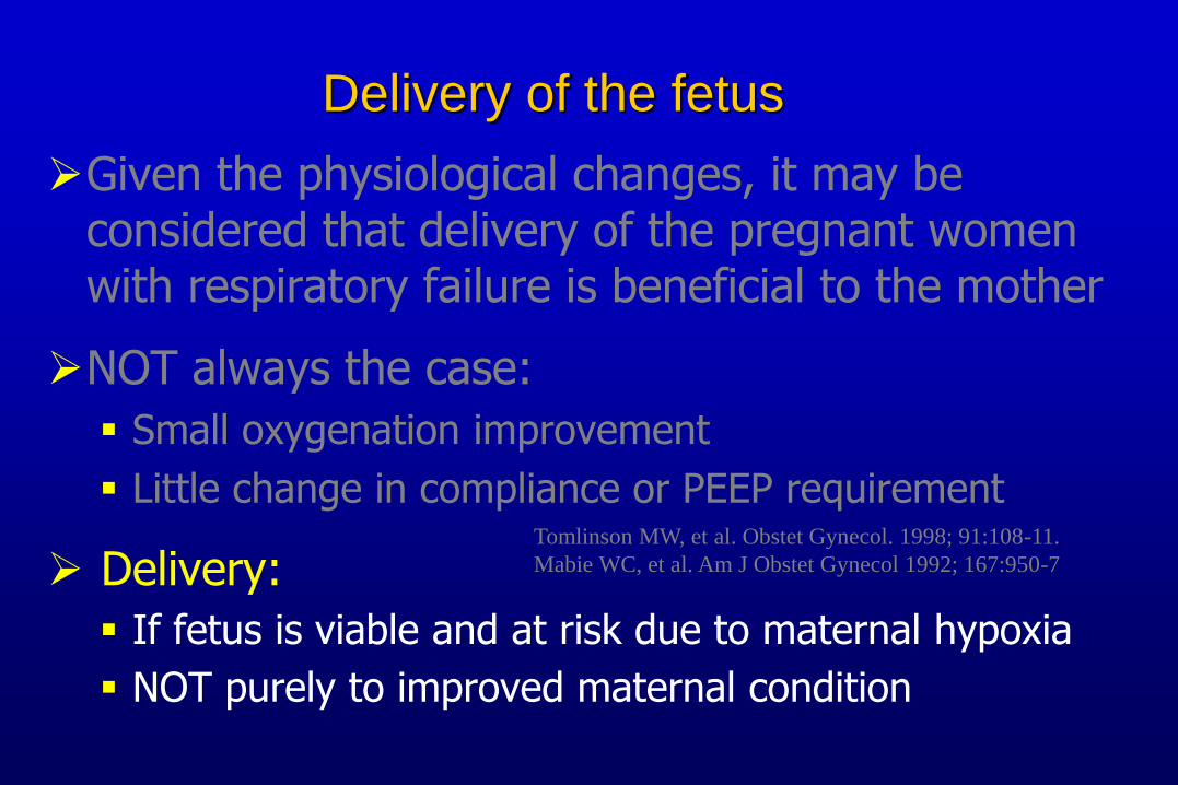
Delivery of the fetus
Given the physiological changes, it may be considered that delivery of the pregnant women with respiratory failure is beneficial to the mother
NOT always the case:
Small oxygenation improvement
Little change in compliance or PEEP requirement
Delivery:
If fetus is viable and at risk due to maternal hypoxia
NOT purely to improved maternal condition
Tomlinson MW, et al. Obstet Gynecol. 1998; 91:108-11.
Mabie WC, et al. Am J Obstet Gynecol 1992; 167:950-7
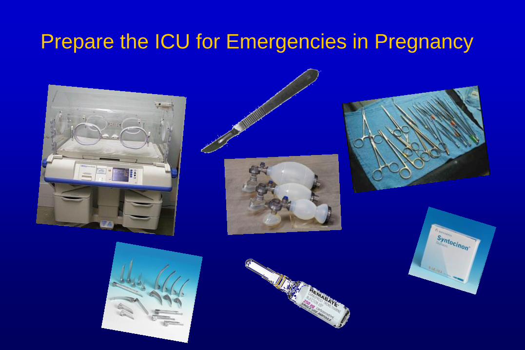
Prepare the ICU for Emergencies in Pregnancy
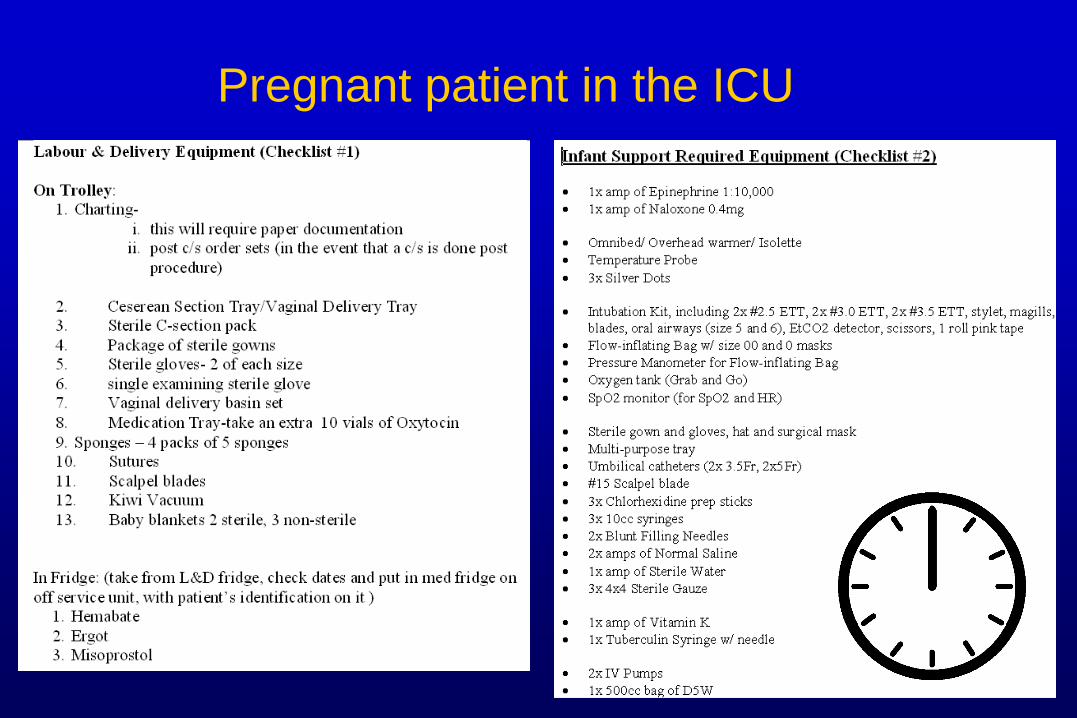
Pregnant patient in the ICU
