case 40-2012: a 43-year-old woman with cardiorespiratory
TRANSCRIPT

case records of the massachusetts general hospital
T h e n e w e ngl a nd j o u r na l o f m e dic i n e
n engl j med 367;26 nejm.org december 27, 20122528
Founded by Richard C. CabotNancy Lee Harris, m.d., Editor Eric S. Rosenberg, m.d., EditorJo-Anne O. Shepard, m.d., Associate Editor Alice M. Cort, m.d., Associate EditorSally H. Ebeling, Assistant Editor Emily K. McDonald, Assistant Editor
Case 40-2012: A 43-Year-Old Woman with Cardiorespiratory Arrest
after a Cesarean SectionJeffrey L. Ecker, M.D., Ken Solt, M.D., Michael G. Fitzsimons, M.D.,
and Thomas E. MacGillivray, M.D.
From the Department of Obstetrics and Gynecology (J.L.E.), the Divisions of Obstetrical Anesthesia (K.S.) and Cardiac Anesthesia (M.G.F.), Department of Anes-thesia, Critical Care, and Pain Medicine, and the Department of Surgery (T.E.M.), Massachusetts General Hospital; and the Departments of Obstetrics, Gynecology, and Reproductive Biology (J.L.E.), Anes-thesia (K.S., M.G.F.), and Surgery (T.E.M.), Harvard Medical School — both in Boston.
N Engl J Med 2012;367:2528-36.DOI: 10.1056/NEJMcpc1201413Copyright © 2012 Massachusetts Medical Society.
PR ESEN TATION OF C A SE
Dr. Britta Panda (Obstetrics and Gynecology): A 43-year-old woman (a multigravida) was admitted to the labor and delivery service of this hospital at 36.4 weeks of ges-tation because of vaginal bleeding.
The patient had had regular prenatal care at this hospital; routine prenatal screening tests were negative. She had had four previous uncomplicated pregnan-cies, with spontaneous vaginal deliveries, the most recent 14 years earlier. She had a history of obesity (prenatal body-mass index [the weight in kilograms divided by the square of the height in meters], 32.1) and migraine headaches; she had no known allergies. She did not smoke, drink alcohol, or use illicit drugs. Marginal placenta previa was seen on obstetrical ultrasonography, and two episodes of bleeding occurred, at 27.7 weeks and 32.7 weeks of gestation, which resolved after the patient was admitted for bed rest, hydration, and the administration of beta-methasone. She was advised to maintain bed rest at home and continue prenatal visits; the medications on discharge were iron sulfate and prenatal vitamins. At a routine prenatal visit 8 days before this presentation (at 35.3 weeks of gestation), obstetrical ultrasonography revealed an anterior placenta with the edge covering the internal os, a finding consistent with placenta previa. Heavy vaginal bleeding developed on the day of admission; the patient came to this hospital and was admitted to labor and delivery.
On examination in the labor and delivery unit, the vital signs and oxygen satu-ration were normal; there was active vaginal bleeding. Plans were made for emer-gency cesarean delivery. The hematocrit was 30.9% (reference range in women who are not pregnant, 36.0 to 46.0), the hemoglobin level was 10.7 g per deciliter (ref-erence range, 12.0 to 16.0), and the ABO blood type was O, Rh-positive, with negative antibody screening; the white-cell and platelet counts were normal. A spinal anesthetic was administered, and a cesarean section was performed, with delivery of a healthy boy; the 1-minute and 5-minute Apgar scores were 8 and 9, respectively. The placenta previa was removed. Twenty minutes after delivery, as the
The New England Journal of Medicine Downloaded from nejm.org by JOHN VOGEL on December 27, 2012. For personal use only. No other uses without permission.
Copyright © 2012 Massachusetts Medical Society. All rights reserved.

case records of the massachusetts gener al hospital
n engl j med 367;26 nejm.org december 27, 2012 2529
abdominal fascia was being closed, the patient’s systolic blood pressure fell to 70 to 80 mm Hg, the pulse to 30 to 39 beats per minute, and oxy-gen saturation to 70 to 80%. The patient re-ported chest pain, and her lips became white; apnea developed rapidly thereafter, and she be-came unresponsive. Electrical activity was pres-ent on electrocardiography (ECG), but radial and carotid pulses were not palpable. The trachea was intubated, and cardiopulmonary resuscita-tion was begun, with closed chest compressions and the administration of pressors.
Diagnostic procedures were performed, and additional management decisions were made.
DIFFER EN TI A L DI AGNOSIS
Dr. Jeffrey L. Ecker: All discussants are aware of the diagnosis in this case. This healthy woman had a sudden and profound cardiorespiratory collapse in the middle of what appeared to be an uncom-plicated cesarean delivery at term. Such situations require clinicians to focus promptly on a most-likely diagnosis in order to direct problem-specific therapies.
Causes of sudden intraoperative circulatory collapse
Primary cardiac events need to be considered as a cause of intraoperative cardiorespiratory collapse. Myocardial ischemia and infarction can cause profound alteration in vital signs, yet this patient was relatively young and had no history of heart disease, and the intraoperative cardiac monitor-ing did not suggest ischemic changes. Arrhythmias may also produce dramatic alterations in physiol-ogy, but the only dysrhythmia seen in this case was a bradycardia, a finding associated with what we soon determined was the likely diagnosis.
Complications associated with anesthesia also need to be considered. Regional anesthesia af-fecting a higher spinal level than expected can cause apnea but would be unlikely to lead imme-diately and concomitantly to loss of respiration and cardiac output. Inadvertent intravascular in-jection of anesthetic agents has been associated with cardiovascular collapse; in this case, how-ever, so much time had passed since the success-ful initiation of anesthesia that it seems unlikely that the subsequent events were directly linked to an anesthetic complication.
Embolic events can produce sudden collapse
of the sort seen in this case. There are three varieties of emboli. A thrombotic pulmonary embolus belongs in the differential diagnosis. However, formation (and migration) of a clot of the necessary size after less than 30 minutes of immobilization seems unlikely, especially since the patient was wearing intermittent pneumatic-compression stockings, as we use in all patients undergoing cesarean deliveries. Air emboli have been reported at the time of cesarean deliveries.1 Although this patient had received no intrave-nous injections close to the time of collapse, the uterus had been exteriorized for repair of the hysterotomy; since it had been raised slightly above the level of the heart, the possibility of an air embolus cannot be entirely ruled out. Embo-lization of amniotic fluid and fetal material is the third type of embolic event we considered.
Amniotic-fluid embolismAmniotic-fluid embolism is thought to result from maternal reaction to fetal material entering the pulmonary circulation and can cause apnea, hypotension, and bradycardia. Amniotic-fluid em-bolism, although rare, is well known and feared by obstetricians, and because it seemed the most likely explanation for this patient’s condition, this diagnosis will be the focus of the discussion here, as it was that day.
Two analyses of administrative data report amniotic-fluid embolism as a complication in 1 in 10,000 and 1 in 100,000 deliveries.2,3 Amniotic-fluid embolisms are described in association with both vaginal and cesarean deliveries and can occur at any time during labor and delivery, as well as during the postpartum period. Risk factors for amniotic-fluid embolism include ad-vanced maternal age, precipitous labor, cesarean delivery, and conditions associated with bleed-ing in pregnancy. This patient was of older ma-ternal age, with placenta previa, and she had just undergone cesarean delivery. However, regard-less of how many risk factors one patient has, each individual risk factor is relatively common, although amniotic-fluid embolism itself is quite rare; therefore, the conclusion of many observers is that amniotic-fluid embolism is unpredictable and unpreventable.
The clinical presentation of amniotic-fluid embolism includes cardiovascular collapse (brady-cardia or hypotension or both), apnea, bleeding, or evidence of fetal compromise (if delivery has
The New England Journal of Medicine Downloaded from nejm.org by JOHN VOGEL on December 27, 2012. For personal use only. No other uses without permission.
Copyright © 2012 Massachusetts Medical Society. All rights reserved.

T h e n e w e ngl a nd j o u r na l o f m e dic i n e
n engl j med 367;26 nejm.org december 27, 20122530
not yet occurred). Often, as in this patient, there is a period of anxiety or agitation preceding the alteration in vital signs and physiology.3
Pathophysiologically, amniotic-fluid embo-lism is thought to be a maternal anaphylactic reaction that unfolds in two phases. A first phase, often lasting less than 30 minutes, is marked by sudden pulmonary vasoconstriction with result-ing pulmonary hypertension and right-sided heart failure. This is followed by a second phase, in-volving left-sided heart failure, endothelial acti-vation and subsequent leakage, and bleeding, all probably caused in part by either the hypoxemia of the first phase or the release of injury-associ-ated agents in the serum. It is not known why some women mount a vigorous reaction and others apparently little reaction at all. However, individual variation, including variation in the time until any response is apparent, most likely accounts for the observation that many cases of amniotic-fluid embolism occur after delivery.4-7
Amniotic-fluid embolism poses a serious risk to a mother’s life and is a leading cause of ma-ternal death in the developed world; in a report from the Centers for Disease Control and Pre-vention, the condition accounts for 5% of mater-nal deaths.8 It was not so long ago that survival of an amniotic-fluid embolism merited a case report; series from the 1970s and 1980s report mortality rates associated with amniotic-fluid embolism of 50% or higher.4 More recent series from both the United States and United Kingdom report mortality rates of approximately 20%.2,3,9
Treatment of an amniotic-fluid embolism is supportive. As was done in this patient, the air-way should be secured and supplemental oxygen delivered. Either crystalloid or blood products are used with pressors as needed to maintain blood pressure. Although volume may be needed acutely, all involved need be mindful of the sec-ond phase of amniotic-fluid embolism, in which peripheral and pulmonary edema are prevalent; central monitoring may be vital. Extraordinary measures, including cardiopulmonary bypass,10 have been used to provide circulatory support and oxygen exchange when such resources are available. A final component of support in cases of amniotic-fluid embolism is treatment of bleed-ing due to either coagulopathy or atony. Replace-ment of blood products is central to treating ei-ther condition. Uterine atony should be treated
first with uterotonic agents, such as ergot de-rivatives and prostaglandins, tamponade from an intrauterine balloon, or some combination of these. If these measures are ineffective, hyster-ectomy may be considered, but performing an operation on women who have this condition and have ongoing coagulopathy is itself fraught with peril.
Unfortunately, there is no quick, standard confirmatory test for the diagnosis of amniotic-fluid embolism. The condition was diagnosed in the past when large amounts of fetal material were seen in a mother’s lungs at the time of autopsy; it was thought that the presence of fetal material in the pulmonary circulation was always pathological.11 However, pulmonary arterial-blood samples analyzed as part of studies of right heart catheterization in pregnant patients con-tained fetal cells more often than previously thought, and the presence of these cells did not always indicate clinical amniotic-f luid embo-lism.12 Therefore, amniotic-fluid embolism is a clinical diagnosis that is made after ruling out other common causes for a patient’s condition. It has been suggested that transesophageal echo-cardiography (TEE) may support the diagnosis.13
Our working diagnosis was amniotic-fluid embolism. The real story is in the resuscitation and the support that an institution like this one can provide to optimize the outcome in an indi-vidual patient. We quickly asked for help, and help quickly came. At the outset, I want to rec-ognize that many people participated in this patient’s care. Not all will speak today, but that in no way diminishes their vital contributions.
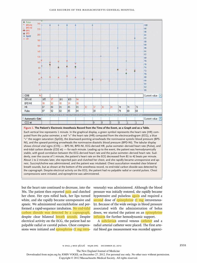
Dr. Ken Solt: Approximately 20 minutes after delivery of the neonate, as the abdominal fascia was being closed, the patient had an acute onset of bradycardia. Over the course of 1 minute, her heart rate decreased from 81 beats per minute to 42 beats per minute (Fig. 1). During cesarean section, a sudden increase in vagal tone may occur in response to manipulation of the uterus, fallopian tubes, or ovaries, resulting in acute bradycardia. However, this patient had bradycar-dia during closure of the abdominal fascia. Oc-casionally, a patient may have a vasovagal re-sponse to the stress of undergoing a major operation while awake, but this patient initially appeared well despite the bradycardia. We ad-ministered glycopyrrolate (0.2 mg intravenously),
The New England Journal of Medicine Downloaded from nejm.org by JOHN VOGEL on December 27, 2012. For personal use only. No other uses without permission.
Copyright © 2012 Massachusetts Medical Society. All rights reserved.
case records of the massachusetts gener al hospital
n engl j med 367;26 nejm.org december 27, 2012 2531
but the heart rate continued to decrease, into the 30s. The patient then reported pain and clutched her chest. Her eyes rolled back, her lips turned white, and she rapidly became unresponsive and apneic. We administered succinylcholine and per-formed a rapid-sequence intubation. No end-tidal carbon dioxide was detected by a capnograph, despite clear bilateral breath sounds. Despite electrical activity on the ECG, the patient had no palpable radial or carotid pulses. Chest compres-sions were initiated and epinephrine (1 mg intra-
venously) was administered. Although the blood pressure was initially restored, she rapidly became hypotensive and pulseless again and required a second dose of epinephrine (1 mg intravenous-ly). Because of the wide swings in blood pressure associated with the administration of bolus doses, we started the patient on an epinephrine infusion for further hemodynamic support.
A subclavian central venous catheter and a radial arterial catheter were placed. The first arte-rial blood gas measurement was recorded approxi-
Figure 1. The Patient’s Electronic Anesthesia Record from the Time of the Event, as a Graph and as a Table.
Each vertical line represents 1 minute. In the graphical display, a green symbol represents the heart rate (HR) com-puted from the pulse oximeter, a red “×” the heart rate (HR) computed from the electrocardiogram (ECG), a blue “+” the oxygen saturation (SpO2), the downward-pointing arrowheads the noninvasive systolic blood pressure (BPS-NI), and the upward-pointing arrowheads the noninvasive diastolic blood pressure (BPD-NI). The tabular display shows clinical vital signs (CVS) — BPS-NI, BPD-NI, ECG-derived HR, pulse oximeter–derived heart rate (Pulse), and end-tidal carbon dioxide (CO2 et) — for each minute. Leading up to the event, the patient was hemodynamically stable, with good correlation between the ECG-derived heart rate and the pulse oximeter–derived heart rate. Sud-denly, over the course of 1 minute, the patient’s heart rate on the ECG decreased from 81 to 42 beats per minute. About 1 to 2 minutes later, she reported pain and clutched her chest, and she rapidly became unresponsive and ap-neic. Succinylcholine was administered, and the patient was intubated. Chest auscultation revealed clear bilateral breath sounds, but as shown at the bottom of the anesthesia record, no end-tidal carbon dioxide was detected by the capnograph. Despite electrical activity on the ECG, the patient had no palpable radial or carotid pulses. Chest compressions were initiated, and epinephrine was administered.
The New England Journal of Medicine Downloaded from nejm.org by JOHN VOGEL on December 27, 2012. For personal use only. No other uses without permission.
Copyright © 2012 Massachusetts Medical Society. All rights reserved.

T h e n e w e ngl a nd j o u r na l o f m e dic i n e
n engl j med 367;26 nejm.org december 27, 20122532
mately 20 minutes after the initial arrest. This revealed a large gap between the partial pressure of arterial carbon dioxide (PaCO2) (48 mm Hg) and the end-tidal carbon dioxide (18 mm Hg). When both the PaCO2 and the end-tidal carbon dioxide are known, one may calculate an esti-mate of the fraction of dead space using the Bohr equation.14 Under normal conditions, ap-proximately 10% of the tidal volume is dead space, representing the anatomical dead space (i.e., the volume of the tracheobronchial tree, which does not participate in gas exchange). In this patient, the calculated dead space was 62.5% of the tidal volume, implying a large com-ponent of alveolar dead space (i.e., 50% of the alveoli were ventilated but not perfused). The end-tidal carbon dioxide of 0 at the time of intu-
bation indicated that the patient’s entire pulmo-nary system was dead space during the initial arrest.
In this patient with the sudden onset of bra-dycardia, chest pain, loss of consciousness, and apnea during cesarean delivery, performed be-cause of bleeding placenta previa, the finding of 100% pulmonary dead space in the context of cardiac arrest presenting as pulseless electrical activity rapidly led us to the preliminary diagno-sis of an amniotic-fluid embolism. We performed TEE to confirm the diagnosis.
Dr . Jeffr e y L . Eck er a nd Dr . K en Solt ’s Di agnosis
Amniotic-fluid embolism.
A
RA
IAS LA
LV
RV
RA
LA
LVRV
RVLVIVS
RATR
RV
B
DC
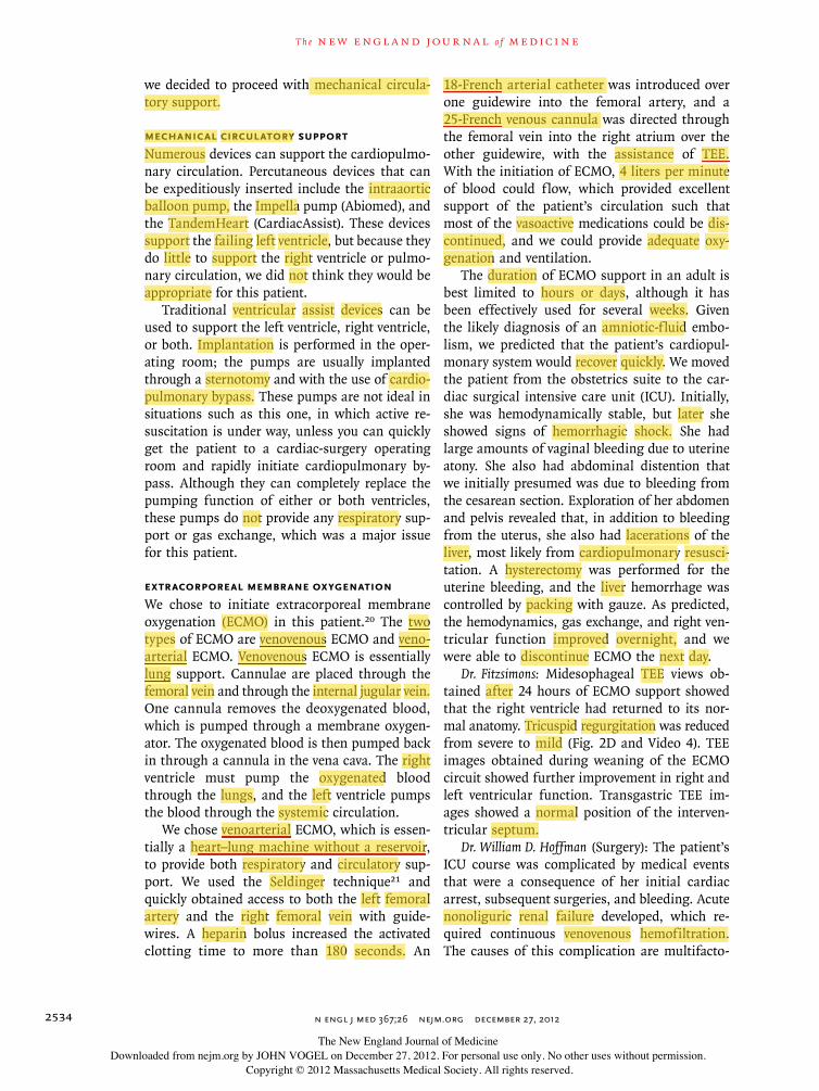
Figure 2. Intraoperative Transesophageal Echocardiograms.
A midesophageal four-chamber image (Panel A) shows dilatation of the right ventricle (RV) and right atrium (RA), with a shift of the interatrial septum (IAS) toward the left atrium (LA). A midesophageal view of the right ventricle and right atrium (Panel B) shows a large jet of tricuspid regurgitation (TR) into the right atrium, indicating severe tricuspid-valve insufficiency. A transgastric view of the right ventricle and left ventricle (LV) (Panel C) shows dilata-tion of the right ventricle, with a shift of the interventricular septum (IVS) toward the left ventricle. A midesophageal four-chamber view after 24 hours of support (Panel D) shows recovery of right ventricular function and resolution of tricuspid regurgitation.
The New England Journal of Medicine Downloaded from nejm.org by JOHN VOGEL on December 27, 2012. For personal use only. No other uses without permission.
Copyright © 2012 Massachusetts Medical Society. All rights reserved.

case records of the massachusetts gener al hospital
n engl j med 367;26 nejm.org december 27, 2012 2533
DI AGNOS TIC DISCUSSION
Dr. Michael G. Fitzsimons: Emergency TEE was per-formed by the cardiac anesthesia service (Fig. 2A, 2B, and 2C; and Videos 1, 2, and 3, available with the full text of this article at NEJM.org), and it revealed a dilated right atrium with a shift of the interatrial septum toward the left, severe tricus-pid valve insufficiency, mild-to-moderate pulmo-nary insufficiency, and shifting of the interven-tricular septum toward the left during systole, resulting in the classic D-shaped left ventricle. The cavity of the left ventricle appeared small. Over time, the right ventricle became more hypo-kinetic, with bulging of the free wall. There was no evidence of aortic dissection, clot in the prox-imal pulmonary artery, patent foramen ovale, or pericardial effusion.
TEE is a quick, portable, and reliable means of identifying the potential causes of hemody-namic collapse during labor and delivery. Amni-otic-fluid embolism causes intense pulmonary vasoconstriction and an acute pressure overload on the right ventricle, leading to dilatation and hypokinesis. Tricuspid regurgitation leads to dilatation of the right atrium and a shift of the interatrial septum toward the left. Hypotension results from impaired filling of the left ventricle associated with pulmonary vasoconstriction and a shift of the interventricular septum toward the
left, resulting in the classic D shape seen in transgastric images. The left ventricle initially appears small and underfilled but hyperkinetic. Worsening left ventricular function may be due to hypoxemia and ischemia. Dilatation of the pulmonary artery and pulmonary regurgitation further support the diagnosis of amniotic-fluid embolism. Several case reports10,13,15-19 have documented such TEE findings at the time of presumed amniotic-fluid embolism (Table 1).
The utility of TEE in acute cardiopulmonary collapse associated with labor and delivery is not limited to diagnosis but may also include man-agement, the evaluation of the placement of ve-nous and arterial cannulae for extracorporeal membranous oxygenation, the placement and effect of intraaortic balloon counterpulsation, or the effects of inotropic agents.
DISCUSSION OF M A NAGEMEN T
Dr. Thomas E. MacGillivray: When I arrived in the obstetrics suite, a well-coordinated resuscitation was under way. The patient was receiving infu-sions of high-dose vasoactive agents and inter-mittent chest compressions to maintain a palpa-ble pulse and blood pressure. Owing to the echocardiographic findings of right ventricular distention and an underfilled left ventricle, com-bined with the marginal hemodynamic status,
Table 1. Transesophageal Echocardiographic Findings in Acute Amniotic-Fluid Embolism.
Year Study Findings Outcome
2010 Lee et al.15 Severe right ventricular dysfunctionFree-floating clot in the right and left atria
Cardiopulmonary bypass and survival
2009 Vellayappan et al.19 Enlarged right ventricleModerate right ventricular hypokinesisLarge mass in the right atrium through patent
foramen ovaleDilated tricuspid valve annulusTrace-to-mild tricuspid regurgitationNormal left ventricle
Cardiopulmonary resuscitation and survival (pathology re-port showed squamous-cell epithelium in the mass)
2004 James et al.13 Normal left ventricular contractilityD-shaped left ventricleEnlarged pulmonary artery and right ventricleSluggish flow in the pulmonary arteries
Cardiopulmonary resuscitation and death
2003 Stanten et al.10 Massive right ventricular dilatation and akinesisVigorous, small left ventricle
Cardiopulmonary bypass and survival
1999 Shechtman et al.18 Right ventricular failureBulging of interatrial septum and interventricular
septum toward the leftSevere tricuspid regurgitationSmall and decompressed left ventricle
Cardiopulmonary resuscitation and death
Videos showing transesophageal echocardiography are available at NEJM.org
The New England Journal of Medicine Downloaded from nejm.org by JOHN VOGEL on December 27, 2012. For personal use only. No other uses without permission.
Copyright © 2012 Massachusetts Medical Society. All rights reserved.
T h e n e w e ngl a nd j o u r na l o f m e dic i n e
n engl j med 367;26 nejm.org december 27, 20122534
we decided to proceed with mechanical circula-tory support.
Mechanical circulatory supportNumerous devices can support the cardiopulmo-nary circulation. Percutaneous devices that can be expeditiously inserted include the intraaortic balloon pump, the Impella pump (Abiomed), and the TandemHeart (CardiacAssist). These devices support the failing left ventricle, but because they do little to support the right ventricle or pulmo-nary circulation, we did not think they would be appropriate for this patient.
Traditional ventricular assist devices can be used to support the left ventricle, right ventricle, or both. Implantation is performed in the oper-ating room; the pumps are usually implanted through a sternotomy and with the use of cardio-pulmonary bypass. These pumps are not ideal in situations such as this one, in which active re-suscitation is under way, unless you can quickly get the patient to a cardiac-surgery operating room and rapidly initiate cardiopulmonary by-pass. Although they can completely replace the pumping function of either or both ventricles, these pumps do not provide any respiratory sup-port or gas exchange, which was a major issue for this patient.
Extracorporeal Membrane OxygenationWe chose to initiate extracorporeal membrane oxygenation (ECMO) in this patient.20 The two types of ECMO are venovenous ECMO and veno-arterial ECMO. Venovenous ECMO is essentially lung support. Cannulae are placed through the femoral vein and through the internal jugular vein. One cannula removes the deoxygenated blood, which is pumped through a membrane oxygen-ator. The oxygenated blood is then pumped back in through a cannula in the vena cava. The right ventricle must pump the oxygenated blood through the lungs, and the left ventricle pumps the blood through the systemic circulation.
We chose venoarterial ECMO, which is essen-tially a heart–lung machine without a reservoir, to provide both respiratory and circulatory sup-port. We used the Seldinger technique21 and quickly obtained access to both the left femoral artery and the right femoral vein with guide-wires. A heparin bolus increased the activated clotting time to more than 180 seconds. An
18-French arterial catheter was introduced over one guidewire into the femoral artery, and a 25-French venous cannula was directed through the femoral vein into the right atrium over the other guidewire, with the assistance of TEE. With the initiation of ECMO, 4 liters per minute of blood could flow, which provided excellent support of the patient’s circulation such that most of the vasoactive medications could be dis-continued, and we could provide adequate oxy-genation and ventilation.
The duration of ECMO support in an adult is best limited to hours or days, although it has been effectively used for several weeks. Given the likely diagnosis of an amniotic-fluid embo-lism, we predicted that the patient’s cardiopul-monary system would recover quickly. We moved the patient from the obstetrics suite to the car-diac surgical intensive care unit (ICU). Initially, she was hemodynamically stable, but later she showed signs of hemorrhagic shock. She had large amounts of vaginal bleeding due to uterine atony. She also had abdominal distention that we initially presumed was due to bleeding from the cesarean section. Exploration of her abdomen and pelvis revealed that, in addition to bleeding from the uterus, she also had lacerations of the liver, most likely from cardiopulmonary resusci-tation. A hysterectomy was performed for the uterine bleeding, and the liver hemorrhage was controlled by packing with gauze. As predicted, the hemodynamics, gas exchange, and right ven-tricular function improved overnight, and we were able to discontinue ECMO the next day.
Dr. Fitzsimons: Midesophageal TEE views ob-tained after 24 hours of ECMO support showed that the right ventricle had returned to its nor-mal anatomy. Tricuspid regurgitation was reduced from severe to mild (Fig. 2D and Video 4). TEE images obtained during weaning of the ECMO circuit showed further improvement in right and left ventricular function. Transgastric TEE im-ages showed a normal position of the interven-tricular septum.
Dr. William D. Hoffman (Surgery): The patient’s ICU course was complicated by medical events that were a consequence of her initial cardiac arrest, subsequent surgeries, and bleeding. Acute nonoliguric renal failure developed, which re-quired continuous venovenous hemofiltration. The causes of this complication are multifacto-
The New England Journal of Medicine Downloaded from nejm.org by JOHN VOGEL on December 27, 2012. For personal use only. No other uses without permission.
Copyright © 2012 Massachusetts Medical Society. All rights reserved.

case records of the massachusetts gener al hospital
n engl j med 367;26 nejm.org december 27, 2012 2535
rial and include renal hypoperfusion and admin-istration of an aminoglycoside antibiotic, which was exchanged for another antibiotic early in the patient’s stay in the ICU. The patient was suffi-ciently awake that we could extubate and remove mechanical ventilation support 7 days after ad-mission.
The patient awoke with an encephalopathy characterized by lack of speech, intact language comprehension, intact nonverbal expression, nor-mal cranial-nerve function, and normal motor and sensory functions. The symptoms could not be attributed to an infarct in the territory of a major cerebral artery. Brain CT revealed no hem-orrhage or infarct. Her speech returned to nor-mal during the next 10 days. She was discharged from the ICU 13 days after admission.
Dr. Ecker: I met this patient only briefly on the morning of delivery. I have subsequently seen her several times and had many conversations with her, which was not something I expected given the initial events. She left the hospital after 32 days and was readmitted very briefly 2 weeks later for a pleural effusion, which resolved. Her baby is healthy.
Dr. Nancy Lee Harris (Pathology): Are there any questions?
A Physician: If initial severe vasoconstriction in the pulmonary vasculature is a trigger for the other events, are there other agents, such as
prostaglandins or sildenafil, that might address this pathophysiological feature?
Dr. MacGillivray: The pathophysiological fea-tures of pulmonary emboli are fascinating. The profound right ventricular dysfunction seems to be caused by more than just a mechanical ob-struction of the pulmonary artery. In thoracic surgery, we can clamp a large branch of the pulmonary artery without circulatory collapse. However, with a pulmonary embolism, there is frequently a severe, diffuse pulmonary vasocon-striction in addition to the mechanical obstruc-tion. Perhaps rapid initiation of a selective pul-monary arterial relaxant, such as nitric oxide, might be beneficial.
Dr. Ecker: One week after this conference, the patient was readmitted to the hospital with mul-tiple pulmonary emboli. A source of the emboli was never identified. The patient was given anti-coagulant agents and was discharged after 2 days. She remains well more than 1 year later.
FINA L DI AGNOSIS
Amniotic-fluid embolus.This case was discussed at Obstetrics and Gynecology
Grand Rounds. Dr. Michael F. Greene assisted with organizing the conference.
No potential conflict of interest relevant to this article was reported. Disclosure forms provided by the authors are avail-able with the full text of this article at NEJM.org.
References
1. Rodgers L, Dangel-Palmer MC, Berner N. Acute circulatory and respiratory col-lapse in obstetrical patients: a case report and review of the literature. AANA J 2000; 68:444-50.2. Abenhaim HA, Azoulay L, Kramer MS, Leduc L. Incidence and risk factors of amniotic fluid embolisms: a population-based study on 3 million births in the United States. Am J Obstet Gynecol 2008; 199:49.e1-49.e8.3. Knight M, Tuffnell D, Brocklehurst P, Spark P, Kurinczuk JJ. Incidence and risk factors for amniotic-fluid embolism. Ob-stet Gynecol 2010;115:910-7.4. Clark SL, Hankins GD, Dudley DA, Dildy GA, Porter TF. Amniotic fluid em-bolism: analysis of the national registry. Am J Obstet Gynecol 1995;172:1158-69.5. Gist RS, Stafford IP, Leibowitz AB, Beilin Y. Amniotic fluid embolism. Anesth Analg 2009;108:1599-602.6. Clark SL, Cotton DB, Gonik B, Greenspoon J, Phelan JP. Central hemody-namic alterations in amniotic fluid embo-
lism. Am J Obstet Gynecol 1988;158: 1124-6.7. Vanmaele L, Noppen M, Vincken W, De Catte L, Huyghens L. Transient left heart failure in amniotic fluid embolism. Intensive Care Med 1990;16:269-71.8. Berg CJ, Callaghan WM, Syverson C, Henderson Z. Pregnancy-related mortali-ty in the United States, 1998 to 2005. Ob-stet Gynecol 2010;116:1302-9.9. Gilbert WM, Danielsen B. Amniotic fluid embolism: decreased mortality in a population-based study. Obstet Gynecol 1999;93:973-7.10. Stanten RD, Iverson LI, Daugharty TM, Lovett SM, Terry C, Blumenstock E. Amniotic fluid embolism causing cata-strophic pulmonary vasoconstriction: diagnosis by transesophageal echocardio-gram and treatment by cardiopulmonary bypass. Obstet Gynecol 2003;102:496-8.11. Roche WD Jr, Norris HJ. Detection and significance of maternal pulmonary amniotic fluid embolism. Obstet Gynecol 1974;43:729-31.
12. Lee W, Ginsburg KA, Cotton DB, Kaufman RH. Squamous and trophoblas-tic cells in the maternal pulmonary circu-lation identified by invasive hemodynam-ic monitoring during the peripartum period. Am J Obstet Gynecol 1986;155: 999-1001.13. James CF, Feinglass NG, Menke DM, Grinton SF, Papadimos TJ. Massive amni-otic fluid embolism: diagnosis aided by emergency transesophageal echocardiog-raphy. Int J Obstet Anesth 2004;13:279-83.14. Respiratory monitoring. In: Miller RD, ed. Miller’s anesthesia. 6th ed. Vol. 1. New York: Churchill Livingstone, 2005: 1437-81.15. Lee PH, Shulman MS, Vellayappan U, Symes JF, Olenchock SA Jr. Surgical treat-ment of an amniotic fluid embolism with cardiopulmonary collapse. Ann Thorac Surg 2010;90:1694-6.16. Porat S, Leibowitz D, Milwidsky A, Valsky DV, Yagel S, Anteby EY. Transient intracardiac thrombi in amniotic fluid embolism. BJOG 2004;111:506-10.
The New England Journal of Medicine Downloaded from nejm.org by JOHN VOGEL on December 27, 2012. For personal use only. No other uses without permission.
Copyright © 2012 Massachusetts Medical Society. All rights reserved.

n engl j med 367;26 nejm.org december 27, 20122536
case records of the massachusetts gener al hospital
17. Saad A, El-Husseini N, Nader GA, Gharzuddine W. Echocardiographically detected mass “in transit” in early amni-otic fluid embolism. Eur J Echocardiogr 2006;7:332-5.18. Shechtman M, Ziser A, Markovits R, Rozenberg B. Amniotic fluid embolism: early findings of transesophageal echocar-diography. Anesth Analg 1999;89:1456-8.
19. Vellayappan U, Attias MD, Shulman MS. Paradoxical embolization by amniotic f luid seen on the transesophageal echo-cardiography. Anesth Analg 2009;108: 1110-2.20. Brodie D, Bacchetta M. Extracorpore-al membrane oxygenation for ARDS in adults. N Engl J Med 2011;365:1905-14.21. Seldinger SI. Catheter replacement of
the needle in percutaneous arteriography; a new technique. Acta Radiol 1953;39: 368-76.Copyright © 2012 Massachusetts Medical Society.
Lantern Slides Updated: Complete PowerPoint Slide Sets from the Clinicopathological Conferences
Any reader of the Journal who uses the Case Records of the Massachusetts General Hospital as a teaching exercise or reference material is now eligible to receive a complete set of PowerPoint slides, including digital images, with identifying legends, shown at the live Clinicopathological Conference (CPC) that is the basis of the Case Record. This slide set contains all of the images from the CPC, not only those published in the Journal. Radiographic, neurologic, and cardiac studies, gross specimens, and photomicrographs, as well as unpublished text slides, tables, and diagrams, are included. Every year 40 sets are produced, averaging 50-60 slides per set. Each set is supplied on a compact disc and is mailed to coincide with the publication of the Case Record.
The cost of an annual subscription is $600, or individual sets may be purchased for $50 each. Application forms for the current subscription year, which began in January, may be obtained from the Lantern Slides Service, Department of Pathology, Massachusetts General Hospital, Boston, MA 02114 (telephone 617-726-2974) or e-mail [email protected].
The New England Journal of Medicine Downloaded from nejm.org by JOHN VOGEL on December 27, 2012. For personal use only. No other uses without permission.
Copyright © 2012 Massachusetts Medical Society. All rights reserved.