case based teaching – respiratory distress ctu · case based teaching – respiratory distress...
TRANSCRIPT

Case Based Teaching – Respiratory Distress CTU Objectives: Medical Expert:
1. Review the different causes of respiratory distress in a newborn
2. Understand the differences between pulmonary and cardiac causes of respiratory distress
3. Review the initial investigations to work come to a diagnosis
4. Understand the benefits and risks of surfactant in RDS 5. Understand the benefits and risks of pre-natal
steroids for respiratory distress syndrome Communicator:
1. Learn how to explain to parents the potential causes of respiratory distress in the newborn period
2. Learn the information needed to counsel around surfactant and injected steroids for newborns with RDS
Resources:
1. Flidel-Rimon, O et al. Respiratory Distress in the Term and Near-term Infant. NeoReviews. 2005;6;e289
2. Warren, JB et al. Respiratory Distress Syndrome. NeoReviews. 2009;10;e351.
3. SOGC Guidelines. Antenatal Corticosteroids for Fetal Maturation. No. 122. January 2003.
4. CPS Position Statement. Recommendations for neonatal surfactant therapy. FN 2005-01.
Case Review: You are on call at St. Joseph’s Hospital for Level 2 Nursery and are called to a delivery at 2:00am. There is a G1 mom about to deliver at 35 weeks and L and D wants pediatrics there for newborn care following delivery. You arrive to the delivery room moments before the baby is born, get a chance to introduce yourself to the parents, and set up your equipment for potential resuscitation including oxygen, bag and mask, suction, towels to dry, intubation equipment. A baby comes out and cries spontaneously. You let out a sigh of relief, and the baby is brought over to the warmer to be dried and examined before going back to parents.

You receive baby, and start to warm, dry and stimulate. She is crying. You check a heart rate which is 120 – ‘nice’ you think. As you get her dried off, and let dad cut the umilical cord she starts to grunt intermittently and you notice some subtle findings of increased work of breathing. Discussion: What are the signs of work of breathing in a newborn? What would you differential be for increased work of breathing in a newborn? Does it change if the baby was 30 weeks GA? What about if baby was 48 hours old? What else do you want to know on history? Physical exam? Case: The baby is now indrawing both subcostally and intercostally quite significantly. She has nasal flaring and is continuously grunting. Her respiratory rate is 85 breaths per minute. You are able to get an O2 sat probe on and fortunately it has a good wavelength. Her saturations are 84% on blow by O2. She has a systolic murmur gr 2/6 loudest at the LUSB. You quickly get some additional antenatal history from mom. This is her first child and was a planned pregnancy. Mom had good antenatal follow up and had a normal IPS screen, normal 20 week U/S and protective serology including rubella immune, HIV negative, VDRL negative, hepatitis negative. GBS status is unknown as mom had not yet had swabs done. Mom had GDM, which was reasonably controlled on diet and exercise alone. She did not have PIH. She did not smoke, drink alcohol, or use any recreational drugs during the pregnancy. Discussion: What initial investigations would you order? What initial management would you initiate? Case: The CXR shows the following image. Blood gas shows 7.23/23/17/-8, CRP 20.1, lactate of 4.8. WBC 20.3, Neuts 12.3, no left shift, Hb 165, platelets 267.

DOI: 10.1542/neo.6-6-e2892005;6;e289Neoreviews
Orna Flidel-Rimon and Eric S. ShinwellRespiratory Distress in the Term and Near-term Infant
http://neoreviews.aappublications.org/content/6/6/e289located on the World Wide Web at:
The online version of this article, along with updated information and services, is
. ISSN:60007. Copyright © 2005 by the American Academy of Pediatrics. All rights reserved. Print
the American Academy of Pediatrics, 141 Northwest Point Boulevard, Elk Grove Village, Illinois,it has been published continuously since . Neoreviews is owned, published, and trademarked by Neoreviews is the official journal of the American Academy of Pediatrics. A monthly publication,
at McMaster University on July 27, 2012http://neoreviews.aappublications.org/Downloaded from
Respiratory Distress in the Termand Near-term InfantOrna Flidel-Rimon,*
Eric S. Shinwell*
Author Disclosure
Drs Flidel-Ramon and
Shinwell did not
disclose any financial
relationships relevant
to this article.
Objectives After completing this article, readers should be able to:
1. Differentiate between cardiac and respiratory causes of cyanosis.2. Describe the primary parenchymal diseases that can cause respiratory distress in the
neonate.3. Describe the primary developmental lung abnormalities that can cause respiratory
distress in the neonate.
IntroductionOne of the most common reasons for admission of term neonates to a neonatal intensivecare unit (NICU) is respiratory distress. (1) The cause may be of pulmonary or nonpul-monary origin. The nonpulmonary causes include cardiac, infectious, metabolic, centralnervous system, and miscellaneous conditions. This review focuses on the major pulmo-nary causes for respiratory distress in term infants, in particular, the first two of the fourgroups that appear in Table 1. (2)
Rule Out Cardiac DiseaseDifferentiating cardiac and respiratory causes of cyanosis is a common clinical problem,particularly in cases in which there is little or no tachypnea or respiratory distress. Themajor signs of neonatal respiratory distress are tachypnea and cyanosis, in which tachypneais defined as a respiratory rate consistently greater than 60 breaths/min. A hyperoxia testmay assist in differentiating between the two. Pulse oximetry may help to decide whethera formal hyperoxic test is useful. A neonate who exhibits cyanosis without markedrespiratory distress and has an O2 saturation of less than 85% in both room air and 100%oxygen likely has an intracardiac shunt. If the O2 saturation increases to more than 85% on100% oxygen, a full hyperoxia test should be performed. The test consists of obtaining abaseline right radial (preductal) arterial blood gas measurement with the child breathingroom air and repeating the measurement while the infant is receiving 100% O2. A PaO2
measurement greater than 300 mm Hg on 100% oxygen is normal, more than 150 mmHg suggests pulmonary disease, and 50 to 150 mm Hg suggests cardiac disease (orsevere pulmonary hypertension). (3) Echocardiography is the definitive investigation,but because it is not immediately available in most units at all hours of the day andnight, it is important for the clinician to be familiar with the previously noted initialapproach.
Hints on the Chest RadiographFor respiratory distress caused by parenchymal disorders, the standard chest radiographremains the most common and useful imaging tool. (4) The location of the stomach, liver,and heart should be determined to rule out dextrocardia and situs inversus. The spectrumof diseases that affect the neonate’s chest have significant overlap in their radiographic andclinical appearances, such that an open exchange of information between the neonatologistand radiologist is critical for intelligent interpretation of the radiologic images in conjunc-tion with the clinical picture. The following is a brief overview of possible diagnostic clues(see also Table 2).
In term (rare) or near-term infants who have respiratory distress syndrome (RDS), themaximum radiographic findings may not be present until 24 to 48 hours after birth. The
*Department of Neonatology, Kaplan Medical Center, Rehovot, Hebrew University, Jerusalem, Israel.
Article pulmonology
NeoReviews Vol.6 No.6 June 2005 e289
at McMaster University on July 27, 2012http://neoreviews.aappublications.org/Downloaded from
characteristic reticular granular pattern and air bron-chograms may develop as the infant uses existing surfac-tant stores in advance of adequate endogenous produc-tion. In addition, exogenous surfactant therapy alters thenatural course of the radiographic findings. Because thesurfactant may not be distributed evenly throughout thelungs, areas of aerated lung may alternate with areas ofunchanged RDS. In addition, surfactant can cause exces-sive distention of multiple acinar units, resulting in pul-monary interstitial emphysema on the chest radiograph.Although this usually resolves spontaneously, it may be aharbinger of other pulmonary air leaks, such as pneumo-thorax.
In neonatal pneumonia, the chest radiograph mayreveal classical patchy infiltrates, but the findings alsomay be indistinguishable from RDS. The presence of apleural effusion supports the diagnosis of pneumonia; ithas been reported in up to 67% of cases, but essentially
never in uncomplicated RDS. Mild cardiac enlargementin the absence of cardiac anomalies also is seen moreoften in pneumonia than in RDS.
The radiographic findings in meconium aspirationsyndrome (MAS) vary with the severity of the aspiration.The typical chest film shows patchy areas of atelectasisdue to complete airway obstruction, interspersed withareas of air trapping due to partial obstruction and aone-way valve phenomenon. There is usually widespreadinvolvement, with no particular area of the lungs beingaffected more often. In severe disease, there may be analmost total white-out, with only large bronchi distin-guishable. Secondary pulmonary air leaks such as pneu-mothorax, pulmonary interstitial emphysema, or pneu-momediastinum frequently are seen.
Transient tachypnea of the newborn (TTN) is charac-terized by the presence of diffuse parenchymal infiltrates,a “wet silhouette” around the heart, or accumulation offluid in the various intralobar spaces that indicate in-creased pulmonary interstitial, alveolar, or pleural watercontent. The lungs usually are affected diffusely, andsometimes it may be difficult to distinguish TTN fromRDS. Similarly, in some cases of TTN, a coarse interstitialpattern may appear similar to pulmonary edema or anirregular opacification may be similar to MAS or neonatalpneumonia. Transient slight cardiac enlargement mayoccur.
In congenital lymphangiectasia, the lungs may appearnormal or exhibit a coarse interstitial infiltrate due to thedistended, abnormal lymphatics. There may be general-ized overinflation. Pleural effusion may be seen in lym-phangiectasia and in traumatic, chylous, or hemorrhagiceffusion.
More Imaging ModalitiesComputed tomography (CT) scan may be useful inconfirming the presence of the lung lesions, determiningthe extent of the lesion, and defining the associatedabnormalities. (5) Reconstructed data from CT exami-nation displayed in either three-dimensional or multipla-nar formats are particularly helpful in delineating abnor-malities of the bronchi and of arterial and venousstructures. (6)
Continuous sophisticated imaging techniques such ashigh-resolution ultrasonography and ultrafast magneticresonance imaging enable intrauterine definition of cer-tain lesions.
In congenital diaphragmatic hernia (CDH), two im-portant features that determine the prognosis are herni-ation of the liver into the chest and the lung-to-head
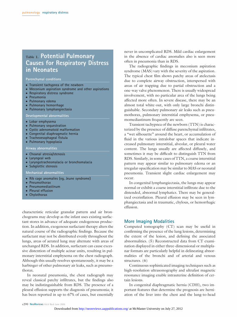
Table 1. Potential PulmonaryCauses for Respiratory Distressin NeonatesParenchymal conditions
● Transient tachypnea of the newborn● Meconium aspiration syndrome and other aspirations● Respiratory distress syndrome● Pneumonia● Pulmonary edema● Pulmonary hemorrhage● Pulmonary lymphangiectasia
Developmental abnormalities
● Lobar emphysema● Pulmonary sequestration● Cystic adenomatoid malformation● Congenital diaphragmatic hernia● Tracheoesophageal fistula● Pulmonary hypoplasia
Airway abnormalities
● Choanal atresia/stenosis● Laryngeal web● Laryngotracheomalacia or bronchomalacia● Subglottic stenosis
Mechanical abnormalities
● Rib cage anomalies (eg, Jeune syndrome)● Pneumothorax● Pneumomediastinum● Pleural effusion● Chylothorax
pulmonology respiratory distress
e290 NeoReviews Vol.6 No.6 June 2005
at McMaster University on July 27, 2012http://neoreviews.aappublications.org/Downloaded from

circumference ratio (LHR). Liver herniation may bedetermined sonographically by Doppler evaluation of theabnormal course of the umbilical, hepatic, and portalveins. The LHR estimates the volume of the contralaterallung, thereby providing a measure of the expected de-gree of pulmonary hypoplasia. When the LHR is less than0.9, the outcome is usually poor; when it is greater than1.4, a good outcome is more likely. (7) This informationmay influence parents to consider delivering at a centerthat has advanced therapeutic modalities, such as extra-corporeal membrane oxygenation (ECMO).
Congenital lobar emphysema may be detected inutero as an echogenic mass on ultrasonography, withassociated mediastinal shift and displacement of the heartresulting in compression of the contralateral lung. Fetal
ultrasonography may diagnose ex-tralobar emphysema as early as19 weeks of gestation. (6)
Parenchymal DiseasesTTN
TTN initially was described byAvery and colleagues in 1966. (8)This relatively benign, self-limiteddisease also is known as RDS type 2or wet lungs. It occurs in approxi-mately 11 per 1,000 live births andappears more often in boys, in in-fants delivered by cesarean section,and in infants who have perinatalasphyxia, umbilical cord prolapse,or maternal complications such asasthma, diabetes, or analgesia oranesthesia during labor. The syn-drome is characterized by tachy-pnea that appears shortly after birthand usually clears within 1 to5 days. The precise cause is un-known, but it is believed to be dueto delayed resorption of fetal lungfluid that may be related to ele-vated central venous pressure anddelayed clearance of pulmonaryliquid by the lymphatics. The rea-son for the delayed absorption isunknown, but it has been sug-gested to be attributed to mildasphyxia resulting in mild pulmo-nary capillary leak and to myocar-dial dysfunction with elevated fill-ing pressure. (9) In most cases,
the clinical course is benign, and mechanical ventila-tion almost never is required.
RDSAlthough RDS is primarily a disease of preterm infants,some near-term infants may be affected. These infants aretypically 34 to 37 weeks of gestation, and risk factorsinclude maternal diabetes, multiple birth, cesarean sec-tion prior to the onset of labor, perinatal asphyxia, coldstress, and infants whose siblings suffered from RDS.Because their surfactant sufficiency is borderline and theyhave larger pulmonary reserves, affected infants may beable to cope without ventilation for longer than smallerpreterm infants. Infants who have RDS may do well withnasal continuous positive airway pressure or may require
Table 2. Possible Diagnoses Related to RadiographicFeatures
Radiographic Features Possible Diagnosis
Air bronchograms ● RDS● Pneumonia
Diffuse parenchymal infiltrates ● TTN● MAS● Pneumonia● Pulmonary lymphangiectasia
Lobar consolidation ● Pneumonia● Lobar sequestration● CCAM
Patchy areas alternating with emphysema ● MASPleural effusion ● Pneumonia
● Pulmonary lymphangiectasiaReticular granular pattern ● RDS
● PneumoniaLoss of lung volume ● RDS
● MASFluid accumulations in interlobar spaces ● TTN
● Pulmonary lymphangiectasiaHyperinflation ● TTN
● MAS● Pulmonary lymphangiectasia
Atelectasis ● MAS● RDS
Pneumothorax/pneumomediastinum ● Spontaneous● MAS● RDS● Pneumonia
“Cystic” mass ● CCAM● CDH● Pulmonary sequestration
RDS�respiratory distress syndrome, TTN�transient tachypnea of the newborn, MAS�meconiumaspiration syndrome, CCAM�congenital cystic adenomatoid malformation, CDH�congenital dia-phragmatic hernia
pulmonology respiratory distress
NeoReviews Vol.6 No.6 June 2005 e291
at McMaster University on July 27, 2012http://neoreviews.aappublications.org/Downloaded from
ventilation. Surfactant often improves pulmonary me-chanics significantly but has little effect on overall out-come, which is favorable in most cases. (10)
Abnormalities of Surfactant ProteinsA small but notable group of term infants who havesevere respiratory distress have congenital abnormalitiesof surfactant proteins. The most common of these con-ditions is deficiency of surfactant protein B (SP-B). Af-fected infants develop severe respiratory distress shortlyafter birth, and chest radiographs show findings identicalto those of RDS. However, infants who have deficiencyof SP-B continue to suffer from extreme respiratoryinsufficiency despite mechanical ventilation, oxygen, re-peated surfactant replacement therapy, and corticoste-roids. The only effective therapy (although neither avail-able for nor consented to by all) is lung transplantation,without which the infants die within 1 to 6 months.
SP-B deficiency is transmitted as an autosomal reces-sive trait and is fatal in homozygotes; heterozygotes areclinically asymptomatic. Compound heterozygotes (twodifferent mutant alleles at the same loci) have a milderform of the disease. The gene for SP-B is located onchromosome 2 and comprises 11 exons. The most com-mon defect (60% to 70%) in the SP-B gene is a frame shiftmutation caused by a base pair insertion that results in apremature stop codon that prevents translation. (11)
The typical pathology in the lung is alveolar proteino-sis. Distended alveoli are filled with proteinaceous mate-rial and detached alveolar epithelial cells, and the alveolarsepta are thickened. In the airways, SP-B is markedlyreduced or absent and, by comparison, there are largeamounts of abnormal SP-A and SP-C. Abnormal pro-cessing of SP-C results in its accumulation within type 2pneumocytes. Ultrastructural examination reveals an ab-sence of normal lamellar bodies and tubular myelin,which are replaced by multivesicular bodies and multi-lamellated structures. There also is an accumulation oflipid vesicles between the alveolar epithelium and itsbasement membrane. (12)
To date, no human infants who lack SP-A have beenidentified. Abnormalities of SP-C and SP-D have beenidentified but do not appear to be associated with respi-ratory distress in human infants.
MASMAS is defined as respiratory distress in an infant bornthrough meconium-stained amniotic fluid whose symp-toms cannot otherwise be explained. Historically, manyof these infants were postmature, although this is seenless often today because obstetricians rarely allow preg-
nancy to continue to more than 41 weeks’ gestation.Approximately 13% of all live births are complicated bymeconium-stained amniotic fluid, and of these, 4% to 5%of infants develop MAS. (13)
The mechanisms of injury include direct toxicity ofthe meconium causing chemical pneumonitis, inactiva-tion of surfactant, activation of complement, and vaso-constriction as well as partial or complete airway obstruc-tion by the thick, particulate meconium. Secondarypulmonary hypertension is a frequent associated finding.
The management of MAS remains a challenge. (14)Before delivery, the infusion of isotonic solution into theamniotic cavity via a catheter is termed amnioinfusion.Studies have shown that this intervention in pregnanciescomplicated by thick meconium and oligohydramnioscan reduce the rate of MAS and fetal distress significantly.However, in view of significant adverse effects, this hasnot become an accepted therapy. (14)
Current recommendations are to perform intrapar-tum oropharyngeal suction before delivery of the body inall cases of meconium-stained amniotic fluid. This ap-proach recently was challenged by a large multicenter,randomized, controlled trial that included more than2,000 infants and showed no beneficial effect of suction-ing on the incidence of MAS. (15) Results of this studymay influence practice significantly. Similarly, electiveintubation and tracheal suction was a standard therapy inthe past, although this practice also has not withstood thetest of time. Wiswell and coworkers, in their large ran-domized study, showed no difference in the rate of MASin neonates who were intubated and had tracheal suc-tioning compared with those who were not intubated.(16) Another prospective randomized study designed todetermine whether routine tracheal suctioning is indi-cated in all meconium-stained healthy term neonatesshowed that the procedure was not harmless and isunnecessary in a vigorous term neonate who hasmeconium-stained fluid. (17)
Because meconium is known to inactivate surfactant,exogenous replacement therapy seems logical. Random-ized, controlled studies have shown that surfactant treat-ment reduces the need for ECMO and may reduce therisk for pneumothorax in neonates who have MAS. (18)Calf lung surfactant therapy was shown to cause signifi-cant, but short-term improvement in the oxygenationindex. (19) This suggests a dose-response relationshipbetween the surfactant inactivation and its replacement.Another method for surfactant administration in MAS isas a lavage with diluted surfactant. The use of lavage canhelp to remove meconium while simultaneously replac-ing the inactivated surfactant. (20)(21)
pulmonology respiratory distress
e292 NeoReviews Vol.6 No.6 June 2005
at McMaster University on July 27, 2012http://neoreviews.aappublications.org/Downloaded from
The use of inhaled nitric oxide (iNO) increases oxy-genation in neonates who have MAS. Since the approvalof iNO by the United States Food and Drug Adminis-tration in 2001, there has been a steady decrease in theuse of ECMO for neonates who have MAS. (22)
Treatment of MAS has improved over the last decadewith new ventilatory modalities, such as different meth-ods of high-frequency ventilation, but no randomizedtrials have compared the different forms of ventilation inthis setting. Experimental studies in animals have com-pared the use of high-frequency ventilation with conven-tional ventilation and have shown enhanced carbon di-oxide elimination, increased lung compliance, anddiminished right-to-left shunts. (14) Despite these ad-vances, MAS remains a challenging condition with asignificant mortality risk.
PneumoniaPneumonia may be acquired in utero, during delivery (orperinatally), or postnatally in the nursery or at home. Itmay be classified as either early- (�7 d of age) or late-onset (�7 d of age).
At autopsies of both stillbirths and liveborn neonataldeaths, pneumonia was found to be present in 20% to60% in different centers. (23)(24) The definition of thepneumonia was based on the presence of polymorpho-nuclear leukocytes in the alveoli or interstitium, althoughthe presence of bacteria was not necessary for the defini-tion.
The causative agent varies, depending on whether theinfection is acquired before, during, or after birth in thenursery or at home. (24) Intrauterine infection is usuallythe result of maternal infection, which may be transmit-ted transplacentally and involves many organs (includingblood, liver, central nervous system, lungs). Pathogensinclude rubella, cytomegalovirus, herpes simplex virus,mumps, adenovirus, Toxoplasma gondii, Treponema pal-lidum, Mycobacterium tuberculosis, Listeria monocyto-genes, Varicella zoster, and human immunodeficiencyvirus.
Pneumonias that are acquired at birth most often arecaused by group B Streptococcus, but Escherichia coli,Klebsiella sp, and Chlamydia trachomatis also are seen.C trachomatis pneumonia typically presents at a later age(3 wk). Pneumonias acquired after birth in the nursery orat home include those caused by respiratory viruses (ad-enovirus, respiratory syncytial virus), gram-positive bac-teria (groups A, B, and G streptococci or Staphylococcusaureus), and gram-negative enteric bacteria (Klebsiellasp, Proteus sp, Pseudomonas aeruginosa, flavobacteria,Serratia marcescens, and E coli). (25)
Congenital pneumonia is a severe disease that fre-quently results in either stillbirth or death within the first24 hours after birth. Pneumonias that are acquired laterpresent most often as systemic disease. Managementincludes oxygen therapy, ventilatory support, antibiotics,and often vasopressor support such as dopamine anddobutamine.
LymphangiectasiaCongenital errors of lymphatic development can lead toprimary pulmonary disorders that include lymphangi-oma, lymphangiectasia, lymphangiomatosis, and lym-phatic dysplasia syndrome. (26) Because of their rarity,they often are misdiagnosed. The origins of these disor-ders are unknown.
Primary lymphangiectasia is a congenital disorder ofthe lymphatic system characterized by marked dilatationof the lymphatic vessels that leads to obstruction andleakage of fluid. (27) This is seen in the visceral pleura aswell as interlobular septa and results in chylothoraces,which lead to respiratory compromise or failure. Intesti-nal and thoracic lymphangiectasia may occur in isolationor simultaneously in the same patient as part of a gener-alized lymphatic dysplasia. Primary lymphangiectasia is arare congenital malformation, with the age of presenta-tion ranging from in utero to early adulthood. Whenpresent in the neonatal period, the clinical course isusually fatal.
The lymphatic vascular system develops during thesixth week of fetal life as an outgrowth of the venoussystem or as a de novo differentiation within the mesen-chymal tissue. They join one another to form the lym-phatic channels. The pulmonary lymphatic channels de-velop before the 20th week of fetal life. Primarycongenital lymphangiectasia results from failure of thepulmonary interstitial connective tissue to regress, lead-ing to dilation of pulmonary lymphatic capillaries. Thelung appears heavy and noncompliant. The visceralpleura have a network of dilated lymphatics that weeplymph fluid when sectioned. Open lung biopsy is re-quired to make the diagnosis. Supportive therapy, in-cluding albumin infusions, diuretics, thoracocentesis,and paracentesis, provide transient relief of symptoms.Dietary modifications are aimed at controlling symptomsand consequences of lymphatic obstruction but do notmodify the underlying disease process. Primary pulmo-nary lymphangiectasia often is associated with a numberof congenital and genetic diseases, including Noonan,Ullrich-Turner, Ehlers-Danlos, and Down syndromes.
pulmonology respiratory distress
NeoReviews Vol.6 No.6 June 2005 e293
at McMaster University on July 27, 2012http://neoreviews.aappublications.org/Downloaded from

Developmental Lung AbnormalitiesCDH
CDH occurs in 1 in 2,000 to 4,000 births. Males areaffected more often (male:female ratio of 1.5:1), and therecurrence risk in future pregnancies is 2%. CDH is adevelopmental abnormality of the diaphragm resulting ina defect that permits abdominal viscera to enter the chest.Usually the defect occurs before the eighth week ofembryonic life. It is seen more often in the posterolateralsegments of the diaphragm and more often on the leftside. Some 95% occur through the posterior foramen ofBochdalek that lies posteriorly and lateral to the spine,and of these, 80% are on the left side. Classic thinking hasbeen that the primary defect is in the diaphragm and thatpulmonary hypoplasia is due to pressure from the ab-dominal viscera in the thoracic cavity. However, infor-mation based on the murine nitrofen-induced diaphrag-matic hernia model suggests that proper formation of thediaphragm requires the normal formation of the lungand that pulmonary hypoplasia is the cause rather thanthe result of the diaphragmatic hernia. It has been shownthat pulmonary hypoplasia occurs before the diaphragmis closed. (28) Cellular mechanisms that appear to beinvolved include altered regulation of expression of vas-cular endothelial growth factor and its receptor, fibro-blast growth factors 7 and 10, insulin-like growth factor,and sonic hedgehog. Glucocorticoid receptor is in-creased, suggesting a protective role for glucocorticoids.Another protective factor appears to be retinoic acid.(29)
Despite the many advances in critical care and venti-lator management, CDH continues to be an extremelychallenging problem in the NICU. The morbidity andmortality remain high and are related primarily to pul-monary hypoplasia and pulmonary hypertension. In thedelivery room, the neonate typically presents with respi-ratory distress shortly after birth. Physical examinationmay show the abdomen to be scaphoid. Air entry isreduced on the affected side, and the heart sounds aredisplaced. Immediate treatment includes intubation andmechanical ventilation, and a nasogastric tube should bepassed for decompression. Bag-and-mask ventilationshould be avoided to prevent gastric dilatation that maycompromise pulmonary function further. New ap-proaches to managing CDH that have been exploredinclude the use of extracorporeal life support (ECMO),high-frequency ventilation, delayed surgical repair, per-missive hypercapnia, nitric oxide, surfactant administra-tion, intratracheal pulmonary ventilation, and liquid ven-tilation. (30) Despite the new approaches, mortality ratesremain high, ranging from 25% to 74% in different
reports. The presence of associated major malformationsincreases the mortality markedly, as does liver herniationnoted at surgery. If there are no other anomalies and thedefect is not part of a genetic syndrome, the prognosisafter neonatal surgical repair usually is good, with overallsurvival rates for liveborn infants of 60% to 80%.
Congenital Cystic Adenomatoid Malformation(CCAM)
CCAM consists of a multicystic mass of dilated bronchio-larlike spaces that proliferate at the expense of alveoli.The result is the formation of a rubbery lesion thatenlarges following air and fluid trapping. The cause isrelated to an abnormal signaling or conjugation betweenthe developing terminal bronchioles and the alveolarmesenchyme.
Males and females are affected equally. Approximately50% of the cases present as life-threatening respiratorydistress in the neonatal period. The condition is morecommon on the right side, and usually only one lobe isinvolved. (31)
CCAM is categorized into four types. Type 1 is char-acterized by a small number of large cysts and is the mostcommon (75%). In type 2, there are evenly spaced cyststhat are less 1 cm in diameter. This type is associated withother congenital anomalies and poor outcome. Type 3 israre and appears more solid on gross examination. (32)A fourth type has been defined that is characterized byacinar-type epithelium rather than the bronchiolar epi-thelium seen in the other three types.
At the cellular level, there is accelerated cell prolifera-tion, with a low apoptotic index. Dysregulation of themesenchymal growth factor, platelet-derived growth fac-tor BB, gene expression has been implicated in thepathogenesis. (28)
Treatment is by surgical resection of the lesion. Thesurvival rate has been reported to be 100% in neonateswho do not have hydrops fetalis, but is much lower inthose who have hydrops.
Congenital Lobar Emphysema (CLE)CLE is characterized by air trapping and overdistentionof segments and lobes of the lungs. It usually is diag-nosed postnatally, and 50% of the cases present by the ageof 6 months. Clinical symptoms include respiratory dis-tress, mediastinal shift, and wheezing due to spontane-ous overinflation of the affected areas. The upper lobesare involved in 90% of the cases. The diagnosis can bemade by simple chest radiograph, but prenatal diagnosiscan be made by high-resolution ultrasonography, mag-netic resonance imaging, or CT. In cases that involve
pulmonology respiratory distress
e294 NeoReviews Vol.6 No.6 June 2005
at McMaster University on July 27, 2012http://neoreviews.aappublications.org/Downloaded from
respiratory distress, the affected area should be removed.Because there are reports of spontaneous resolution,asymptomatic cases may be followed expectantly. CLEaccounts for 50% of structural lesions causing respiratorydistress in the newborn. It is more common in males(2:1). Sometimes the lesion can be mistaken for pneu-mothorax or CDH. There are associated anomalies in14% to 40% of cases, most of which are cardiovascular.(31)(33) The prognosis is favorable, but depends on theassociated abnormalities.
Pulmonary SequestrationLobar sequestration is composed of abnormal lung tissuethat has no connection with the normal tracheobronchialtree. There are two types of lesions, and both receivetheir arterial blood supply from the systemic circulation,usually a branch of the aorta.
With extralobar sequestration, the discrete mass ofpulmonary parenchyma is outside the pleural investmentof the lung. The lesion is found on the left side, proximalto the esophagus and between the lower lobe and thediaphragm in 66% of the cases. In 80% of the cases, theblood supply derives from the descending thoracic orabdominal aorta, and the venous drainage is to theazygous or hemiazygous vein (80%) and the rest to thepulmonary venous system. It is more common in males(3 to 4 times), and 50% of patients have respiratorydistress due to compression of the rest of the lungparenchyma. In more than 65% of cases, there are asso-ciated anomalies, including CDH (20% to 30%), pericar-dial defects, and total anomalous pulmonary venous re-turn.
Abnormal expression of the homeobox gene Hoxb-5,which is necessary for normal airway branching anddevelopment, has been implicated in the etiology. (34)
Intralobar sequestration is characterized by the lesionresting within the lobe of the lung without separatepleura. It is usually in the lower lobe (95%), and in 55% ofcases is on the left side. The arterial supply comes fromthe abdominal aorta or celiac axis, and there may bemultiple feeding arteries. The venous drainage is throughthe pulmonary vein. Intralobar sequestration is three tosix times more common than extralobar sequestrationand can be an acquired lesion that results from recurrentinfection. In both types, the definitive treatment is resec-tion of the lesion. (31)(33)
SummaryAlthough most term infants who have respiratory distresshave either TTN or infection, the differential diagnosis is
extensive, and the rarer causes need to be considered inatypical circumstances.
References1. Boyle KM, Baker VL, Cassaday CJ. Neonatal pulmonary disor-ders. In: Barnhart SL, Czervinske MP, eds. Perinatal and PediatricRespiratory Care. 2nd ed. Philadelphia, Pa: WB Saunders Co;2001:4452. Marino BS, Bird GL, Wernovsky G. Diagnosis and managementof the newborn with suspected congenital heart disease. Clin Peri-natol. 2001;28:91–1363. Sasidharan P. An approach to diagnosis and management ofcyanosis and tachypnea in term infants. Pediatr Clin North Am.2004;54:999–10214. Cleavland RH. A radiologic update on medical diseases of thenewborn chest. Pediatr Radiol. 1995;25:631–6375. Daltro P, Fricke BL, Kuroki I, Domingues R, Donnelly LF. CTof congenital lung lesions in pediatric patients. AJR Am J Roent.2004;183:1497–15066. Johnson AM, Hubbard AM. Congenital anomalies of the fetal/neonatal chest. Semin Roentg. 2004;39:197–2147. Metkus AP, Filly RA, Stringer MD, et al. Sonographic predictorsof survival in fetal diaphragmatic hernia. J Pediatr Surg. 1996;31:148–1518. Avery ME, Gatewood OB, Brumly G. Transient tachypnea ofthe newborn. Am J Dis Child. 1966;111:3809. Haliday H, McClure G, Reid M. Transient tachypnoea of thenewborn: two distinct clinical entities? Arch Dis Child. 1981;56:322–32510. Golombek SG, Truog WE. Effects of surfactant on gas ex-change and clinical course in near-term newborns with RDS. JPerinat Med. 2000;28:436–44211. Cole FS, Hamvas A, Nogee LM. Genetic disorders of neonatalrespiratory function. Pediatr Res. 2001;50:157–16212. deMello DE. Pulmonary pathology. Semin Neonatol. 2004;9:311–32913. Cleary GM, Wiswell TE. Meconium-stained amniotic fluid andthe meconium aspiration syndrome: an update. Pediatr Clin NorthAm. 1998;45:511–52914. Gelfand SL, Fanaroff JM, Walsh MC. Controversies in thetreatment of meconium aspiration syndrome Clin Perinatol. 2004;31:445–45215. Vain NE, Szyld EG, Prudent LM, Wiswell TE, Augilar AM,Vivas N, for the Meconium Study Network. Oropharyngeal andnasopharyngeal suctioning of meconium-stained neonates beforedelivery of their shoulders: multicentre, randomized controlledtrial. Lancet. 2004;364:597–60216. Wiswell TE, Gannon CM, Jacob J, et al. Delivery room man-agement of the apparently vigorous meconium-stained neonate:results of the multicenter, international collaborative trial. Pediat-rics. 2000;105:1–717. Linder N, Aranda V, Tsur M, et al. Need for endotrachealintubation in meconium stained neonates. J Pediatr. 1988;112:613–61518. Findlay RD, Taeusch HW, Walther FJ. Surfactant replacementtherapy for meconium aspiration syndrome. Pediatrics. 1996;97:48–5219. Auten RL, Notter RH, Kendig JW, Davis JM, Shapiro DL.
pulmonology respiratory distress
NeoReviews Vol.6 No.6 June 2005 e295
at McMaster University on July 27, 2012http://neoreviews.aappublications.org/Downloaded from

Surfactant treatment of full-term newborns with respiratory failure.Pediatrics. 1991;87:101–10720. Lam BCC, Yeung CY. Surfactant lavage for meconium aspira-tion syndrome. Pediatrics. 1999;103:1014–101821. Wiswell TE, Knight GR, Finer NN, et al. A multicenter,randomized, controlled trial comparing Surfaxin (lucinactant) la-vage with standard care for treatment of meconium aspirationsyndrome. Pediatrics. 2002;109:1081–108722. Roberts JD, Fineman JR, Morin FC, et al. Inhaled nitric oxideand persistent pulmonary hypertension of the newborn. N EnglJ Med. 1997;336:605–61023. Whitsett JA, Pryhuber GS, Rice WR. Acute respiratory disor-ders. In: Avery GE, Fletcher MA, MacDonald MG, eds. Neonatol-ogy: Pathophysiology and Management of the Newborn. 4th ed. Phil-adelphia, Pa: JB Lippincott; 199424. Remington JS, Klein JO. Bacterial infections of the respiratorytract. In: Infectious Diseases of the Fetus and the Newborn Infant. 4thed. Philadelphia, Pa: WB Saunders; 199525. Fujikura T, Froehlich LA. Intrauterine pneumonia in relationto birth weight and race. Am J Obstet Gynecol. 1967;97:8126. Faul JL, Berry GJ, Colby TV, et al Thoracic lymphangiomas,lymphangiectasis, lymphangiomatosis and lymphatic dysplasia syn-drome: state of the art Am J Respir Crit Care. 2000;161:1037–1046
27. Barker PM, Esther CR, Fordham LA, Maygarden SJ, Funk-houser WK. Primary pulmonary lymphangiectasia in infancy andchildhood. Eur Respir J. 2004;24:413–41928. Mahta SS, Gittes GK. Impact of advances in developmentalbiology on the management of neonatal surgical anomalies. SeminPerinatol. 2004;28:152–16329. Doyle NM, Lally KP. The CDH study group and advances inthe clinical care of the patient with congenital diaphragmatic hernia.Semin Perinatol. 2004;28:174–18430. Ivascu FA, Hirschel RB. New approaches to managing con-genital diaphragmatic hernia. Semin Perinatol. 2004;28:185–19831. Mendeloff EN. Sequestrations, congenital cystic adenomatoidmalformations and congenital lobar emphysema. Semin ThoracCardiovasc Surg. 2004;16:204–21432. Stocker JT. The respiratory system. In: Textbook of PediatricPathology. Philadelphia, Pa: Lippincott Williams & Wilkins; 1992:505–53233. Ankermann T, Oppermann HC, Engler S, Leuschner, VonKaisenberg CS. Congenital masses of the lung, cystic adenomatoidmalformation versus congenital lobar emphysema. J UltrasoundMed. 2004;23:1379–138434. Volpe MV, Archivachotikul K, Bhan I. Association of broncho-pulmonary sequestration with expression of the homeobox proteinHoxb-5. J Pediatr Surg. 2000;35:1817–1819
pulmonology respiratory distress
e296 NeoReviews Vol.6 No.6 June 2005
at McMaster University on July 27, 2012http://neoreviews.aappublications.org/Downloaded from

NeoReviews Quiz
9. A newborn is delivered at an estimated gestational age of 36 weeks by emergent cesarean section for fetaldistress. The maternal history is significant for prolonged rupture of membranes. The infant has evidenceof respiratory distress, and the chest radiograph shows patchy infiltrates and pleural effusion, as indicatedby obliteration of both costophrenic angles. Of the following, the most likely cause of these chestradiographic findings in this infant is:
A. Hyaline membrane disease.B. Meconium aspiration syndrome.C. Neonatal pneumonia.D. Pulmonary edema.E. Pulmonary hemorrhage.
10. A rare cause of respiratory distress among term newborns is a congenital abnormality of surfactantproteins. The most common of these conditions is deficiency of surfactant protein B (SP-B). Of thefollowing, the most accurate statement regarding SP-B is that:
A. SP-B deficiency is accompanied by reductions in SP-A and SP-C in airways.B. SP-B deficiency is transmitted as an autosomal dominant trait.C. The gene for SP-B is located on chromosome 22.D. The most common defect in SP-B deficiency is a frame shift mutation.E. The typical pathologic finding in SP-B deficiency is generalized alveolar atelectasis.
11. Several developmental abnormalities of lung structure can cause respiratory distress in the newborn. Ofthe following, the most common structural lesion that can cause respiratory distress in the newborn is:
A. Congenital cystic adenomatoid malformation.B. Congenital lobar emphysema.C. Primary pulmonary lymphangiectasia.D. Pulmonary hypoplasia.E. Pulmonary sequestration.
pulmonology respiratory distress
NeoReviews Vol.6 No.6 June 2005 e297
at McMaster University on July 27, 2012http://neoreviews.aappublications.org/Downloaded from

DOI: 10.1542/neo.6-6-e2892005;6;e289Neoreviews
Orna Flidel-Rimon and Eric S. ShinwellRespiratory Distress in the Term and Near-term Infant
ServicesUpdated Information &
http://neoreviews.aappublications.org/content/6/6/e289including high resolution figures, can be found at:
References
http://neoreviews.aappublications.org/content/6/6/e289#BIBLat: This article cites 29 articles, 9 of which you can access for free
Subspecialty Collections
orn_infanthttp://neoreviews.aappublications.org/cgi/collection/fetus_newbFetus and Newborn Infantfollowing collection(s): This article, along with others on similar topics, appears in the
Permissions & Licensing
/site/misc/Permissions.xhtmltables) or in its entirety can be found online at: Information about reproducing this article in parts (figures,
Reprints/site/misc/reprints.xhtmlInformation about ordering reprints can be found online:
at McMaster University on July 27, 2012http://neoreviews.aappublications.org/Downloaded from

Core Concepts:Respiratory Distress SyndromeJamie B. Warren, MD,*
JoDee M. Anderson, MD,
MSEd(c)*
Author Disclosure
Drs Warren and
Anderson have
disclosed no financial
relationships relevant
to this article. This
commentary does not
include a discussion
of an unapproved/
investigative use of a
commercial
product/device.
Objectives After completing this article, readers should be able to:
1. Define respiratory distress syndrome (RDS).2. Discuss the epidemiology, pathophysiology, and diagnosis of RDS.3. Create a differential diagnosis for respiratory distress in the neonate.4. Describe the proven treatments for RDS, with particular attention to antenatal steroids
and surfactant replacement therapy (SRT), their benefits and possible complications.5. Discuss ventilation strategies that can be used in the infant who has RDS.6. Describe long-term complications of RDS and its treatments.
AbstractRespiratory distress syndrome (RDS) is seen primarily in the preterm neonate and isdue mostly to pulmonary surfactant deficiency. Lung atelectasis leads to ventilation-perfusion mismatching, hypoxia, and eventual respiratory failure in the untreatedinfant who has RDS. RDS is diagnosed by physical findings consistent with respiratorydistress and characteristic radiographic findings. Treatment of RDS begins antenatallywith the administration of maternal steroids to women at risk of preterm deliverybetween 24 and 34 weeks’ gestation. The use of repeat doses of antenatal steroids isunder investigation but is currently not recommended outside of randomized, con-trolled trials. SRT has been approved for use since 1990 and has been successful indecreasing rates of RDS. Natural surfactant is currently recommended for use, butsynthetic surfactant that contains proteins to mimic surfactant proteins is beinginvestigated. In general, prophylactic use of surfactant is recommended over rescuetreatment in infants at high risk for developing RDS, but the determination of whichinfants are at high risk for developing RDS remains a clinical one. The push toward useof less invasive ventilation strategies in the treatment of RDS has led to several trials ofnasal continuous positive airway pressure (nCPAP). Results of the SUPPORT trial arepending, but the COIN trial has concluded that nCPAP use in infants who have RDSis not detrimental. Inhaled nitric oxide for RDS still requires investigation on safetyand efficacy. Several other treatments have been studied, but as of yet, only inositoladministration shows promise in the treatment of RDS. Several complications of therecommended treatments for RDS have been identified, but the benefits far outweighthe risks. Finally, there remains a need for long-term follow-up studies on preterminfants treated for RDS to assess neurodevelopmental outcomes.
DefinitionRDS, formerly known as hyaline membrane disease, occurs in incompletely developedlungs and is, therefore, a disease of prematurity. Immature lungs are functionally deficientin mature surfactant. (1) The absence of surfactant in the liquid film lining of alveoli causesan increase in surface tension and alveolar collapse. (2) If not treated, such atelectasis causesan increased work of breathing, intrapulmonary shunting, ventilation-perfusion mismatch,hypoxia, and eventual respiratory failure. (1)
*Oregon Health and Science University, Portland, Ore.
core concepts
NeoReviews Vol.10 No.7 July 2009 e351
EpidemiologyRDS is seen almost exclusively in preterm infants, beforethe lungs begin to manufacture adequate amounts ofsurfactant. (2) In fact, the risk of RDS decreases withincreasing gestational age: 60% of babies born at fewerthan 28 weeks’ gestation, 30% of babies born between28 and 34 weeks’ gestation, and fewer than 5% of babiesborn after 34 weeks’ gestation develop RDS. (3) Otherfactors that increase the risk of RDS include male sex,maternal gestational diabetes, perinatal asphyxia, hypo-thermia, and multiple gestations. (4) Antenatal steroidsand prolonged rupture of membranes decrease the risk ofRDS. (5) With the advent of therapies for RDS, includ-ing antenatal steroids and SRT, mortality from RDS hasdecreased from nearly 100% to less than 10% in recentyears. (6)
Differential DiagnosisThe differential diagnosis of respiratory distress in thenewborn encompasses upper respiratory obstruction,pulmonary disease, cardiac disease, thoracic causes, met-abolic disorders, diaphragmatic causes, neuromusculardiseases, infectious causes, hemolytic/vascular causes,and miscellaneous causes (Table 1). (7)(8)
PathophysiologyNormal Lung Development
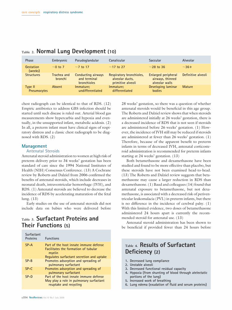
The period of viability begins at around 23 weeks’ ges-tation, when the fetal lung begins to transition from thecanalicular to the saccular stage of development (Table2). (9) During the saccular stage, peripheral airways
enlarge and distal airways begin to dilate while their wallsbegin to thin. (10) Type II pneumocytes, the cells re-sponsible for surfactant production, are present and ma-turing. (10) Although gas exchange is possible duringthis stage, total surface area for gas exchange is low anddiffusion distance for gas exchange is high in relation tobody weight and metabolic rate. (9) Secondary septa-tion, or alveolarization, begins at about 32 weeks’ gesta-tion. (9) During this phase, alveoli form and mature andalveolar walls thin. (10) All cell types proliferate duringthis phase, including type II pneumocytes. (10) Theoverall result is a maturing lung with a larger surface areaand a minimal diffusion distance for gas exchange. (10)
Surfactant Composition and Life CycleSurfactant is a mixture of phospholipids and proteins. (2)The most abundant surface-active phospholipid in ma-ture lungs is phosphatidylcholine. (11) Phosphatidylcho-line forms a monolayer on the liquid film lining of thealveolus, lowering the surface tension of that film. (2) Inaddition to phospholipids, surfactant contains four majorproteins: surfactant proteins (SPs) A, B, C, and D (Table3). (11) SP-A helps to regulate surfactant secretion anduptake; SP-B and SP-C facilitate adsorption and spread-ing of phospholipids on the liquid film lining of thealveoli. (2) SP-D may play a role in surfactant reuptakeand recycling. (5)
Pulmonary surfactant is manufactured in the Golgiapparatus and stored in lamellar bodies of type II pneu-mocytes. (5) Once secreted by the lamellar bodies intothe extracellular space, surfactant is organized into tubu-lar myelin, adsorbed into the air-water interface, andformed into a lipid monolayer. (5)(6) The surface-activeproperties of the lipid monolayer decrease the surfacetension of the air-water interface and prevent alveolarcollapse. (6) The majority of surfactant constituents arebelieved to be recycled, either through reuptake by typeII pneumocytes or by alveolar macrophages. (9)
RDSAn infant born before the alveolarization stage of lungdevelopment has underdevelopment of alveolar sacsand difficulty with oxygenation and ventilation. (9) Sim-ilarly, an infant born before this stage of lung develop-ment experiences a delay in production and secretion offunctional surfactant. (9) Such surfactant deficiency isthe major reason for poor lung function in the pretermneonate (Table 4). (2)
Although the preterm neonate does produce a smallamount of surfactant, this surfactant contains lowamounts of phospholipids and SPs. (9) It is estimated
Abbreviations
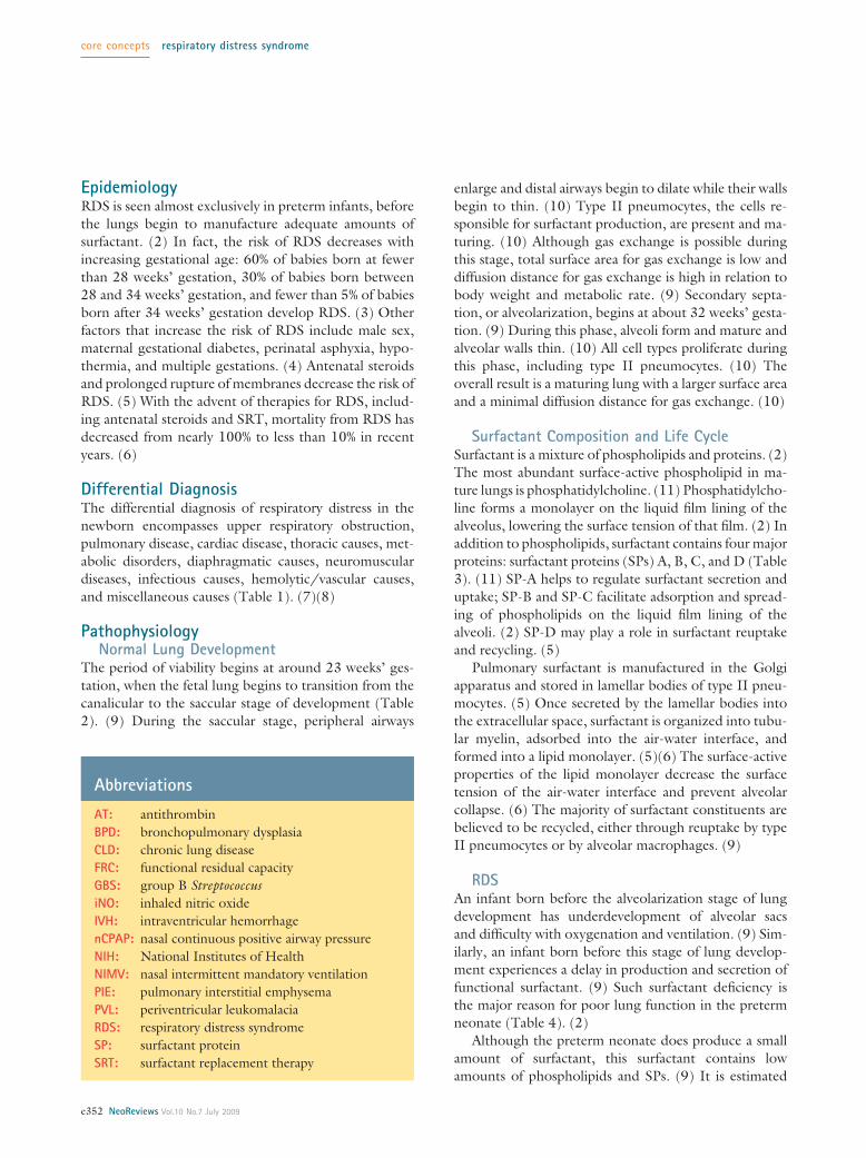
AT: antithrombinBPD: bronchopulmonary dysplasiaCLD: chronic lung diseaseFRC: functional residual capacityGBS: group B StreptococcusiNO: inhaled nitric oxideIVH: intraventricular hemorrhagenCPAP: nasal continuous positive airway pressureNIH: National Institutes of HealthNIMV: nasal intermittent mandatory ventilationPIE: pulmonary interstitial emphysemaPVL: periventricular leukomalaciaRDS: respiratory distress syndromeSP: surfactant proteinSRT: surfactant replacement therapy
core concepts respiratory distress syndrome
e352 NeoReviews Vol.10 No.7 July 2009

that infants who have RDS have surfactant pools ofless than 10 mg/kg compared with pools of up to100 mg/kg in term infants. Such surfactant deficiencynecessitates increased work of breathing to distend alve-oli, which the preterm neonate may not be able toprovide. (2) Diffuse atelectasis ensues and leads to anoverall decrease in functional residual capacity (FRC) ofthe lungs. (2) If an infant is allowed to breathe from an
inadequate FRC, lung injury canoccur. (9) Lung injury leadsto protein exudation and edema,which can inactivate surfactant fur-ther. The acidosis and hypoxia thatresults from atelectasis and lung in-jury further interferes with surfac-tant production. The combinationof these events leads to respiratoryfailure.
DiagnosisClinical Evaluation
RDS presents at the time of or soonafter birth, and symptoms worsenover time. (2) Clinical symptomsof RDS are the same as those ofany other respiratory distress:tachypnea, nasal flaring, chest wallretractions, expiratory grunting,and central cyanosis. (2) In the ex-tremely preterm infant, the onlyclinical symptom of RDS may beapnea. (2) It is important to re-member that some infants whohave RDS exhibit all of these symp-toms, and others may show none.
An accurate history is importantin diagnosing RDS. As stated, RDSis more prevalent in earlier gesta-tional ages, so an accurate estima-tion of gestational age is necessary.Other historical factors must bediscerned, such as antenatal steroidtherapy; maternal history of gesta-tional diabetes; course of labor,including prolonged rupture ofmembranes, maternal fever, groupB Streptococcus (GBS) status andantibiotic therapy; method of deliv-ery; and need for resuscitation.
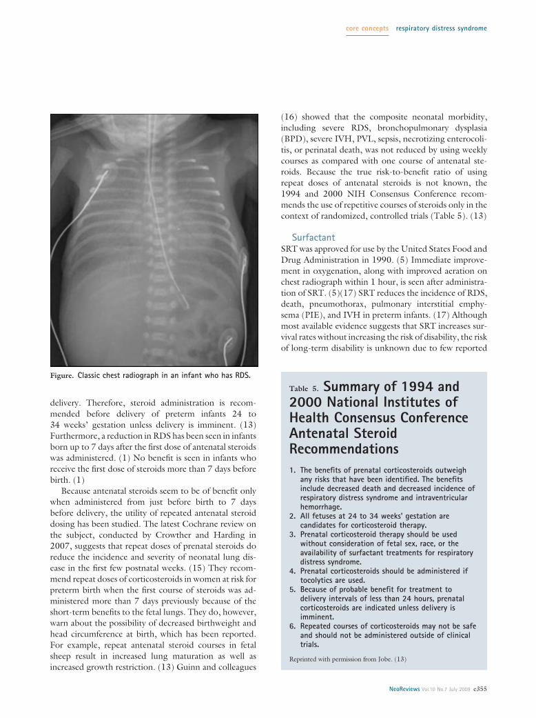
Diagnostic StudiesAlong with the history and physical examination, a chestradiograph is needed for the diagnosis of RDS. Thetypical chest radiograph shows diffuse atelectasis and theclassic “ground glass” appearance of the lung fields (Fig-ure). (2) Air bronchograms, which are air-filled bronchisuperimposed on the relatively airless parenchyma of thelung tissue, also are seen commonly on chest radiograph.(2) Importantly, the appearance of GBS pneumonia on
Table 1. Differential Diagnosis of RespiratoryDistress in the NewbornUpper Airway Obstruction
Choanal atresia, nasal stenosis, Pierre Robin sequence, laryngeal stenosis or atresia,hemangioma, vocal cord paralysis, vascular rings, tracheobronchial stenosis,masses, cleft palate, nasal stuffiness
Pulmonary Diseases
Respiratory distress syndrome, retained fetal lung liquid syndrome (transienttachypnea of the newborn), aspiration (including meconium aspirationsyndrome), pneumonia, pneumothorax, pneumomediastinum, primary pulmonaryhypertension, tracheoesophageal fistula, pulmonary hemorrhage, pulmonaryhypoplasia, pulmonary agenesis, cystic disease, pleural effusion, chylothorax,neoplasm, bronchopulmonary sequestration, pulmonary arteriovenousmalformation, pulmonary interstitial emphysema, pulmonary edema, congenitalalveolar proteinosis, congenital lobar emphysema
Cardiac Diseases
Cyanotic congenital heart disease, acyanotic congenital heart disease, arrhythmia,increased intravascular volume, high output failure, pneumopericardium,cardiomyopathy
Thoracic Causes
Chest wall deformity, mass
Metabolic Disorders
Hypoglycemia, infant of a diabetic mother, inborn errors of metabolism
Diaphragmatic Causes
Hernia, paralysis
Neuromuscular Diseases
Central nervous system damage (birth trauma, hemorrhage), medication (maternalsedation, narcotic withdrawal), muscular disease (myasthenia gravis),intraventricular hemorrhage, meningitis, hypoxic-ischemic encephalopathy,seizure disorder, obstructed hydrocephalus, infantile botulism, spinal cord injury
Infectious Causes
Sepsis, pneumonia (especially group B Streptococcus)
Hemolytic/vascular Causes
Anemia, polycythemia, abnormal hemoglobin
Miscellaneous Causes
Asphyxia, acidosis, hypo/hyperthermia, hypo/hypernatremia
core concepts respiratory distress syndrome
NeoReviews Vol.10 No.7 July 2009 e353
chest radiograph can be identical to that of RDS. (12)Empiric antibiotics to address GBS infection should bestarted until such disease is ruled out. Arterial blood gasmeasurements show hypercarbia and hypoxia and even-tually, in the unsupported infant, metabolic acidosis. (2)In all, a preterm infant must have clinical signs of respi-ratory distress and a classic chest radiograph to be diag-nosed with RDS. (2)
ManagementAntenatal Steroids
Antenatal steroid administration to women at high risk ofpreterm delivery prior to 34 weeks’ gestation has beenstandard of care since the 1994 National Institutes ofHealth (NIH) Consensus Conference. (13) A Cochranereview by Roberts and Dalziel from 2006 confirmed thebenefits of antenatal steroids, which include decreases inneonatal death, intraventricular hemorrhage (IVH), andRDS. (1) Antenatal steroids are believed to decrease theincidence of RDS by accelerating maturation of the fetallung. (13)
Early studies on the use of antenatal steroids did notinclude data on babies who were delivered before
28 weeks’ gestation, so there was a question of whetherantenatal steroids would be beneficial in this age group.The Roberts and Dalziel review shows that when steroidsare administered initially at 26 weeks’ gestation, there isa decreased incidence of RDS that is not seen if steroidsare administered before 26 weeks’ gestation. (1) How-ever, the incidence of IVH still may be reduced if steroidsare administered at fewer than 26 weeks’ gestation. (1)Therefore, because of the apparent benefit to preterminfants in terms of decreased IVH, antenatal corticoste-roid administration is recommended for preterm infantsstarting at 24 weeks’ gestation. (13)
Both betamethasone and dexamethasone have beenstudied and found to be more effective than placebo, butthese steroids have not been examined head-to-head.(13) The Roberts and Dalziel review suggests that beta-methasone may cause a larger reduction in RDS thandexamethasone. (1) Baud and colleagues (14) found thatantenatal exposure to betamethasone, but not dexa-methasone, is associated with a decreased risk of periven-tricular leukomalacia (PVL) in preterm infants, but thereis no difference in the incidence of cerebral palsy. (1)With this limited evidence, two doses of betamethasoneadministered 24 hours apart is currently the recom-mended steroid for antenatal use. (13)
Antenatal steroid administration has been shown tobe beneficial if provided fewer than 24 hours before
Table 2. Normal Lung Development (10)
Phase Embryonic Pseudoglandular Canalicular Saccular Alveolar
Gestation(weeks)
�0 to 7 �7 to 17 �17 to 27 �28 to 36 �36�
Structures Trachea andbronchi
Conducting airwaysand terminalbronchioles
Respiratory bronchioles,alveolar ducts,primitive alveoli
Enlarged peripheralairways, thinnedalveolar walls
Definitive alveoli
Type IIPneumocytes
Absent Immature;undifferentiated
Immature;differentiated
Developing laminarbodies
Mature
Table 3. Surfactant Proteins andTheir Functions (5)
SurfactantProteins Functions
SP-A Part of the host innate immune defenseFacilitates the formation of tubular
myelinRegulates surfactant secretion and uptake
SP-B Promotes adsorption and spreading ofpulmonary surfactant
SP-C Promotes adsorption and spreading ofpulmonary surfactant
SP-D Part of the host innate immune defenseMay play a role in pulmonary surfactant
reuptake and recycling
Table 4. Results of SurfactantDeficiency (2)
1. Decreased lung compliance2. Unstable alveoli3. Decreased functional residual capacity4. Hypoxia (from shunting of blood through atelectatic
portions of the lung)5. Increased work of breathing6. Lung edema (exudation of fluid and serum proteins)
core concepts respiratory distress syndrome
e354 NeoReviews Vol.10 No.7 July 2009
delivery. Therefore, steroid administration is recom-mended before delivery of preterm infants 24 to34 weeks’ gestation unless delivery is imminent. (13)Furthermore, a reduction in RDS has been seen in infantsborn up to 7 days after the first dose of antenatal steroidswas administered. (1) No benefit is seen in infants whoreceive the first dose of steroids more than 7 days beforebirth. (1)
Because antenatal steroids seem to be of benefit onlywhen administered from just before birth to 7 daysbefore delivery, the utility of repeated antenatal steroiddosing has been studied. The latest Cochrane review onthe subject, conducted by Crowther and Harding in2007, suggests that repeat doses of prenatal steroids doreduce the incidence and severity of neonatal lung dis-ease in the first few postnatal weeks. (15) They recom-mend repeat doses of corticosteroids in women at risk forpreterm birth when the first course of steroids was ad-ministered more than 7 days previously because of theshort-term benefits to the fetal lungs. They do, however,warn about the possibility of decreased birthweight andhead circumference at birth, which has been reported.For example, repeat antenatal steroid courses in fetalsheep result in increased lung maturation as well asincreased growth restriction. (13) Guinn and colleagues
(16) showed that the composite neonatal morbidity,including severe RDS, bronchopulmonary dysplasia(BPD), severe IVH, PVL, sepsis, necrotizing enterocoli-tis, or perinatal death, was not reduced by using weeklycourses as compared with one course of antenatal ste-roids. Because the true risk-to-benefit ratio of usingrepeat doses of antenatal steroids is not known, the1994 and 2000 NIH Consensus Conference recom-mends the use of repetitive courses of steroids only in thecontext of randomized, controlled trials (Table 5). (13)
SurfactantSRT was approved for use by the United States Food andDrug Administration in 1990. (5) Immediate improve-ment in oxygenation, along with improved aeration onchest radiograph within 1 hour, is seen after administra-tion of SRT. (5)(17) SRT reduces the incidence of RDS,death, pneumothorax, pulmonary interstitial emphy-sema (PIE), and IVH in preterm infants. (17) Althoughmost available evidence suggests that SRT increases sur-vival rates without increasing the risk of disability, the riskof long-term disability is unknown due to few reported
Figure. Classic chest radiograph in an infant who has RDS.
Table 5. Summary of 1994 and2000 National Institutes ofHealth Consensus ConferenceAntenatal SteroidRecommendations1. The benefits of prenatal corticosteroids outweigh
any risks that have been identified. The benefitsinclude decreased death and decreased incidence ofrespiratory distress syndrome and intraventricularhemorrhage.
2. All fetuses at 24 to 34 weeks’ gestation arecandidates for corticosteroid therapy.
3. Prenatal corticosteroid therapy should be usedwithout consideration of fetal sex, race, or theavailability of surfactant treatments for respiratorydistress syndrome.
4. Prenatal corticosteroids should be administered iftocolytics are used.
5. Because of probable benefit for treatment todelivery intervals of less than 24 hours, prenatalcorticosteroids are indicated unless delivery isimminent.
6. Repeated courses of corticosteroids may not be safeand should not be administered outside of clinicaltrials.
Reprinted with permission from Jobe. (13)
core concepts respiratory distress syndrome
NeoReviews Vol.10 No.7 July 2009 e355
follow-up studies on the preterm infants who have re-ceived surfactant. (17)
Surfactant is administered directly into the lungs viaan endotracheal tube. (5) Other methods of surfactantadministration, including aerosolization, nebulization,and instillation via bronchoalveolar lavage, have beenfound to be ineffective. (5) Surfactant administration vialaryngeal mask airway is being studied. (5) Surfactant canbe administered as either two or four fractional doses ineither two or four different body positions; clinical evi-dence is not sufficient to recommend an optimal numberof fractional doses. (17) Surfactant can be administeredas either a bolus or an infusion into the endotrachealtube; again, data in humans are insufficient to recom-mend an optimal method of surfactant administration.(17) Interestingly, data examining the distribution ofsurfactant in mechanically ventilated rabbits showed thatbolus instillation resulted in reasonably homogenous pul-monary surfactant distribution, while tracheal infusion re-sulted in extremely uneven pulmonary distribution. (18)
Natural and synthetic surfactant preparations exist,and both are effective in the treatment and prevention ofRDS. (19) Natural surfactants are derived from animallungs (bovine or porcine) and contain phospholipidswith SP-B and SP-C; first-generation synthetic surfac-tants contain only phospholipids without proteins. (19)A Cochrane meta-analysis by Soll and Blanco conductedin 2001 comparing natural surfactant to first-generationsynthetic surfactant confirmed that natural surfactantmore effectively reduces the risk of pneumothorax andlowers mortality rates in infants treated for RDS. (20)There is also a marginal decrease in the risk of BPD whenusing natural surfactant. Although natural surfactantsappear to be associated with higher rates of IVH, grade3 and 4 IVH rates are not increased. The conclusion ofthis meta-analysis is that natural surfactants are the moredesirable choice over the first-generation synthetic sur-factants, which is likely due to the inclusion of the SPs inthe natural surfactant. (20)
Synthetic surfactants containing peptides that mimicSPs recently have been developed and tested. (21) In ameta-analysis of two studies comparing protein-containing synthetic surfactant to natural surfactant, nostatistically significant differences were found betweenthe two groups in terms of death or chronic lung disease(CLD), and clinical outcomes were generally similar.(21) Further studies comparing these two groups areneeded.
The use of prophylactic versus selective administra-tion of surfactant has been studied thoroughly. Prophy-lactic SRT involves intubation and surfactant administra-
tion in preterm infants at high risk for RDS and usuallyoccurs after the initial resuscitation and within 10 to30 minutes of birth. (17) Prophylactic SRT has theadvantage of establishing a normal surfactant pool beforedamage due to a low FRC, and an increased work ofbreathing can occur. (5) Its major disadvantage is thepossibility that an infant who would not have developedRDS may be intubated and treated with surfactant. (5)Selective, or rescue, SRT is the administration of sur-factant to preterm infants who already have developedRDS. (17) The two types of selective SRT are early andlate. (17) Early selective SRT is administered within 1 to2 hours of birth; late selective SRT occurs 2 or morehours after birth. The advantage of selective SRT is theavoidance of overtreatment, but in those infants whodevelop RDS, the delay in treatment allows lung inflam-mation and damage to occur. (5)
In the Cochrane review by Soll and Morley in 2001,the use of prophylactic surfactant in infants at high risk ofdeveloping RDS was compared with selective surfactanttreatment at the time of respiratory failure. (22) Prophy-lactic surfactant treatment was associated with a signifi-cant reduction in the risk of pneumothorax, PIE, mor-tality, and BPD or death. (22) A secondary analysis ofinfants of fewer than 30 weeks’ gestation found a signif-icant decrease in the risk of mortality and the risk of BPDor death. The conclusion of this study is that prophylacticsurfactant is beneficial in preterm infants believed to be athigh risk for developing RDS, but the best method ofdetermining if an infant is at high risk for developingRDS remains unclear. (22)
Because the incidence of RDS decreases with increas-ing gestational age, it becomes likely that prophylactictreatment with surfactant once gestational ages approach28 to 30 weeks results in a good percentage of overtreat-ment. (5) In these cases, it may make more sense to treatselectively with surfactant. The most recent Cochranereview examining early versus late selective surfactantadministration found that early selective SRT decreasedneonatal mortality, pneumothoraces, PIE, and the inci-dence of CLD and death at 36 weeks’ postmenstrual agewhen compared with late selective SRT. (5)
Finally, in 1999, a Cochrane review compared multi-ple versus single doses of natural surfactant for the treat-ment of RDS. (23) The reason for this comparison wasthe observation that some infants seemed to relapseafter initial surfactant treatment. In this meta-analysis, amore sustained response in the treatment of RDS wasseen in the group of infants allowed to have multipledoses of surfactant. (23) A decreased risk of pneumotho-
core concepts respiratory distress syndrome
e356 NeoReviews Vol.10 No.7 July 2009
rax and a trend toward a decreased risk of mortality alsowas reported.
Overall, survival without BPD has increased sinceSRT began, although the incidence of BPD in verylow-birthweight infants is unchanged. (17) The risk ofrespiratory problems later in infancy or childhood (in-cluding asthma and infection) remains high for preterminfants who were treated with surfactant and mechanicalventilation. (17) Long-term studies are needed to assessthe respiratory function of children who received surfac-tant as preterm infants. (17)
Antenatal Steroids and SurfactantNo randomized, controlled trials have been conductedto address whether antenatal steroids reduce the need forprophylactic or rescue SRT in preterm infants. (17) Onsubgroup analyses of observational studies and clinicaltrials, infants born before 32 weeks’ gestation who re-ceived both antenatal steroids and SRT had significantreductions in mortality, severity of respiratory distress,and frequency of air leaks compared with infants whoreceived neither treatment, only antenatal steroids, oronly SRT. (17) Infants born before 27 weeks’ gestationdid not have a lower incidence of RDS, but the severity ofRDS may have been decreased. Therefore, it is generallyaccepted that the effects of antenatal steroids and SRTare additive, and it is not expected that trials will beconducted to verify this.
Ventilatory ManagementSeveral methods can be used to ventilate the pretermneonate at risk for RDS. Surfactant administration fol-lowed by conventional ventilation has historically beenthe management of choice, but concerns that both pos-itive pressure ventilation via the endotracheal tube andthe duration of mechanical ventilation have direct effectson the incidence of BPD have prompted investigators tosearch for less harsh ventilatory strategies. (24)(25) Be-cause most preterm infants who have RDS require ven-tilatory support and BPD is a major morbidity of manyforms of ventilatory support, the hope is to find a nonin-vasive method of ventilation for RDS that is both safe andeffective.
The initial belief was that more complex ventilationstrategies, such as high-frequency oscillatory ventilation,might decrease the risk of developing BPD. However,when optimal lung volume strategies are used, there is nodifference between conventional ventilators and high-frequency ventilators in terms of pulmonary and nonpul-monary outcomes. (24)(25) A Cochrane review on this
subject from 2007 confirmed the lack of clear evidencefor elective use of high-frequency ventilation over con-ventional ventilation because no difference was docu-mented in mortality between the two modes of ventila-tion at 30 days or at term-equivalent age. (26) Patient-triggered ventilation is a form of conventional ventilationthat includes synchronized intermittent mandatory ven-tilation, assist control, and pressure support. (24)(25)Studies have shown that patient-triggered ventilation hasbenefits over conventional ventilation and high-frequency ventilation in terms of a decreased duration ofmechanical ventilation and decreased number of days onoxygen. (24)(25) However, there was no significantdifference in terms of a decrease in lung injury betweenthe three ventilation strategies.
The noninvasive ventilation strategy of nCPAP is be-lieved to work by improving oxygenation without in-creasing PaCO2 through the stabilization and recruitmentof collapsed alveoli. (27) The idea is that nCPAP will helpto achieve the adequate FRC that is necessary to avoidthe development of RDS because increased FRC meansincreased alveolar surface area and less intrapulmonaryshunt. (27) The avoidance of endotracheal intubationsaves the infant from the barotrauma and volutraumaseen with the use of mechanical ventilators. A CochraneReview from 2002 states that although a higher rate ofpneumothorax was seen, there was an overall reductionin respiratory failure and mortality in preterm infantswho had RDS and were treated with nCPAP. (28) Largerandomized, controlled trials to evaluate this possibilityare underway.
The COIN trial (Continuous Positive Airway Pres-sure or Intubation at Birth) is a recently published ran-domized trial addressing whether the use of nCPAPshortly after birth would decrease the rates of death andBPD (defined as the need for oxygen at 36 weeks gesta-tional age). (29) A total of 610 infants from gestationalages 25 to 28 and 6/7 weeks were randomized at 5 min-utes after birth to receive either nCPAP or intubationand mechanical ventilation. Outcomes between the twogroups were assessed at 28 days, 36 weeks gestationalage, and before discharge. There was a significantly lowerrisk of death or need for oxygen at 28 days in thenCPAP-treated infants, but early nCPAP did not signif-icantly decrease the rates of death or BPD compared withintubation and ventilation at 36 weeks gestational age.Infants in the nCPAP group required fewer overall daysof ventilation, but also had a significant increase in pneu-mothoraces compared with mechanically ventilated in-fants. The overall conclusion of the study was that early
core concepts respiratory distress syndrome
NeoReviews Vol.10 No.7 July 2009 e357

nCPAP was not detrimental to preterm infants whosegestational ages were between 25 and 28 and 6/7 weeks.(29)
The SUPPORT trial (Surfactant Positive Airway Pres-sure and Pulse Oximetry Trial in Extremely Low Birth-weight Infants) is currently ongoing. This trial is ran-domizing infants of gestational ages between 24 weeksand 27 and 6/7 weeks to either a treatment group ofCPAP and permissive ventilation management or a con-trol group of prophylactic/early surfactant and conven-tional ventilator management as well as either a low (85%to 89%) or high (91% to 95%) SpO2 group. Results of thisstudy are pending. (30)
Finally, nasal intermittent mandatory ventilation(NIMV) has been studied in the treatment of RDS. Therationale for NIMV use is that the administration of“sighs” to the neonate can help to open microatelectasisand recruit more alveoli. (31) Kugelman and associates(31) showed that NIMV was more successful thannCPAP in the initial treatment of RDS among infantsyounger than 35 weeks’ gestation by reducing the rate ofendotracheal intubation and the incidence of BPD. (31)As with other studies, failure of nasal respiratory sup-port was associated with lower birthweight. Furtherstudy is needed, but NIMV may be a promising non-invasive method of ventilation for preterm infants at riskfor RDS.
Ventilatory Management and SurfactantSurfactant is a proven treatment for RDS that must beadministered via endotracheal tube. As ventilation strat-egies become more sophisticated and less invasive, theuse of surfactant may become more complicated. Thepush toward less invasive ventilation strategies does notallow an opportunity for surfactant administration. Toallow for use of surfactant, many centers have started tointubate, administer surfactant, and extubate to mini-mally invasive respiratory support (“in-and-out surf”).A recent Cochrane Review compared early surfactantadministration with brief ventilation to selective surfac-tant with continued mechanical ventilation in preterminfants who had or were at risk for RDS. (32) The use ofsurfactant followed by early extubation to nCPAP wasmore effective than selective intubation and surfactantadministration followed by mechanical ventilation in pre-venting the need for mechanical ventilation as well asdecreasing the incidence of BPD and pneumothorax.(32) No investigators currently are examining the use ofearly and continuous nCPAP versus surfactant adminis-tration followed by early extubation and nCPAP.
Inhaled Nitric Oxide (iNO)NO is a vascular endothelial relaxing factor that success-fully causes local smooth muscle cell relaxation in thepulmonary circulation when delivered by inhalation.(33) iNO has been used as a treatment in illnesses inwhich pulmonary vasodilation would be of benefit. Pul-monary hypertension is recognized as a complicatingfactor that may contribute to RDS. (33) The ability ofiNO to dilate the blood vessels in the pulmonary vascu-lature, reduce pulmonary hypertension, and improveventilation-perfusion matching (a known problem inRDS) has led to clinical trials on the use of iNO in thepreterm infant who has RDS. (33) Of note, one majorconcern for the use of iNO in preterm infants is theadverse effect of bleeding complications.
In the Cochrane Review conducted by Barringtonand Finer in 2007, iNO possibly improved outcome inmildly ill infants, with a possible decrease in ICH. (34)However, when administered to very ill preterm infants,iNO did not improve outcome and may have contrib-uted to an increase in ICH. (34) Overall, the benefit ofiNO in the preterm infant is largely unknown, and fur-ther randomized, controlled trials with subsequent meta-analyses are needed to answer this question. (33)
OtherSeveral other therapies have been studied as possibletreatments for RDS. The following therapies have beenreviewed in meta-analyses and published in the CochraneDatabase of Systematic Reviews.
DIURETICS. RDS may be complicated by lung edema,so studies have been performed to determine if adminis-tration of diuretics may improve the course of RDS.A Cochrane Review by Brion and Soll in 2007 (35)analyzed seven studies with the aim of assessing risks andbenefits of diuretic use in preterm infants who had RDS.Six of these studies used furosemide and were conductedbefore the era of prenatal steroids and surfactant. Al-though a transient furosemide-induced improvement inpulmonary function was seen, this benefit did not out-weigh the risk for patent ductus arteriosus and hemody-namic instability. There were no long-term benefits. Theother study assessed theophylline use and found no long-term benefits. Overall, the reviewers of these studies didnot find data to support the routine administration offurosemide or theophylline in preterm infants who hadRDS. (35)
ANTITHROMBIN (AT). AT is produced by the liver andis important in both blood clotting and clot lysis. Infants
core concepts respiratory distress syndrome
e358 NeoReviews Vol.10 No.7 July 2009
who have RDS, as well as infants who have other criticalillnesses, have low serum AT concentrations. It was hy-pothesized that increased thrombin formation due tolow AT concentrations might contribute to the patho-physiology of RDS and that administration of AT mayimprove the clinical course of affected infants. A reviewby Bassler and associates (36) found a trend towardincreased mortality as well as a significantly prolongedduration of mechanical ventilation and oxygen therapy inthe AT-treated group. Therefore, due to the lack ofbenefit, as well as the potential harm, AT is not a recom-mended treatment for infants who have RDS.
DIGOXIN. It has been suggested that pulmonaryedema due to congestive heart failure may contribute toRDS in the neonate. Based on this suggestion, digoxinhas been studied as a potential treatment in RDS. Tworandomized, controlled trials were analyzed by Soll, (37)who found that digoxin did not result in improved RDSsymptoms. Therefore, digoxin is not recommended foruse in infants solely affected with RDS.
INOSITOL. Inositol is a nutrient required by cells forgrowth and survival that also has been found to promotematuration of several components of surfactant. A 2003review by Howlett and Ohlsson (38) includes threerandomized, controlled trials of the use of inositol inpreterm infants who had RDS. A significant reduction indeath or BPD, stage 4 retinopathy of prematurity, andgrade 3 or 4 IVH was seen in the inositol-treated group.No significant increase in adverse effects was reported.Due to the relatively small number of infants in thesereviewed trials, multicenter randomized, controlled trialsare recommended. However, these early results onthe use of inositol in preterm infants with RDS arepromising.
POSTNATAL THYROID HORMONE. Animal research hasshown that antenatal administration of thyroid hormonestimulates surfactant production and reduces the inci-dence and severity of RDS. A review by Osborn andHunt (39) examined trials that used postnatal thyroidhormone in preterm infants who had RDS. The conclu-sion was that administration of thyroid hormone therapywithin the first hours after birth had no significant effecton the severity of RDS, morbidity, or mortality in suchpreterm infants and, therefore, is not recommended.
Complications and Treatment of RDSA major pulmonary complication of RDS is the develop-ment of BPD, which is generally defined as the need for
oxygen supplementation at 36 weeks’ corrected gesta-tional age. (11) Importantly, BPD is not caused by RDS;rather, it can be the result of the many treatments ofRDS. (40) The “new BPD,” a term coined by Jobe in1999, describes a syndrome that results from processesthat interfere with lung development, not a syndromeresulting only from injury. (40) These processes caninclude chorioamnionitis, oxygen administration, hightidal volumes, mechanical ventilation, postnatal sepsis,and postnatal corticosteroids. Accordingly, it is possibleto develop BPD without having RDS, but BPD abso-lutely can occur in preterm infants who developed andwere treated for RDS. (40) Other complications of RDSin the preterm infant include IVH, patent ductus arteri-osus, sepsis, and pulmonary hemorrhage, which likelyresult from a combination of prematurity, RDS, and itstreatments.
Complications from the treatments for RDS are inev-itable, but based on risk-to-benefit ratios of the treat-ments, the complications are mostly tolerable. Antenatalsteroids do not have true short-term complications whenexamined in meta-analyses; there has been no associatedincrease in maternal death, maternal infection, fetaldeath, neonatal CLD, or neonatal birthweight. (1) Con-cerns of decreased birthweight (15) as well as trendstoward increased incidence of IVH and long-term ad-verse behaviors have been voiced with the use of multiplerepeat doses of antenatal steroids, but never consistentlyproven. (16) Interestingly, in a 30-year follow-up ofinfants who received antenatal corticosteroids, no changein adult size or blood lipid or cortisol concentrations wasdoumented, but there was a slight increase in the inci-dence of insulin resistance. (13) These results may haveimplications for the hypothesis of the fetal origins ofadult disease. (1)
Mild complications of surfactant administration mayinclude transient oxygen desaturation, apnea, and brady-cardia, but such complications typically improve rapidly.(5) More serious complications include endotrachealtube blockage and pulmonary hemorrhage. (5) Afteradministration, surfactant may distribute unevenly toonly one lung or certain lobes. A second dose generallyfollows the same course as the first, which can lead tocontinued atelectasis of certain areas of the lungs. (9) Asmentioned, natural surfactant administration causes anincrease in grade 1 and 2 IVH compared with syntheticsurfactant. (20) Finally, after surfactant administration,the clinical signs of a PDA may develop earlier in theclinical course. (17)
Complications of mechanical ventilation are not spe-cific to infants being treated for RDS. Air leak syn-
core concepts respiratory distress syndrome
NeoReviews Vol.10 No.7 July 2009 e359

dromes, including PIE and pneumothorax, are morecommon when the poorly compliant lungs in RDS aremechanically ventilated. (2) Pneumothorax is also asso-ciated with the use of nCPAP. (29)
Long-term PrognosisSurvival of infants who have RDS has improved greatlywith the use of antenatal steroids and SRT. Preliminarydata in infants treated with antenatal steroids suggestthe possibility of less neurodevelopmental delay. (1)Overall, however, information regarding neurodevelop-mental outcomes in the preterm infants treated for RDSis lacking, and long-term follow-up studies are needed.
References1. Roberts D, Dalziel S. Antenatal corticosteroids for acceleratingfetal lung maturation for women at risk of preterm birth. CochraneDatabase Syst Rev. 2006;3: CD0044542. Kopelman AE, Matthew OP. Common respiratory disorders ofthe newborn. Pediatr Rev. 1995;16:209–2173. American Lung Association. Respiratory Distress Syndrome of theNewborn Fact Sheet. 2008. Available at: http://www.lungusa.org/site/apps/nlnet/content3.aspx?c�dvLUK9O0E&b� 2060721&content_id�{552A7003-4621-43E5-82B4-1678D9A6D963}¬oc�1. Accessed August 20084. Fraser J, Walls M, McGuire W. ABC of preterm birth: respira-tory complications of preterm birth. Br Med J. 2004;329:962–9655. Stevens TP, Sinkin RA. Surfactant replacement therapy. Chest.2007;131:1577–15826. Hallman M. Lung surfactant, respiratory failure, and genes.N Engl J Med. 2004;350:1278–12807. Aly H. Respiratory disorders in the newborn: identification anddiagnosis. Pediatr Rev. 2004;25:201–2088. Schreiner RL, Bradburn NC. Newborns with acute respiratorydistress: diagnosis and management. Pediatr Rev. 1988;9:279–2859. Jobe AH. Why surfactant works for respiratory distress syn-drome. NeoReviews. 2006;7:e95–e10610. Joshi S, Kotecha S. Lung growth and development. EarlyHuman Dev. 2007;83:789–79411. Rodriguez RJ. Management of respiratory distress syndrome:an update. Respir Care. 2003;48:279–28712. Tumbaga PF, Philip AGS. Perinatal group B streptococcalinfections: past, present, and future. NeoReviews. 2003;4:e65–e72
13. Jobe AH. Prenatal corticosteroids: a neonatologist’s perspec-tive. NeoReviews. 2006;7:e259–e26814. Baud O, Foix-L’Helias L, Kaminski M, et al. Antenatal glu-cocorticoid treatment and cystic periventricular leukomalacia invery premature infants. N Engl J Med. 1999;341:1190–119615. Crowther CA, Harding JE. Repeat doses of prenatal cortico-steroids for women at risk of preterm birth for preventing neonatalrespiratory disease. Cochrane Database Syst Rev. 2007;3: CD00393516. Guinn DA, Atkinson MW, Sullivan L, et al. Single vs weeklycourses of antenatal corticosteroids for women at risk of pretermdelivery. JAMA. 2001;286:1581–158717. Engle WA, Committee on Fetus and Newborn. Surfactant-replacement therapy for respiratory distress in the preterm and termneonate. Pediatrics. 2008;121:419–43218. Segerer H, van Gelder W, Angenent FWM, et al. Pulmonarydistribution and efficacy of exogenous surfactant in lung-lavagedrabbits are influenced by the instillation technique. Pediatr Res.1993;34:490–49419. Moya F, Maturana A. Animal-derived surfactants versus pastand current synthetic surfactants: current status. Clin Perinatol.2007;34:145–17720. Soll RF, Blanco F. Natural surfactant extract versus syntheticsurfactant for neonatal respiratory distress syndrome. CochraneDatabase Syst Rev. 2001;2:CD00014421. Pfister RH, Soll RF, Wiswell T. Protein containing syntheticsurfactant versus animal derived surfactant extract for the preven-tion and treatment of respiratory distress syndrome. CochraneDatabase Syst Rev. 2007;4:CD00606922. Soll RF, Morley CJ. Prophylactic versus selective use of surfac-tant in preventing morbidity and mortality in preterm infants.Cochrane Database Syst Rev. 2001;2:CD00051023. Soll RF. Multiple versus single dose natural surfactant extractfor severe neonatal respiratory distress syndrome. Cochrane Data-base Syst Rev. 1999;2:CD00014124. Ramanathan R, Sardesai S. Lung protective ventilatory strate-gies in very low birth weight infants. J Perinatol. 2008;28:S41–S4625. Ramanathan R. Optimal ventilatory strategies and surfactant toprotect the preterm lung. Neonataolgy. 2008;93:302–30826. Henderson-Smart DJ, Cools F, Bhuta T, Offringa M. Electivehigh frequency oscillatory ventilation versus conventional ventila-tion for acute pulmonary dysfunction in preterm infants. CochraneDatabase Syst Rev. 2007;3:CD00010427. Thomson MA. Early nasal continuous positive airway pressureto minimize the need for endotracheal intubation and ventilation.NeoReviews. 2005;6:e184–e18828. Ho JJ, Subramaniam P, Henderson-Smart DJ, Davis PG.Continuous distending pressure for respiratory distress syndrome inpreterm infants. Cochrane Database Syst Rev. 2002;2:CD00227129. Morley CJ, Davis PG, Doyle LW, Brion LP, Hascoet JM,Carlin JB, for the COIN Trial Investigators. Nasal CPAP of intu-bation at birth for very preterm infants. N Engl J Med. 2008;358:700–70830. Surfactant Positive Airway Pressure and Pulse Oximetry Trial(SUPPORT). ClinicalTrials.gov. Available at: http://clinicaltrials.gov/ct2/show/NCT00233324. Accessed March 200931. Kugelman A, Feferkorn I, Riskin A, Chistyakov I, Kaufman B,Bader D. Nasal intermittent mandatory ventilation versus nasalcontinuous positive airway pressure for respiratory distress syn-drome: a randomized, controlled, prospective study. J Pediatr.2007;150:521–52632. Stevens TP, Blennow M, Myers EH, Soll R. Early surfactant
American Board of Pediatrics Neonatal-PerinatalMedicine Content Specifications● Know the pathophysiology and risk factors
for RDS.● Recognize the clinical, imaging, and
laboratory features of RDS.● Recognize the pathologic features of RDS.● Know the clinical strategies and therapies used to decrease
the risk and severity of RDS.● Know the management of RDS, including surfactant
replacement.
core concepts respiratory distress syndrome
e360 NeoReviews Vol.10 No.7 July 2009

administration with brief ventilation vs. selective surfactant andcontinued mechanical ventilation for preterm infants with or at riskfor respiratory distress syndrome. Cochrane Database Syst Rev.2007;4:CD00306333. Van Meurs KP. Inhaled nitric oxide therapy in the preterminfant who has respiratory distress syndrome. NeoReviews. 2005;6:e268–e27734. Barrington KJ, Finer NN. Inhaled nitric oxide for respiratoryfailure in preterm infants. Cochrane Database Syst Rev. 2007;3:CD00050935. Brion LP, Soll RF. Diuretics for respiratory distress syndromein preterm infants. Cochrane Database Syst Rev. 2008;1:CD001454
36. Bassler D, Millar D, Schmidt B. Antithrombin for respiratorydistress syndrome in preterm infants. Cochrane Database Syst Rev.2006;4:CD00538337. Soll RF. Digoxin for preventing or treating neonatal respira-tory distress syndrome. Cochrane Database Syst Rev. 1998;2:CD00108038. Howlett A, Ohlsson A. Inositol for respiratory distress syn-drome in preterm infants. Cochrane Database Syst Rev. 2003;4:CD00036639. Osborn DA, Hunt RW. Postnatal thyroid hormones for respi-ratory distress syndrome in preterm infants. Cochrane Database SystRev. 2007;1:CD00594640. Jobe AH. The new BPD. NeoReviews. 2006;7:e531–e545
NeoReviews Quiz
10. Normal lung development during fetal life occurs through a series of sequential phases that leads to amature lung with a large surface area and a minimal diffusion distance for gas exchange. Of thefollowing, the presence of respiratory bronchioles, alveolar ducts, and primitive alveoli in the developinglung is most characteristic of the:
A. Alveolar phase.B. Canalicular phase.C. Embryonic phase.D. Pseudoglandular phase.E. Saccular phase.
11. In addition to phospholipids, pulmonary surfactant contains four major proteins: surfactant protein (SP)-A, SP-B, SP-C, and SP-D. Each surfactant protein has a specific function. Of the following, SP-B is mostimportant for:
A. Facilitating formation of tubular myelin.B. Participating in host innate immune defense.C. Promoting adsorption and spreading of surfactant.D. Regulating surfactant reuptake and recycling.E. Regulating surfactant secretion and uptake.
12. Surfactant replacement therapy has been approved by the United States Food and Drug Administration forthe treatment of respiratory distress syndrome (RDS) since 1990. Several other therapeutic approacheshave been studied as possible adjunct treatments for RDS, as reviewed in meta-analyses published in theCochrane Database of Systematic Reviews. Of the following, the most promising adjunct treatment forRDS in preterm infants is the administration of:
A. Antithrombin.B. Digoxin.C. Furosemide.D. Inositol.E. Thyroxin.
core concepts respiratory distress syndrome
NeoReviews Vol.10 No.7 July 2009 e361
JOGC JANUARY 2003
AbstractObjectives: To assess the benefits and risks of antenatal corti-
costeroid therapy for fetal maturation.Options: To administer antenatal corticosteroids or not to
women at risk of preterm birth.Outcomes: Perinatal morbidity, including: respiratory distress
syndrome, intraventricular hemorrhage, infection, adrenal sup-pression, somatic and brain growth; perinatal mortality; andmaternal morbidity, including infection and adrenal suppression.
Evidence: MEDLINE and PubMed searches 1996 to August 2002for English-language articles related to antenatal corticosteroidtherapy for fetal maturation, the Cochrane Library, and nation-al statements including that of the National Institutes ofHealth (NIH), the American College of Obstetricians andGynecologists, and the Royal College of Obstetricians andGynaecologists.
Values: The evidence obtained was reviewed and evaluated bythe Maternal-Fetal Medicine Committee of the Society ofObstetricians and Gynaecologists of Canada (SOGC) and rec-ommendations were made according to guidelines developedby the Canadian Task Force on the Periodic Health Exam.
Benefits and Harms: A single course of corticosteroidsreduces perinatal mortality, respiratory distress syndrome, and
intraventricular hemorrhage. Information regarding repeatcourses of corticosteroids is limited and conflicting, with manystudies being retrospective and non-randomized. Some studiessuggested a reduction in respiratory distress syndrome withrepeat courses, but some found increased rates of neonataland maternal infection; fetal, neonatal, and maternal adrenalsuppression; decreased fetal or neonatal somatic and braingrowth; and increased perinatal mortality.
Recommendations: The SOGC supports the recommendationsof the NIH Consensus Development Panel:
1. All pregnant women between 24 and 34 weeks’ gestationwho are at risk of preterm delivery within 7 days should beconsidered candidates for antenatal treatment with a singlecourse of corticosteroids. (I-A)
2. Treatment should consist of two 12 mg doses of betametha-sone given IM 24 hours apart, or four 6 mg doses of dexa-methasone given IM 12 hours apart (I-A).There is no proof ofefficacy for any other regimen.
3. Because of insufficient scientific data from randomized clinicaltrials regarding efficacy and safety, repeat courses of corti-costeroids should not be used routinely (II-2E) but be reservedfor women participating in randomized controlled trials.
Validation: This Committee Opinion has been reviewed andapproved by the Maternal-Fetal Medicine Committee of theSOGC and approved by SOGC Council.
J Obstet Gynaecol Can 2003;25(1):45–8.
1
S O G C C O M M I T T E E O P I N I O N
ANTENATAL CORTICOSTEROID THERAPY
FOR FETAL MATURATIONThis Committee Opinion has been approved by the Executive Committee of the Society of Obstetricians and Gynaecologists of Canada and replaces Committee Opinion No. 53 dated December 1995.
No. 122, January 2003
These guidelines reflect emerging clinical and scientific advances as of the date issued and are subject to change.The information should not be construed asdictating an exclusive course of treatment or procedure to be followed. Local institutions can dictate amendments to these opinions.They should be well doc-umented if modified at the local level. None of the contents may be reproduced in any form without prior written permission of SOGC.
PRINCIPAL AUTHORJoan Crane, MD, FRCSC, St. John’s NL
MATERNAL-FETAL MEDICINE COMMITTEEJoan Crane, MD, FRCSC (Chair), St. John’s NLAnthony Armson, MD, FRCSC, Halifax NS Monica Brunner, MD, Montreal QCSandra de la Ronde, MD, FRCSC, Calgary ABDan Farine, MD, FRCSC,Toronto ONLisa Keenan-Lindsay, RN, Oakville ONLine Leduc, MD, FRCSC, Montreal QCCarol Schneider, MD, FRCSC,Winnipeg MBJohn Van Aerde, MD, FRCPC, Edmonton AB
Key WordsCorticosteroid, preterm birth, respiratory distress syndrome,perinatal morbidity and mortality

INTRODUCTION
Preterm birth is a significant cause of perinatal morbidity and mor-
tality,1 accounting for up to 85% of neonatal mortality not caused
by lethal malformations.2 It is a major determinant of serious
neonatal and infant morbidity including respiratory distress syn-
drome (RDS), necrotizing enterocolitis, intraventricular hemor-
rhage (IVH), and long-term neurodevelopmental handicap.3
In addition to its medical importance, preterm delivery has
an important economic effect on both short- and long-term
care of these preterm infants.4 The lifetime cost of a surviving
preterm infant weighing less than 2500 g, including initial hos-
pitalization, rehospitalization in the first years of life, and long-
term morbidity with and without institutionalization, is over
$600,000, with an annual cost in Canada of over eight billion
dollars attributed to prematurity.5
Despite improvements in perinatal care, the preterm birth
rate in Canada has increased from 6.3% in 1981 to 6.8% in
1992 through 1994, a relative increase of 9%.6
The quality of evidence reported in this document has been
described using the Evaluation of Evidence criteria outlined in
the Report of the Canadian Task Force on the Periodic Health
Exam (Table).7
EVIDENCE AND OPINION
A meta-analysis of studies evaluating the use of corticosteroids in
women at increased risk of preterm birth has concluded that a
single course of corticosteriods reduced perinatal mortality
(OR 0.60, 95% CI 0.48–0.75), respiratory distress syndrome
(OR 0.53, 95% CI 0.44–0.63), and intraventricular hemorrhage
(OR 0.48, 95% CI 0.32–0.72).8 In 1994, the National Institutes
of Health (NIH) sponsored a Consensus Development Confer-
ence on the effect of corticosteroids for fetal lung maturation on
perinatal outcomes, concluding that a single course of cortico-
steroids should be considered for women at risk of preterm deliv-
ery.9 It concluded that the optimal benefit of corticosteroids lasted
for 7 days, and further research was needed to determine the pos-
sible benefit of repeat corticosteroid doses 7 days after the initial
course.9 Despite this call for further research, repeat and “rescue”
courses of corticosteroids have been increasingly used in clinical
practice outside of clinical trials.9-14
The NIH again convened a consensus panel in 2000 to
address the issue of antenatal corticosteroid use for preterm
women, reaffirming the benefit of a single course of cortico-
steroids, but concluding that current data did not support the
routine use of repeat courses.14
Previous studies in animal models have tried to evaluate
the benefits and risks of repeat courses of corticosteroids. The
findings include improved lung mechanics, gas exchange, and
maturation,15-17 but also increased risk of reduced lung, brain,
and overall body growth,14-30 delayed cerebral myelination,20
as well as deleterious effects on the hypothalamic-pituitary-
adrenal axis.17,21,26,27,30
Studies in humans of repeat corticosteroids use are limited,
many being non-randomized and retrospective, and therefore
subject to methodologic problems.15,30 Some studies suggested
a reduction in the incidence and severity of RDS,30-34 increased
JOGC JANUARY 20032
QUALITY OF EVIDENCE ASSESSMENT
The quality of evidence reported in this document has beendescribed using the Evaluation of Evidence criteria outlined in the Report of the Canadian Task Force on the PeriodicHealth Exam.7
I: Evidence obtained from at least one properly random-ized controlled trial.
II-1: Evidence from well-designed controlled trials withoutrandomization.
II-2: Evidence from well-designed cohort (prospective or retrospective) or case-control studies, preferably frommore than one centre or research group.
II-3: Evidence obtained from comparisons between times orplaces with or without the intervention. Dramaticresults in uncontrolled experiments (such as the resultsof treatment with penicillin in the 1940s) could also beincluded in this category.
III: Opinions of respected authorities, based on clinicalexperience, descriptive studies, or reports of expertcommittees.
1
CLASSIFICATION OF RECOMMENDATIONS
Recommendations included in this document have been adaptedfrom the ranking method described in the Classification ofRecommendations found in the Report of the Canadian TaskForce on the Periodic Health Exam.7
A. There is good evidence to support the recommendationthat the condition be specifically considered in a periodichealth examination.
B. There is fair evidence to support the recommendationthat the condition be specifically considered in a periodichealth examination.
C. There is poor evidence regarding the inclusion or exclu-sion of the condition in a periodic health examination,but recommendations may be made on other grounds.
D. There is fair evidence to support the recommendationthat the condition not be considered in a periodic healthexamination.
E. There is good evidence to support the recommendationthat the condition be excluded from consideration in aperiodic health examination.

JOGC JANUARY 20033
rates of maternal and neonatal infection,30,31,35,37 maternal and
fetal adrenal suppression,38-40 decreased fetal or neonatal somat-
ic and brain growth,31,38,41 and increased perinatal mortality.36,38
A randomized trial of single versus weekly courses of ante-
natal corticosteroids for women at risk of preterm delivery was
stopped after an interim analysis found that weekly courses did
not reduce composite neonatal morbidity (RR 0.80, 95% CI
0.59–1.10), but resulted in a trend toward more cases of severe
IVH (9 vs 2 cases, p = 0.06) and chorioamnionitis (24.1% vs
17.8%, p = 0.09) in the weekly group.42 This study has been
criticized for being terminated before its calculated sample size
could have adequate power to find possible reduction in
adverse perinatal outcome.43,44
Both betamethasone and dexamethasone have been shown
to have benefit for the fetus.8,14 A recent retrospective study
comparing betamethasone and dexamethasone found that
betamethasone, but not dexamethasone, reduced the risk of
periventricular leukomalacia.45 This finding has not been
reported by other investigators. The NIH consensus panel did
not feel that there was enough evidence to recommend
betamethasone over dexamethasone.14 Betamethasone use has
been associated with transient reduction in fetal heart rate vari-
ability and fetal movement.46,47
RECOMMENDATIONS
The SOGC Maternal-Fetal Medicine Committee, consistent
with recommendations of the American College of Obstetri-
cians and Gynecologists48 and the Royal College of Obstetri-
cians and Gynaecologists,49 supports the recommendations of
the NIH Consensus Development panel:14
1. All pregnant women between 24 and 34 weeks’ gestationwho are at risk of preterm delivery within 7 days shouldbe considered candidates for antenatal treatment with asingle course of corticosteroids. (I-A)
2. Treatment should consist of two 12 mg doses ofbetamethasone given IM 24 hours apart, or four 6 mgdoses of dexamethasone given IM 12 hours apart (I-A).There is no proof of efficacy for any other regimen.
3. Because of insufficient scientific data from randomizedclinical trials regarding efficacy and safety, repeat coursesof corticosteroids should not be used routinely (II-2E) butbe reserved for women participating in randomized con-trolled trials.
REFERENCES
1. Morrison JC. Preterm birth: a puzzle worth solving. Obstet Gynecol1990;76:5S–12S.
2. Rush RW, Keirse MJNC, Howat P, Baum JD, Anderson ABM,Turnbull AC. Contribution of preterm delivery to perinatal mortality.Br Med J 1976;2:965–8.
3. McCormick MC.The contribution of low birth weight to infant mortality and childhood morbidity. N Eng J Med 1985;312:82–90.
4. Creasy RK, Merkatz IR. Prevention of preterm birth: clinical opinion.Obstet Gynecol 1990;76:2S–4S.
5. Moutquin JM, Milot-Roy V, Irion O. Preterm birth prevention: effective-ness of current strategies. J Soc Obstet Gynaecol Can 1996;18:571–88.
6. Joseph KS, Kramer MS, Marcoux S, Ohlsson A,Wen SW, Allen A, et al.Determinants of preterm birth rates in Canada from 1981 through1983 and from 1992 through 1994. N Engl J Med 1998;339:1434–9.
7. Woolf SH, Battista RN, Angerson GM, Logan AG, Eel W. Canadian TaskForce on the Periodic Health Exam. Ottawa: Canada CommunicationGroup; 1994. p. xxxvii.
8. Crowley P. Prophylactic corticosteroids for preterm birth (Cochrane Re-view). In:The Cochrane Library. Issue 4.Oxford:Update Software; 2002.
9. National Institutes of Health Consensus Development Panel. NationalInstitutes of Health Consensus Development Conference Statement.Effects of corticosteroid for fetal maturation on perinatal outcomes,February 28–March 2, 1994. J Am Med Assoc 1995;273:413–7.
10. Brocklehurst P, Gates S, McKenzie-McHarg K, Alfirevic Z,Chamberlain G. Are we prescribing multiple courses of antenatal corticosteroids? A survey of practice in the UK. Br J Obstet Gynaecol1999;106(9):977–9.
11. O’Connell MP. Comment on: Brocklehurst P, et al. Are we prescribingmultiple courses of antenatal corticosteroids? A survey of practice inthe UK [Br J Obstet Gynaecol 1999;106(9):977–9]. Br J ObstetGynaecol 2000;107(4):577–8.
12. Spencer C, Pakarian F. Comment on: Brocklehurst P, et al. Are we prescribing multiple courses of antenatal corticosteroids? A survey of practice in the UK [Br J Obstet Gynaecol 1999;106(9):977–9].Br J Obstet Gynaecol 2000;107(3):434–5.
13. Quinlivan JA, Evans SF, Dunlop SA, Beazley LD, Newnham JP. Use of corticosteroids by Australian obstetricians – a survey of clinical practice. Aust N Z J Obstet Gynaecol 1998;38:1–7.
14. National Institutes of Health Consensus Development Panel. Antenatalcorticosteroids revisited: repeat courses – National Institutes of HealthConsensus Development Conference Statement, August 17–18, 2000.Obstet Gynecol 2001:98:144–50.
15. Smith GN, Kingdom JCP, Penning DH, Matthews SG. Antenatal corti-costeroids: is more better? Lancet 2000;355:251–2.
16. Stewart JD, Sienko AE, Gonzalez CL, Christensen HD, Rayburn WF.Placebo-controlled comparison between a single dose and a multidoseof betamethasone in accelerating lung maturation of mice offspring.Am J Obstet Gynecol 1998;179:1241–7.
17. Pratt L, Magness RR, Phernetton T, Hendricks SK, Abbott DH, Bird IM.Repeated use of betamethasone in rabbits: effect of treatment variationon adrenal suppression, pulmonary maturation, and pregnancyoutcome. Am J Obstet Gynecol 1999;180:995–1005.
18. Jobe AH,Wada N, Berry LM, Ikegami M, Ervin MG. Single and repetitivematernal glucocorticoid exposures reduce fetal growth in sheep.Am J Obstet Gynecol 1998;178:880–5.
19. Huang WL, Beazley LD, Quinlivan JA, Evans SF, Newnham JP, Dunlop SA.Effect of corticosteroids on brain growth in fetal sheep. ObstetGynecol 1999;94:213–8.
20. Quinlivan JA, Dunlop SA, Newnham J, Evans SF, Beazley LD. Repeated,but not single, maternal administration of corticosteroids delays myeli-nation in the brain of fetal sheep. Prenat Neonat Med 1999;4:47–55.
21. Uno H, Lohmiller L,Thieme C, Kemnitz JW, Engle MJ, Roecker EB, et al.Brain damage induced by prenatal exposure to dexamethasone in fetalrhesus macaques, I: Hippocampus. Dev Brain Res 1990;53:157–67.
22. Uno H, Eisele S, Sakai A, Shelton S, Baker E, Dejesus O, et al.Neurotoxicity of glucocorticoids in the primate brain.Horm Behav 1994;28:336–48.
23. Stewart JD, Gonzalez CL, Christensen HD, Rayburn WF. Impact of multiple antenatal doses of betamethasone on growth and develop-ment of mice offspring. Am J Obstet Gynecol 1997;177:1138–44.
24. Jobe AH, Newnham J,Willet K, Sly P, Ikegami M. Fetal versus maternaland gestational age effects of repetitive antenatal glucocorticoids.Pediatrics 1998;102:1116–25.

25. Ikegami M, Jobe AH, Newnham J, Polk DH, Willet KE, Sly P. Repetitiveprenatal glucocorticoids improve lung function and decrease growth in preterm lambs. Am J Respir Crit Care Med 1997;156:178–84.
26. Levitt NS, Lindsay RS, Holmes MC, Seckl JR. Dexamethasone in the lastweek of pregnancy attenuates hippocampal glucocorticoid receptorgene expression and elevates blood pressure in the adult offspring inthe rat. Neuroendocrinology 1996;64:412–8.
27. Benediktsson R, Lindsay RS, Noble J, Seckl JR, Edwards CR. Gluco-corticoid exposure in utero: new model for adult hypertension.Lancet 1993;341:339–41.
28. Pratt L, Magness RR, Phernetton T, Hendricks SK, Abbott DH, Bird IM.Repeated use of betamethasone in rabbits: effects of treatment varia-tion on adrenal suppression, pulmonary maturation, and pregnancy outcome. Am J Obstet Gynecol 1999;180:995–1005.
29. Tabor BL, Rider ED, Ikegami M, Jobe AH, Lewis JF. Dose effects of antenatal corticosteroids for induction of lung maturation in pretermrabbits. Am J Obstet Gynecol 1991;164:675–81.
30. Walfisch A, Hallak M, Major M. Multiple courses of antenatal steroids:risks and benefits. Obstet Gynecol 2001;98:491–7.
31. Abbasi S, Hirsch D, Davis J,Tolasa J, Stouffer N, Debbs R, et al. Effect ofsingle versus multiple courses of antenatal corticosteroids on maternaland neonatal outcome. Am J Obstet Gynecol 2000;182:1243–9.
32. Vermillion ST, Bland ML, Soper DE. Effectiveness of a rescue dose ofantenatal betamethasone after an initial single course. Am J ObstetGynecol 2001;185:1086–9.
33. Pratt L,Waschbusch L, Ladd W, Gangnon R, Hendricks SK. Multiple vs.single betamethasone therapy. Neonatal and maternal effects. J ReprodMed 1999;44:257–64.
34. Elimian A, Verma U, Visintainer P, Tejani N. Effectiveness of multidoseantenatal steroids. Obstet Gynecol 2000;95:34–6.
35. Vermillion ST, Soper DE, Chasedunn-Roark J. Neonatal sepsis afterbetamethasone administration to patients with preterm prematurerupture of membranes. Am J Obstet Gynecol 1999;181:320–7.
36. Vermillion ST, Soper DE, Newman RB. Neonatal sepsis and death aftermultiple courses of antenatal betamethasone therapy. Am J ObstetGynecol 2000;183:810–4.
37. Rotmensch S, Vishne TH, Celentano C, Dan M, BenRafael Z. Maternalinfectious morbidity following multiple courses of betamethasone.J Infect 1999;39:49–54.
38. Banks BA, Cnaan A, Morgan MA, Parer JT, Merrill JD, Ballard PL, et al.Multiple courses of antenatal corticosteroids and outcome of pre-mature neonates. North American Thyrotropin-Releasing HormoneStudy Group. Am J Obstet Gynecol 1999;181:709–17.
39. Halel KJ, Gordon MC, Lightner CR, Barth WH Jr. Adrenal suppressioninduced by betamethasone in women at risk for premature delivery.Obstet Gynecol 2000;96:287–90.
40. McKenna DS,Wittber GM, Nagaraja HN, Samuels P. The effect ofrepeat doses of antenatal corticosteroids on maternal adrenal function.Am J Obstet Gynecol 2000;183:669–73.
41. French NP, Hagan R, Evans SF, Godfrey M, Newnham JP. Repeated antenatal corticosteroids: size at birth and subsequent development.Am J Obstet Gynecol 1999;180:114–21.
42. Guinn DA, Atkinson MW, Sullivan L, Lee MJ, MacGregor S, Parilla BV,et al. Single vs weekly courses of antenatal corticosteroids for womenat risk of preterm delivery. J Am Med Assoc 2001;286:1581–7.
43. Murphy KE, Hannah M, Brocklehurst P. Are weekly courses of antenatal steroids beneficial or dangerous [letter]. J Am Med Assoc 2002:287:188–90.
44. Jenkins TM, Wapner RJ, Thom EA, Spong CY. Are weekly courses of antenatal steroids beneficial or dangerous [letter].J Am Med Assoc 2002;287:187–8.
45. Baud O, Foix-L’Helias L, Kaminski M, Audibert F, Jarreau PH,Papiernik E, et al. Antenatal glucocorticosteroid treatment and cysticperiventricular leukomalacia in very premature infants. N Engl J Med1999; 341:1190–6.
46. Senat MV, Minoui S, Multon O, Fernadez H, Frydonan R, Ville Y.Effect of dexamethasone and betamethasone on fetal heart rate variability in preterm labour: a randomised study. Br J Obstet Gynaecol 1998; 105:749–55.
47. Milder EJ, Derks JB,Visser GH. Antenatal corticosteroid therapy andfetal behavior: a randomised study of the effects of betamethasone and dexamethasone. Br J Obstet Gynaecol 1997;104:1239–47.
48. American College of Obstetricians and Gynecologists. Obstetric Practice Committee. Antenatal corticosteroid therapy for fetal maturation. ACOG Committee Opinion, No. 273. Obstet Gynecol2002;99(5 Pt 1):871–3.
49. Penney GC, on behalf of the Guidelines and Audit Sub-Committee.Royal College of Obstetricians and Gynaecologists. Antenatal corti-costeroids to prevent respiratory distress syndrome. RCOG; 1999.Available on-line at<http://www.rcog.org.uk/guidelines.asp?PageID=106&GuidelineID=3>.Cited November 25, 2002.
JOGC JANUARY 20034
Paediatr Child Health Vol 10 No 2 February 2005 109
Exogenous surfactant therapy has become well estab-lished in newborn infants with respiratory distress. Many
aspects of its use have been well evaluated in high-qualitytrials and systematic reviews. This statement summarizesthe evidence and gives recommendations for the use of sur-factant therapy in a variety of clinical situations.
BACKGROUNDIn 1959, Avery and Mead (1) reported on the deficiency ofsurface-active material in the lungs of preterm babies withrespiratory distress syndrome (RDS). This led to clinical tri-als of artificial surface-active materials in babies with RDS(2,3). Surfactants have since been studied in several large,well-designed randomized controlled trials (RCTs), whichmake possible this comprehensive analysis of indications,risks and benefits.
METHODS OF STATEMENT DEVELOPMENTSystematic reviews were sought from the CochraneDatabase of Systematic Reviews (Cochrane Collaboration)(4) and the Database of Abstracts of Reviews ofEffectiveness (DARE) (University of York, York, UnitedKingdom). For aspects of surfactant replacement that werenot investigated in reviews, MEDLINE was searched for theyears 1986 to 2003, and all available RCTs addressing theseaspects were reviewed. The specific issues included the roleof surfactants in pulmonary hemorrhage and neonatal pneu-monias; the use of antenatal steroids in combination withsurfactant therapy; and the frequency of, and indicationsfor, retreatment. The search was limited to articles address-ing human newborns in English, French, German orSpanish. The following questions about the optimal use ofsurfactant replacement therapy were addressed using infor-mation from the literature review described above.
WHAT ARE THE INDICATIONS FOR AND BENEFITS OFSURFACTANT REPLACEMENT THERAPY?
RDS is usually defined by the presence of acute respiratorydistress with disturbed gas exchange in a preterm infant witha typical clinical course or x-ray (ground glass appearance, airbronchograms and reduced lung volume). The lungs ofpreterm babies with RDS are both anatomically and bio-chemically immature; they neither synthesize nor secrete
surfactant well. Surfactant normally lines the alveolar sur-faces in the lung, thereby reducing surface tension and pre-venting atelectasis. Surfactant replacement therapy, either asa rescue treatment or a prophylactic natural surfactant therapy,reduces mortality (evidence level 1a [Table 1]) and severalaspects of morbidity in babies with RDS (5-13). These mor-bidities include deficits in oxygenation, the incidence of pul-monary air leaks (pneumothorax and pulmonary interstitialemphysema) and the duration of ventilatory support (evi-dence level 1a). Surfactant replacement increases the likeli-hood of surviving without bronchopulmonary dysplasia(BPD, also known as chronic lung disease of the preterm)largely by improving survival rather than the incidence ofBPD. Babies treated with surfactants have shorter hospitalstays and lower costs of intensive care treatment (14-19)compared with randomized control infants receiving no sur-factants. The increase in survival is achieved with no increasein adverse neurodevelopmental outcome (evidence level 1a).
Recommendations for neonatal surfactant therapy
POSITION STATEMENT (FN 2005-01)
Français en page 119
Correspondence: Canadian Paediatric Society, 2305 St. Laurent Blvd., Ottawa, Ontario K1G 4J8. Telephone 613-526-9397, fax 613-526-3332, Web sites www.cps.ca, www.caringforkids.cps.ca
TABLE 1Levels of evidence used in this statementLevel of evidence
1a Systematic review (with homogeneity) of randomized controlled trials
1b Individual randomized controlled trial (with narrow CI)
2a Systematic review (with homogeneity) of cohort studies
2b Individual cohort study (or low-quality randomized controlled trial, eg, <80% follow-up)
3a Systematic review (with homogeneity) of case-control studies
3b Individual case-control study
4 Case-series (and poor quality cohort and case-control studies)
5 Expert opinion without explicit critical appraisal, or based on physiology, bench research or ‘first principles’
Grade of recommendation
A Consistent level 1 studies
B Consistent level 2 or 3 studies
C Level 4 studies
D Level 5 evidence or troublingly inconsistent or inconclusive studies of any level
Recommendation• Intubated infants with RDS should receive exogenous
surfactant therapy (grade A).
Secondary surfactant deficiency or dysfunction occurs inother newborn respiratory disorders, including meconiumaspiration syndrome, pneumonia and pulmonary hemor-rhage. A variety of substances, including albumin, meconiumand blood inhibit surfactant function (20-22). Two RCTs(23,24) in babies with severe meconium aspiration syn-drome have shown the benefits of surfactant replacementtherapy. One studied infants requiring 100% oxygen withan oxygenation index greater than 15 (24), and the otherstudied infants requiring more than 50% oxygen with anarterial/alveolar O2 tension ratio of less than 0.22 (23). Asystematic review reported no differences in mortality orpneumothorax but it showed a decrease in the requirementfor extracorporeal oxygenation (25) (evidence level 1a).
Recommendation• Intubated infants with meconium aspiration syndrome
requiring more than 50% oxygen should receiveexogenous surfactant therapy (grade A).
Surfactant lavage for meconium aspiration syndromecould be effective but requires further study because therehas been only one small controlled trial (26) showing possi-ble short-term physiological benefits and no clinically sig-nificant benefits when compared with a group withrestricted rescue surfactant therapy.
The use of surfactant replacement therapy in neonatalpneumonia has not been adequately studied. A subgroupanalysis of near-term babies with respiratory failure from theprospective RCT of Lotze et al (24), showed that those whohad sepsis and were treated with surfactants had a 40%decrease in the need for extracorporeal membrane oxygena-tion. Other case series of neonatal bacterial pneumoniaappear to show surfactant therapy to be beneficial (27-29)(evidence level 4).
Recommendation• Sick newborn infants with pneumonia and an
oxygenation index greater than 15 should receiveexogenous surfactant therapy (grade C).
In controlled trials, exogenous surfactant therapy increasesthe incidence of pulmonary hemorrhage (30). However,because haemoglobin and other blood components such asfibrinogen have been shown to have serious adverse effectson surfactant function (31), surfactant replacement therapyhas also been used to treat pulmonary hemorrhage. Thereare no RCTs examining the use of surfactant replacementtherapy in this condition. Pulmonary hemorrhage is oftenvery acute and unpredictable, and leads to rapid deteriora-tion, which would make a formal RCT difficult. However,the incidence of pulmonary hemorrhage in the most imma-ture infants is as high as 28%, suggesting that there may beopportunity for a focussed trial in the future (32). One retro-spective cohort study showed a substantial acute improvement
in oxygenation in babies with pulmonary hemorrhage whohad significant clinical compromise when they were givensurfactant replacement therapy (33) (evidence level 4).
Recommendation• Intubated newborn infants with pulmonary
hemorrhage which leads to clinical deteriorationshould receive exogenous surfactant therapy as oneaspect of clinical care (grade C).
Finally, for lung hypoplasia and congenital diaphragmatichernia, only small case series have been reported (34,35)and no conclusions can be made.
WHAT ARE THE RISKS OF EXOGENOUSSURFACTANT THERAPY?
The short-term risks of surfactant replacement therapyinclude bradycardia and hypoxemia during instillation, as wellas blockage of the endotracheal tube (36). There may also bean increase in pulmonary hemorrhage following surfactanttreatment; however, mortality ascribed to pulmonary hemor-rhage is not increased (37) and overall mortality is lower aftersurfactant therapy. The RR for pulmonary hemorrhage fol-lowing surfactant treatment has been reported at approxi-mately 1.47 (95% CI 1.05 to 2.07) in trials (30) but,unfortunately, many of the RCTs on surfactant replacementhave not reported this outcome, nor have the data fromautopsy studies clearly defined the magnitude of this risk(38-40) (evidence level 1a). No other adverse clinical out-come has been shown to be increased by surfactant therapy.
There is often a very rapid improvement in gas exchangein surfactant-treated infants who are surfactant deficient. Thisis accompanied by dramatic improvements in static pul-monary compliance (41,42). In contrast, when dynamic com-pliance is measured, there is little acute change detected (43).This discrepancy is explained by the large increase in func-tional residual capacity due to the recruitment of lung volume(evidence level 1b). Therefore, the pressure volume loops ofthe lung are normalized, but unless administered pressures arereduced, overdistension can occur. Hyperventilation withvery low PCO2 can also sometimes accidentally occur. Thus,weaning of administered airway pressures and ventilator set-tings should be expected within a few minutes of the adminis-tration of natural surfactants, and the caregivers must beaware of the nature and speed of these changes.
Natural surfactants contain proteins (surfactant protein-A,surfactant protein-B) from bovine or porcine sources andquestions have been raised about the immunologicaleffects. To date, there is no evidence that there areimmunological changes of clinical concern. Babies withRDS have detectable circulating immune complexes directedtoward surfactant proteins, but these do not appear to bemore frequent in babies that are treated with surfactants(44-48). One study (44) showed a lower incidence of anti-surfactant protein-A and antisurfactant protein-B in babiestreated with surfactant compared with controls. The smallnumber of patients that have been followed long term do
Paediatr Child Health Vol 10 No 2 February 2005110
CPS Statement: FN 2005-01
not show detectable levels of antibodies to exogenous sur-factant proteins (49). There may be family preferences forparticular sources of surfactants, given the animal nature ofthe sources. This is rarely a problem in day-to-day practicebut should be approached sensitively.
Approved surfactants are produced in accordance withregulated standards of microbiological safety. However, giventhe uncertainty about the methods of transmission ofemerging pathogens such as prions, no comment can bemade at the present time about the potential transmissionof such agents.
WHICH IS BETTER: NATURAL OR SYNTHETIC SURFACTANTS?
A total of 11 randomized studies comparing natural to syn-thetic surfactants for babies with RDS have been subject tosystematic review (10). The review showed that overall mor-tality is decreased by the use of natural surfactants comparedwith synthetic surfactants (RR of death = 0.86, 95% CI 0.75 to0.99; absolute risk difference (ARD) = 0.025, 95% CI –0.047to –0.003; number needed to treat (NNT) with natural sur-factants rather than synthetic surfactants to prevent onedeath = 40, 95% CI 21 to 333). Most of the studies showedthat babies treated with natural surfactants have lower needsfor oxygen and ventilatory support for at least three days fol-lowing dosing compared with babies treated with syntheticsurfactants. Pulmonary air leak syndrome is less common inbabies treated with natural surfactants (RR of pneumotho-rax = 0.63, 95% CI 0.52 to 0.76; ARD=0.044, 95% CI –0.061to –0.027; NNT=23, 95% CI 16 to 37; evidence level 1a). Theincidence of BPD is not different in babies given natural orsynthetic surfactants, but because mortality is reduced inbabies given natural surfactants, the combined outcome ofdeath or BPD is reduced (RR=0.95, 95% CI 0.90 to 1.01).There is a paucity of information about long-term outcomescomparing babies treated with natural or synthetic surfactants.
Therefore, natural surfactants improve survival withoutBPD and with a lower incidence of airleak, and they are tobe preferred over synthetic surfactants (evidence level 1a).However, it must be noted that all studies comparing natu-ral with synthetic surfactants have been done using syntheticpreparations that did not contain surfactant protein ana-logues. New synthetic surfactants have been developedwhich may have enhanced efficacy and they are presentlybeing investigated in clinical trials.
Recommendation• Natural surfactants should be used in preference to any
of the synthetic surfactants available at the time ofpublication of this statement (grade A).
WHICH IS BETTER: SURFACTANTS GIVEN ASPROPHYLAXIS OR RESCUE THERAPY FOR
PRETERM BABIES WITH RDS?A number of studies have evaluated whether surfactantshould be given to all babies at significant risk for developingRDS or only after the development of significant disease. Soll
and Morley (50) reviewed seven RCTs of prophylactic versusrescue therapy. These were all trials that used natural surfac-tants. Six of the RCTs enrolled babies less than 30 weeks ofgestation and one enrolled babies of 29 to 32 weeks of gesta-tion. Mortality, both before 28 days and before hospital dis-charge, was reduced by prophylactic surfactant treatment(evidence level 1a) (RR of neonatal mortality = 0.61, 95% CI 0.48 to 0.77; ARD=–0.046, 95% CI –0.067 to–0.024; NNT=22, 95% CI 15 to 42). The incidence of RDS,pneumothorax (RR=0.62, 95% CI 0.42 to 0.89; ARD=–0.021,95% CI –0.037 to –0.005; NNT=50, 95% CI 27 to 200) andpulmonary interstitial emphysema (RR=0.54, 95% CI 0.36 to0.82; ARD=–0.026, 95% CI –0.043 to –0.009; NNT=38,95% CI 23 to 111) were all decreased in babies treated pro-phylactically. There was no difference in the incidence ofBPD, although the combined outcome of BPD or death didshow a decrease in babies treated prophylactically. No differ-ences were noted in the incidences of patent ductus arteriosus,necrotizing enterocolitis, retinopathy of prematurity or severeintraventricular hemorrhage. The meta-analysis (50) indicatedthat there would be two fewer pneumothoraces and five fewerdeaths for every 100 babies treated prophylactically with sur-factant. If a prophylactic treatment approach is used for allinfants of less than 32 weeks gestation, approximately twice asmany babies at risk for RDS will receive surfactant therapythan if a rescue approach is used.
In a multicentre RCT with 651 infants, Kendig et al (51)showed that there was no clinically significant difference inoutcome between immediate administration of prophylacticsurfactant and administration at 10 min after birth after a briefperiod of stabilization (evidence level 1b). However, givingthe surfactant as soon as possible once stabilization hasoccurred seems to be important. The open study of infants athigh risk of or with respiratory insufficiency – the role of sur-factant (OSIRIS) (52) demonstrated that the combined inci-dence of death or BPD was reduced by about 11% whensurfactant was given at a mean postnatal age of 2 h rather than3 h (RR=0.89, 95% CI 0.79 to 1.00, evidence level 1b), show-ing that even fairly short delays in therapy worsen outcomes(evidence level 1b).
It should be noted that the RR of death appears to be verysimilar whatever the underlying risk. However, the absoluterisk of death, and, therefore, the NNT, will differ accordingto the absolute risk among untreated patients. For example,in the Cochrane meta-analysis (50), the RR is identical forthe whole group (0.61) and for infants of less than 30 weeksof gestation (0.62), despite different ARDs (0.11 versus0.16). The decision to intervene with prophylactic surfactantshould depend on the availability of competent personneland centre-specific mortality rates. It should be noted thatvery early rescue (eg, 30-min- to 45-min-old) has not beenadequately studied, specifically in comparison with a trulyprophylactic approach.
The usage of antenatal steroids was not reported for twoof the studies reviewed by Soll and Morley (50). In theremainder of the studies, antenatal steroid use ranged from14% to 50%, which is considerably less than current usage.
Paediatr Child Health Vol 10 No 2 February 2005 111
CPS Statement: FN 2005-01

The RR of death for prophylactic compared with rescue sur-factant therapy does not appear to be related to the fre-quency of steroid treatment; however, the ARD that can beexpected will differ based on the underlying risk, which isaffected by antenatal steroids. With the current mortalityrates at tertiary centres, a reasonable option would be togive surfactant prophylactically to all infants less than 26 weeks gestation, and to those of 26 to 27 weeks gestationwho have not received the benefit of antenatal steroids.
No prospective RCTs have evaluated prophylactic syn-thetic surfactant.
Recommendation• Infants who are at a significant risk of RDS should
receive prophylactic natural surfactant therapy as soonas they are stable within a few minutes after intubation(grade A).
For patients not considered candidates for prophylaxis,other supplemental rapid tests of surfactant deficiency maybe beneficial. There are no randomized comparisons of theuse of such tests in the determination of who would benefitfrom surfactant treatment.
HOW SHOULD THE SURFACTANTREPLACEMENT THERAPY BE GIVEN?
For all of the surfactant replacement therapy trials, surfac-tant was instilled in liquid form via the endotracheal tube.Some trials instilled all of the surfactant at once, whileothers instilled it in smaller aliquots. Only one very smalltrial (53) compared a slow infusion with bolus administra-tion of surfactant. It concluded that slow infusion was atleast as effective as bolus therapy.
There is no evidence to support the practice of placingthe infant in multiple different positions during the admin-istration of surfactant.
WHAT DOSAGE SHOULD BE USED?Dosages have varied from 25 mg to 200 mg phospholipids/kgbody weight as single doses in the different clinical trials.Studies of different dosing regimens are limited. Surfactant-TA(a natural bovine surfactant) was more effective at a dose of120 mg/kg than 60 mg/kg (54). Curosurf (ChiesiPharmaceuticals, Italy) (a natural porcine surfactant) wasmore effective acutely at 200 mg/kg than 100 mg/kg (55). Itmay well be that lower doses would be appropriate for pro-phylaxis while higher doses might be required for treatmentof established RDS when surfactant inhibitors are present inthe airspaces. This has not been empirically evaluated, butwould be consistent with data showing a lower total doserequirement in infants treated prophylactically comparedwith rescue therapy. Thus, it appears that improvements inoutcomes are seen up to a dose of about 120 mg phospho-lipids/kg body weight for the first dose, larger initial doses donot lead to further improvements in outcomes (evidencelevel 1b, from summation of results of various trials, withoutformal systematic review).
SHOULD MULTIPLE OR SINGLE DOSES OFSURFACTANT BE USED?
Two trials of multiple versus single doses of surfactantreplacement therapy (which included 394 babies in total)have been reviewed (56). These studies compared infantstreated with a single dose with either retreatment with upto three doses within the first 72 h for infants who had adeterioration (shown by a 0.1 increase in the fraction ofinspired oxygen [FiO2] after an initial response) (57) orretreatment with up to three doses at 12 h and 24 h afterthe initial dose for infants who remained intubated andrequired oxygen (58). It should be noted that the babiesstudied were a heterogeneous group with gestational agesthat ranged from 30 to 36 weeks in one study and a birth-weight range of 700 g to 2000 g in the other. Meta-analy-sis of the trials showed a reduction in the risk ofpneumothorax (RR=0.51, 95% CI 0.30 to 0.88;ARD=–0.09, 95% CI –0.15 to –0.02) and a trend towarda reduction in mortality (RR=0.63, 95% CI 0.39 to 1.02;ARD=–0.07, 95% CI –0.14 to 0.0). No complicationsassociated with multiple dose treatment were identified(evidence level 1a).
Recommendation• Infants with RDS who have persistent or recurrent
oxygen and ventilatory requirements within the first72 h of life should have repeated doses of surfactant.Administering more than three doses has not beenshown to have a benefit (grade A).
One RCT (59) showed that for synthetic surfactants,babies who received three prophylactic doses rather thanone had decreased oxygen and ventilatory needs in the firstweek of life and lower mortality at 28 days and one year oflife (evidence level 1b).
WHAT ARE THE CRITERIA FOR, AND TIMING OF, RETREATMENT?
There are extremely limited data comparing the differentcriteria for retreatment (they were decided arbitrarily in thetwo trials referenced above) (57). Kattwinkel et al (60)compared the relative efficacy of administering second andsubsequent doses of a natural surfactant at low (FiO2 greaterthan 0.30, still requiring intubation) and high (FiO2 greaterthan 0.40, mean airway pressure greater than 7 cm H2O)thresholds after a minimum of 6 h. They noted no benefitsfrom retreating at the lower threshold, except in thosebabies with complicated RDS (evidence of perinatal com-promise or sepsis) who had a lower mortality with lowthreshold retreatment (evidence level 1b).
Retreatment strategies may be dependent on whichpreparation is used, as some are more prone to protein inac-tivation. The timing of retreatment has been fairly arbitrarilydetermined in most of the surfactant trials, but comparisonsof the timing of retreatment have been limited and therehave been no comparisons of the timing of retreatmentbetween surfactant preparations.
Paediatr Child Health Vol 10 No 2 February 2005112
CPS Statement: FN 2005-01

Figueras-Aloy et al (61) randomly compared retreatmentat 2 h and 6 h after the initial dose. There appeared to besome short-term advantages to earlier redosing in the small-est infants, but the study was small and no clinically impor-tant benefits were shown (evidence level 2).
Recommendation• Retreatment should be considered when there is a
persistent or recurrent oxygen requirement of 30% ormore and it may be given as early as 2 h after theinitial dose or, more commonly, 4 h to 6 h after theinitial dose (grade A).
HOW SHOULD VENTILATORY MANAGEMENTAFTER SURFACTANT THERAPY BE
APPROACHED?Because of the rapid changes in lung mechanics and theventilation/perfusion matching that occurs after rescuesurfactant therapy, and the prevention of serious lung dis-ease by the prophylactic use of natural surfactants, manyinfants can be very rapidly weaned and extubated to nasalcontinuous positive airway pressure (CPAP) within 1 h ofintubation and surfactant administration. To do this, thepremedication used for intubation should only cause a briefduration of respiratory depression and staff must be trainedand skilled in rapid ventilator weaning. Such weaning isoften performed with few or no blood gases, relying insteadon the infant’s clinical condition and spontaneous respira-tory effort and with consideration of the oxygen require-ments as determined from pulse oximetry and sometimes withthe use of transcutaneous CO2 measurements.
There is currently no proof that a rapid wean and extu-bation approach improves long-term outcomes comparedwith the more traditional weaning approach. In two smallrandomized trials (62,63), such an approach led to adecrease in the need for more than 1 h of mechanical ven-tilation (evidence level 2b). Definitive recommendationswill require further evidence.
Recommendation• Options for ventilatory management that are to be
considered after prophylactic surfactant therapyinclude very rapid weaning and extubation to CPAPwithin 1 h (grade B).
IF WE CAN GIVE SURFACTANT THERAPY, DO WE STILL NEED TO USE
ANTENATAL STEROIDS?According to current guidelines (64), expectant motherswith threatened preterm labour should be given a singlecourse of steroids. Large cohort studies indicate that thecombination of surfactant and steroids is more effectivethan exogenous surfactant alone (65) (evidence level 2b).A secondary analysis of data from surfactant trials also indi-cates a reduction in disease severity in babies who receivedantenatal steroids (66) (evidence level 4). Two other RCTs
(67,68) have confirmed that antenatal steroids continue toreduce the risk of poor outcome, even in centres where sur-factant is available; one (69) showed a reduction in RDS aswell as an increase in survival without ventilatory supportand both showed significant reductions in severe intraven-tricular hemorrhage.
Recommendation• According to established guidelines (64), mothers at risk
of delivering babies with less than 34 weeks gestationshould be given antenatal steroids regardless of theavailability of postnatal surfactant therapy (grade A).
SHOULD SURFACTANT THERAPY BE GIVENBEFORE THE TRANSPORT OF A BABY WITH RDS?Administration of surfactants to preterm babies beforetransport has been studied retrospectively and was found tobe safe (69,70). There were no major improvements in mor-bidity or mortality, although in one of the studies (70) therewere lower oxygen requirements during transport and fewerdays of ventilation compared with a concurrent retrospec-tive control group (evidence level 3b). Prospective studieswould be required to clearly determine whether outcomesare improved if surfactant is given before transport. The sig-nificantly reduced risk for pneumothorax after surfactanttherapy is a potential benefit, given the difficulties of man-aging this complication during transport.
Recommendation• Intubated infants with RDS should receive exogenous
surfactant therapy before transport (grade C).
If surfactant therapy is to be given before transport,which may be beneficial given the distances to referral hos-pitals in some parts of Canada, the health care workers mustbe skilled in neonatal intubation, understand the changesin lung compliance and ventilation that can occur follow-ing surfactant use, and know the potential short-term sideeffects of surfactant replacement therapy. Constant on-siteavailability of personnel trained and licensed to deal withthe possible complications is essential (evidence level 5).
Recommendation• Centres administering surfactant therapy to newborn
infants must ensure the continuous on-site availabilityof personnel that are competent and licensed to dealwith the acute complications of assisted ventilationand surfactant therapy (grade D).
Given that not many at-risk babies are born in peripheralhospitals, regional networks should develop surfactantexchange programs so that this medication can be used in acost-effective manner. For example, surfactants nearingexpiration could be exchanged with the local tertiary cen-tre for a new vial.
Although surfactant therapy can be very beneficial inthe stabilization of these babies, it does not alleviate themultisystem dysfunction that these babies may have.
Paediatr Child Health Vol 10 No 2 February 2005 113
CPS Statement: FN 2005-01
Surfactant replacement therapy should not change the fre-quency of referral of high-risk, low birth weight babies tonurseries that have the full range of expertise and resourcesto care for them.
HOW SHOULD SURFACTANT REPLACEMENTTHERAPY BE USED OUTSIDE A
TERTIARY CENTRE?Very preterm infants (less than 32 weeks gestation) deliv-ered outside of a tertiary centre have increased mortalityand long-term morbidity (71) (evidence level 2b). Everyeffort should be made to give antenatal steroids (accordingto current recommendations) and transfer mothers withthreatened delivery before 32 weeks gestation to a centrewith a level 3 neonatal intensive care unit before delivery,regardless of the availability of surfactants. If this is not pos-sible because of pending delivery, the decision to intubateprophylactically for surfactant administration should followthe principles outlined above. Mothers delivering outside atertiary centre will usually have insufficient time for ante-natal steroids to be effective and, therefore, the infantshave a greatly increased odds (OR=4.6, 95% CI 3.6 to 6.3)of developing RDS. On the other hand, the availability ofexperienced, competent and appropriately licensed person-nel needs to be considered, as does the delay in the atten-dance of the tertiary transport team.
Randomized trials of surfactant prophylaxis were gener-ally performed in tertiary centres where the risk-benefitratio may differ from other centres. Therefore, we make apragmatic recommendation that after unavoidable deliver-ies at a level 2 centre at less than 29 weeks gestation,infants should be considered for prophylactic natural surfac-tant therapy as soon as they are stable within a few minutesafter intubation.
Considering that the immaturity of their other organfunctions requires the close monitoring and specialized careavailable in tertiary centres to ensure optimal outcomes,infants who receive surfactant therapy should be transferredto a tertiary centre afterward, even though many suchinfants will have little residual lung disease.
Recommendation• Mothers with threatened delivery before 32 weeks
gestation should be transferred to a tertiary centre if atall possible (grade B).
• Infants who deliver at less than 29 weeks gestationoutside of a tertiary centre should be considered forimmediate intubation followed by surfactantadministration after stabilization, if competentpersonnel are available (grade A).
SUMMARYExogenous surfactant therapy is safe and has major benefitsin the treatment of several respiratory diseases in the new-born. It has been well studied in RCTs of excellent quality,which have clearly documented that its administration
should be standard in the treatment of RDS and as prophy-laxis in identified groups of preterm babies. Evidence con-tinues to be accumulated for its use in other newbornrespiratory diseases. The Canadian Paediatric Societymakes the following recommendations.
RECOMMENDATIONS• Mothers at risk of delivering babies with less than
34 weeks gestation should be given antenatal steroids according to established guidelines (64)regardless of the availability of postnatal surfactanttherapy (grade A).
• Intubated infants with RDS should receive exogenoussurfactant therapy (grade A).
• Intubated infants with meconium aspiration syndromerequiring more than 50% oxygen should receiveexogenous surfactant therapy (grade A).
• Sick newborn infants with pneumonia and anoxygenation index greater than 15 should receiveexogenous surfactant therapy (grade C).
• Intubated newborn infants with pulmonaryhemorrhage which leads to clinical deteriorationshould receive exogenous surfactant therapy as oneaspect of clinical care (grade C).
• Natural surfactants should be used in preference to any ofthe artificial surfactants available at the time ofpublication of this statement (grade A).
• Infants who are at a significant risk for RDS should receiveprophylactic natural surfactant therapy as soon as they arestable within a few minutes after intubation (grade A).
• Infants with RDS who have persistent or recurrentoxygen and ventilatory requirements within the first72 h of life should have repeated doses of surfactant.Administering more than three doses has not beenshown to have a benefit (grade A).
• Retreatment should be considered when there is apersistent or recurrent oxygen requirement of 30% ormore, and it may be given as early as 2 h after theinitial dose or, more commonly, 4 h to 6 h after theinitial dose (grade A).
• Options for ventilatory management that are to beconsidered after prophylactic surfactant therapyinclude very rapid weaning and extubation to CPAPwithin 1 h (grade B).
• Intubated infants with RDS should receive exogenoussurfactant therapy before transport (grade C).
• Centres administering surfactant to newborn infantsmust ensure the continuous on-site availability ofpersonnel competent and licensed to deal with theacute complications of assisted ventilation andsurfactant therapy (grade D).
Paediatr Child Health Vol 10 No 2 February 2005114
CPS Statement: FN 2005-01

• Mothers with threatened delivery before 32 weeksgestation should be transferred to a tertiary centre if atall possible (grade B).
• Infants who deliver at less than 29 weeks gestationoutside of a tertiary centre should be considered forimmediate intubation followed by surfactantadministration after stabilization, if competentpersonnel are available (grade A).
• Further research into retreatment criteria and theoptimal timing of prophylactic therapy is required.
Paediatr Child Health Vol 10 No 2 February 2005 115
CPS Statement: FN 2005-01
REFERENCES1. Avery ME, Mead J. Surface properties in relation to atelectasis and
hyaline membrane disease. Am J Dis Child 1959;97:517-23.2. Robillard E, Alarie Y, Dagenais-Perusse P, Baril E, Guilbeault A.
Microaerosol administration of synthetic dipalmitoyl lecithin in the respiratory distress syndrome: A preliminary report. CMAJ1964;90:55-7.
3. Chu J, Clements JA, Cotton EK, et al. Neonatal pulmonary ischemia:Clinical and physiologic studies. Pediatrics 1967;40(Suppl):709-82.
4. The Cochrane Library. Oxford: Update Software, 2003.5. Soll RF. Synthetic surfactant for respiratory distress syndrome in
preterm infants (Cochrane Review). In: The Cochrane Library, Issue 4, 2004. Chichester: John Wiley & Sons, Ltd.
6. Soll RF. Prophylactic synthetic surfactant for preventing morbidity andmortality in preterm infants (Cochrane Review). In: The CochraneLibrary, Issue 4, 2004. Chichester: John Wiley & Sons, Ltd.
7. Soll RF. Prophylactic natural surfactant extract for preventing morbidityand mortality in preterm infants (Cochrane Review). In: The CochraneLibrary, Issue 4, 2004. Chichester, UK: John Wiley & Sons, Ltd.
8. Halliday HL. Overview of clinical trials comparing natural andsynthetic surfactants. Biol Neonate 1995;67(Suppl 1):32-47.
9. Halliday, HL. Natural vs synthetic surfactants in neonatal respiratorydistress syndrome. Drugs 1996;51:226-37.
10. Soll RF, Blanco F. Natural surfactant extract versus syntheticsurfactant for neonatal respiratory distress syndrome (CochraneReview). In: The Cochrane Library, Issue 4, 2004. Chichester, UK:John Wiley & Sons, Ltd.
11. Robertson B, Halliday HL. Principles of surfactant replacement.Biochim Biophys Acta 1998;1408:346-61.
12. Kattwinkel J. Surfactant: Evolving issues. Clin Perinatol 1998;25:17-32.13. Rodriguez RJ, Martin RJ. Exogenous surfactant therapy in newborns.
Respir Care Clin N Am 1999;5:595-616. 14. Backhouse ME, Mauskopf JA, Jones D, et al. Economic outcomes of
colfosceril palmitate rescue therapy in infants weighing 1250g or morewith respiratory distress syndrome: Results from a randomised trial.Pharmacoeconomics 1994;6:358-69.
15. The Victorian Infant Collaborative Study Group. Economic outcomefor intensive care of infants of birthweight 500-999 g born in Victoriain the post surfactant era. J Paediatr Child Health 1997;33:202-8.
16. Doyle LW, Davis P, Dharmalingam A, Bowman E. Assisted ventilationand survival of extremely low birthweight infants. J Paediatr Child Health 1996;32:138-42.
17. Phibbs CS, Phibbs RH, Wakeley A, Schlueter MA, Sniderman S,Tooley WH. Cost effects of surfactant therapy for neonatal respiratorydistress syndrome. J Pediatr 1993;123:953-62.
18. Soll RF, Jacobs J, Pashko S, Thomas R. Cost effectiveness of beractantin the prevention of respiratory distress syndrome.Pharmacoeconomics 1993;4:278-86.
19. Bryson HM, Whittington R. Colfosceril palmitate. A pharmacoeconomic evaluation of a synthetic surfactant preparation(Exosurf Neonatal) in infants with respiratory distress syndrome.Pharmacoeconomics 1994;6:563-77.
20. Dargaville PA, South M, McDougall PN. Surfactant and surfactantinhibitors in meconium aspiration syndrome. J Pediatr 2001;138:113-5.
21. Dargaville PA, Morley CJ. Overcoming surfactant inhibition withpolymers. Acta Paediatr 2000;89:1397-400.
22. Tashiro K, Kobayashi T, Robertson B. Dextran reduces surfactantinhibition by meconium. Acta Paediatr 2000;89:1439-45.
23. Findlay RD, Taeusch HW, Walther FJ. Surfactant replacement therapyfor meconium aspiration syndromes. Pediatrics 1996;97:48-52.
24. Lotze A, Mitchell BR, Bulas DI, Zola EM, Shalwitz RA, Gunkel JH.Multicenter study of surfactant (Beractant) use in the treatment ofterm infants with severe respiratory failure. J Pediatr 1998;132:40-7.
25. Soll RF, Dargaville P. Surfactant for meconium aspiration syndrome infull term infants (Cochrane Review). In: The Cochrane Library, Issue 4, 2004. Chichester, UK: John Wiley & Sons, Ltd.
26. Wiswell TE, Knight GR, Finer NN, et al. A multicenter, randomized,controlled trial comparing Surfaxin (Lucinactant) lavage withstandard care for treatment of meconium aspiration syndrome.Pediatrics 2002;109:1081-7.
27. Herting E, Gefeller O, Land M, van Sonderen L, Harms K, Robertson B.Surfactant treatment of neonates with respiratory failure and group Bstreptococcal infection. Members of the Collaborative EuropeanMulticenter Study Group. Pediatrics 2000;106:957-64.
28. Auten RL, Notter RH, Kendig JW, Davis JM, Shapiro DL. Surfactanttreatment of full-term newborns with respiratory failure. Pediatrics1991;87:101.
29. Fetter WP, Baerts W, Bos AP, van Lingen RA. Surfactant replacementtherapy in neonates with respiratory failure due to bacterial sepsis.Acta Paediatr 1995;84:14-6.
30. Raju TN, Langenberg P. Pulmonary hemorrhage and exogenoussurfactant therapy: A metaanalysis. J Pediatr 1993;123:603-10.
31. Seeger W, Grube C, Gunther A, Schmidt R. Surfactant inhibition byplasma proteins: Differential sensitivity of various surfactantpreparations. Eur Respir J 1993;6:971-7.
32. Johnson AH, Peacock JL, Greenough A, et al. High-frequencyoscillatory ventilation for the prevention of chronic lung disease ofprematurity. N Engl J Med 2002;347:633-42.
33. Pandit PB, Dunn MS, Colucci EA. Surfactant therapy in neonateswith respiratory deterioration due to pulmonary hemorrhage.Pediatrics 1995;95:32-6.
34. Bos AP, Tibboel D, Hazebroek FW, Molenaar JC, Lachmann B,Gommers D. Surfactant replacement therapy in high-risk congenitaldiaphragmatic hernia. Lancet 1991;338:1279.
35. Glick PL, Leach CL, Besner GE, et al. Pathophysiology of congenitaldiaphragmatic hernia III: Exogenous surfactant therapy for the high-risk neonate with CDH. J Pediatr Surg 1992;27:866-9.
36. Hentschel R, Jorch G. Acute side effects of surfactant treatment. J Perinat Med 2002;30:143-8.
37. Long W, Corbet A, Cotton R, et al. A controlled trial of syntheticsurfactant in infants weighing 1250 g or more with respiratory distresssyndrome. The American Exosurf Neonatal Study Group I, and theCanadian Exosurf Neonatal Study Group. N Engl J Med1991;325:1696-703.
38. Pappin A, Shenker N, Hack M, Redline RW. Extensive intraalveolarpulmonary hemorrhage in infants dying after surfactant therapy. J Pediatr 1994;124:621-6.
39. Thornton CM, Halliday HL, O’Hara MD. Surfactant replacementtherapy in preterm neonates: A comparison of postmortem pulmonaryhistology in treated and untreated infants. Pediatr Pathol1994;14:945-53.
40. Toti P, Buonocore G, Rinaldi G, Catella AM, Bracci R. Pulmonarypathology in surfactant-treated preterm infants with respiratorydistress syndrome: An autopsy study. Biol Neonate 1996;70:21-8.
41. Baraldi E, Pettenazzo A, Filippone M, Magagnin GP, Saia OS,Zacchello F. Rapid improvement of static compliance after surfactanttreatment in preterm infants with respiratory distress syndrome.Pediatr Pulmonol 1993;15:157-62.
42. Dimitriou G, Greenough A, Laubscher B. Appropriate positive endexpiratory pressure level in surfactant-treated preterm infants. Eur J Paediatr 1999;158:888-91.
43. Couser RJ, Ferrara TB, Ebert J, Hoekstra RE, Fangman JJ. Effects ofexogenous surfactant therapy on dynamic compliance duringmechanical breathing in preterm infants with hyaline membranedisease. J Pediatr 1990;116:119-24.
44. Chida S, Phelps DS, Soll RF, Taeusch HW. Surfactant proteins andanti-surfactant antibodies in sera from infants with respiratory distresssyndrome with and without surfactant treatment. Pediatrics1991;88:84-9.
45. Bartmann P, Bamberger U, Pohlandt F, Gortner L. Immunogenicityand immunomodulatory activity of bovine surfactant (SF-RI 1). Acta Paediatr 1992;81:383-8.
46. Bambang Oetomo S, Bos AF, de Lei L, et al. Immune response aftersurfactant treatment of newborn infants with respiratory distresssyndrome. Biol Neonate 1993;64:341-5.
Paediatr Child Health Vol 10 No 2 February 2005116
CPS Statement: FN 2005-01
47. Strayer DS, Merritt TA, Hallman M. Surfactant replacement:Immunological considerations. Eur Respir J Suppl 1989;3:91S-6S.
48. Merritt TA, Strayer DS, Hallman M, Spragg RD, Wozniak P.Immunologic consequences of exogenous surfactant administration.Semin Perinatol 1989;12:221-30.
49. Survanta Multidose Study Group. Two year follow-up of infantstreated for neonatal respiratory distress syndrome with bovinesurfactant. J Pediatr 1994;124:962-7.
50. Soll RF, Morley CJ. Prophylactic versus selective use of surfactant inpreventing morbidity and mortality in preterm infants (CochraneReview). In: The Cochrane Library, Issue 4, 2004. Chichester, UK:John Wiley & Sons, Ltd.
51. Kendig JW, Ryan RM, Sinkin RA, et al. Comparison of twostrategies for surfactant prophylaxis in very premature infants: A multicenter randomized trial. Pediatrics 1998;101:1006-12.
52. The OSIRIS Collaborative Group. Early versus delayed neonataladministration of a synthetic surfactant – the judgment of OSIRIS.Lancet 1992;340:1363-9.
53. Hentschel R, Brune T, Franke N, Harms E, Jorch G. Sequential changesin compliance and resistance after bolus administration or slow infusionof surfactant in preterm infants. Intensive Care Med 2002;28:622-8.
54. Konishi M, Fujiwara T, Naito T, et al. Surfactant replacementtherapy in neonatal respiratory distress syndrome. A multi-centre,randomized clinical trial: Comparison of high- versus low-dose ofsurfactant TA. Eur J Pediatr 1988;147:20-5.
55. Halliday HL, Tarnow-Mordi WO, Corcoran JD, Patterson CC.Multicentre randomised trial comparing high and low dose surfactant regimens for the treatment of respiratory distress syndrome (the Curosurf 4 trial). Arch Dis Child 1993;69:276-80.
56. Soll RF. Multiple versus single dose natural surfactant extract forsevere neonatal respiratory distress syndrome (Cochrane Review). In: The Cochrane Library, Issue 4, 2004. Chichester, UK: John Wiley& Sons, Ltd.
57. Dunn MS, Shennan AT, Possmayer F. Single- versus multiple-dosesurfactant replacement therapy in neonates of 30 to 36 weeks’gestation with respiratory distress syndrome. Pediatrics 1990;86:564-71.
58. Speer CP, Robertson B, Curstedt T, et al. Randomized Europeanmulticenter trial of surfactant replacement therapy for severeneonatal respiratory distress syndrome: Single versus multiple dosesof Curosurf. Pediatrics 1992;89:13-20.
59. Corbet A, Gerdes J, Long W, et al. Double-blind, randomized trial of oneversus three prophylactic doses of synthetic surfactant in 826 neonatesweighing 700 to 1100 grams: Effects on mortality rate. American ExosurfNeonatal Study Groups I and IIa. J Pediatr 1995;126:969-78.
60. Kattwinkel J, Bloom BT, Delmore P, et al. High-versus low-thresholdsurfactant retreatment for neonatal respiratory distress syndrome.Pediatrics 2000;106:282-8.
61. Figueras-Aloy J, Quero J, Carbonell-Estrany X, et al. Earlyadministration of the second dose of surfactant (beractant) in thetreatment of severe hyaline membrane disease. Acta Paediatr2001;90:296-301.
62. Verder H, Albertsen P, Ebbesen F, et al. Nasal continuous positiveairway pressure and early surfactant therapy for respiratory distresssyndrome in newborns of less than 30 weeks’ gestation. Pediatrics1999;103:E24.
63. Tooley J, Dyke M. Randomized study of nasal continuous positiveairway pressure in the preterm infant with respiratory distresssyndrome. Acta Paediatr 2003;92:1170-4.
64. Crane J, Armson A, Brunner M, et al, for the Executive Committeeof the Society of Obstetricians and Gynaecologists of Canada.Antenatal corticosteroid therapy for fetal maturation. J ObstetGynaecol Can 2003;25:45-52.
65. Chien LY, Ohlsson A, Seshia MM, Boulton J, Sankaran K, Lee SK,for the Canadian Neonatal Network. Variations in antenatalcorticosteroid therapy: A persistent problem despite 30 years ofevidence. Obstet Gynecol 2002;99:401-8.
66. Farrell EE, Silver RK, Kimberlin LV, Wolf ES, Dusik JM. Impact ofantenatal dexamethasone administration on respiratory distresssyndrome in surfactant treated infants. Am J Obstet Gynecol1989;161:628-33.
67. Kari MA, Hallman M, Eronen M, et al. Prenatal dexamethasonetreatment in conjunction with rescue therapy of human surfactant:A randomized placebo-controlled multicenter study. Pediatr1994;93:730-6.
68. Silver RK, Vyskocil C, Solomon SL, Ragin A, Neerhof MG, Farrell EE.Randomized trial of antenatal dexamethasone in surfactant-treatedinfants delivered before 30 weeks’ gestation. Obstet Gynecol1996;87:683-91.
69. Costakos D, Allen D, Krauss A, et al. Surfactant therapy prior tointerhospital transport of preterm infants. Am J Perinatol1996;13:309-16.
70. Mildenhall LF, Pavuluri NN, Bowman ED. Safety of syntheticsurfactant use before preterm newborn transport. J Paediatr ChildHealth 1999;35:530-5.
71. Chien LY, Whyte R, Aziz K, Thiessen P, Matthew D, Lee SK, for the Canadian Neonatal Network. Improved outcome of preterminfants when delivered in tertiary care centers. Obstet Gynecol2001;98:247-52.
FETUS AND NEWBORN COMMITTEE (2004-2005)Members: Drs Khalid Aziz, Department of Pediatrics (Neonatology), Janeway Children’s Health and Rehabilitation Centre, St John’s,Newfoundland and Labrador (board representative); Keith J Barrington, Royal Victoria Hospital, Montreal, Quebec (chair); Haresh Kirpalani,Children’s Hospital – Hamilton HSC, Hamilton, Ontario; Shoo K Lee, BC Children’s Hospital, Vancouver, British Columbia; KoravangattuSankaran, Royal University Hospital, Saskatoon, Saskatchewan; John Van Aerde, Walter Mckenzie Health Sciences Centre, Edmonton, Alberta(1998-2004); Robin Whyte, IWK Health Centre, Halifax, Nova ScotiaLiaisons: Drs Lillian Blackmon, University of Maryland School of Medicine, Baltimore, Maryland (USA) (Committee on Fetus and Newborn,American Academy of Pediatrics); Catherine McCourt, Health Surveillance and Epidemiology, Health Canada, Ottawa, Ontario (Health Canada);David Price, Hamilton, Ontario (Fetus and Newborn Committee, College of Family Physicians of Canada); Alfonso Solimano, BC Children’sHospital, Vancouver, British Columbia (Neonatology Section, Canadian Paediatric Society); Ms Amanda Symington, Hamilton Health SciencesCentre – McMaster Site, Hamilton, Ontario (Neonatal nurses)Principal authors: Drs Deborah J Davis, Bispebjerg Hospital, Kobanhaun NV Denmark; Keith J Barrington, Royal Victoria Hospital, Montreal,Quebec
The recommendations in this statement do not indicate an exclusive course of treatment or procedure to be followed. Variations, takinginto account individual circumstances, may be appropriate.