case report a case of rathke s cleft cyst associated with
TRANSCRIPT

Case ReportA Case of Rathke’s Cleft Cyst Associated with Transient CentralAdrenal Insufficiency and Masked Diabetes Insipidus
Masahiro Asakawa,1 Rina Chin,2 Yoshihiro Niitsu,1 Tetsuo Sekine,1
Arisa Niwa,1 Atsuko Miyake,1 Naoko Inoshita,3 Mitsunobu Kawamura,1
Yoshihiro Ogawa,4 and Yukio Hirata5
1 Department of Endocrinology and Metabolism, Tokyo Teishin Hospital, 2-14-23, Fujimi, Chiyoda-ku, Tokyo 102-0071, Japan2Department of Endocrinology and Metabolism, Tokyo Kyosai Hospital, 2-3-8, Nakameguro, Meguro-ku, Tokyo 153-0061, Japan3Department of Pathology, Toranomon Hospital, 2-2-2, Toranomon, Minato-ku, Tokyo 105-0001, Japan4Department of Molecular Endocrinology and Metabolism, Tokyo Medical and Dental University Hospital, 1-5-45, Yushima,Bunkyo-ku, Tokyo 113-8510, Japan
5 Institute of Biochemical Research and Innovation Hospital, 2-2 Minatoshima-Minamicho, Chuo-ku, Kobe, Hyogo 650-0047, Japan
Correspondence should be addressed to Masahiro Asakawa; [email protected]
Received 23 August 2014; Revised 18 October 2014; Accepted 20 October 2014; Published 6 November 2014
Academic Editor: Osamu Isozaki
Copyright © 2014 Masahiro Asakawa et al. This is an open access article distributed under the Creative Commons AttributionLicense, which permits unrestricted use, distribution, and reproduction in any medium, provided the original work is properlycited.
A 73-year-old woman admitted to our hospital because of headache, poor appetite, malaise, weight loss, and vomiting wasfound to have central adrenal insufficiency and thyrotoxicosis due to silent thyroiditis. Polyuria developed after replacementwith glucocorticoid (masked diabetes insipidus), which was controlled with nasal administration of desmopressin. Magneticresonance imaging of the brain showed a large cystic pituitary mass (18 × 18 × 12mm) extending suprasellarly to the optic chiasm.Transsphenoidal surgery revealed that the pituitary tumor was Rathke’s cleft cyst. Following surgery, replacement with neitherglucocorticoid nor desmopressin was needed any more. Therefore, it is suggested that Rathke’s cleft cyst is responsible for themasked diabetes insipidus and the central insufficiency. Furthermore, it is speculated that thyrotoxicosis with painless thyroiditismight induce changes from subclinical adrenal insufficiency to transiently overt insufficiency.
1. Introduction
Rathke’s cleft cyst (RCC) is defined as cystic sellar and sup-rasellar lesions derived from remnants of Rathke’s pouchwhich is lined with cuboidal or columnar epithelium. Nor-mally, Rathke’s pouch is closed off by cell proliferation ofthe anterior and the posterior lobes of the pituitary glandduring fetal development, leaving a thin residual cleft in thegland. A failure of pouch obliteration results in cysts or cysticremnants at the interface between the anterior and posteriorpituitary lobe [1, 2]. RCC, found frequently (13–22%) in nor-mal pituitary glands at autopsy [2], is sometimes identifiedincidentally by magnetic resonance imaging (MRI) of thehead [3]. RCC, although usually asymptomatic, may accom-pany various symptoms when it enlarges to compress theoptic chiasm, hypothalamus, and pituitary gland [3–5].When
inflammation within the cysts involves the adjacent pituitarygland, it may cause symptoms even without mechanicalcompression [4–6]. The most common symptoms includeheadache, visual disturbance, and hypopituitarism, and, lesscommonly, diabetes insipidus [2, 5, 7, 8]. Such symptomsmay resolve or be improved by transsphenoidal surgery withcomplete cyst evacuation and partial wall excision [3, 5–9].Herein,we report a case of RCCassociatedwith transient cen-tral adrenal insufficiency and masked diabetes insipidus pos-sibly induced by silent thyroiditis, both of which improvedafter surgical removal of RCC.
2. Case Presentation
A 73-year-old woman was admitted to our hospital becauseof severe headache, anorexia, malaise, weight loss, and
Hindawi Publishing CorporationCase Reports in EndocrinologyVolume 2014, Article ID 693294, 6 pageshttp://dx.doi.org/10.1155/2014/693294
2 Case Reports in Endocrinology
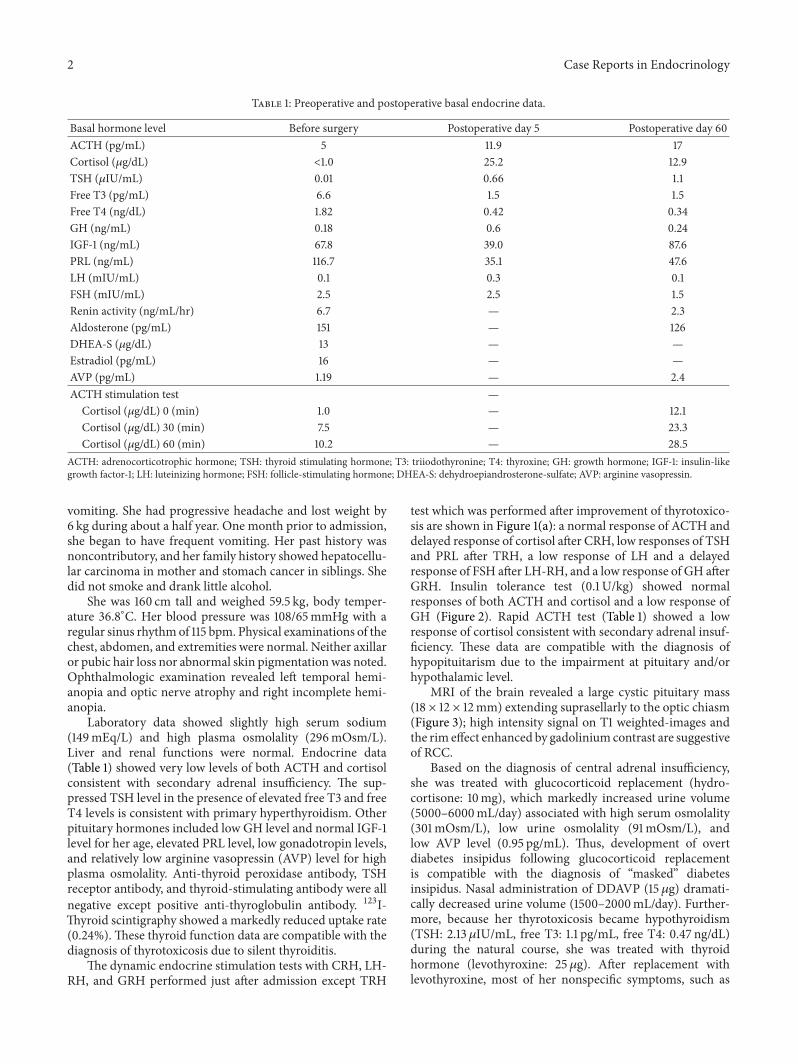
Table 1: Preoperative and postoperative basal endocrine data.
Basal hormone level Before surgery Postoperative day 5 Postoperative day 60ACTH (pg/mL) 5 11.9 17Cortisol (𝜇g/dL) <1.0 25.2 12.9TSH (𝜇IU/mL) 0.01 0.66 1.1Free T3 (pg/mL) 6.6 1.5 1.5Free T4 (ng/dL) 1.82 0.42 0.34GH (ng/mL) 0.18 0.6 0.24IGF-1 (ng/mL) 67.8 39.0 87.6PRL (ng/mL) 116.7 35.1 47.6LH (mIU/mL) 0.1 0.3 0.1FSH (mIU/mL) 2.5 2.5 1.5Renin activity (ng/mL/hr) 6.7 — 2.3Aldosterone (pg/mL) 151 — 126DHEA-S (𝜇g/dL) 13 — —Estradiol (pg/mL) 16 — —AVP (pg/mL) 1.19 — 2.4ACTH stimulation test —
Cortisol (𝜇g/dL) 0 (min) 1.0 — 12.1Cortisol (𝜇g/dL) 30 (min) 7.5 — 23.3Cortisol (𝜇g/dL) 60 (min) 10.2 — 28.5
ACTH: adrenocorticotrophic hormone; TSH: thyroid stimulating hormone; T3: triiodothyronine; T4: thyroxine; GH: growth hormone; IGF-1: insulin-likegrowth factor-1; LH: luteinizing hormone; FSH: follicle-stimulating hormone; DHEA-S: dehydroepiandrosterone-sulfate; AVP: arginine vasopressin.
vomiting. She had progressive headache and lost weight by6 kg during about a half year. One month prior to admission,she began to have frequent vomiting. Her past history wasnoncontributory, and her family history showed hepatocellu-lar carcinoma in mother and stomach cancer in siblings. Shedid not smoke and drank little alcohol.
She was 160 cm tall and weighed 59.5 kg, body temper-ature 36.8∘C. Her blood pressure was 108/65mmHg with aregular sinus rhythmof 115 bpm. Physical examinations of thechest, abdomen, and extremities were normal. Neither axillaror pubic hair loss nor abnormal skin pigmentationwas noted.Ophthalmologic examination revealed left temporal hemi-anopia and optic nerve atrophy and right incomplete hemi-anopia.
Laboratory data showed slightly high serum sodium(149mEq/L) and high plasma osmolality (296mOsm/L).Liver and renal functions were normal. Endocrine data(Table 1) showed very low levels of both ACTH and cortisolconsistent with secondary adrenal insufficiency. The sup-pressed TSH level in the presence of elevated free T3 and freeT4 levels is consistent with primary hyperthyroidism. Otherpituitary hormones included low GH level and normal IGF-1level for her age, elevated PRL level, low gonadotropin levels,and relatively low arginine vasopressin (AVP) level for highplasma osmolality. Anti-thyroid peroxidase antibody, TSHreceptor antibody, and thyroid-stimulating antibody were allnegative except positive anti-thyroglobulin antibody. 123I-Thyroid scintigraphy showed a markedly reduced uptake rate(0.24%).These thyroid function data are compatible with thediagnosis of thyrotoxicosis due to silent thyroiditis.
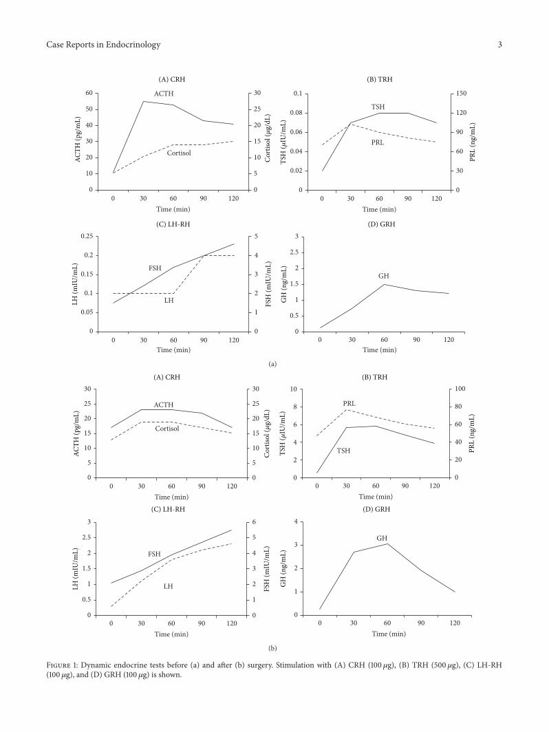
The dynamic endocrine stimulation tests with CRH, LH-RH, and GRH performed just after admission except TRH
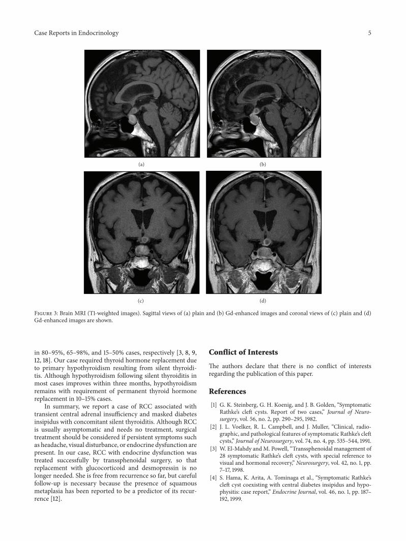
test which was performed after improvement of thyrotoxico-sis are shown in Figure 1(a): a normal response of ACTH anddelayed response of cortisol after CRH, low responses of TSHand PRL after TRH, a low response of LH and a delayedresponse of FSH after LH-RH, and a low response of GH afterGRH. Insulin tolerance test (0.1 U/kg) showed normalresponses of both ACTH and cortisol and a low response ofGH (Figure 2). Rapid ACTH test (Table 1) showed a lowresponse of cortisol consistent with secondary adrenal insuf-ficiency. These data are compatible with the diagnosis ofhypopituitarism due to the impairment at pituitary and/orhypothalamic level.
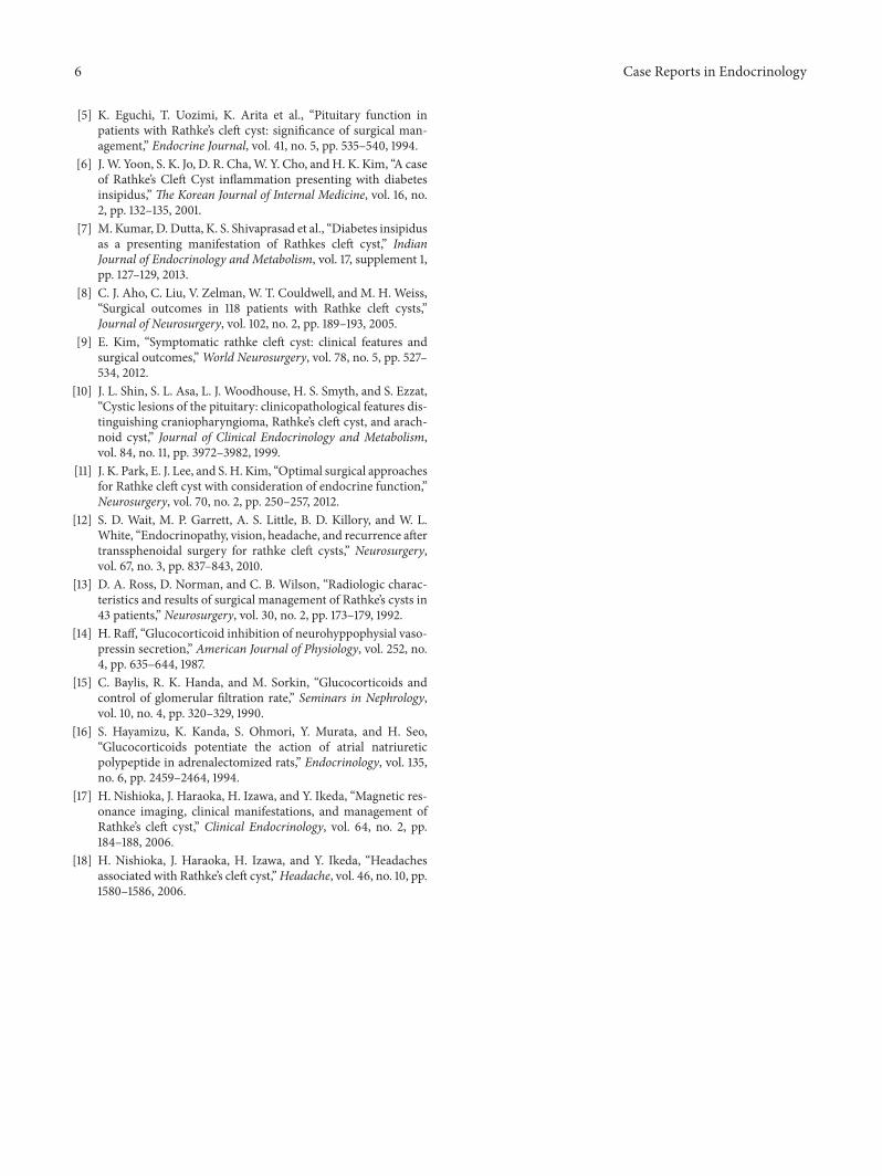
MRI of the brain revealed a large cystic pituitary mass(18 × 12 × 12mm) extending suprasellarly to the optic chiasm(Figure 3); high intensity signal on T1 weighted-images andthe rim effect enhanced by gadolinium contrast are suggestiveof RCC.
Based on the diagnosis of central adrenal insufficiency,she was treated with glucocorticoid replacement (hydro-cortisone: 10mg), which markedly increased urine volume(5000–6000mL/day) associated with high serum osmolality(301mOsm/L), low urine osmolality (91mOsm/L), andlow AVP level (0.95 pg/mL). Thus, development of overtdiabetes insipidus following glucocorticoid replacementis compatible with the diagnosis of “masked” diabetesinsipidus. Nasal administration of DDAVP (15 𝜇g) dramati-cally decreased urine volume (1500–2000mL/day). Further-more, because her thyrotoxicosis became hypothyroidism(TSH: 2.13 𝜇IU/mL, free T3: 1.1 pg/mL, free T4: 0.47 ng/dL)during the natural course, she was treated with thyroidhormone (levothyroxine: 25 𝜇g). After replacement withlevothyroxine, most of her nonspecific symptoms, such as
Case Reports in Endocrinology 3
0
5
10
15
20
25
30
0
10
20
30
40
50
60
0 30 60 90 120
0
0.5
1
1.5
2
2.5
3
0
1
2
3
4
5
0
0.05
0.1
0.15
0.2
0.25
0
30
60
90
120
150
0
0.02
0.04
0.06
0.08
0.1ACTH
Cortisol
Time (min)
TSH
PRL
LH
FSHGH
ACTH
(pg/
mL)
Cor
tisol
(𝜇g/
dL)
TSH
(𝜇IU
/mL)
PRL
(ng/
mL)
GH
(ng/
mL)
0 30 60 90 120Time (min)
LH (m
IU/m
L)
FSH
(mIU
/mL)
0 30 60 90 120Time (min)
0 30 60 90 120Time (min)
(A) CRH (B) TRH
(C) LH-RH (D) GRH
(a)
0
5
10
15
20
25
30
0 30 60 90 120
ACTH
Cortisol
Time (min)
Cor
tisol
(𝜇g/
dL)
ACTH
(pg/
mL)
0
5
10
15
20
25
30
0
20
40
60
80
100
0
2
4
6
8
10
TSH
PRL
TSH
(𝜇IU
/mL)
PRL
(ng/
mL)
0 30 60 90 120Time (min)
0
0.5
1
1.5
2
2.5
3
0
1
2
3
4
6
5
LH
FSH
LH (m
IU/m
L)
FSH
(mIU
/mL)
0 30 60 90 120Time (min)
0
1
2
3
4
GH
GH
(ng/
mL)
0 30 60 90 120Time (min)
(A) CRH (B) TRH
(C) LH-RH (D) GRH
(b)
Figure 1: Dynamic endocrine tests before (a) and after (b) surgery. Stimulation with (A) CRH (100𝜇g), (B) TRH (500𝜇g), (C) LH-RH(100𝜇g), and (D) GRH (100 𝜇g) is shown.

4 Case Reports in Endocrinology
0
0.1
0.2
0.3
0.4
0.5
0
10
20
30
40
50
0 30 60 90Time (min)
ACTH
Cortisol
GHCor
tisol
(𝜇g/
dL)
ACTH
(pg/
mL)
GH
(ng/
mL)
Figure 2: Insulin tolerance test. Regular insulin (0.1 U/Kg) wasintravenously administered as a bolus with a nadir plasma glucoselevel (28mg/dL) at 30 minutes.
poor appetite, malaise, weight loss, and vomiting, graduallyimproved, but her headache persisted.
She underwent transsphenoidal surgery with completecyst evacuation and partial wall excision; RCC was con-firmed histopathologically. Postoperatively, her headacheresolved, but bitemporal hemianopia persisted. Postoperativeendocrine data (Table 1) showed elevated levels of ACTH,cortisol, TSH, and AVP but low levels of thyroid hormonesand gonadotropin and normal PRL level. Postoperativedynamic endocrine stimulation tests (Figure 1(b)) showednormal responses of ACTH and cortisol after CRH, a delayedresponse of TSH and a low response of PRL after TRH,delayed responses of LH and FSH after LH-RH, and a normalresponse of GH after GRH. Postoperative rapid ACTH test(Table 1) showed a normal response of cortisol after ACTHstimulation. Based on the postoperative endocrine data,replacements with glucocorticoid and desmopressin werestopped, while thyroid hormone was continued.
3. Discussion
RCC, a benign cystic pituitary mass, is often detected inci-dentally by MRI of the brain. RCC is usually asymptomatic,but when it enlarges and/or is associated with inflammation,various symptoms, such as headache, visual disturbance, andendocrine dysfunction, may occur. Endocrine dysfunction isobserved in 40–60% of patients with a symptomatic RCC[9–12], among which hypopituitarism is most common (75–100%), but diabetes insipidus is less common (0–20%) [3, 5,8]. A key mechanism for pituitary dysfunction caused by alarge cystic RCC has been considered to be due to its com-pression to normal pituitary gland [13], although intrasellarRCC seems unlikely to cause such mechanical compression.Alternatively, RCC-associated inflammatory change in thepituitary gland may have caused pituitary dysfunction [5]. Infact, such inflammatory change has been assumed to resultfrom the leakage of its cyst contents into the pituitary gland[5].
The major symptoms and signs of headache and visualdisturbance in this case are due to the suprasellar pituitarytumor compressing optic chiasm as demonstrated by brainMRI. The concomitant clinical manifestations of anorexia,
malaise, and weight loss suggest the possible hypopituitarismby the pituitary tumor as evidenced by the endocrine datawith low basal levels of pituitary hormones (ACTH, TSH,GH, LH, and FSH) and hyperprolactinemia. The dynamicendocrine data with low and/or delayed responses of GH byGRH, LH and FSH by LH-RH, and TSH by TRH suggest thepituitary and/or the hypothalamic lesions. On the other hand,normal responses of ACTH and cortisol by CRH stimulationand insulin tolerance test and a low response of cortisol afterACTH stimulation are consistent with hypothalamic ratherthan pituitary lesion. Thus, hypopituitarism in this case ismost likely caused by the impairment by pituitary and/orhypothalamic lesions. Her nonspecific symptoms (anorexia,malaise, and weight loss) are due to either adrenal insuffi-ciency or thyrotoxicosis due to silent thyroiditis. Since thy-rotoxicosis accelerates the metabolism of cortisol to induceadrenal insufficiency, it is possible to speculate that subclini-cal adrenal insufficiency was actualized in overt insufficiency,triggered by thyrotoxicosis in our case.
Soon after replacement with glucocorticoid, she hadmarkedly increased urine volume with elevated serum osmo-lality, lowurine osmolality, and decreased vasopressin release,suggesting the onset of overt diabetes insipidus. Masked dia-betes insipidus is defined as the condition in which polyuriadue to diabetes insipidus is masked by glucocorticoid defi-ciency. The underlying mechanisms of masked diabetesinsipidus are accounted by various effects of glucocorticoid,such as inhibition of vasopressin release [14], reduced waterabsorption at the proximal tubule [15], and stimulation ofatrial natriuretic peptide secretion to enhance the diureticeffect [16]. In the present case, vasopressin release impaired bythe compression and/or inflammation of the pituitary stalk byRCC should have caused overt diabetes insipidus, whichcould have been masked by the transient glucocorticoiddeficiency due to central adrenal insufficiency.
The cyst wall of RCC is histopathologically characterizedby lining by cuboidal or columnar epithelial cells. However,the presence of a small amount of squamous epithelium inthis case suggests that squamous metaplasia occurs due toinflammation of the RCC. In fact, it has been reported thatsquamous epithelium is identified in 45% with symptomaticRCC [9]. Furthermore, the presence of inflammatory cells inthe cyst wall in this case was suspected by the high-intensitymass on the T1-weighted images, reflecting mucous materialswithin the cyst, often associated with chronic inflammation,which may potentially cause irreversible endocrine dysfunc-tion [17].Therefore, it is suggested that the pituitary dysfunc-tionwas caused not only by compression by themass, but alsoby inflammation.
After surgery, pituitary endocrine dysfunctions improvedpromptly. In particular, replacement with glucocorticoidbecame unnecessary on postoperative 5th day and thenthat with desmopressin became unnecessary on postopera-tive 60th day, suggesting reversible changes of compressionand/or inflammation by RCC. In contrast, her headacheresolved after surgery without any improvement of visualdisturbance (temporal hemianopia). It has been reportedthat, during the clinical course of RCC, headache, visual dis-turbance, and pituitary dysfunction improved after surgery
Case Reports in Endocrinology 5
(a) (b)
(c) (d)
Figure 3: Brain MRI (T1-weighted images). Sagittal views of (a) plain and (b) Gd-enhanced images and coronal views of (c) plain and (d)Gd-enhanced images are shown.
in 80–95%, 65–98%, and 15–50% cases, respectively [3, 8, 9,12, 18]. Our case required thyroid hormone replacement dueto primary hypothyroidism resulting from silent thyroidi-tis. Although hypothyroidism following silent thyroiditis inmost cases improves within three months, hypothyroidismremains with requirement of permanent thyroid hormonereplacement in 10–15% cases.
In summary, we report a case of RCC associated withtransient central adrenal insufficiency and masked diabetesinsipidus with concomitant silent thyroiditis. Although RCCis usually asymptomatic and needs no treatment, surgicaltreatment should be considered if persistent symptoms suchas headache, visual disturbance, or endocrine dysfunction arepresent. In our case, RCC with endocrine dysfunction wastreated successfully by transsphenoidal surgery, so thatreplacement with glucocorticoid and desmopressin is nolonger needed. She is free from recurrence so far, but carefulfollow-up is necessary because the presence of squamousmetaplasia has been reported to be a predictor of its recur-rence [12].
Conflict of Interests
The authors declare that there is no conflict of interestsregarding the publication of this paper.
References
[1] G. K. Steinberg, G. H. Koenig, and J. B. Golden, “SymptomaticRathke’s cleft cysts. Report of two cases,” Journal of Neuro-surgery, vol. 56, no. 2, pp. 290–295, 1982.
[2] J. L. Voelker, R. L. Campbell, and J. Muller, “Clinical, radio-graphic, and pathological features of symptomatic Rathke’s cleftcysts,” Journal of Neurosurgery, vol. 74, no. 4, pp. 535–544, 1991.
[3] W. El-Mahdy andM. Powell, “Transsphenoidal management of28 symptomatic Rathke’s cleft cysts, with special reference tovisual and hormonal recovery,” Neurosurgery, vol. 42, no. 1, pp.7–17, 1998.
[4] S. Hama, K. Arita, A. Tominaga et al., “Symptomatic Rathke’scleft cyst coexisting with central diabetes insipidus and hypo-physitis: case report,” Endocrine Journal, vol. 46, no. 1, pp. 187–192, 1999.
6 Case Reports in Endocrinology
[5] K. Eguchi, T. Uozimi, K. Arita et al., “Pituitary function inpatients with Rathke’s cleft cyst: significance of surgical man-agement,” Endocrine Journal, vol. 41, no. 5, pp. 535–540, 1994.
[6] J.W. Yoon, S. K. Jo, D. R. Cha,W. Y. Cho, andH. K. Kim, “A caseof Rathke’s Cleft Cyst inflammation presenting with diabetesinsipidus,” The Korean Journal of Internal Medicine, vol. 16, no.2, pp. 132–135, 2001.
[7] M. Kumar, D. Dutta, K. S. Shivaprasad et al., “Diabetes insipidusas a presenting manifestation of Rathkes cleft cyst,” IndianJournal of Endocrinology and Metabolism, vol. 17, supplement 1,pp. 127–129, 2013.
[8] C. J. Aho, C. Liu, V. Zelman, W. T. Couldwell, and M. H. Weiss,“Surgical outcomes in 118 patients with Rathke cleft cysts,”Journal of Neurosurgery, vol. 102, no. 2, pp. 189–193, 2005.
[9] E. Kim, “Symptomatic rathke cleft cyst: clinical features andsurgical outcomes,”World Neurosurgery, vol. 78, no. 5, pp. 527–534, 2012.
[10] J. L. Shin, S. L. Asa, L. J. Woodhouse, H. S. Smyth, and S. Ezzat,“Cystic lesions of the pituitary: clinicopathological features dis-tinguishing craniopharyngioma, Rathke’s cleft cyst, and arach-noid cyst,” Journal of Clinical Endocrinology and Metabolism,vol. 84, no. 11, pp. 3972–3982, 1999.
[11] J. K. Park, E. J. Lee, and S. H. Kim, “Optimal surgical approachesfor Rathke cleft cyst with consideration of endocrine function,”Neurosurgery, vol. 70, no. 2, pp. 250–257, 2012.
[12] S. D. Wait, M. P. Garrett, A. S. Little, B. D. Killory, and W. L.White, “Endocrinopathy, vision, headache, and recurrence aftertranssphenoidal surgery for rathke cleft cysts,” Neurosurgery,vol. 67, no. 3, pp. 837–843, 2010.
[13] D. A. Ross, D. Norman, and C. B. Wilson, “Radiologic charac-teristics and results of surgical management of Rathke’s cysts in43 patients,” Neurosurgery, vol. 30, no. 2, pp. 173–179, 1992.
[14] H. Raff, “Glucocorticoid inhibition of neurohyppophysial vaso-pressin secretion,” American Journal of Physiology, vol. 252, no.4, pp. 635–644, 1987.
[15] C. Baylis, R. K. Handa, and M. Sorkin, “Glucocorticoids andcontrol of glomerular filtration rate,” Seminars in Nephrology,vol. 10, no. 4, pp. 320–329, 1990.
[16] S. Hayamizu, K. Kanda, S. Ohmori, Y. Murata, and H. Seo,“Glucocorticoids potentiate the action of atrial natriureticpolypeptide in adrenalectomized rats,” Endocrinology, vol. 135,no. 6, pp. 2459–2464, 1994.
[17] H. Nishioka, J. Haraoka, H. Izawa, and Y. Ikeda, “Magnetic res-onance imaging, clinical manifestations, and management ofRathke’s cleft cyst,” Clinical Endocrinology, vol. 64, no. 2, pp.184–188, 2006.
[18] H. Nishioka, J. Haraoka, H. Izawa, and Y. Ikeda, “Headachesassociated with Rathke’s cleft cyst,”Headache, vol. 46, no. 10, pp.1580–1586, 2006.

Submit your manuscripts athttp://www.hindawi.com
Stem CellsInternational
Hindawi Publishing Corporationhttp://www.hindawi.com Volume 2014
Hindawi Publishing Corporationhttp://www.hindawi.com Volume 2014
MEDIATORSINFLAMMATION
of
Hindawi Publishing Corporationhttp://www.hindawi.com Volume 2014
Behavioural Neurology
EndocrinologyInternational Journal of
Hindawi Publishing Corporationhttp://www.hindawi.com Volume 2014
Hindawi Publishing Corporationhttp://www.hindawi.com Volume 2014
Disease Markers
Hindawi Publishing Corporationhttp://www.hindawi.com Volume 2014
BioMed Research International
OncologyJournal of
Hindawi Publishing Corporationhttp://www.hindawi.com Volume 2014
Hindawi Publishing Corporationhttp://www.hindawi.com Volume 2014
Oxidative Medicine and Cellular Longevity
Hindawi Publishing Corporationhttp://www.hindawi.com Volume 2014
PPAR Research
The Scientific World JournalHindawi Publishing Corporation http://www.hindawi.com Volume 2014
Immunology ResearchHindawi Publishing Corporationhttp://www.hindawi.com Volume 2014
Journal of
ObesityJournal of
Hindawi Publishing Corporationhttp://www.hindawi.com Volume 2014
Hindawi Publishing Corporationhttp://www.hindawi.com Volume 2014
Computational and Mathematical Methods in Medicine
OphthalmologyJournal of
Hindawi Publishing Corporationhttp://www.hindawi.com Volume 2014
Diabetes ResearchJournal of
Hindawi Publishing Corporationhttp://www.hindawi.com Volume 2014
Hindawi Publishing Corporationhttp://www.hindawi.com Volume 2014
Research and TreatmentAIDS
Hindawi Publishing Corporationhttp://www.hindawi.com Volume 2014
Gastroenterology Research and Practice
Hindawi Publishing Corporationhttp://www.hindawi.com Volume 2014
Parkinson’s Disease
Evidence-Based Complementary and Alternative Medicine
Volume 2014Hindawi Publishing Corporationhttp://www.hindawi.com