case report a rare mullerian duct anomaly not included...
TRANSCRIPT
Hindawi Publishing CorporationCase Reports in Obstetrics and GynecologyVolume 2013, Article ID 569480, 3 pageshttp://dx.doi.org/10.1155/2013/569480
Case ReportA Rare Mullerian Duct Anomaly Not Included inthe Classification System by the American Society forReproductive Medicine
Shereene J. Brown and Shawky Z. A. Badawy
Department of Obstetrics and Gynecology, State University of New York Upstate Medical University, 736 Irving Avenue, Syracuse,NY 13210, USA
Correspondence should be addressed to Shawky Z. A. Badawy; [email protected]
Received 31 January 2013; Accepted 27 February 2013
Academic Editors: J. Awwad, P. De Franciscis, C. Ficicioglu, and D. Hellberg
Copyright © 2013 S. J. Brown and S. Z. A. Badawy. This is an open access article distributed under the Creative CommonsAttribution License, which permits unrestricted use, distribution, and reproduction in any medium, provided the original work isproperly cited.
This is a case report of a 37-year-old female with a uterine septum (two cavities), a normal single fundal contour, two cervices, and alongitudinal vaginal septum.This is a rare finding that is not described in the current classification system by the American Societyfor Reproductive Medicine.
1. Introduction
Mullerian duct anomalies have become more easily diag-nosed due to better imaging modalities over the last fewdecades.The incidence has been quoted as low as 0.001% andas high as 10% [1]. It is theorized that the mullerian ductsfuse around the eleventh through the thirteenth weeks inutero and that fusion and absorption are unidirectional fromcaudal to cephalad [1–3]. Our current classification systemfor uterine anomalies is based on this unidirectional theory[4]. The majority of uterine anomalies can be explainedby this theory; however, there are a number of cases thatdo not fit this paradigm [4]. The case described here is amullerian duct anomaly with a longitudinal vaginal septum,two cervices, a uterine septum, and a single normal fundus.This represents failure of absorption of the septum thatseparates the mullerian ducts after fusion.
2. A Case Report
37-year-old G1P0101 was referred to the ReproductiveEndocrinology Division because of chronic dyspareunia andsecondary infertility. She had a prior Caesarean deliverywhere it was discovered that she had an unspecifiedmullerianduct anomaly. She now had a new partner and had been
trying to get pregnant for three years. She had regular cycleswith normal flow. On physical examination, a vaginal septumand two cervices were observed.
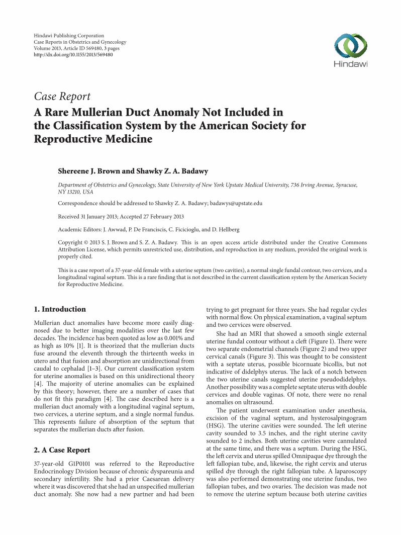
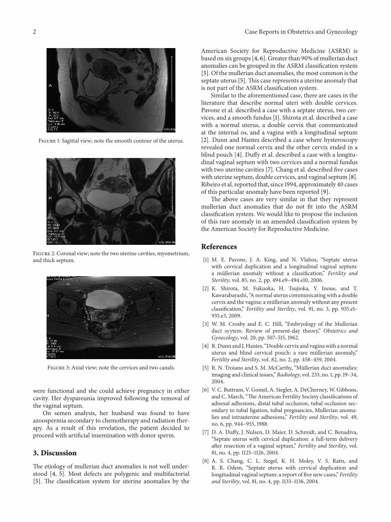
She had an MRI that showed a smooth single externaluterine fundal contour without a cleft (Figure 1). There weretwo separate endometrial channels (Figure 2) and two uppercervical canals (Figure 3). This was thought to be consistentwith a septate uterus, possible bicornuate bicollis, but notindicative of didelphys uterus. The lack of a notch betweenthe two uterine canals suggested uterine pseudodidelphys.Another possibility was a complete septate uteruswith doublecervices and double vaginas. Of note, there were no renalanomalies on ultrasound.
The patient underwent examination under anesthesia,excision of the vaginal septum, and hysterosalpingogram(HSG). The uterine cavities were sounded. The left uterinecavity sounded to 3.5 inches, and the right uterine cavitysounded to 2 inches. Both uterine cavities were cannulatedat the same time, and there was a septum. During the HSG,the left cervix and uterus spilled Omnipaque dye through theleft fallopian tube, and, likewise, the right cervix and uterusspilled dye through the right fallopian tube. A laparoscopywas also performed demonstrating one uterine fundus, twofallopian tubes, and two ovaries. The decision was made notto remove the uterine septum because both uterine cavities
2 Case Reports in Obstetrics and Gynecology
Figure 1: Sagittal view; note the smooth contour of the uterus.
Figure 2: Coronal view; note the two uterine cavities, myometrium,and thick septum.
Figure 3: Axial view; note the cervices and two canals.
were functional and she could achieve pregnancy in eithercavity. Her dyspareunia improved following the removal ofthe vaginal septum.
On semen analysis, her husband was found to haveazoospermia secondary to chemotherapy and radiation ther-apy. As a result of this revelation, the patient decided toproceed with artificial insemination with donor sperm.
3. Discussion
The etiology of mullerian duct anomalies is not well under-stood [4, 5]. Most defects are polygenic and multifactorial[5]. The classification system for uterine anomalies by the
American Society for Reproductive Medicine (ASRM) isbased on six groups [4, 6]. Greater than 90%ofmullerian ductanomalies can be grouped in the ASRM classification system[5]. Of themullerian duct anomalies, themost common is theseptate uterus [5].This case represents a uterine anomaly thatis not part of the ASRM classification system.
Similar to the aforementioned case, there are cases in theliterature that describe normal uteri with double cervices.Pavone et al. described a case with a septate uterus, two cer-vices, and a smooth fundus [1]. Shirota et al. described a casewith a normal uterus, a double cervix that communicatedat the internal os, and a vagina with a longitudinal septum[2]. Dunn and Hantes described a case where hysteroscopyrevealed one normal cervix and the other cervix ended in ablind pouch [4]. Duffy et al. described a case with a longitu-dinal vaginal septum with two cervices and a normal funduswith two uterine cavities [7]. Chang et al. described five caseswith uterine septum, double cervices, and vaginal septum [8].Ribeiro et al. reported that, since 1994, approximately 40 casesof this particular anomaly have been reported [9].
The above cases are very similar in that they representmullerian duct anomalies that do not fit into the ASRMclassification system. We would like to propose the inclusionof this rare anomaly in an amended classification system bythe American Society for Reproductive Medicine.
References
[1] M. E. Pavone, J. A. King, and N. Vlahos, “Septate uteruswith cervical duplication and a longitudinal vaginal septum:a mullerian anomaly without a classification,” Fertility andSterility, vol. 85, no. 2, pp. 494.e9–494.e10, 2006.
[2] K. Shirota, M. Fukuoka, H. Tsujioka, Y. Inoue, and T.Kawarabayashi, “Anormal uterus communicatingwith a doublecervix and the vagina: a mullerian anomaly without any presentclassification,” Fertility and Sterility, vol. 91, no. 3, pp. 935.e1–935.e3, 2009.
[3] W. M. Crosby and E. C. Hill, “Embryology of the Mullerianduct syytem. Review of present-day theory,” Obstetrics andGynecology, vol. 20, pp. 507–515, 1962.
[4] R.Dunn and J.Hantes, “Double cervix and vaginawith a normaluterus and blind cervical pouch: a rare mullerian anomaly,”Fertility and Sterility, vol. 82, no. 2, pp. 458–459, 2004.
[5] R. N. Troiano and S. M. McCarthy, “Mullerian duct anomalies:imaging and clinical issues,”Radiology, vol. 233, no. 1, pp. 19–34,2004.
[6] V. C. Buttram, V. Gomel, A. Siegler, A. DeCherney,W. Gibbons,and C.March, “The American Fertility Society classifications ofadnexal adhesions, distal tubal occlusion, tubal occlusion sec-ondary to tubal ligation, tubal pregnancies, Mullerian anoma-lies and intrauterine adhesions,” Fertility and Sterility, vol. 49,no. 6, pp. 944–955, 1988.
[7] D. A. Duffy, J. Nulsen, D. Maier, D. Schmidt, and C. Benadiva,“Septate uterus with cervical duplication: a full-term deliveryafter resection of a vaginal septum,” Fertility and Sterility, vol.81, no. 4, pp. 1125–1126, 2004.
[8] A. S. Chang, C. L. Siegel, K. H. Moley, V. S. Ratts, andR. R. Odem, “Septate uterus with cervical duplication andlongitudinal vaginal septum: a report of five new cases,” Fertilityand Sterility, vol. 81, no. 4, pp. 1133–1136, 2004.

Case Reports in Obstetrics and Gynecology 3
[9] S. C. Ribeiro, L. Y. S. Yamakami, R. A. Tormena, W. S. Pinheiro,J. A. M. de Almeida, and E. C. Baracat, “Septate uterus withcervical duplication and longitudinal vaginal septum,” Revistada Associacao Medica Brasileira, vol. 56, no. 2, pp. 254–256,2010.

Submit your manuscripts athttp://www.hindawi.com
Stem CellsInternational
Hindawi Publishing Corporationhttp://www.hindawi.com Volume 2014
Hindawi Publishing Corporationhttp://www.hindawi.com Volume 2014
MEDIATORSINFLAMMATION
of
Hindawi Publishing Corporationhttp://www.hindawi.com Volume 2014
Behavioural Neurology
EndocrinologyInternational Journal of
Hindawi Publishing Corporationhttp://www.hindawi.com Volume 2014
Hindawi Publishing Corporationhttp://www.hindawi.com Volume 2014
Disease Markers
Hindawi Publishing Corporationhttp://www.hindawi.com Volume 2014
BioMed Research International
OncologyJournal of
Hindawi Publishing Corporationhttp://www.hindawi.com Volume 2014
Hindawi Publishing Corporationhttp://www.hindawi.com Volume 2014
Oxidative Medicine and Cellular Longevity
Hindawi Publishing Corporationhttp://www.hindawi.com Volume 2014
PPAR Research
The Scientific World JournalHindawi Publishing Corporation http://www.hindawi.com Volume 2014
Immunology ResearchHindawi Publishing Corporationhttp://www.hindawi.com Volume 2014
Journal of
ObesityJournal of
Hindawi Publishing Corporationhttp://www.hindawi.com Volume 2014
Hindawi Publishing Corporationhttp://www.hindawi.com Volume 2014
Computational and Mathematical Methods in Medicine
OphthalmologyJournal of
Hindawi Publishing Corporationhttp://www.hindawi.com Volume 2014
Diabetes ResearchJournal of
Hindawi Publishing Corporationhttp://www.hindawi.com Volume 2014
Hindawi Publishing Corporationhttp://www.hindawi.com Volume 2014
Research and TreatmentAIDS
Hindawi Publishing Corporationhttp://www.hindawi.com Volume 2014
Gastroenterology Research and Practice
Hindawi Publishing Corporationhttp://www.hindawi.com Volume 2014
Parkinson’s Disease
Evidence-Based Complementary and Alternative Medicine
Volume 2014Hindawi Publishing Corporationhttp://www.hindawi.com