case report delayed diagnosis of post‑traumatic aneurysm...
TRANSCRIPT
© 2018 Surgical Neurology International | Published by Wolters Kluwer - Medknow
Editor:Kazuhiro Hongo, M.D.,Shinsui University, Matsomoto, Japan
OPEN ACCESSFor entire Editorial Board visit : http://www.surgicalneurologyint.com
SNI: Neurovascular
Case Report
Delayed diagnosis of post‑traumatic aneurysm of distal anterior cerebral arteryDomenico Policicchio, Giampiero Muggianu, Giosuè Dipellegrini, Riccardo Boccaletti1
Department of Neurosurgery, Azienda Ospedaliero Universitaria di Sassari, Via Enrico De Nicola 1, 07100 Sassari (SS), Department of Neurosurgery, 1Regina Elena National Cancer Institute, Neurosurgery Department, Via Elio Chianesi 53, 00144, Rome, Italy
E‑mail: *Domenico Policicchio ‑ [email protected]; Giampiero Muggianu ‑ [email protected]; Giosuè Dipellegrini ‑ [email protected]; Riccardo Boccaletti ‑ [email protected] *Corresponding author
Received: 23 July 18 Accepted: 27 September 18 Published: 01 November 18
Access this article onlineWebsite:www.surgicalneurologyint.comDOI: 10.4103/sni.sni_252_18Quick Response Code:
AbstractBackground: Traumatic intracranial aneurysms (TICA) are often associated with poor prognosis and should be diagnosed as soon as possible to prevent delayed intracranial hemorrhage and high rates of morbidity/mortality related to bleeding. Diagnosis requires a high index of suspicion. The goal of treatment is to exclude the aneurysm issue with surgical or endovascular methods.Case Description: We report the case of a 19‑year‑old boy who suffered a cranio‑orbital trauma; 2 weeks after initial trauma he deteriorates with a new intracranial bleeding. Immediate angiography resulted negative. Delayed follow‑up by magnetic resonance angiography showed an unruptured aneurysm of anterior cerebral artery that was successfully clipped.Conclusions: A TICA should be suspected in case of delayed deterioration in head‑injured patient, prompt diagnosis and treatment could improve prognosis and reduce morbidity and mortality.
Key Words: Digital subtraction angiography, magnetic resonance angiography, penetrating trauma, post‑traumatic aneurysm
INTRODUCTION
Traumatic intracranial aneurysm (TICA) is considered a rare entity, constituting <1% of all cerebral aneurysms.[11] A TICA can occur after both blunt and penetrating head trauma and is more frequently described in the pediatric population. Delayed intracranial hemorrhage is typical in these patients and is associated with a significant mortality rate, as high as 50%.[4] Diagnosis of TICAs requires a high index of suspicion and is based on intracranial vascular study.[14] Aggressive surgical management is associated with a better outcome than conservative treatment.[11] We report a case of a delayed diagnosis of TICA of the anterior cerebral artery in a 19‑year‑old boy.
CASE DESCRIPTION
A 19‑year‑old boy was admitted to our Emergency Department for Cranio‑facial Trauma; while he was
How to cite this article: Policicchio D, Muggianu G, Dipellegrini G, Boccaletti R. Delayed diagnosis of post-traumatic aneurysm of distal anterior cerebral artery. Surg Neurol Int 2018;9:222.http://surgicalneurologyint.com/Delayed-diagnosis-of-post-traumatic-aneurysm-of-distal-anterior-cerebral-artery/
This is an open access journal, and articles are distributed under the terms of the Creative Commons Attribution-NonCommercial-ShareAlike 4.0 License, which allows others to remix, tweak, and build upon the work non-commercially, as long as appropriate credit is given and the new creations are licensed under the identical terms.
For reprints contact: [email protected]
Surgical Neurology International 2018, 9:222 http://www.surgicalneurologyint.com/content/9/1/222
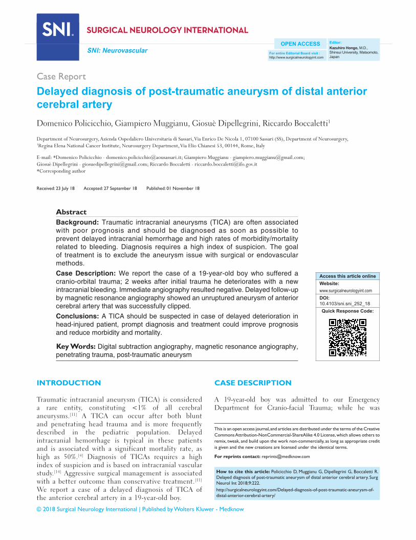
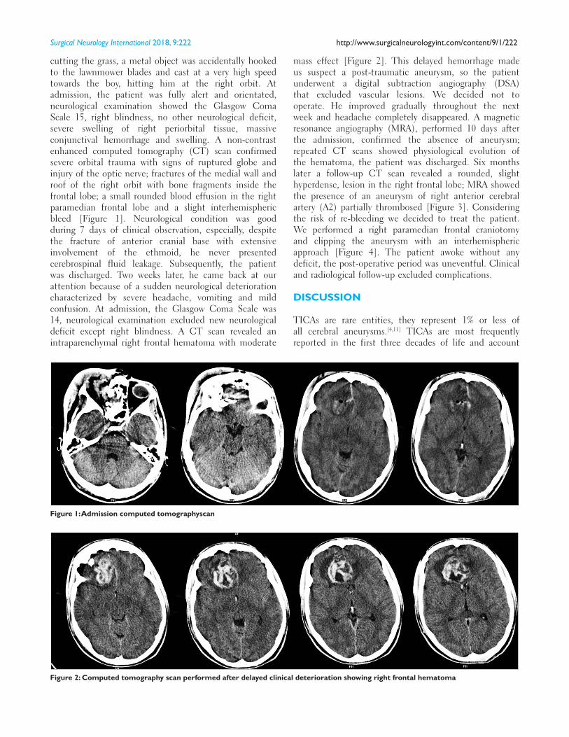
cutting the grass, a metal object was accidentally hooked to the lawnmower blades and cast at a very high speed towards the boy, hitting him at the right orbit. At admission, the patient was fully alert and orientated, neurological examination showed the Glasgow Coma Scale 15, right blindness, no other neurological deficit, severe swelling of right periorbital tissue, massive conjunctival hemorrhage and swelling. A non‑contrast enhanced computed tomography (CT) scan confirmed severe orbital trauma with signs of ruptured globe and injury of the optic nerve; fractures of the medial wall and roof of the right orbit with bone fragments inside the frontal lobe; a small rounded blood effusion in the right paramedian frontal lobe and a slight interhemispheric bleed [Figure 1]. Neurological condition was good during 7 days of clinical observation, especially, despite the fracture of anterior cranial base with extensive involvement of the ethmoid, he never presented cerebrospinal fluid leakage. Subsequently, the patient was discharged. Two weeks later, he came back at our attention because of a sudden neurological deterioration characterized by severe headache, vomiting and mild confusion. At admission, the Glasgow Coma Scale was 14, neurological examination excluded new neurological deficit except right blindness. A CT scan revealed an intraparenchymal right frontal hematoma with moderate
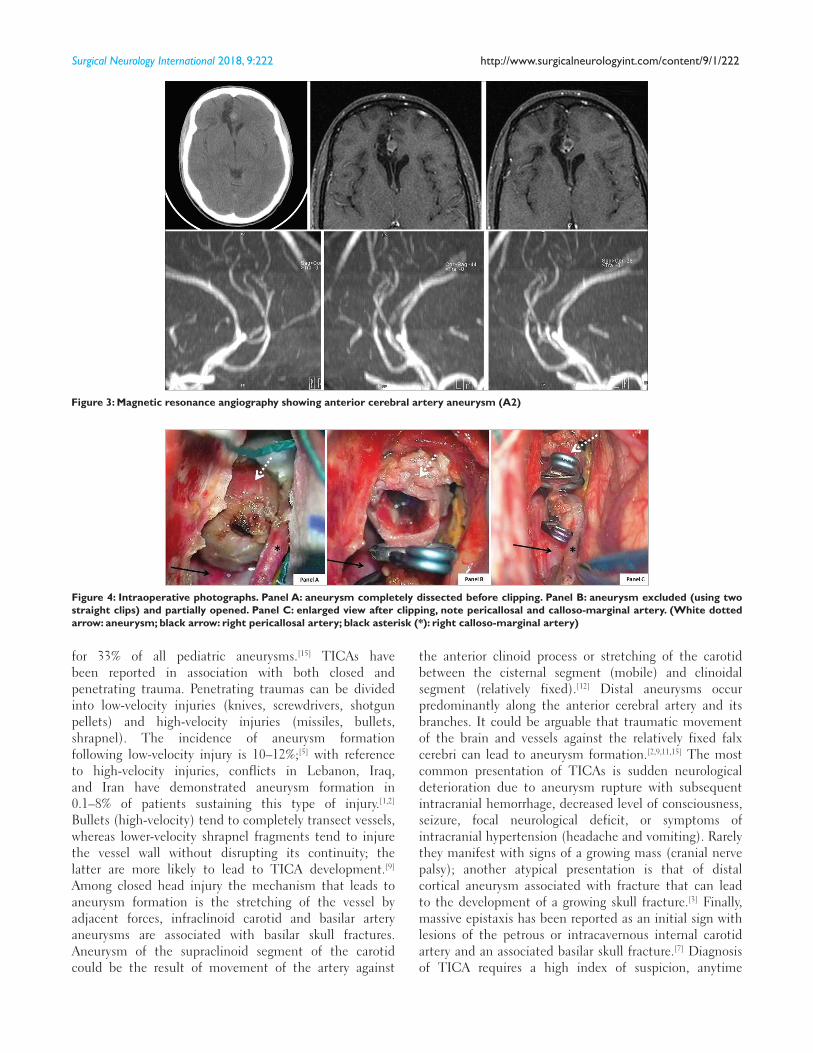
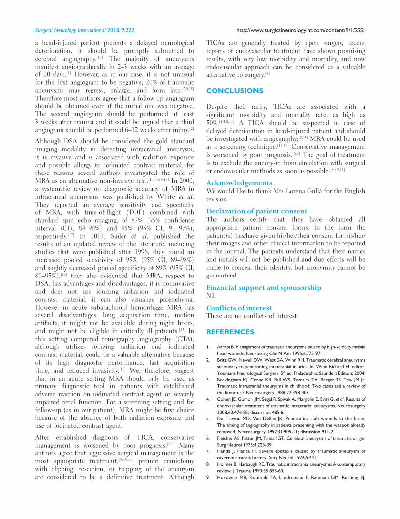
mass effect [Figure 2]. This delayed hemorrhage made us suspect a post‑traumatic aneurysm, so the patient underwent a digital subtraction angiography (DSA) that excluded vascular lesions. We decided not to operate. He improved gradually throughout the next week and headache completely disappeared. A magnetic resonance angiography (MRA), performed 10 days after the admission, confirmed the absence of aneurysm; repeated CT scans showed physiological evolution of the hematoma, the patient was discharged. Six months later a follow‑up CT scan revealed a rounded, slight hyperdense, lesion in the right frontal lobe; MRA showed the presence of an aneurysm of right anterior cerebral artery (A2) partially thrombosed [Figure 3]. Considering the risk of re‑bleeding we decided to treat the patient. We performed a right paramedian frontal craniotomy and clipping the aneurysm with an interhemispheric approach [Figure 4]. The patient awoke without any deficit, the post‑operative period was uneventful. Clinical and radiological follow‑up excluded complications.
DISCUSSION
TICAs are rare entities, they represent 1% or less of all cerebral aneurysms.[4,11] TICAs are most frequently reported in the first three decades of life and account
Figure 1: Admission computed tomographyscan
Figure 2: Computed tomography scan performed after delayed clinical deterioration showing right frontal hematoma
Surgical Neurology International 2018, 9:222 http://www.surgicalneurologyint.com/content/9/1/222
for 33% of all pediatric aneurysms.[15] TICAs have been reported in association with both closed and penetrating trauma. Penetrating traumas can be divided into low‑velocity injuries (knives, screwdrivers, shotgun pellets) and high‑velocity injuries (missiles, bullets, shrapnel). The incidence of aneurysm formation following low‑velocity injury is 10–12%;[5] with reference to high‑velocity injuries, conflicts in Lebanon, Iraq, and Iran have demonstrated aneurysm formation in 0.1–8% of patients sustaining this type of injury.[1,2] Bullets (high‑velocity) tend to completely transect vessels, whereas lower‑velocity shrapnel fragments tend to injure the vessel wall without disrupting its continuity; the latter are more likely to lead to TICA development.[9] Among closed head injury the mechanism that leads to aneurysm formation is the stretching of the vessel by adjacent forces, infraclinoid carotid and basilar artery aneurysms are associated with basilar skull fractures. Aneurysm of the supraclinoid segment of the carotid could be the result of movement of the artery against
the anterior clinoid process or stretching of the carotid between the cisternal segment (mobile) and clinoidal segment (relatively fixed).[12] Distal aneurysms occur predominantly along the anterior cerebral artery and its branches. It could be arguable that traumatic movement of the brain and vessels against the relatively fixed falx cerebri can lead to aneurysm formation.[2,9,11,15] The most common presentation of TICAs is sudden neurological deterioration due to aneurysm rupture with subsequent intracranial hemorrhage, decreased level of consciousness, seizure, focal neurological deficit, or symptoms of intracranial hypertension (headache and vomiting). Rarely they manifest with signs of a growing mass (cranial nerve palsy); another atypical presentation is that of distal cortical aneurysm associated with fracture that can lead to the development of a growing skull fracture.[3] Finally, massive epistaxis has been reported as an initial sign with lesions of the petrous or intracavernous internal carotid artery and an associated basilar skull fracture.[7] Diagnosis of TICA requires a high index of suspicion, anytime
Figure 3: Magnetic resonance angiography showing anterior cerebral artery aneurysm (A2)
Figure 4: Intraoperative photographs. Panel A: aneurysm completely dissected before clipping. Panel B: aneurysm excluded (using two straight clips) and partially opened. Panel C: enlarged view after clipping, note pericallosal and calloso‑marginal artery. (White dotted arrow: aneurysm; black arrow: right pericallosal artery; black asterisk (*): right calloso‑marginal artery)
Surgical Neurology International 2018, 9:222 http://www.surgicalneurologyint.com/content/9/1/222
a head‑injured patient presents a delayed neurological deterioration, it should be promptly submitted to cerebral angiography.[15] The majority of aneurysms manifest angiographically in 2–3 weeks with an average of 20 days.[1] However, as in our case, it is not unusual for the first angiogram to be negative; 20% of traumatic aneurysms may regress, enlarge, and form late.[2,9,15] Therefore most authors agree that a follow‑up angiogram should be obtained even if the initial one was negative. The second angiogram should be performed at least 3 weeks after trauma and it could be argued that a third angiogram should be performed 6–12 weeks after injury.[2]
Although DSA should be considered the gold standard imaging modality in detecting intracranial aneurysm, it is invasive and is associated with radiation exposure and possible allergy to iodinated contrast material; for these reasons several authors investigated the role of MRA as an alternative non‑invasive test.[10,13,14,17] In 2000, a systematic review on diagnostic accuracy of MRA in intracranial aneurysms was published by White et al. They reported an average sensitivity and specificity of MRA, with time‑of‑flight (TOF) combined with standard spin echo imaging, of 87% [95% confidence interval (CI), 84–90%] and 95% (95% CI, 91–97%), respectively.[17] In 2013, Sailer et al. published the results of an updated review of the literature, including studies that were published after 1998, they found an increased pooled sensitivity of 95% (95% CI, 89–98%) and slightly decreased pooled specificity of 89% (95% CI, 80–95%);[13] they also evidenced that MRA, respect to DSA, has advantages and disadvantages, it is noninvasive and does not use ionizing radiation and iodinated contrast material, it can also visualize parenchyma. However in acute subarachnoid hemorrhage MRA has several disadvantages, long acquisition time, motion artifacts, it might not be available during night hours, and might not be eligible in critically ill patients.[13] In this setting computed tomography angiography (CTA), although utilizes ionizing radiation and iodinated contrast material, could be a valuable alternative because of its high diagnostic performance, fast acquisition time, and reduced invasivity.[16] We, therefore, suggest that in an acute setting MRA should only be used as primary diagnostic tool in patients with established adverse reaction on iodinated contrast agent or severely impaired renal function. For a screening setting and for follow‑up (as in our patient), MRA might be first choice because of the absence of both radiation exposure and use of iodinated contrast agent.
After established diagnosis of TICA, conservative management is worsened by poor prognosis.[6,8] Many authors agree that aggressive surgical management is the most appropriate treatment,[3,8,9,11] prompt craniotomy with clipping, resection, or trapping of the aneurysm are considered to be a definitive treatment. Although
TICAs are generally treated by open surgery, recent reports of endovascular treatment have shown promising results, with very low morbidity and mortality, and now endovascular approach can be considered as a valuable alternative to surgery.[4]
CONCLUSIONS
Despite their rarity, TICAs are associated with a significant morbidity and mortality rate, as high as 50%.[1,4,8,11] A TICA should be suspected in case of delayed deterioration in head‑injured patient and should be investigated with angiography;[2,15] MRA could be used as a screening technique.[13,17] Conservative management is worsened by poor prognosis.[6,8] The goal of treatment is to exclude the aneurysm from circulation with surgical or endovascular methods as soon as possible.[4,8,9,11]
AcknowledgementsWe would like to thank Mrs Lorena Gullà for the English revision.
Declaration of patient consentThe authors certify that they have obtained all appropriate patient consent forms. In the form the patient(s) has/have given his/her/their consent for his/her/their images and other clinical information to be reported in the journal. The patients understand that their names and initials will not be published and due efforts will be made to conceal their identity, but anonymity cannot be guaranteed.
Financial support and sponsorshipNil.
Conflicts of interestThere are no conflicts of interest.
REFERENCES
1. Aarabi B. Management of traumatic aneurysms caused by high-velocity missile head wounds. Neurosurg Clin N Am 1995;6:775-97.
2. Britz GW, Newell DW, West GA, Winn RH. Traumatic cerebral aneurysms secondary to penetrating intracranial injuries. In: Winn Richard H. editor. Youmans Neurological Surgery. 5th ed. Philadelphia: Saunders Edition; 2004.
3. Buckingham MJ, Crone KR, Ball WS, Tomsick TA, Berger TS, Tew JM Jr. Traumatic intracranial aneurysms in childhood: Two cases and a review of the literature. Neurosurgery 1988;22:398-408.
4. Cohen JE, Gomori JM, Segal R, Spivak A, Margolin E, Sviri G, et al. Results of endovascular treatment of traumatic intracranial aneurysms. Neurosurgery 2008;63:476-85; discussion 485-6.
5. Du Trevou MD, Van Dellen JR. Penetrating stab wounds to the brain: The timing of angiography in patients presenting with the weapon already removed. Neurosurgery 1992;31:905-11; discussion 911-2.
6. Fleisher AS, Patton JM, Tindall GT. Cerebral aneurysms of traumatic origin. Surg Neurol 1975;4:223-39.
7. Handa J, Handa H. Severe epistaxis caused by traumatic aneurysm of cavernous carotid artery. Surg Neurol 1976;5:241.
8. Holmes B, Harbaugh RE. Traumatic intracranial aneurysms: A contemporary review. J Trauma 1993;35:855-60.
9. Horowitz MB, Kopitnik TA, Landreneau F, Ramnani DM, Rushing EJ,

Surgical Neurology International 2018, 9:222 http://www.surgicalneurologyint.com/content/9/1/222
George E, et al. Multidisciplinary approach to intracranial traumatic aneurysms secondary to shotgun and handgun wounds. Surg Neurol 1999;51:31-42.
10. Ida M, Kurisu Y, Yamashita M. MR angiography of ruptured aneurysms in acute subarachnoid hemorrhage. Am J Neuroradiol 1997;18:1025-32.
11. Larson PS, Reisner A, Morassutti DJ, Abdulhadi B, Harpring JE. Traumatic intracranial aneurysms. Neurosurg Focus 2000;8:e4.
12. Pozzati E, Gaist G, Servadei F. Traumatic aneurysms of the supraclinoid internal carotid artery. J Neurosurg 1982;57:418-22.
13. Sailer AM, Wagemans BA, Nelemans PJ, de Graaf R, van Zwam WH. Diagnosing intracranial aneurysms with MR angiography: Systematic review and meta-analysis. Stroke 2014;45:119-26.
14. Schuierer G, Huk WJ, Laub G. Magnetic resonance angiography of intracranial
aneurysms: Comparison with intra-arterial digital subtraction angiography. Neuroragiology 1992;35:50-4.
15. Ventureyra EC, Higgins MJ. Traumatic intracranial aneurysms in childhood and adolescence. Case reports and review of the literature. Childs Nerv Syst 1994;10:361-79.
16. Westerlaan HE, van Dijk JM, Jansen-van der Weide MC, de Groot JC, Groen RJ, Mooij JJ, et al. Intracranial aneurysms in patients with subarachnoid hemorrhage: CT angiography as a primary examination tool for diagnosis–systematic review and meta-analysis. Radiology 2011;258:134-45.
17. White PM, Wardlaw JM, Easton V. Can noninvasive imaging accurately depict intracranial aneurysms? A systematic review. Radiology 2000;217:361-70.