case report extraction of a large central airway...
TRANSCRIPT
Case ReportExtraction of a Large Central Airway ForeignBody Using Flexible Bronchoscopy Combined with anEndobronchial Blocker
Tyler Paradis,1 Michael Wollenberg,1 and Brandon Tieu2
1Oregon Health and Science University, Portland, OR 97239, USA2Division of Cardiothoracic Surgery, Oregon Health and Science University, Portland, OR 97239, USA
Correspondence should be addressed to Tyler Paradis; [email protected]
Received 2 February 2016; Revised 29 March 2016; Accepted 13 April 2016
Academic Editor: Baran Tokar
Copyright © 2016 Tyler Paradis et al. This is an open access article distributed under the Creative Commons Attribution License,which permits unrestricted use, distribution, and reproduction in any medium, provided the original work is properly cited.
Adult foreign body (FB) aspiration is an uncommonbut potentially fatal event. Options for extraction include flexible bronchoscopy(FLXB), rigid bronchoscopy (RB), and surgical extraction. We report the case of a large, smooth aspirated rock causing airwayobstruction in an elderly male. RB is generally the preferred approach for extraction of a large complex FB; however, due to its size,the FB had to be removed using FLXB combined with an endobronchial blocker. In this report, we describe the anesthetic andsurgical considerations and the novel technique used to extract the FB.
1. Introduction
Foreign body (FB) aspiration uncommonly occurs in adultpopulations. Patients typically present with subtle, subacutesymptoms with acute-on-chronic cough being the mostcommonpresenting symptom [1–3].With adult FB aspirationbeing uncommon, standardized management is not welldocumented in the literature.Operative interventions includerigid bronchoscopy (RB), flexible bronchoscopy (FLXB), orretrieval via thoracotomy [3, 4]. FLXB is a reasonable optionin cases of non-life-threatening FB aspiration. It is typicallypreferred for smaller objects in the lower airway, beyond thereach of rigid instruments. It is also useful in patients whoare intubated and for those who have cervical instability orfacial trauma [4–6]. Although not commonly used for thispurpose in adults, FLXB has been found to be effective inrecent studies. Sehgal et al. [7] reviewed over 25,998 flexiblebronchoscopies in adults and found that 65 were done for FBretrieval, and when performed for this reason, their successrate was near 90%. Goyal et al. [3] reported similar successrates.Wepresent a case of an adult patientwhopresentedwithrespiratory distress and radiologic evidence of a large FB atthe carina occluding 80% of the airway.The FB was retrievedusing FLXB combined with an endobronchial blocker as apreviously undescribed technique.
2. Case Report
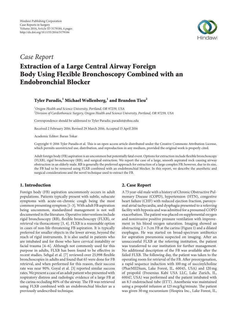
A73 year-oldmale with a history of ChronicObstructive Pul-monary Disease (COPD), hypertension (HTN), congestiveheart failure (CHF) with reduced ejection fraction, paroxys-mal atrial tachycardia, and dysphagia presented to a referringfacility with hypoxia andwas admitted for a presumedCOPDexacerbation.Thepatientwas placed on supplemental oxygenand noninvasive positive pressure ventilation with improve-ment in his blood oxygen saturation. Imaging showed anobstructing 2 × 3 cm FB at the carina (Figure 1) and a dilatedesophagus. He was started on broad-spectrum antibioticsfor aspiration pneumonia suspected on imaging. After anunsuccessful FLXB at the referring institution, the patientwas transferred to our institution for further management.No additional description of the FB was available after thefailed FLXB. The following day, the patient was taken to theoperating room for retrieval of the FB. After preoxygenation,a rapid sequence induction with 100mg of succinylcholine(PharMEDium, Lake Forest, IL, 60045, USA) and 120mgof propofol (Fresenius Kabi USA LLC, Lake Zurich, IL,60047, USA) was performed and the patient intubated withan 8.5 endotracheal tube (ETT). Anesthesia was maintainedusing a propofol infusion at 125mcg/kg/minute. The patientwas given 30mg rocuronium (Hospira Inc., Lake Forest, IL,
Hindawi Publishing CorporationCase Reports in SurgeryVolume 2016, Article ID 3179184, 4 pageshttp://dx.doi.org/10.1155/2016/3179184
2 Case Reports in Surgery
Figure 1: CT chest showing foreign body.
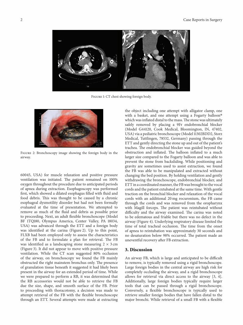
Figure 2: Bronchoscopy image showing the foreign body in theairway.
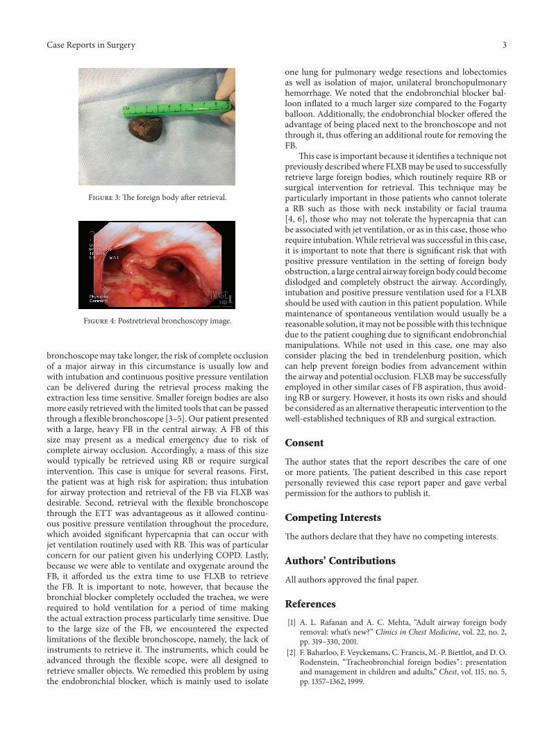
60045, USA) for muscle relaxation and positive pressureventilation was initiated. The patient remained on 100%oxygen throughout the procedure due to anticipated periodsof apnea during extraction. Esophagoscopy was performedfirst, which showed a dilated esophagus filled with fluid andfood debris. This was thought to be caused by a chronicesophageal dysmotility disorder but had not been formallyevaluated at the time of presentation. We attempted toremove as much of the fluid and debris as possible priorto proceeding. Next, an adult flexible bronchoscope (ModelBF 1TQ180, Olympus America, Center Valley, PA 18034,USA) was advanced through the ETT and a foreign bodywas identified at the carina (Figure 2). Up to this point,FLXB had been employed only to assess the characteristicsof the FB and to formulate a plan for retrieval. The FBwas identified as a landscaping stone measuring 2 × 3 cm(Figure 3). It did not appear to move with positive pressureventilation. While the CT scan suggested 80% occlusionof the airway, on bronchoscopy we found the FB mainlyobstructed the right mainstem bronchus only. The presenceof granulation tissue beneath it suggested it had likely beenpresent in the airway for an extended period of time. Whilewe were prepared to perform a RB, it was determined thatthe RB accessories would not be able to retrieve the FBdue the size, shape, and smooth surface of the FB. Priorto proceeding with thoracotomy, a decision was made toattempt retrieval of the FB with the flexible bronchoscopethrough an ETT. Several attempts were made at extracting
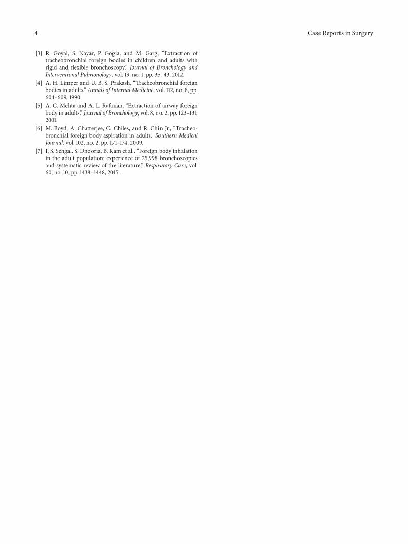
the object including one attempt with alligator clamp, onewith a basket, and one attempt using a Fogarty balloon�whichwas inflated distal to themass.The stonewas ultimatelysafely removed by placing a 9Fr endobronchial blocker(Model G44120, Cook Medical, Bloomington, IN, 47402,USA) via a pediatric bronchoscope (Model 11302BDD2, StorzMedical, Tuttlingen, 78532, Germany) passing through theETT and gently directing the stone up and out of the patient’strachea. The endobronchial blocker was guided beyond theobstruction and inflated. The balloon inflated to a muchlarger size compared to the Fogarty balloon and was able toprevent the stone from backsliding. While positioning andgravity are sometimes used to assist extraction, we foundthe FB was able to be manipulated and extracted withoutchanging the bed position. By holding ventilation and gentlywithdrawing the bronchoscope, endobronchial blocker, andETT in a coordinatedmanner, the FBwas brought to the vocalcords and the patient extubated at the same time.With gentletraction on the bronchial blocker and relaxation of the vocalcords with an additional 20mg rocuronium, the FB camethrough the cords and was removed from the oropharynxwith Magill forceps. The patient was reintubated withoutdifficulty and the airway examined. The carina was notedto be edematous and friable but there was no defect in theairway (Figure 4). Underlying respiratory disease limited thetime of total tracheal occlusion. The time from the onsetof apnea to reintubation was approximately 30 seconds andno desaturation below 98% occurred. The patient made anuneventful recovery after FB extraction.
3. Discussion
An airway FB, which is large and anticipated to be difficultto remove, is typically removed using a rigid bronchoscope.Large foreign bodies in the central airway are high risk forcompletely occluding the airway, and a rigid bronchoscopeallows for retrieval via direct access to the airway [3, 4].Additionally, large foreign bodies typically require largertools that can be passed through a rigid bronchoscope.Conversely, a flexible bronchoscope is typically used toretrieve smaller foreign bodies that have fallen distal to themajor bronchi. While retrieval of a small FB with a flexible
Case Reports in Surgery 3
Figure 3: The foreign body after retrieval.
Figure 4: Postretrieval bronchoscopy image.
bronchoscopemay take longer, the risk of complete occlusionof a major airway in this circumstance is usually low andwith intubation and continuous positive pressure ventilationcan be delivered during the retrieval process making theextraction less time sensitive. Smaller foreign bodies are alsomore easily retrieved with the limited tools that can be passedthrough a flexible bronchoscope [3–5]. Our patient presentedwith a large, heavy FB in the central airway. A FB of thissize may present as a medical emergency due to risk ofcomplete airway occlusion. Accordingly, a mass of this sizewould typically be retrieved using RB or require surgicalintervention. This case is unique for several reasons. First,the patient was at high risk for aspiration; thus intubationfor airway protection and retrieval of the FB via FLXB wasdesirable. Second, retrieval with the flexible bronchoscopethrough the ETT was advantageous as it allowed continu-ous positive pressure ventilation throughout the procedure,which avoided significant hypercapnia that can occur withjet ventilation routinely used with RB. This was of particularconcern for our patient given his underlying COPD. Lastly,because we were able to ventilate and oxygenate around theFB, it afforded us the extra time to use FLXB to retrievethe FB. It is important to note, however, that because thebronchial blocker completely occluded the trachea, we wererequired to hold ventilation for a period of time makingthe actual extraction process particularly time sensitive. Dueto the large size of the FB, we encountered the expectedlimitations of the flexible bronchoscope, namely, the lack ofinstruments to retrieve it. The instruments, which could beadvanced through the flexible scope, were all designed toretrieve smaller objects. We remedied this problem by usingthe endobronchial blocker, which is mainly used to isolate
one lung for pulmonary wedge resections and lobectomiesas well as isolation of major, unilateral bronchopulmonaryhemorrhage. We noted that the endobronchial blocker bal-loon inflated to a much larger size compared to the Fogartyballoon. Additionally, the endobronchial blocker offered theadvantage of being placed next to the bronchoscope and notthrough it, thus offering an additional route for removing theFB.
This case is important because it identifies a technique notpreviously describedwhere FLXBmay be used to successfullyretrieve large foreign bodies, which routinely require RB orsurgical intervention for retrieval. This technique may beparticularly important in those patients who cannot toleratea RB such as those with neck instability or facial trauma[4, 6], those who may not tolerate the hypercapnia that canbe associated with jet ventilation, or as in this case, those whorequire intubation.While retrieval was successful in this case,it is important to note that there is significant risk that withpositive pressure ventilation in the setting of foreign bodyobstruction, a large central airway foreign body could becomedislodged and completely obstruct the airway. Accordingly,intubation and positive pressure ventilation used for a FLXBshould be used with caution in this patient population.Whilemaintenance of spontaneous ventilation would usually be areasonable solution, itmaynot be possiblewith this techniquedue to the patient coughing due to significant endobronchialmanipulations. While not used in this case, one may alsoconsider placing the bed in trendelenburg position, whichcan help prevent foreign bodies from advancement withinthe airway and potential occlusion. FLXBmay be successfullyemployed in other similar cases of FB aspiration, thus avoid-ing RB or surgery. However, it hosts its own risks and shouldbe considered as an alternative therapeutic intervention to thewell-established techniques of RB and surgical extraction.
Consent
The author states that the report describes the care of oneor more patients. The patient described in this case reportpersonally reviewed this case report paper and gave verbalpermission for the authors to publish it.
Competing Interests
The authors declare that they have no competing interests.
Authors’ Contributions
All authors approved the final paper.
References
[1] A. L. Rafanan and A. C. Mehta, “Adult airway foreign bodyremoval: what’s new?” Clinics in Chest Medicine, vol. 22, no. 2,pp. 319–330, 2001.
[2] F. Baharloo, F. Veyckemans, C. Francis, M.-P. Biettlot, and D. O.Rodenstein, “Tracheobronchial foreign bodies∗: presentationand management in children and adults,” Chest, vol. 115, no. 5,pp. 1357–1362, 1999.
4 Case Reports in Surgery
[3] R. Goyal, S. Nayar, P. Gogia, and M. Garg, “Extraction oftracheobronchial foreign bodies in children and adults withrigid and flexible bronchoscopy,” Journal of Bronchology andInterventional Pulmonology, vol. 19, no. 1, pp. 35–43, 2012.
[4] A. H. Limper and U. B. S. Prakash, “Tracheobronchial foreignbodies in adults,” Annals of Internal Medicine, vol. 112, no. 8, pp.604–609, 1990.
[5] A. C. Mehta and A. L. Rafanan, “Extraction of airway foreignbody in adults,” Journal of Bronchology, vol. 8, no. 2, pp. 123–131,2001.
[6] M. Boyd, A. Chatterjee, C. Chiles, and R. Chin Jr., “Tracheo-bronchial foreign body aspiration in adults,” Southern MedicalJournal, vol. 102, no. 2, pp. 171–174, 2009.
[7] I. S. Sehgal, S. Dhooria, B. Ram et al., “Foreign body inhalationin the adult population: experience of 25,998 bronchoscopiesand systematic review of the literature,” Respiratory Care, vol.60, no. 10, pp. 1438–1448, 2015.

Submit your manuscripts athttp://www.hindawi.com
Stem CellsInternational
Hindawi Publishing Corporationhttp://www.hindawi.com Volume 2014
Hindawi Publishing Corporationhttp://www.hindawi.com Volume 2014
MEDIATORSINFLAMMATION
of
Hindawi Publishing Corporationhttp://www.hindawi.com Volume 2014
Behavioural Neurology
EndocrinologyInternational Journal of
Hindawi Publishing Corporationhttp://www.hindawi.com Volume 2014
Hindawi Publishing Corporationhttp://www.hindawi.com Volume 2014
Disease Markers
Hindawi Publishing Corporationhttp://www.hindawi.com Volume 2014
BioMed Research International
OncologyJournal of
Hindawi Publishing Corporationhttp://www.hindawi.com Volume 2014
Hindawi Publishing Corporationhttp://www.hindawi.com Volume 2014
Oxidative Medicine and Cellular Longevity
Hindawi Publishing Corporationhttp://www.hindawi.com Volume 2014
PPAR Research
The Scientific World JournalHindawi Publishing Corporation http://www.hindawi.com Volume 2014
Immunology ResearchHindawi Publishing Corporationhttp://www.hindawi.com Volume 2014
Journal of
ObesityJournal of
Hindawi Publishing Corporationhttp://www.hindawi.com Volume 2014
Hindawi Publishing Corporationhttp://www.hindawi.com Volume 2014
Computational and Mathematical Methods in Medicine
OphthalmologyJournal of
Hindawi Publishing Corporationhttp://www.hindawi.com Volume 2014
Diabetes ResearchJournal of
Hindawi Publishing Corporationhttp://www.hindawi.com Volume 2014
Hindawi Publishing Corporationhttp://www.hindawi.com Volume 2014
Research and TreatmentAIDS
Hindawi Publishing Corporationhttp://www.hindawi.com Volume 2014
Gastroenterology Research and Practice
Hindawi Publishing Corporationhttp://www.hindawi.com Volume 2014
Parkinson’s Disease
Evidence-Based Complementary and Alternative Medicine
Volume 2014Hindawi Publishing Corporationhttp://www.hindawi.com