case report interdisiplinary collaboration of orthodontics
TRANSCRIPT

IP Indian Journal of Orthodontics and Dentofacial Research 2020;6(3):149–156
Content available at: https://www.ipinnovative.com/open-access-journals
IP Indian Journal of Orthodontics and Dentofacial Research
Journal homepage: www.ipinnovative.com
Case Report
Interdisiplinary collaboration of orthodontics and oral and maxillofacial surgeryfor the correction of severe class III skeletal pattern in an adult male with anhapsburg jaw - A case report on surgical orthodontics
Bhushan Jawale1, Lishoy Rodrigues1,*, J B Garde2, Anup Belludi3, Anand Patil4,Pranita Palande1
1Dept. of Orthodontics and Dentofacial Orthopedics, Sinhgad Dental College and Hospital, Pune, Maharashtra, India2Dept. of Oral and Maxillofacial Surgery, Rangoonwala Dental College and Hospital, Pune, Maharashtra, India3Dept. of Orthodontics and Dentofacial Orthopedics, KLE Dental College and Hospital, Bangalore, Karnataka, India4Dept. of Orthodontics and Dentofacial Orthopedics, SDM Dharwad, Karnataka, India
A R T I C L E I N F O
Article history:Received 27-05-2020Accepted 13-06-2020Available online 04-09-2020
Keywords:Hapsburg JawBilateral sagittal split osteotomyBSSOPrognathic lower jawClass III skeletal patternDecompensationOrthognathic surgery
A B S T R A C T
This case report, aims to present a combined orthodontic and surgical approach in the treatment of anadult male patient with skeletal class III malocclusion with an Hapsburg jaw. The malocclusion wasdecompensated by pre-surgical orthodontic treatment and then normal jaw relationship was achieved byBilateral Sagittal Split Osteotomy Setback followed by debonding after 4 months. The treatment lasted forover 14months, it improved facial esthetics and profile of the patient significantly and resulted in a normalocclusion, overjet, and overbite and a pleasant smile.
© 2020 Published by Innovative Publication. This is an open access article under the CC BY-NC license(https://creativecommons.org/licenses/by-nc/4.0/)
1. Introduction
Recently, the number of adults seeking orthodontictreatment has increased significantly.1,2 Treatmentalternatives of correction of a skeletal class III in adults areeither Orthodontic camouflage or a Combined orthodontic-orthognathic surgical therapy. It eventually dependsmainly upon the severity of the malocclusion3,4 and theamount of needed tooth movements.3,5 If the skeletaldiscrepancy6 cannot be corrected by camouflage, anydental compensation may produce a reasonably goodocclusion7but at the expense of compromised esthetics. Foradult patients having severe orthodontic problems, surgeryto realign the jaws or reposition dentoalveolar segmentsis the only possible treatment option left. One indication
* Corresponding author.E-mail address: [email protected] (L. Rodrigues).
for surgery is a malocclusion so severe that it cannot becorrected by orthodontics alone.8Class III malocclusionpatients frequently show a combinations of skeletaland dentoalveolar components. Many cephalometricpecularities have been reported in class III patients,such as an acute cranial base angle, a retrusive maxilla,proclined maxillary and retroclined mandibular incisors,an increased lower anterior face height and obtuse gonialangle.9,10 Prevalence of class 3 malocclusion in caucasiansranges from 0.8% to 4.0% and increases up to 12-13%in Chinese and Japanese population, while in Americanpopulation class 3 malocclusion ranges from 3-4% of thepopulation.9–11 The surgical correction of prognathism is aprocedure that dates back more than 100 years. In121849Hullihen described a technique for the correction of sucha deformity. Since that time refinements of technique and
https://doi.org/10.18231/j.ijodr.2020.0302581-9356/© 2020 Innovative Publication, All rights reserved. 149
150 Jawale et al. / IP Indian Journal of Orthodontics and Dentofacial Research 2020;6(3):149–156
various methods have13 been described. At the turn of thecentury Blair published several articles on this particularsubject. Interest in the subject and in the various techniquesused in its correction became widespread. After14,15 Blair,came reports from Kazanjian, Dingman,16,17 Reiter,”Caldwell and Letterman, Moose, and many others. Thiscase report is about the integrated efforts of Orthodonticsand Oral and Maxillofacial surgery in the correction of anadult male patient with Severe Class III Skeletal patternwith a Hapsburg jaw.
2. Case Report
An adult male patient aged 30 years presented at Smileand Shine Orthodontic Clinic, Pune, Maharashtra, India,with the chief complaint of forwardly placed lower jawand a forwardly placed chin. He was very unsatisfied withhis facial profile. He desired a straight profile. He hadan unesthetic facial and dental appearance. However, thepatient presented with no relevant medical or dental historyand was himself internally motivated for the treatment withextreme desire to improve his facial aesthetics. He had asevere class 3 malocclusion with 4mm of reverse overjet and1mm of reverse overbite.
2.1. Clinical examination
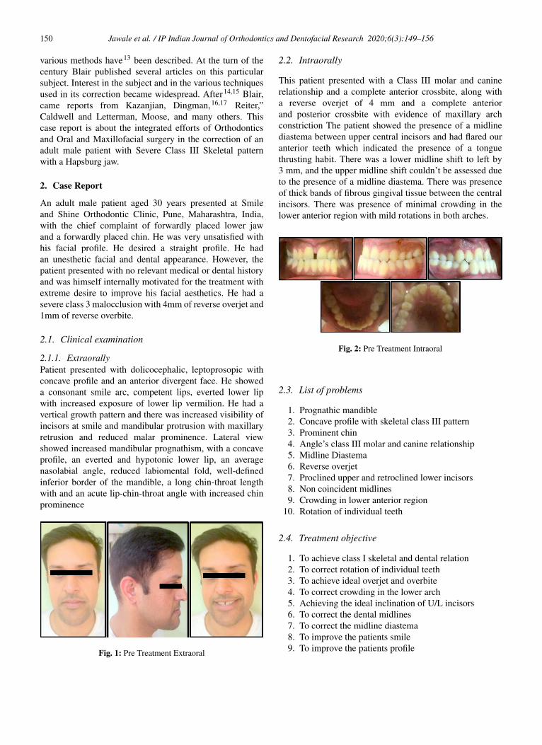
2.1.1. ExtraorallyPatient presented with dolicocephalic, leptoprosopic withconcave profile and an anterior divergent face. He showeda consonant smile arc, competent lips, everted lower lipwith increased exposure of lower lip vermilion. He had avertical growth pattern and there was increased visibility ofincisors at smile and mandibular protrusion with maxillaryretrusion and reduced malar prominence. Lateral viewshowed increased mandibular prognathism, with a concaveprofile, an everted and hypotonic lower lip, an averagenasolabial angle, reduced labiomental fold, well-definedinferior border of the mandible, a long chin-throat lengthwith and an acute lip-chin-throat angle with increased chinprominence
Fig. 1: Pre Treatment Extraoral
2.2. Intraorally
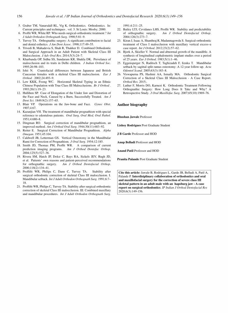
This patient presented with a Class III molar and caninerelationship and a complete anterior crossbite, along witha reverse overjet of 4 mm and a complete anteriorand posterior crossbite with evidence of maxillary archconstriction The patient showed the presence of a midlinediastema between upper central incisors and had flared ouranterior teeth which indicated the presence of a tonguethrusting habit. There was a lower midline shift to left by3 mm, and the upper midline shift couldn’t be assessed dueto the presence of a midline diastema. There was presenceof thick bands of fibrous gingival tissue between the centralincisors. There was presence of minimal crowding in thelower anterior region with mild rotations in both arches.
Fig. 2: Pre Treatment Intraoral
2.3. List of problems
1. Prognathic mandible2. Concave profile with skeletal class III pattern3. Prominent chin4. Angle’s class III molar and canine relationship5. Midline Diastema6. Reverse overjet7. Proclined upper and retroclined lower incisors8. Non coincident midlines9. Crowding in lower anterior region
10. Rotation of individual teeth
2.4. Treatment objective
1. To achieve class I skeletal and dental relation2. To correct rotation of individual teeth3. To achieve ideal overjet and overbite4. To correct crowding in the lower arch5. Achieving the ideal inclination of U/L incisors6. To correct the dental midlines7. To correct the midline diastema8. To improve the patients smile9. To improve the patients profile

Jawale et al. / IP Indian Journal of Orthodontics and Dentofacial Research 2020;6(3):149–156 151
Fig. 3: Pre treatment radiographs
2.5. Treatment plan
Pre-surgical orthodontics was performed followed bya bilateral surgical split osteotomy for mandibularsetback was planned in order to achieved goals of facialaesthetics, functionally optimum occlusion, minimumtrauma to the patient and to achieve a straight facialprofile. Cephalometric analysis was performed withpretreatment radiographic records which includedlateral cephalograms, orthopantomogram (OPG) andPA cephalogram. Radiographs revealed a Class III skeletalpattern with mandibular prognathism, increased verticalchin height, an increased maxillary dental and skeletalheight and upper anterior proclination with lower anteriorretroclination. After a combined clinic discussion with theOral and Maxillofacial Surgeon, it was planned to performa mandibular setback with bilateral sagittal split osteotomy(BSSO), with presurgical and postsurgical orthodonticsto obtain facial aesthetics and an optimum functionalocclusion. To relieve maxillary and mandibular crowding,presurgical extraction was not needed in this case and hencewas not performed. Also one more unique feature was thealthough 3rd molars were present in this patient, we didnot find the need to extract them before surgery as enoughspace was available to perform the BSSO incision cuts.After discussion with the Oral & Maxillofacial surgeon, amandibular setback of 6.0 mm was planned to achieve aclass I molar and canines relationship and an estheticallypleasing profile .
Fig. 4: MID treatment extraoral after decompensation
2.6. Pre-surgical orthodontics
Pre-surgical orthodontics was done to achievedecompensation of the malocclusion. Firstly a 0.016NiTi wire in upper and 0.014 NiTi in lower arch was placed,followed by 0.017x0.025 NiTi wires in upper and lowerarch.
Both arches were aligned till 0.019 × 0.025 SS wirewith 0.022 slot edgewise brackets. The lower incisors weredecompensated by proclining them and the both arches werecoordinated. An 0.021 X 0.025 inch S.S archwires wereplaced for stabilization 2 weeks before surgery
Fig. 5: MID treatment intraoral after decompensation
2.7. Pre surgical orthodontic objectives
1. Alignment and leveling of both arches2. Decompensation of proclined upper incisors and
retroclined lower incisors3. To correct angulations of every tooth on their
respective bases.
Fig. 6: MID treatment radiographs
2.8. Cephalometric Prediction Technique
Cephalometric prediction tracing was done both withcomputer image prediction software and also manuallyusing the template method. In template method, maxillaryand mandibular profiles were traced on an acetate paper.Then from these templates, trial sections were made andamount for osteotomy were detected.18 Cut sections ofboth arches were fitted back to desired occlusal relation.At last, soft tissue outline in regard to the reference ratioswere traced, and the postsurgical changes were evaluated.Computer-based analysis in which cephalometric landmarkswere digitized and the surgical positioning was monitoredwas then done. Calculations, measurements and analysesweredone using cephalometric digital software.

152 Jawale et al. / IP Indian Journal of Orthodontics and Dentofacial Research 2020;6(3):149–156
Fig. 7:
2.9. Mock surgery
Fabrication of occlusal splints with Model surgery forsurgery was the next step in the planning sequence. Lowerdental cast was then repositioned simulating the movementof the jaws as directed by digital and manual prediction.The final occlusal splint was then fabricated, based on thisposition. Both upper and lower dental casts were mountedon a semiadjustable articulator with the help of a facebowtransfer and bite registration taken with the patient’s jawsin the posteriorly retruded contact position also called thecentric relation.
Fig. 8: Splint During Surgery
2.10. Surgical procedure
Retromolar posterior area was exposed and made visibleusing a modified third molar incision. A BSSO withshort lingual split was done using surgical saws. Medialpterygoid muscle was detached by performing the split,with 6 mm of mandibular setback and 7◦ of counter-clockwise rotation, was achieved successfully. The BSSOsetback was performed symmetrically thereby improvedfacial and dental symmetry. Rigid type fixations using
Fig. 9: Splint provided post surgery
fourhole miniplates and screws on both sides were usedin both jaws. Intermaxillary elastics on braces were placedfor 14 days in the immediate postoperative phase. Goodbone healing and tolerance was seen. The goal was toachieve ideal occlusal relationships, with respect to canineand molar class, coincidence of the dental midlines andoverjet and overbite. Cooperation of Patient was very goodthroughout the treatment. The splint and archwires withintermaxillary fixation done continued for three weeks andthe patient was advised to use elastics. Cephalometricfinding post treatment showed normal jaw relationship
Fig. 10: Surgical Photographs
2.11. Surgical correction objectives
1. Mandibular setback of 5mm and rigid fixation withsplints
2. To achieve optimal facial esthetics, and to obtain anoptimal overbite-overbite relationship

Jawale et al. / IP Indian Journal of Orthodontics and Dentofacial Research 2020;6(3):149–156 153
2.12. Post-surgical orthodontics
Orthodontic treatment actively was resumed 4 weeks aftersurgery when a good range of jaw movement was achievedand good bone healing and tolerance was seen. The patientwas observed closely after the surgery and was advised toperform opening as well as lateral jaw movements. Goalwas to achieve an ideal occlusal relationship ideal overjet,ideal overbite and coincidence of the dental midlines.Detailing of occlusion was accomplished by settling usingshort inter-maxillary elastics. In postsurgical orthodonticsthe arch wires were changed from 0.017×0.025 NiTi to0.019× 0.025 to 0.021× 0.025 in SS wires. Any residualdiastema closure was achieved, and occlusion was perfectedby segmental settling with short inter maxillary elastics.After approximately 4 months of treatment, fixed applianceswere debonded and the post treatment retention phase wasinitiated with fixed retainer in both arches and additionalremovable retainer plate in upper arch was given. In botharches, a 0.018 inch multistranded coaxial 3-to-3 retainerwas bonded. Post treatment radiographs were then taken andevaluated for treatment changes by superimposition.
Fig. 11: Post treatment radiographs
2.13. Post surgical orthodontic objectives
1. Settling of Occlusion2. Finishing and detailing.
Fig. 12: Post surgical extraoral photographs
Fig. 13: Post surgical intraoral photographs
3. Discussion
This case report emphasizes on the treatment of an adultIndian male patient with dental and skeletal class IIIrelationships. Surgical–orthodontic treatment for achievingan acceptable occlusion and a good esthetic result wasthe best option in this case. Rivera et al reportedthat patients underwent othognathic surgery to improveesthetic, functional problems. But, these benefits fromthe orthognathic surgery are not always realized.19 Mainreasons for not a very satisfactory treatment outcome couldbe relapse of surgical changes. It has been reported thatthe relapse following setback are one of the highest fora surgical procedure.20–22Establishing common objectivesconcerning the outcome of proposed surgical orthodontictherapy is a very important part of the treatment planningprocess. Hence an experienced multidisciplinary teamapproach delivers a satisfactory outcome.23 Most relapseafter setback surgery occurs during the postsurgical phasein the first two months following surgery. Similar findingswere reported by Mobarak.24An additional minor relapseduring the span from two months to a year after surgery.Minimal relapse beyond the first post-postoperative year,similar to that reported by Eggensperger et al wasobserved.25 This rebound tendency affects not only thefinal occlusion, but also the facial esthetics. In Class IIIloer jaw setback surgery, many surgeons tend to pushthe segments backward during the fixation procedure.However this seems to be the main reason for the forwardrelapse of mandible in the majority of the mandibularsetback surgery subjects. All the dental compensationsare removed by presurgical orthodontics. After rigorousevaluation, presurgical phase of orthodontic interventionwas initiated with the aim to achieve ideal inter- and intra-arch coordination, always keeping in mind the goals ofthe subsequent surgical repositioning.18 Both manual anddigital cephalometric predictions were employed in this caseat the end of presurgical phase. Ideal skeletal relationshipis achieved by surgical osteotomy and BSSO setback ofthe prognathic mandible. Postsurgical orthodontics guidesthe normal occlusal position by correcting any emergingdental discrepancies.18 Post-surgical orthodontics was donein this case for 4 months, and it primarily involvedfinalization of the occlusion and retention. The duration ofthe final orthodontic phase greatly depends on the degreeof preparation achieved during pre-surgical treatment.Good dental retention is a very important contributor to

154 Jawale et al. / IP Indian Journal of Orthodontics and Dentofacial Research 2020;6(3):149–156
Table 1: Cephalometric Values
Variable Pre-treatment Post-treatment Change One yearfollow-up
SNA 83◦ 82◦ -1◦ 82◦
SNB 84◦ 79◦ -5◦ 79◦
ANB -2◦ 2◦ 4◦ 2◦
WITS -8mm 2mm 10mm 2mmN ⊥ Pt A 1mm 0mm -1mm 0mmN ⊥ POG 4 mm -4 mm -8 mm -4 mmAngle of inclination 85◦ 85◦ 0◦ 85◦
Go-Gn to SN 32.5◦ 32◦ -0.5◦ 32◦
Eff. Max. Length 91 mm 89 mm -2 mm 89 mmEff. Man. Length 128 mm 121 mm -7 mm 121 mmY- Axis 67◦ 69◦ +2◦ 69◦
Facial axis -2◦ -3◦ -1◦ -3◦
Upper incisor – NA 22 mm 15 mm -7 mm 15 mmUpper incisor – NA 53.5◦ 32◦ -21.5◦ 32◦
Upper incisor – SN 130◦ 113◦ -17◦ 113◦
Upper incisor to maxillary plane angle 138◦ 120◦ -18◦ 120◦
Lower incisor to mandibular plane angle 101◦ 94◦ -7◦ 94◦
Lower incisor to NB 15 mm 8 mm -7 mm 8 mmLower incisor to NB 38◦ 27◦ -11◦ 27◦
Interincisal angle 94◦ 127◦ +33◦ 127◦
Maxillary mandibular planes angle 32◦ 27◦ 0◦ 27◦
Upper anterior face height 55 mm 58 mm +3 mm 58 mmLower anterior face height 81 mm 77.5 mm -3.5 mm 80 mmFace height ratio 46% 47% +1% 47%Lower incisor to APo line 19 mm 7 mm -12 mm 7 mmLower lip to Ricketts E Plane 10 mm 3 mm -7 mm 3 mm
retain the final occlusion that was achieved surgically,ensuring occlusal stability, which will surely have positiverepercussions on the final tissue stability. The facial changesthat resulted from the treatment were pronounced andgreatly improved the patient’s self image. One year afterretention, extra-and intraoral photographs indicated that thetreatment results were stable.
Fig. 14: Post treatment extraoral photographs
Fig. 15: Post surgical intraoral photographs
4. Results
4.1. Skeletal changes
There was a reduction in the SNB angle and mandibularlength with improvement of profile on lateral cephalogram.
4.2. Dental changes
Midline diastema was corrected, class I molar and caninerelation was achieved, crowding was relieved, optimaloverbite and overjet were achieved and all spaces wereclosed.
4.3. Radiographic changes
Cephalometrically, a significant decrease in mandible by -5 degrees (SNA- 84 degrees to 79 degrees), ANB angle (2degrees), WITS analysis showed a significant improvementfrom -8mm to 2mm and it remained constant even 1

Jawale et al. / IP Indian Journal of Orthodontics and Dentofacial Research 2020;6(3):149–156 155
Fig. 17: One year follow up radiographs
Fig. 18: One year follow up extraoral photographs
year post treatment. Main finding wa a change in theeffective mandibular length from 128 mm to 121 mm.Growth improvement was seen in the Y axis value whichchanged from 67 degree to 69 degrees. Upper incisorproclination changed from 22mm to 15 mm and from53.5 degree to 32 degree. Lower incisor proclinationchanged from 14mm to 8 mm and from 39 degree to27 degree. Interincisal angle changed from 94 degrees to127degrees, thus significantly decreasing the proclinationand the increased lower anterior facial height reduced from80 mm to 77.5mm. The maxillomandibular plane anglechanged from 32 degree to 27degree. Cephalometrically,following changes were observed— A superimposition ofthe pre- and post treatment cephalometric tracings showsthe decrease in proclination of upper and lower anteriorteeth with a normal inter-incisal angle. The shortening ofmandible due to BSSO setback can also be seen. Acceptableroot parallelism was achieved and bone loss did not occur
4.4. Soft tissue changes
The procumbent lower lip before treatment was changedinto ideal form, nasolabial angle improved, chin throatangle improved, facial profile was changed to a straightprofile and the aim of achieving a pleasing smile andprofile was achieved. All pretreatment objectives weremet in this case. The facial appearance was improved asa result of both skeletal and dental changes and classI skeletal relationship was achieved at the end of surgicaland orthodontic treatment.
Fig. 16: Pre and post comparison
5. Conclusion
It is very Imperative that Class III discrepancy bediagnosed and evaluated according to its etiology andtreatment be done with appropriate surgery, including notonly mandibular surgery, but also maxillary surgery, inorder to attain a normal facial profile. As orthodonticsprogressesto develop techically and wholely, we do expectthat advances in diagnosis and treatment planning areinevitable.26Duration of final treatment phase dependson the amount of preparation that is achieved duringpresurgical phase of treatment.27 It is important howeverto emphasize that good retention does contribute tomaintaining the appropriate occlusion that is achievedsurgically, ensuring and guaranteeing the occlusal stability,which will surely have positive repercussions on the finaltissue stability.
6. Source of Funding
None.
7. Conflict of Interest
None.
References1. Boyd RL, Leggott PJ, Quinn RS, Eakle WS, Chambers D. Periodontal
implications of orthodontic treatment in adults with reduced ornormal periodontal tissues versus those of adolescents. Am J OrthodDentofacial Orthop. 1989;96(3):191–8.
2. Gottleib EL, Nelson AH, Vogels DS. JCO study of orthodonticdiagnosis and treatment procedures. Part 1: Results and trends. J ClinOrthod. 1990;25:145–56.
3. Bailey LJ, Proffit WR, White R. Assessment of patients fororthognathicsurgery. Semin Orthod . 1999;5(4):209–22.
4. Jf T, Lenz BE, Phillips C. Surgical Versus orthodontic correction forclass II patients: age and severity in treatment planning and treatmentoutcome. Semin Orthod. 1977;72:1–22.
156 Jawale et al. / IP Indian Journal of Orthodontics and Dentofacial Research 2020;6(3):149–156
5. Graber TM, Vanarsdall RL, Vig K, Orthodontics. Orthodontics. In:Current principles and techniques . vol. 3. St Louis: Mosby; 2000.
6. Proffit WR, White RP. Who needs surgical-orthodontic treatment ? IntJ Adult Orthodon Orthognath Surg. 1990;5:81–9.
7. Turvey TA. Orthognathic surgery: A significant contribution to facialand dental esthetics. J Am Dent Assoc. 1998;117:49–55.
8. Trivedi B, Mahadevia S, Shah R, Thakker D. Combined Orthodonticand Surgical Approach in an Adult Patient with Skeletal Class IIIMalocclusion. J Adv Oral Res. 2014;5(3):24–7.
9. Kharbanda OP, Sidhu SS, Sundaram KR, Shukla DK. Prevelance ofmalocclusion and its traits in Delhi children. J Indian Orthod Soc.1995;26:98–103.
10. Ishii N. Craniofacial differences between Japanese and BritishCaucasian females with a skeletal Class III malocclusion. Eur JOrthod . 2002;24:493–9.
11. Lew KKK, Foong WC. Horizontal Skeletal Typing in an EthnicChinese Population with True Class III Malocclusions. Br J Orthod .1993;20(1):19–23.
12. Hullihen SP. Case of Elongation of the Under Jaw and Distortion ofthe Face and Neck, Caused by a Burn, Successfully Treated. Am JDent Sci. 1849;9(2):157–65.
13. Blair VP. Operations on the Jaw-bone and Face. Gynec Obst.1907;4:67.
14. Kazanjian VH. The treatment of mandibular prognathism with specialreference to edentulous patients. Oral Surg, Oral Med, Oral Pathol.1951;4:680–8.
15. Dingman RO. Surgical correction of mandibular prognathism, animproved method. Am J Orthod Oral Surg. 1944;30(11):683–92.
16. Reiter E. Surgical Correction of Mandibular Prognathism. AlphaOmegan. 1951;45:104.
17. Caldwell JB, Letterman GS. Vertical Osteotomy in the MandibularRami for Correction of Prognathism. J Oral Surg. 1954;12:185.
18. Smith JD, Thomas PM, Proffit WR. A comparison of currentprediction imaging programs. Am J Orthod Dentofac Orthop.2004;125(5):527–36.
19. Rivera SM, Hatch JP, Dolce C, Bays RA, Sickels JEV, Rugh JD,et al. Patients’ own reasons and patient-perceived recommendationsfor orthognathic surgery. Am J Orthod Dentofacial Orthop.2000;118(2):134–41.
20. Proffifit WR, Philips C, Dann C, Turvey TA. Stability aftersurgical orthodontic correction of skeletal Class III malocclusion. I.Mandibular setback. Int J Adult Orthodon Orthognath Surg. 1991;6:7–18.
21. Proffifit WR, Philips C, Turvey TA. Stability after surgical orthodonticcorrection of skeletal Class III malocclusion. III. Combined maxillaryand mandibular procedures. Int J Adult Orthodon Orthognath Surg.
1991;4:211–25.22. Bailey LTJ, Cevidanes LHS, Proffit WR. Stability and predictability
of orthognathic surgery. Am J Orthod Dentofacial Orthop.2004;126(3):273–7.
23. Kiran J, Isaac A, Shanthraj R, Madannagowda S. Surgical-orthodontictreatment of Class I malocclusion with maxillary vertical excess–acase report. Int J Orthod. 2012;23(2):57–62.
24. Bjork A, Skieller V. Normal and abnormal growth of the mandible. Asynthesis of longitudinal cephalometric implant studies over a periodof 25 years. Eur J Orthod. 1983;5(1):1–46.
25. Eggensperger N, Raditsch T, Taghizadeh F, Iizuka T. Mandibularsetback by sagittal split ramus osteotomy: A 12-year follow-up. ActaOdontol Scand. 2005;63(3):183–8.
26. Viswapurna PS, Hashmi AA, Ismaily MA. Orthodontic SurgicalCorrection of a Skeletal Class III Malocclusion - A Case Report.Orthod Res. 2015;.
27. Luther F, Morris DO, Karnezi K. Orthodontic Treatment FollowingOrthognathic Surgery: How Long Does It Take and Why? ARetrospective Study. J Oral Maxillofac Surg. 2007;65(10):1969–76.
Author biography
Bhushan Jawale Professor
Lishoy Rodrigues Post Graduate Student
J B Garde Professor and HOD
Anup Belludi Professor and HOD
Anand Patil Professor and HOD
Pranita Palande Post Graduate Student
Cite this article: Jawale B, Rodrigues L, Garde JB, Belludi A, Patil A,Palande P. Interdisiplinary collaboration of orthodontics and oraland maxillofacial surgery for the correction of severe class IIIskeletal pattern in an adult male with an hapsburg jaw - A casereport on surgical orthodontics. IP Indian J Orthod Dentofacial Res2020;6(3):149-156.