case report nodular lymphocyte predominant hodgkin...
TRANSCRIPT
Case ReportNodular Lymphocyte Predominant HodgkinLymphoma versus T-Cell/Histiocyte-Rich Large B-CellLymphoma: A Diagnostic Challenge
Anton V. Rets and Susan R. S. Gottesman
Department of Pathology, State University of New York Downstate Medical Center, 450 Clarkson Avenue, Brooklyn, NY 11203, USA
Correspondence should be addressed to Susan R. S. Gottesman; [email protected]
Received 1 May 2014; Accepted 25 June 2014; Published 7 July 2014
Academic Editor: Piero Tosi
Copyright © 2014 A. V. Rets and S. R. S. Gottesman. This is an open access article distributed under the Creative CommonsAttribution License, which permits unrestricted use, distribution, and reproduction in any medium, provided the original work isproperly cited.
Lymphomas with overlapping histological features of two distinct entities cause difficulty in classification. Their classification is ofparticular significancewhen the two alternatives require different treatmentmodalities.Wepresent a diagnostically challenging caseof a nodular lymphocyte predominant Hodgkin lymphoma (NLPHL) with features of T-cell/histiocyte-rich large B-cell lymphoma(THRLBCL). Our patient is a 39-year-old woman who presented with painless subclavicular and axillary lymphadenopathy. Thebiopsied lymph node showed diffuse architectural effacement and scattered large neoplastic cells with large irregular nuclei andprominent nucleoli. These cells were positive for CD20 and Bcl-6 and negative for CD15, CD30, IgD, and Bcl-2. The backgroundcells were predominantly T lymphocytes, whereas B cells were markedly depleted. The lymph node was interpreted as NLPHL,consistent with THRLBCL-like variant. NLPHL, especially THRLBC-like variant, and de novo THRLBCL are characterized bysignificant morphologic and immunophenotypic overlap. Our case demonstrates a rare predominance of background T-cells inNLPHL and emphasizes the importance of thorough evaluation of multiple morphologic and immunophenotypic features as anessential approach for arriving at the correct diagnosis.
1. Introduction
Current classification of lymphomas is based on a complex ofclinical, morphological, immunophenotypic, and molecularcriteria. Such an approach provides an accurate classificationof the majority of lymphomas. There are, however, caseswhich are difficult to fit into a distinct category.They demon-strate overlapping features between groups of lymphomas ofdifferent prognostic significance and treatment modalities.In such cases, making a correct diagnosis is a challengingtask. Commonly these neoplasms show features of Hodgkinlymphomas (classical or nodular lymphocyte predominant(NLPHL)) and diffuse large B-cell lymphomas (DLBCL) orthe latter and Burkitt lymphoma [1, 2]. For some, the dilemmacannot be resolved and a term “gray zone lymphoma” hasbeen used to designate such cases. Here we present adiagnostically challenging case of a lymphoma with featuresof NLPHL and T-cell/histiocyte-rich large B-cell lymphoma
(THRLBCL) with an attempt to distinguish between themand summarize their unique and overlapping diagnosticfeatures.
2. Case Presentation
A 39-year-old obese woman presented with a painless 2 ×2 cm left subclavicular lymph node that she had had for 1-2 years. She denied any complaints, including B-symptoms,and her past medical and social histories were unremarkable.Physical examination revealed left supraclavicular and axil-lary lymphadenopathy and absence of hepatosplenomegaly.Peripheral blood was remarkable for mild normocytic nor-mochromic anemia and mild neutropenia.
PET scan demonstrated bulky supraclavicular and axil-lary lymphadenopathy (SUV 8.01 and 11.89 resp.) with aneoplastic range hypermetabolic activity in the spleen and
Hindawi Publishing CorporationCase Reports in PathologyVolume 2014, Article ID 956217, 5 pageshttp://dx.doi.org/10.1155/2014/956217
2 Case Reports in Pathology
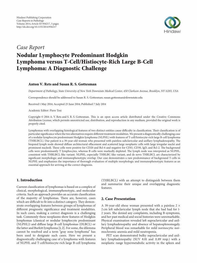
Figure 1: Biopsied lymph node architecture (hematoxylin and eosinstain, 40x). Vague nodules are present in the background of diffusearchitectural effacement.
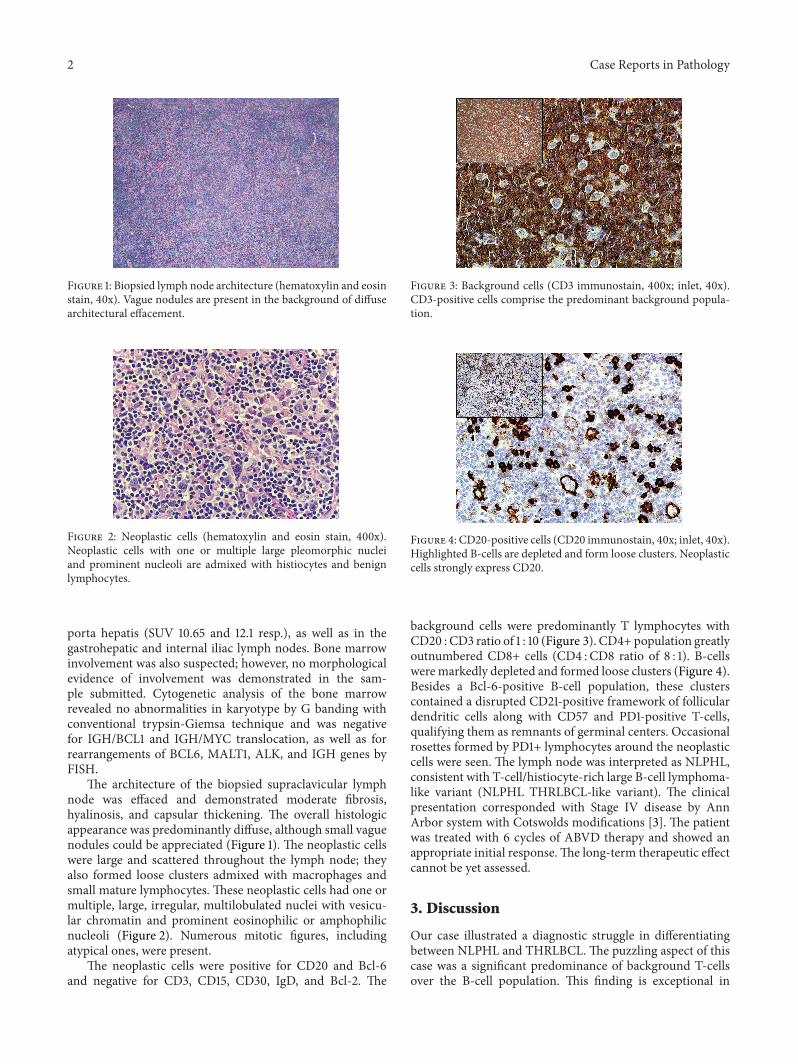
Figure 2: Neoplastic cells (hematoxylin and eosin stain, 400x).Neoplastic cells with one or multiple large pleomorphic nucleiand prominent nucleoli are admixed with histiocytes and benignlymphocytes.
porta hepatis (SUV 10.65 and 12.1 resp.), as well as in thegastrohepatic and internal iliac lymph nodes. Bone marrowinvolvement was also suspected; however, no morphologicalevidence of involvement was demonstrated in the sam-ple submitted. Cytogenetic analysis of the bone marrowrevealed no abnormalities in karyotype by G banding withconventional trypsin-Giemsa technique and was negativefor IGH/BCL1 and IGH/MYC translocation, as well as forrearrangements of BCL6, MALT1, ALK, and IGH genes byFISH.
The architecture of the biopsied supraclavicular lymphnode was effaced and demonstrated moderate fibrosis,hyalinosis, and capsular thickening. The overall histologicappearance was predominantly diffuse, although small vaguenodules could be appreciated (Figure 1). The neoplastic cellswere large and scattered throughout the lymph node; theyalso formed loose clusters admixed with macrophages andsmall mature lymphocytes. These neoplastic cells had one ormultiple, large, irregular, multilobulated nuclei with vesicu-lar chromatin and prominent eosinophilic or amphophilicnucleoli (Figure 2). Numerous mitotic figures, includingatypical ones, were present.
The neoplastic cells were positive for CD20 and Bcl-6and negative for CD3, CD15, CD30, IgD, and Bcl-2. The
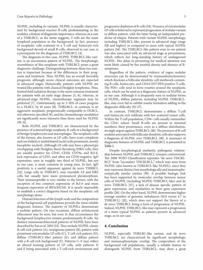
Figure 3: Background cells (CD3 immunostain, 400x; inlet, 40x).CD3-positive cells comprise the predominant background popula-tion.
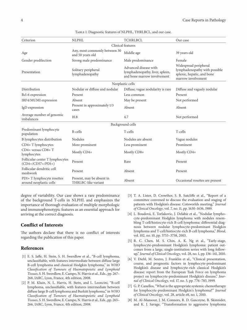
Figure 4: CD20-positive cells (CD20 immunostain, 40x; inlet, 40x).Highlighted B-cells are depleted and form loose clusters. Neoplasticcells strongly express CD20.
background cells were predominantly T lymphocytes withCD20 : CD3 ratio of 1 : 10 (Figure 3). CD4+ population greatlyoutnumbered CD8+ cells (CD4 : CD8 ratio of 8 : 1). B-cellsweremarkedly depleted and formed loose clusters (Figure 4).Besides a Bcl-6-positive B-cell population, these clusterscontained a disrupted CD21-positive framework of folliculardendritic cells along with CD57 and PD1-positive T-cells,qualifying them as remnants of germinal centers. Occasionalrosettes formed by PD1+ lymphocytes around the neoplasticcells were seen. The lymph node was interpreted as NLPHL,consistent with T-cell/histiocyte-rich large B-cell lymphoma-like variant (NLPHL THRLBCL-like variant). The clinicalpresentation corresponded with Stage IV disease by AnnArbor system with Cotswolds modifications [3]. The patientwas treated with 6 cycles of ABVD therapy and showed anappropriate initial response.The long-term therapeutic effectcannot be yet assessed.
3. Discussion
Our case illustrated a diagnostic struggle in differentiatingbetween NLPHL and THRLBCL. The puzzling aspect of thiscase was a significant predominance of background T-cellsover the B-cell population. This finding is exceptional in
Case Reports in Pathology 3
NLPHL, including its variants. NLPHL is usually character-ized by background reactive B-cells predominating in thenodules, a feature of diagnostic importance, whereas, in a caseof a THRLBCL, as the name suggests, T-cells are the mainlymphoid population in the background. In fact, presenceof neoplastic cells scattered in a T-cell and histiocyte-richbackground devoid of small B-cells, observed in our case, isa prerequisite for the diagnosis of THRLBCL [4].
The diagnosis in this case, NLPHL THRLBCL-like vari-ant, is an uncommon pattern of NLPHL. The morphologicresemblance of this neoplasm with THRLBCL poses a greatdiagnostic challenge. Distinguishing between these two enti-ties is important because of the differences in their prog-nosis and treatment. Thus, NLPHL has an overall favorableprognosis, although worse clinical outcomes are expectedin advanced stages. Historically, patients with NLPHL aretreated like patients with classical Hodgkin lymphoma.Thus,limited field radiation therapy is themost common treatmentfor patients with an early stage disease [5, 6], whereas, inadvanced-stage NLPHL, chemotherapy regimens may bepreferred [7]. Unfortunately, up to 3–10% of cases progressto a DLBCL by 10 years [8]. THRLBCL, in contrast, is anaggressive neoplasm, prognostically comparable to DLBCL,not otherwise specified [9], and the chemotherapymodalitiesare significantly more intensive than those used for NLPHL[10, 11].
Both NLPHL and THRLBCL are characterized by thepresence of scattered large neoplastic B-cells in a backgroundof benign lymphocytes andmacrophages.The neoplastic cellsof the former, also known as LP cells, have scant cytoplasmandone folded ormultilobated nuclei with prominent,mostlybasophilic nucleoli. Although LP cells may have a phenotypeoverlapping with Hodgkin-Reed-Sternberg (HRS) cells, theyare usually positive for CD45, CD20, CD79a, and Bcl-6,lack expression of CD15, and often are CD30-negative. IgDexpression, seen in roughly one third of NLPHL, but notin our case, is more common in young men. In fact, IgDpositivity is a useful argument against de novo THRBCL[12]. Large cells in THRLBCL may resemble LP and HRScells but usually have more pronounced pleomorphism.Their immunoprofile is very similar to the former, with theexception of less common expression of Bcl-6 and morefrequent expression of IRF4/MUM1. It is nearly impossibleto establish a correct diagnosis based on the neoplastic cellmorphology alone.
Histoarchitecture of the lymph node and the compositionof the background cell populations provide the most reliablediagnostic features. The majority of NLPHLs demonstratea nodular pattern. On rare occasions, diffuse architecturaleffacement may be seen, but even in that circumstance thebackground lymphocytes remain predominantly B-cells. Sixdistinct immunoarchitectural patterns of NLPHL have beendescribed by Fan et al. 2003 [13].They includeNLPHL-classicB-cell-rich pattern (A), serpiginous pattern (B), pattern withprominent extranodular LP cells (C), T-cell-rich pattern (D),diffuse (THRLBCL-like) pattern (E), and diffuse patternwith a B-cell rich background (F). Patterns C–F may reflectan altered homing pattern of LP cells, with patterns Dand E being associated with a T-cell-rich environment and
progressive depletion of B-cells [14]. Presence of extranodularLP cells is believed to represent progression of nodular variantto diffuse pattern, with the latter being an independent pre-dictor of relapse. Patients with variant NLPHL morphology,including THRLBCL-like, present in advanced stage (stageIIB and higher) as compared to cases with typical NLPHLpattern [14]. The THRLBCL-like pattern seen in our patientwas also associated with an advanced stage at presentation,which reflects her long-standing history of undiagnosedNLPHL. Her delay in presenting for medical attention wasmost likely caused by her morbid obesity and absence of B-symptoms.
Regardless of the pattern, evidence of vague nodularstructures can be demonstrated by immunohistochemistrywhich discloses a follicular dendritic cell meshwork contain-ing B-cells, histiocytes, and CD4/CD57/PD1-positive T-cells.The PD1+ cells tend to form rosettes around the neoplasticcells, which can be used as a diagnostic feature of NLPHL, asin our case. Although it is frequently seen in nodular formsof NLPHL, diffuse patterns, including NLPHL THRLBCL-like, may even fail to exhibit rosette formation adding to thediagnostic difficulty [15–17].
In contrast, THRLBCL demonstrates a diffuse T-celland histiocyte-rich infiltrate with few scattered tumor cells.Within the T-cell population, CD8+ cells usually outnumberthe CD4+ subset. Small B-cells are present in minimalnumbers; their prominence, even in a diffuse distribution,strongly argues against THRLBCL [18].The presence of B-cellnodules associatedwith follicular dendritic cells also supportsa diagnosis of NLPHL over THRLBCL [18]. A summary ofdiagnostic features of NLPHL and THRLBCL is presented inTable 1.
Despite morphological similarity, pathogenic relation-ship between NLPHL and THRLBCL remains controversial.The 2008 WHO Classification separates “de novo THCRL-BCL” from “secondary THCRLBCL,” which may arise fromNLPHL (also known as THRLBCL-like NLPHL), since theymay represent distinct butmorphologically and immunophe-notypically similar entities [19]. A possible biologic linkhas been supported by molecular overlap between tumorcells of NLPHL (including NLPHL-THRLBCL-like) and denovo THRLBCL [17], a lack of disease specific pattern ofgene expression, and similarities in their gene expressionprofile [20]. On the other hand, NLPHL demonstrates higheraverage number of genomic imbalances (10.8 versus 4.7 inTHRLBCL) [21], which does not support the theory of ade novo THRLBCL being a form of progression of NLPHL.Indeed, NLPHL THRLBCL-like may represent a progressionof a more typical NLPHL as patients present in advancedstage, as in our case.
4. Conclusion
NLPHL, especially THRLBC-like variant, and de novoTHRLBCL are characterized by significant morphologicand immunophenotypic overlap. The composition of thebackground cell populations, usually a reliable feature todistinguish NLPHL from THRLBCL, may show a great
4 Case Reports in Pathology
Table 1: Diagnostic features of NLPHL, THRLBCL, and our case.
Criterion NLPHL TCHRLBCL Our caseClinical features
Age Any, most commonly between 30and 50 years old Middle age 39 years old
Gender predilection Strong male predominance Male predominance Female
Presentation Solitary peripherallymphadenopathy
Advanced disease withlymphadenopathy, liver, spleen,and bone marrow involvement
Widespread peripherallymphadenopathy with possiblesplenic, hepatic, and bonemarrow involvement
Neoplastic cellsDistribution Nodular or diffuse and nodular Diffuse; vague nodularity is rare Diffuse and vaguely nodularBcl-6 expression Present Less common PresentIRF4/MUM1 expression Absent May be present Not performed
IgD expression Present in approximately 1/3cases Absent Absent
Average number of genomicimbalances 10.8 4.7 Not performed
Background cellsPredominant lymphocytepopulation B-cells T-cells T-cells
B lymphocytes distribution Nodules Nodules are absent Vague nodulesCD4+ T lymphocytes More prominent Less prominent ProminentCD4+ versus CD8+ Tlymphocytes Mostly CD4+ Mostly CD8+ Mostly CD4+
Follicular center T lymphocytes(CD4+/CD57+/PD1+) Present Rare Present
Follicular dendritic cellmeshwork Present Absent Present
PD1+ T lymphocyte rosettesaround neoplastic cells
Present, may be absent inTHRLBC-like variant Absent Occasional rosettes are present
degree of variability. Our case shows a rare predominanceof the background T-cells in NLPHL and emphasizes theimportance of thorough evaluation of multiple morphologicand immunophenotypic features as an essential approach forarriving at the correct diagnosis.
Conflict of Interests
The authors declare that there is no conflict of interestsregarding the publication of this paper.
References
[1] E. S. Jaffe, H. Stein, S. H. Swerdlow et al., “B-cell lymphoma,unclassifiable, with features intermediate between diffuse largeB-cell lymphoma and classical Hodgkin lymphoma,” in WHOClassification of Tumours of Haematopoietic and LymphoidTissues, S. H. Swerdlow, E. Campo,N.Harris et al., Eds., pp. 267–268, IARC, Lyon, France, 4th edition, 2008.
[2] P. M. Kluin, N. L. Harris, H. Stein, and L. Leoncini, “B-celllymphoma, unclassifiable, with features intermediate betweendiffuse large B-cell lymphoma and Burkitt lymphoma,” inWHOClassification of Tumours of Haematopoietic and LymphoidTissues, S.H. Swerdlow, E. Campo,N.Harris et al., Eds., pp. 265–266, IARC, Lyon, France, 4th edition, 2008.
[3] T. A. Lister, D. Crowther, S. B. Sutcliffe et al., “Report of acommittee convened to discuss the evaluation and staging ofpatients with Hodgkin’s disease: Cotsworlds meeting,” Journalof Clinical Oncology, vol. 7, no. 11, pp. 1630–1636, 1989.
[4] L. Boudova, E. Torlakovic, J. Delabie et al., “Nodular lympho-cyte-predominant Hodgkin lymphoma with nodules resem-bling T-cell/histiocyte-rich B-cell lymphoma: differential diag-nosis between nodular lymphocyte-predominant Hodgkinlymphoma and T-cell/histiocyte-rich B-cell lymphoma,” Blood,vol. 102, no. 10, pp. 3753–3758, 2003.
[5] R. C. Chen, M. S. Chin, A. K. Ng et al., “Early-stage,lymphocyte-predominant Hodgkin’s lymphoma: patient out-comes from a large, single-institution series with long follow-up,” Journal of Clinical Oncology, vol. 28, no. 1, pp. 136–141, 2010.
[6] V. Diehl, M. Sextro, J. Franklin et al., “Clinical presentation,course, and prognostic factors in lymphocyte-predominantHodgkin’s disease and lymphocyte-rich classical Hodgkin’sdisease: report from the European Task Force on lymphomaproject on lymphocyte-predominant Hodgkin’s disease,” Jour-nal of Clinical Oncology, vol. 17, no. 3, pp. 776–783, 1999.
[7] G. P. Canellos, “What is the appropriate systemic chemotherapyfor lymphocyte-predominant Hodgkin's lymphoma?” Journalof Clinical Oncology, vol. 28, article e8, no. 1, 2010.
[8] M. Al-Mansour, J. M. Connors, R. D. Gascoyne, B. Skinnider,and K. J. Savage, “Transformation to aggressive lymphoma
Case Reports in Pathology 5
in nodular lymphocyte-predominant Hodgkin’s lymphoma,”Journal of Clinical Oncology, vol. 28, no. 5, pp. 793–799, 2010.
[9] S. Monti, K. J. Savage, J. L. Kutok et al., “Molecular profilingof diffuse large B-cell lymphoma identifies robust subtypesincluding one characterized by host inflammatory response,”Blood, vol. 105, no. 5, pp. 1851–1861, 2005.
[10] S. J. Horning, E. Weller, K. Kim et al., “Chemotherapy with orwithout radiotherapy in limited-stage diffuse aggressive non-Hodgkin’s lymphoma: Eastern Cooperative Oncology GroupStudy 1484,” Journal of Clinical Oncology, vol. 22, no. 15, pp.3032–3038, 2004.
[11] F. Reyes, E. Lepage, G. Ganem et al., “ACVBP versus CHOPplus radiotherapy for localized aggressive lymphoma,”The NewEngland Journal ofMedicine, vol. 352, no. 12, pp. 1197–1205, 2005.
[12] L. Quintanilla-Martinez, D. de Jong, A. deMascarel et al., “Grayzones around diffuse large B cell lymphoma. conclusions basedon the workshop of the XIV meeting of the European Associa-tion for Hematopathology and the Society of Hematopathologyin Bordeaux, France,” Journal of Hematopathology, vol. 2, no. 4,pp. 211–236, 2009.
[13] Z. Fan, Y. Natkunam, E. Bair, R. Tibshirani, and R. A. Warnke,“Characterization of variant patterns of nodular lymphocytepredominant Hodgkin lymphoma with immunohistologic andclinical correlation,”TheAmerican Journal of Surgical Pathology,vol. 27, no. 10, pp. 1346–1356, 2003.
[14] S. Hartmann, D. A. Eichenauer, A. Plutschow et al., “Theprognostic impact of variant histology in nodular lymphocyte-predominant Hodgkin lymphoma: a report from the GermanHodgkin study group,” Blood, vol. 122, no. 26, pp. 4246–4252,2013.
[15] S. H. Nam-Cha, G. Roncador, L. Sanchez-Verde et al., “PD-1, A follicular T-cell marker useful for recognizing nodularlymphocyte-predominant Hodgkin lymphoma,” The AmericanJournal of Surgical Pathology, vol. 32, no. 8, pp. 1252–1257, 2008.
[16] H. R. O. Churchill, G. Roncador, R. A. Warnke, and Y. Natku-nam, “Programmed death 1 expression in variant immuno-architectural patterns of nodular lymphocyte predominantHodgkin lymphoma: comparison with CD57 and lymphomasin the differential diagnosis,” Human Pathology, vol. 41, no. 12,pp. 1726–1734, 2010.
[17] S. Hartmann, C.Doring, C. Jakobus et al., “Nodular lymphocytepredominant Hodgkin lymphoma and T cell/histiocyte richlarge B cell lymphoma endpoints of a spectrum of one disease?”PLoS ONE, vol. 8, no. 11, Article ID e78812, 2013.
[18] N. L. Harris, “Shades of gray between large B-cell lymphomasand Hodgkin lymphomas: Differential diagnosis and biologicalimplications,” Modern Pathology, vol. 26, no. 1, pp. S57–S70,2013.
[19] C. De Wolf-Peeters, J. Delabie, E. Campo et al., “T cell/histio-cyte-rich large B-cell lymphoma,” in WHO Classification ofTumours of Haematopoietic and Lymphoid Tissues, S. H. Swerd-low, N. Harris, E. S. Jaffe et al., Eds., pp. 238–239, IARC, Lyon,France, 4th edition, 2008.
[20] V. Brune, E. Tiacci, I. Pfeil et al., “Origin and pathogenesisof nodular lymphocyte-predominant Hodgkin lymphoma asrevealed by global gene expression analysis,” Journal of Exper-imental Medicine, vol. 205, no. 10, pp. 2251–2268, 2008.
[21] S. Franke, I. Wlodarska, B. Maes et al., “Comparative genomichybridization pattern distinguishes T-cell/histiocyte-rich B-celllymphoma from nodular lymphocyte predominance Hodgkin’slymphoma,” The American Journal of Pathology, vol. 161, no. 5,pp. 1861–1867, 2002.

Submit your manuscripts athttp://www.hindawi.com
Stem CellsInternational
Hindawi Publishing Corporationhttp://www.hindawi.com Volume 2014
Hindawi Publishing Corporationhttp://www.hindawi.com Volume 2014
MEDIATORSINFLAMMATION
of
Hindawi Publishing Corporationhttp://www.hindawi.com Volume 2014
Behavioural Neurology
EndocrinologyInternational Journal of
Hindawi Publishing Corporationhttp://www.hindawi.com Volume 2014
Hindawi Publishing Corporationhttp://www.hindawi.com Volume 2014
Disease Markers
Hindawi Publishing Corporationhttp://www.hindawi.com Volume 2014
BioMed Research International
OncologyJournal of
Hindawi Publishing Corporationhttp://www.hindawi.com Volume 2014
Hindawi Publishing Corporationhttp://www.hindawi.com Volume 2014
Oxidative Medicine and Cellular Longevity
Hindawi Publishing Corporationhttp://www.hindawi.com Volume 2014
PPAR Research
The Scientific World JournalHindawi Publishing Corporation http://www.hindawi.com Volume 2014
Immunology ResearchHindawi Publishing Corporationhttp://www.hindawi.com Volume 2014
Journal of
ObesityJournal of
Hindawi Publishing Corporationhttp://www.hindawi.com Volume 2014
Hindawi Publishing Corporationhttp://www.hindawi.com Volume 2014
Computational and Mathematical Methods in Medicine
OphthalmologyJournal of
Hindawi Publishing Corporationhttp://www.hindawi.com Volume 2014
Diabetes ResearchJournal of
Hindawi Publishing Corporationhttp://www.hindawi.com Volume 2014
Hindawi Publishing Corporationhttp://www.hindawi.com Volume 2014
Research and TreatmentAIDS
Hindawi Publishing Corporationhttp://www.hindawi.com Volume 2014
Gastroenterology Research and Practice
Hindawi Publishing Corporationhttp://www.hindawi.com Volume 2014
Parkinson’s Disease
Evidence-Based Complementary and Alternative Medicine
Volume 2014Hindawi Publishing Corporationhttp://www.hindawi.com