case report open access bronchioloalveolar carcinoma as … · case report open access...
TRANSCRIPT
WORLD JOURNAL OF SURGICAL ONCOLOGY
Okui et al. World Journal of Surgical Oncology 2013, 11:135http://www.wjso.com/content/11/1/135
CASE REPORT Open Access
Bronchioloalveolar carcinoma as a secondmalignancy in a pediatric osteosarcoma survivor:case reportMasayuki Okui1, Taichiro Goto1*, Yuichiro Hayashi2, Robert Nakayama3 and Mitsutomo Kohno1
Abstract
Background: Primary lung cancer is extremely rare in children, while secondary malignancies reportedly develop in2% to 3% of pediatric osteosarcoma survivors.
Case presentation: A 14-year-old girl was found to have two pulmonary lesions on computed tomography. Thesetumors had developed 1 year after osteosarcoma surgery. Segmentectomy of right segment 1 and wedge resectionof right segment 9 were performed. Both lesions were completely resected and postoperative histopathologicalexamination revealed metastasis of osteosarcoma and bronchioloalveolar carcinoma, respectively.
Conclusion: Bronchioloalveolar carcinoma may present as a solitary pulmonary lesion indistinguishable from ametastatic lesion and should be included in the differential diagnosis of pulmonary lesions in survivors of pediatriccancer. Thus, pulmonary lesions identified in these patients should be biopsied or resected to establish ahistological diagnosis.
BackgroundOsteosarcoma accounts for approximately 3% of allpediatric malignancies. With current multi-modal ap-proaches, 70% of patients can achieve long-term survival[1]. The lungs are a common site of recurrence, andclose follow-up with imaging studies is mandatory [2].The differential diagnosis of new lung lesions in survi-vors of osteosarcoma usually includes disease recurrenceversus inflammatory lesions, such as granulomatous dis-ease [3], but the possibility of a second malignancy isseldom considered. Herein, we present a surgical case ofbronchioloalveolar carcinoma (BAC) found simultan-eously with pulmonary metastasis of osteosarcoma. Thepatient was a 14-year-old girl.
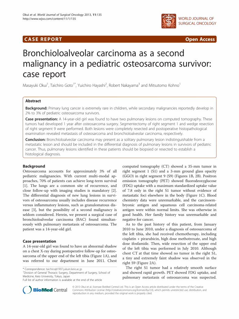
Case presentationA 14-year-old girl was found to have an abnormal shadowon a chest X-ray during postoperative follow-up for osteo-sarcoma of the upper end of the left tibia (Figure 1A), andwas referred to our department in June 2011. Chest
* Correspondence: [email protected] of General Thoracic Surgery, Department of Surgery, School ofMedicine, Keio University, Tokyo, JapanFull list of author information is available at the end of the article
© 2013 Okui et al.; licensee BioMed Central LtCommons Attribution License (http://creativecreproduction in any medium, provided the or
computed tomography (CT) showed a 35-mm tumor inright segment 1 (S1) and a 3-mm ground glass opacity(GGO) in right segment 9 (S9) (Figures 1B, 2B). Positronemission tomography (PET) showed fluorodeoxyglucose(FDG) uptake with a maximum standardized uptake valueof 7.8 only in the right S1 tumor without evidence ofmetastatic foci elsewhere in the body (Figure 1C). Bloodchemistry data were unremarkable, and the carcinoem-bryonic antigen and squamous cell carcinoma-relatedantigen were within normal limits. She was otherwise ingood health. Her family history was unremarkable andnegative for cancer.As to the past history of this patient, from January
2010 to June 2010, under a diagnosis of osteosarcoma ofthe left tibia, she had received chemotherapy, includingcisplatin + pirarubicin, high dose methotrexate, and highdose ifosfamide. Then, wide resection of the upper endof the left tibia was performed in July 2010. Althoughchest CT at that time showed no tumor in the right S1,a tiny and extremely faint shadow was observed in theright S9 (Figure 2A).The right S1 tumor had a relatively smooth surface
and showed rapid growth. PET showed FDG uptake, andpulmonary metastasis of osteosarcoma was suspected.
d. This is an Open Access article distributed under the terms of the Creativeommons.org/licenses/by/2.0), which permits unrestricted use, distribution, andiginal work is properly cited.
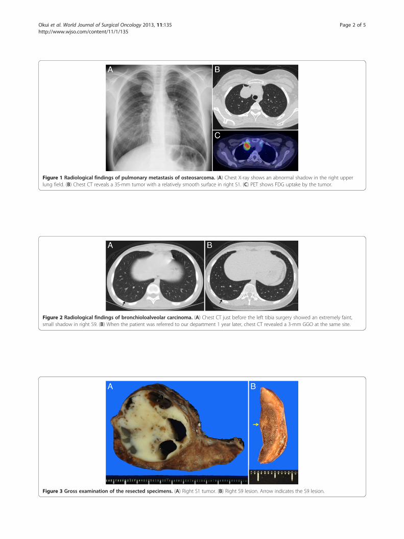
Figure 1 Radiological findings of pulmonary metastasis of osteosarcoma. (A) Chest X-ray shows an abnormal shadow in the right upperlung field. (B) Chest CT reveals a 35-mm tumor with a relatively smooth surface in right S1. (C) PET shows FDG uptake by the tumor.
Figure 2 Radiological findings of bronchioloalveolar carcinoma. (A) Chest CT just before the left tibia surgery showed an extremely faint,small shadow in right S9. (B) When the patient was referred to our department 1 year later, chest CT revealed a 3-mm GGO at the same site.
Figure 3 Gross examination of the resected specimens. (A) Right S1 tumor. (B) Right S9 lesion. Arrow indicates the S9 lesion.
Okui et al. World Journal of Surgical Oncology 2013, 11:135 Page 2 of 5http://www.wjso.com/content/11/1/135
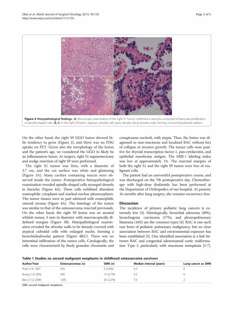
Figure 4 Histopathological findings. (A) Microscopic examination of the right S1 tumor confirmed a sarcoma composed of fascicular proliferationof spindle-shaped cells. (B, C) In the right S9 lesion, atypical cuboidal cells grew densely along alveolar walls, forming a bronchioloalveolar pattern.
Okui et al. World Journal of Surgical Oncology 2013, 11:135 Page 3 of 5http://www.wjso.com/content/11/1/135
On the other hand, the right S9 GGO lesion showed lit-tle tendency to grow (Figure 2), and there was no FDGuptake on PET. Given also the morphology of the lesionand the patient’s age, we considered the GGO to likely bean inflammatory lesion. At surgery, right S1 segmentectomyand wedge resection of right S9 were performed.The right S1 tumor was firm, with a diameter of
3.7 cm, and the cut surface was white and glistening(Figure 3A). Many cavities containing mucus were ob-served inside the tumor. Postoperative histopathologicalexamination revealed spindle-shaped cells arranged denselyin fascicles (Figure 4A). These cells exhibited abundanteosinophilic cytoplasm and marked nuclear pleomorphism.The tumor tissues were in part admixed with eosinophilicosteoid stroma (Figure 4A). The histology of the tumorwas similar to that of the osteosarcoma resected previously.On the other hand, the right S9 lesion was an aeratedwhitish tumor, 3 mm in diameter with macroscopically ill-defined margins (Figure 3B). Histopathological examin-ation revealed the alveolar walls to be densely covered withatypical cuboidal cells with enlarged nuclei, forming abronchioloalveolar pattern (Figure 4B,C). There was nointerstitial infiltration of the tumor cells. Cytologically, thecells were characterized by finely granular chromatin and
Table 1 Studies on second malignant neoplasms in childhood
Author/Year Osteosarcomas (n) SMN (n)
Pratt [14] 1997 334 9 (2.6%)
Anug [13] 2002 509 14 (2.7%)
Bacci [12] 2006 1205 26 (2.2%)
SMN, second malignant neoplasms.
conspicuous nucleoli, with atypia. Thus, the lesion was di-agnosed as non-mucinous and localized BAC without fociof collapse or invasive growth. The tumor cells were posi-tive for thyroid transcription factor-1, pan-cytokeratin, andepithelial membrane antigen. The MIB-1 labeling indexwas low at approximately 1%. The resected margins ofboth the right S1 and the right S9 tumor were free of ma-lignant cells.The patient had an uneventful postoperative course, and
was discharged on the 7th postoperative day. Chemother-apy with high-dose ifosfamide has been performed atthe Department of Orthopedics of our hospital. At present,16 months after lung surgery, she remains recurrence free.
DiscussionThe incidence of primary pediatric lung cancers is ex-tremely low [4]. Histologically, bronchial adenoma (40%),bronchogenic carcinoma (17%), and pleuropulmonaryblastoma (16%) are the common types [4]. BAC is one suchrare form of pediatric pulmonary malignancy, but no clearassociation between BAC and environmental exposure hasbeen established [5]. One identified association is a link be-tween BAC and congenital adenomatoid cystic malforma-tion Type I, particularly with mucinous metaplasia [5-7].
osteosarcoma survivors
Median interval (years) Lung cancer as SMN
6.3 0
5.5 0
7.6 2
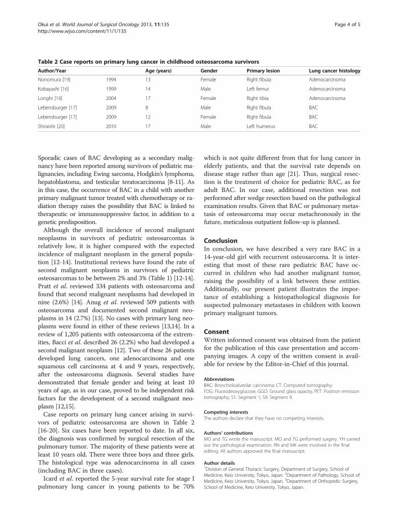
Table 2 Case reports on primary lung cancer in childhood osteosarcoma survivors
Author/Year Age (years) Gender Primary lesion Lung cancer histology
Nonomura [19] 1994 13 Female Right fibula Adenocarcinoma
Kobayashi [16] 1999 14 Male Left femur Adenocarcinoma
Longhi [18] 2004 17 Female Right tibia Adenocarcinoma
Lebensburger [17] 2009 8 Male Right fibula BAC
Lebensburger [17] 2009 12 Female Right fibula BAC
Shiraishi [20] 2010 17 Male Left humerus BAC
Okui et al. World Journal of Surgical Oncology 2013, 11:135 Page 4 of 5http://www.wjso.com/content/11/1/135
Sporadic cases of BAC developing as a secondary malig-nancy have been reported among survivors of pediatric ma-lignancies, including Ewing sarcoma, Hodgkin’s lymphoma,hepatoblastoma, and testicular teratocarcinoma [8-11]. Asin this case, the occurrence of BAC in a child with anotherprimary malignant tumor treated with chemotherapy or ra-diation therapy raises the possibility that BAC is linked totherapeutic or immunosuppressive factor, in addition to agenetic predisposition.Although the overall incidence of second malignant
neoplasms in survivors of pediatric osteosarcomas isrelatively low, it is higher compared with the expectedincidence of malignant neoplasm in the general popula-tion [12-14]. Institutional reviews have found the rate ofsecond malignant neoplasms in survivors of pediatricosteosarcomas to be between 2% and 3% (Table 1) [12-14].Pratt et al. reviewed 334 patients with osteosarcoma andfound that second malignant neoplasms had developed innine (2.6%) [14]. Anug et al. reviewed 509 patients withosteosarcoma and documented second malignant neo-plasms in 14 (2.7%) [13]. No cases with primary lung neo-plasms were found in either of these reviews [13,14]. In areview of 1,205 patients with osteosarcoma of the extrem-ities, Bacci et al. described 26 (2.2%) who had developed asecond malignant neoplasm [12]. Two of these 26 patientsdeveloped lung cancers, one adenocarcinoma and onesquamous cell carcinoma at 4 and 9 years, respectively,after the osteosarcoma diagnosis. Several studies havedemonstrated that female gender and being at least 10years of age, as in our case, proved to be independent riskfactors for the development of a second malignant neo-plasm [12,15].Case reports on primary lung cancer arising in survi-
vors of pediatric osteosarcoma are shown in Table 2[16-20]. Six cases have been reported to date. In all six,the diagnosis was confirmed by surgical resection of thepulmonary tumor. The majority of these patients were atleast 10 years old. There were three boys and three girls.The histological type was adenocarcinoma in all cases(including BAC in three cases).Icard et al. reported the 5-year survival rate for stage I
pulmonary lung cancer in young patients to be 70%
which is not quite different from that for lung cancer inelderly patients, and that the survival rate depends ondisease stage rather than age [21]. Thus, surgical resec-tion is the treatment of choice for pediatric BAC, as foradult BAC. In our case, additional resection was notperformed after wedge resection based on the pathologicalexamination results. Given that BAC or pulmonary metas-tasis of osteosarcoma may occur metachronously in thefuture, meticulous outpatient follow-up is planned.
ConclusionIn conclusion, we have described a very rare BAC in a14-year-old girl with recurrent osteosarcoma. It is inter-esting that most of these rare pediatric BAC have oc-curred in children who had another malignant tumor,raising the possibility of a link between these entities.Additionally, our present patient illustrates the impor-tance of establishing a histopathological diagnosis forsuspected pulmonary metastases in children with knownprimary malignant tumors.
ConsentWritten informed consent was obtained from the patientfor the publication of this case presentation and accom-panying images. A copy of the written consent is avail-able for review by the Editor-in-Chief of this journal.
AbbreviationsBAC: Bronchioloalveolar carcinoma; CT: Computed tomography;FDG: Fluorodeoxyglucose; GGO: Ground glass opacity; PET: Positron emissiontomography; S1: Segment 1; S9: Segment 9.
Competing interestsThe authors declare that they have no competing interests.
Authors’ contributionsMO and TG wrote the manuscript. MO and TG performed surgery. YH carriedout the pathological examination. RN and MK were involved in the finalediting. All authors approved the final manuscript.
Author details1Division of General Thoracic Surgery, Department of Surgery, School ofMedicine, Keio University, Tokyo, Japan. 2Department of Pathology, School ofMedicine, Keio University, Tokyo, Japan. 3Department of Orthopedic Surgery,School of Medicine, Keio University, Tokyo, Japan.
Okui et al. World Journal of Surgical Oncology 2013, 11:135 Page 5 of 5http://www.wjso.com/content/11/1/135
Received: 4 October 2012 Accepted: 1 June 2013Published: 12 June 2013
References1. Meyers PA, Schwartz CL, Krailo MD, Healey JH, Bernstein ML, Betcher D,
Ferguson WS, Gebhardt MC, Goorin AM, Harris M, et al: Osteosarcoma: theaddition of muramyl tripeptide to chemotherapy improves overallsurvival–a report from the Children's Oncology Group. J Clin Oncol 2008,26:633–638.
2. Briccoli A, Rocca M, Salone M, Bacci G, Ferrari S, Balladelli A, Mercuri M:Resection of recurrent pulmonary metastases in patients withosteosarcoma. Cancer 2005, 104:1721–1725.
3. McCarville MB, Kaste SC, Cain AM, Goloubeva O, Rao BN, Pratt CB:Prognostic factors and imaging patterns of recurrent pulmonary nodulesafter thoracotomy in children with osteosarcoma. Cancer 2001,91:1170–1176.
4. Weldon CB, Shamberger RC: Pediatric pulmonary tumors: primary andmetastatic. Semin Pediatr Surg 2008, 17:17–29.
5. Ohye RG, Cohen DM, Caldwell S, Qualman SJ: Pediatric bronchioloalveolarcarcinoma: a favorable pediatric malignancy? J Pediatr Surg 1998,33:730–732.
6. Granata C, Gambini C, Balducci T, Toma P, Michelazzi A, Conte M, Jasonni V:Bronchioloalveolar carcinoma arising in congenital cystic adenomatoidmalformation in a child: a case report and review on malignanciesoriginating in congenital cystic adenomatoid malformation. PediatrPulmonol 1998, 25:62–66.
7. Kaslovsky RA, Purdy S, Dangman BC, McKenna BJ, Brien T, Ilves R:Bronchioloalveolar carcinoma in a child with congenital cysticadenomatoid malformation. Chest 1997, 112:548–551.
8. Kowalski P, Rodziewicz B, Pejcz J: Bilateral bronchioloalveolar carcinoma ofthe lungs in a 7 year old girl treated for Hodgkin’s disease. Tumori 1989,75:449–451.
9. Kuttesch JF Jr, Wexler LH, Marcus RB, Fairclough D, Weaver-McClure L, WhiteM, Mao L, Delaney TF, Pratt CB, Horowitz ME, Kun LE: Second malignanciesafter Ewing’s sarcoma: radiation dose-dependency of secondarysarcomas. J Clin Oncol 1996, 14:2818–2825.
10. Spaner SJ, Raymond G, Puttagunta L, Bhargava R: Bronchioloalveolar cellcarcinoma in a child with hepatoblastoma: case report. Can Assoc Radiol J1999, 50:343–345.
11. Travis WD, Linnoila RI, Horowitz M, Becker RL Jr, Pass H, Ozols R, Gazdar A:Pulmonary nodules resembling bronchioloalveolar carcinoma inadolescent cancer patients. Mod Pathol 1988, 1:372–377.
12. Bacci G, Ferrari C, Longhi A, Ferrari S, Forni C, Bacchini P, Palmerini E,Briccoli A, Pignotti E, Balladelli A, Picci P: Second malignant neoplasmin patients with osteosarcoma of the extremities treated withadjuvant and neoadjuvant chemotherapy. J Pediatr Hematol Oncol2006, 28:774–780.
13. Aung L, Gorlick RG, Shi W, Thaler H, Shorter NA, Healey JH, Huvos AG,Meyers PA: Second malignant neoplasms in long-term survivors ofosteosarcoma: memorial sloan-kettering cancer center experience.Cancer 2002, 95:1728–1734.
14. Pratt CB, Meyer WH, Luo X, Cain AM, Kaste SC, Pappo AS, Rao BN, FlemingID, Jenkins JJ 3rd: Second malignant neoplasms occuring in survivors ofosteosarcoma. Cancer 1997, 80:960–965.
15. Knowling MA, Basco VE: Breast cancer after treatment for osteosarcoma.Med Pediatr Oncol 1986, 14:51–53.
16. Kobayashi H, Mori T, Yoshioka M, Tanaka M, Okuma T, Iida SI, Isogai M,Tabira Y, Kitamura N: A 14-year-old boy with small primary lung cancer.J Jpn Assoc Chest Surg 1999, 13:144–147.
17. Lebensburger J, Katzenstein H, Jenkins JJ, Rodriguez-Galindo C:Bronchioloalveolar carcinoma as a second malignancy in osteosarcomasurvivors. Pediatr Blood Cancer 2009, 53:499–501.
18. Longhi A, Bertoni F, Bacchini P, Albisinni U, Mercati U, Bacci G:Simultaneous osteosarcoma lung metastasis and second primary lungcancer. J Pediatr Hematol Oncol 2004, 26:457–461.
19. Nonomura A, Mizukami Y, Shimizu J, Watanabe Y, Kamimura R, Takashima T,Tsuchiya H, Tomita K: Simultaneous occurrence of lung adenocarcinomaand fibular osteosarcoma in a 13-year-old girl. Thorac Cardiovasc Surg1994, 42:61–63.
20. Shiraishi K, Mori T, Ohba Y, Iwatani K, Yoshimoto K, Iyama K: Three youngosteosarcoma patients with small adenocarcinoma or atypicaladenomatous hyperplasia of the lung. Ann Thorac Cardiovasc Surg 2010,16:358–361.
21. Icard P, Regnard JF, de Napoli S, Rojas-Miranda A, Dartevelle P, Levasseur P:Primary lung cancer in young patients: a study of 82 surgically treatedpatients. Ann Thorac Surg 1992, 54:99–103.
doi:10.1186/1477-7819-11-135Cite this article as: Okui et al.: Bronchioloalveolar carcinoma as a secondmalignancy in a pediatric osteosarcoma survivor: case report. WorldJournal of Surgical Oncology 2013 11:135.
Submit your next manuscript to BioMed Centraland take full advantage of:
• Convenient online submission
• Thorough peer review
• No space constraints or color figure charges
• Immediate publication on acceptance
• Inclusion in PubMed, CAS, Scopus and Google Scholar
• Research which is freely available for redistribution
Submit your manuscript at www.biomedcentral.com/submit