case report open access disease flare of ankylosing
TRANSCRIPT

CASE REPORT Open Access
Disease flare of ankylosing spondylitis presentingas reactive arthritis with seropositivity: a casereportEM Manoj* and MK Ragunathan
Abstract
Introduction: Concurrent rheumatoid factor seropositivity is occasionally detected in ankylosing spondylitis andoften causes confusion in clinical routine. Overlap between various seronegative arthritides is a known butuncommon association. Differentiation of spondyloarthropathy from rheumatoid arthritis is important, since thenatural history, complications, treatments and prognosis of the two diseases differ significantly.
Case presentation: Here, we report the case of a 47-year-old Sri Lankan man who had a long history ofintermittent joint pains worsening following a recent episode of self-resolving non-bloody diarrhea. Subsequently,he developed a skin rash suggestive of keratoderma blenorrhagica and circinate balanitis. He had classicalradiological evidence of ankylosing spondylosis (previously undiagnosed) associated with human leukocyte antigenB27 antigen, but was positive for rheumatoid factor.
Conclusions: A disease flare of ankylosing spondylitis prompted by a minor diarrheal illness showing welldocumented features of reactive arthritis is remarkable. The prognostic implications of seropositivity inspondyloarthritis are discussed.
IntroductionEstimates of the prevalence of spondyloarthritis rangefrom 1% to 2% of the population, and are similar to theprevalence of rheumatoid arthritis (RA). The lack of anyassociation with autoantibodies including rheumatoidfactor (hence the term ‘seronegative’), an increased inci-dence of ankylosing spondylitis (AS) in the whole groupand the association with human leukocyte antigen(HLA)-B27 are the features common to this group ofdiseases [1]. The prognostic implications of early admin-istration of disease-modifying drugs in RA, but not inspondyloarthropathy, are well established. The unusualpresentation of reactive arthritis as a flare up of AS fol-lowing a minor diarrheal illness associated with seropo-sitivity prompted us to report this case.
Case presentationA 47-year-old man who worked as a driver wasadmitted to our facility with a three-week history of
worsening peripheral joint pains following an episode ofwatery diarrhea. The joint pains mainly involved theright side of his shoulder girdle, right wrist, right secondtoe and right Achilles’ tendon. He had morning stiffnessin the involved joints lasting for around one hour. Hedid not have significant backache, urinary symptoms orred eyes. In the past 20 years he had experienced inter-mittent episodes of joint pains and swelling involvinghis right knee joint, right elbow joint and right shoulderjoint. Those episodes were not preceded by gastrointest-inal or urinary infections and he denied any episodessimilar to his current presentation. He had no history tosuggest inflammatory bowel disease or psoriasis.A physical examination revealed an averagely built man
with a stiff posture. Our patient was not dyspneic but hischest expansion (1.7 cm) was impaired. Swelling and ten-derness of the right side of the shoulder girdle was asso-ciated with marked restriction of the anterior and lateralflexion of spine. There was a demonstrable right-sidedAchilles tendinitis. His swollen, tender right second toe wassuggestive of ‘sausage finger’. He had a non-scaly, reddishbrown discrete papular eruption on his palms and soles* Correspondence: [email protected]
Ward 42, National Hospital of Sri Lanka, Colombo, Sri Lanka
Manoj and Ragunathan Journal of Medical Case Reports 2012, 6:60http://www.jmedicalcasereports.com/content/6/1/60 JOURNAL OF MEDICAL
CASE REPORTS
© 2012 Manoj and Ragunathan; licensee BioMed Central Ltd. This is an Open Access article distributed under the terms of the CreativeCommons Attribution License (http://creativecommons.org/licenses/by/2.0), which permits unrestricted use, distribution, andreproduction in any medium, provided the original work is properly cited.

suggestive of keratoderma blenorrhagica (Figure 1). Evi-dence for lung fibrosis or cardiac valvular involvement wasabsent. He developed circinate balanitis some weeks later.Laboratory investigations revealed mild neutrophil leu-
kocytosis with markedly elevated inflammatory markers(erythrocyte sedimentation rate (ESR) = 115 mm/hour,C-reactive protein (CRP) = 96 mg/dL). A test for rheu-matoid factor was positive (64 IU/mL; latex method, cut-off < 8 IU/mL) but an anti-cyclic citrullinated peptide(CCP) antibody test (medical image analysis method) wasnegative. A urine microscopy study and routine culturetests were also negative. A stool culture test was alsonegative. An enzyme-linked immunosorbent assay(ELISA) test for HIV, VDRL test and Treponema palli-dum particle agglutination (TPPA) assay were also allnegative. A test for human leukocyte antigen (HLA)-B27was also positive (lymphocytotoxicity method).Radiography of his hands (Figure 2), wrists, right
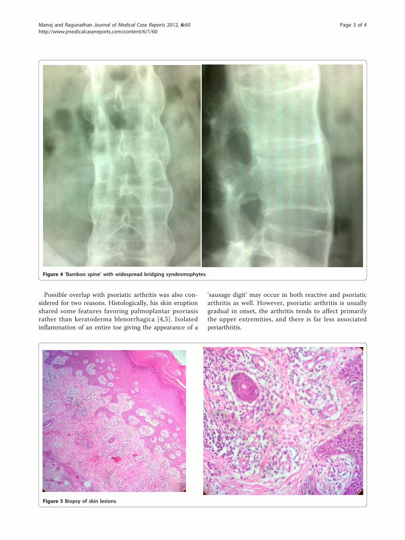
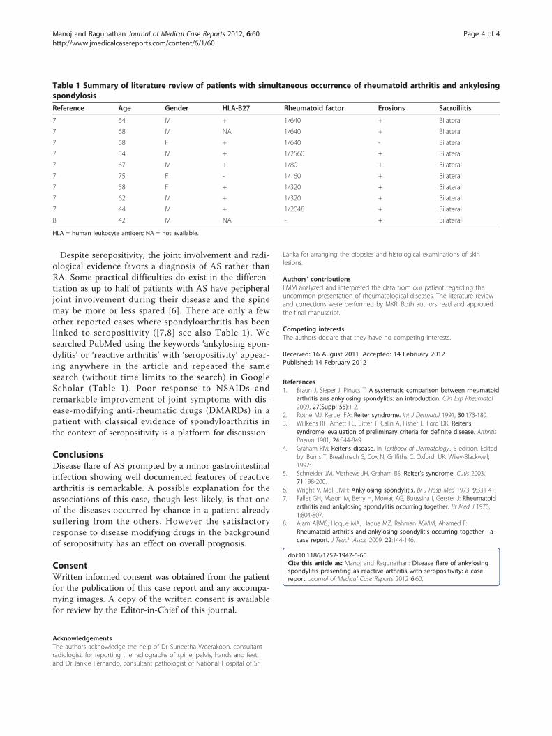
shoulder joint and foot did not reveal any erosivechanges. Both sacroiliac joints were markedly sclerosed(Figure 3) and the typical bamboo spine with widespreadbridging syndesmophytes (Figure 4) was seen. Biopsy ofhis skin lesions (Figure 5) revealed hyperkeratosis form-ing a thin horny layer and psoriasiform hyperplasia, andpapillary dermis showed chronic inflammatory cell infil-trates and superficial dermal edema.Treatment with systemic steroids and non-steroidal anti-
inflammatory drugs (NSAIDs) was not successful. Conse-quently our patient was started on sulfasalazine. Intra-articular injections of steroids and sulfasalazine lead toremission of his current episode. He managed to re-engagein his occupation as a driver with satisfactory improvementof joint symptoms while on sulfasalazine therapy.
DiscussionTo the best of our knowledge, the case of our patient isunique in the clinical, radiological and serological
associations of oligoarthritis aggravated following a diar-rheal episode. Keratoderma blenorrhagica, circinate bala-nitis, ankylosing spondylitis, presence of HLA-B27antigen and seropositivity are unorthodox associations[2]. Though 3% of the normal population may haverheumatoid factor positivity, its occurrence in this caseis less likely to be coincidental, considering the hightiter of rheumatoid factor.Peripheral oligoarthritis aggravated following a diarrheal
episode followed by keratoderma blenorrhagica and circi-nate balanitis suggested a diagnosis of reactive arthritis [3],whereas only one-third of patients show the completetriad of arthritis, conjunctivitis and urethritis. The classicalradiological features of more symmetrical bilateral sacroi-liitis and typical ‘bamboo spine’ in our patient, associatedwith limitation of movement of the lumbar spine in thesagittal and lumbar planes, was adequate to diagnose ASaccording to the modified New York criteria.
Figure 1 Palmoplantar pustulosis, suggestive of keratodermablenorrhagica.
Figure 2 X-ray of hands.
Figure 3 Advanced symmetrical sacroiliitis.
Manoj and Ragunathan Journal of Medical Case Reports 2012, 6:60http://www.jmedicalcasereports.com/content/6/1/60
Page 2 of 4
Possible overlap with psoriatic arthritis was also con-sidered for two reasons. Histologically, his skin eruptionshared some features favoring palmoplantar psoriasisrather than keratoderma blenorrhagica [4,5]. Isolatedinflammation of an entire toe giving the appearance of a
‘sausage digit’ may occur in both reactive and psoriaticarthritis as well. However, psoriatic arthritis is usuallygradual in onset, the arthritis tends to affect primarilythe upper extremities, and there is far less associatedperiarthritis.
Figure 4 ’Bamboo spine’ with widespread bridging syndesmophytes.
Figure 5 Biopsy of skin lesions.
Manoj and Ragunathan Journal of Medical Case Reports 2012, 6:60http://www.jmedicalcasereports.com/content/6/1/60
Page 3 of 4
Despite seropositivity, the joint involvement and radi-ological evidence favors a diagnosis of AS rather thanRA. Some practical difficulties do exist in the differen-tiation as up to half of patients with AS have peripheraljoint involvement during their disease and the spinemay be more or less spared [6]. There are only a fewother reported cases where spondyloarthritis has beenlinked to seropositivity ([7,8] see also Table 1). Wesearched PubMed using the keywords ‘ankylosing spon-dylitis’ or ‘reactive arthritis’ with ‘seropositivity’ appear-ing anywhere in the article and repeated the samesearch (without time limits to the search) in GoogleScholar (Table 1). Poor response to NSAIDs andremarkable improvement of joint symptoms with dis-ease-modifying anti-rheumatic drugs (DMARDs) in apatient with classical evidence of spondyloarthritis inthe context of seropositivity is a platform for discussion.
ConclusionsDisease flare of AS prompted by a minor gastrointestinalinfection showing well documented features of reactivearthritis is remarkable. A possible explanation for theassociations of this case, though less likely, is that oneof the diseases occurred by chance in a patient alreadysuffering from the others. However the satisfactoryresponse to disease modifying drugs in the backgroundof seropositivity has an effect on overall prognosis.
ConsentWritten informed consent was obtained from the patientfor the publication of this case report and any accompa-nying images. A copy of the written consent is availablefor review by the Editor-in-Chief of this journal.
AcknowledgementsThe authors acknowledge the help of Dr Suneetha Weerakoon, consultantradiologist, for reporting the radiographs of spine, pelvis, hands and feet,and Dr Jankie Fernando, consultant pathologist of National Hospital of Sri
Lanka for arranging the biopsies and histological examinations of skinlesions.
Authors’ contributionsEMM analyzed and interpreted the data from our patient regarding theuncommon presentation of rheumatological diseases. The literature reviewand corrections were performed by MKR. Both authors read and approvedthe final manuscript.
Competing interestsThe authors declare that they have no competing interests.
Received: 16 August 2011 Accepted: 14 February 2012Published: 14 February 2012
References1. Braun J, Sieper J, Pinucs T: A systematic comparison between rheumatoid
arthritis ans ankylosing spondylitis: an introduction. Clin Exp Rheumatol2009, 27(Suppl 55):1-2.
2. Rothe MJ, Kerdel FA: Reiter syndrome. Int J Dermatol 1991, 30:173-180.3. Willkens RF, Arnett FC, Bitter T, Calin A, Fisher L, Ford DK: Reiter’s
syndrome: evaluation of preliminary criteria for definite disease. ArthritisRheum 1981, 24:844-849.
4. Graham RM: Reiter’s disease. In Textbook of Dermatology.. 5 edition. Editedby: Burns T, Breathnach S, Cox N, Griffiths C. Oxford, UK: Wiley-Blackwell;1992:.
5. Schneider JM, Mathews JH, Graham BS: Reiter’s syndrome. Cutis 2003,71:198-200.
6. Wright V, Moll JMH: Ankylosing spondylitis. Br J Hosp Med 1973, 9:331-41.7. Fallet GH, Mason M, Berry H, Mowat AG, Boussina I, Gerster J: Rheumatoid
arthritis and ankylosing spondylitis occurring together. Br Med J 1976,1:804-807.
8. Alam ABMS, Hoque MA, Haque MZ, Rahman ASMM, Ahamed F:Rheumatoid arthritis and ankylosing spondylitis occurring together - acase report. J Teach Assoc 2009, 22:144-146.
doi:10.1186/1752-1947-6-60Cite this article as: Manoj and Ragunathan: Disease flare of ankylosingspondylitis presenting as reactive arthritis with seropositivity: a casereport. Journal of Medical Case Reports 2012 6:60.
Table 1 Summary of literature review of patients with simultaneous occurrence of rheumatoid arthritis and ankylosingspondylosis
Reference Age Gender HLA-B27 Rheumatoid factor Erosions Sacroiliitis
7 64 M + 1/640 + Bilateral
7 68 M NA 1/640 + Bilateral
7 68 F + 1/640 - Bilateral
7 54 M + 1/2560 + Bilateral
7 67 M + 1/80 + Bilateral
7 75 F - 1/160 + Bilateral
7 58 F + 1/320 + Bilateral
7 62 M + 1/320 + Bilateral
7 44 M + 1/2048 + Bilateral
8 42 M NA - + Bilateral
HLA = human leukocyte antigen; NA = not available.
Manoj and Ragunathan Journal of Medical Case Reports 2012, 6:60http://www.jmedicalcasereports.com/content/6/1/60
Page 4 of 4