case report rare cause of seizures, renal failure, and...
TRANSCRIPT

Hindawi Publishing CorporationCase Reports in ImmunologyVolume 2013, Article ID 523865, 5 pageshttp://dx.doi.org/10.1155/2013/523865
Case ReportRare Cause of Seizures, Renal Failure, and Gangrene inan 83-Year-Old Diabetic Male
Stalin Viswanathan and Kandan Balamurugesan
Indira Gandhi Medical College, Kadhiramam, Pondicherry 605009, India
Correspondence should be addressed to Stalin Viswanathan; [email protected]
Received 12 May 2013; Accepted 11 June 2013
Academic Editors: A. M. Mansour and H. Narimatsu
Copyright © 2013 S. Viswanathan and K. Balamurugesan. This is an open access article distributed under the Creative CommonsAttribution License, which permits unrestricted use, distribution, and reproduction in any medium, provided the original work isproperly cited.
We report an 83-year-old diabeticmale who presented with acute-onset renal failure, seizures, psychosis, pneumonia, and right footgangrene. Investigations revealed thrombocytopenia, CSF lymphocytosis, ANA and dsDNA positivity, hypocomplementemia, andpneumonitis following which he was treated with pulse methylprednisolone. He was treated for Pseudomonas-related ventilator-associated pneumonia, candiduria, and E. coli-related bedsore infection prior to discharge. He was discharged at request and died17 days later due to a respiratory infection.
1. Introduction
Onset of systemic lupus erythematosus (SLE) after the age of50 (late-onset SLE) constitutes 6–18% of the lupus population[1]. Most cases of lupus over 65 years have been described ascase reports. Renal failure is the initial presentation only in25% patients of SLE [2]. Neuropsychiatric SLE (NSLE) in theelderly is very rare. Infections, malignancies, and atheroscle-rotic disease account for most deaths in SLE patients [3].Here we describe an 83-year-old diabetic who presented withacute-onset seizures, psychosis, pneumonitis, foot gangrene,and renal failure and improved with immunosuppressivetherapy for SLE but succumbed to another respiratory infec-tion 17 days after discharge from hospital.
2. Case
This 83-year-old diabetic of 10 years’ duration (on metformin750mg OD) was brought from another hospital by hisrelatives formechanical ventilation. Fifteen days prior, he hadcomplained of fatigue and anorexia and was admitted in alocal nursing home where he was told to have early renalfailure (creatinine 202𝜇mol/L). Four days later he had had ageneralized tonic-clonic seizure for which he was taken to thereferring hospital for management. Computed tomography
(CT) of brain was normal and the patient was commencedon phenytoin; he was uncooperative for magnetic resonanceimaging (MRI). He had developed acute psychosis anddelirium in hospital and was managed with risperidone.Three days later he was intubated for altered sensorium andrespiratory distress following acute cough, breathlessness,and fever. He was mechanically ventilated and administeredceftriaxone and metronidazole; his seizures remained undercontrol but altered sensorium persisted. During his stay inintensive care, he developed discoloration of his right footand warfarin had been initiated. His renal parameters hadcontinued toworsen (creatinine 350 𝜇mol/L) and his relativesrequested discharge and brought him to our hospital.
He was a cigar smoker (>40 years) and drank occasion-ally. He had had a left hip fracture six years ago which hadbeen treated conservatively. On admission, his pulse was104 bpm, BP 92/60mmHg, respiratory rate 42 breaths/minwith SpO
292% on FiO2 of 100, temperature 99∘F, and central
venous pressure (CVP)was 6 cm.Chest examination revealedleft-sided coarse crackles. The Glasgow Coma Scale (GCS) of2T/15, bilaterally 3mm sluggishly reacting pupils, generalizedhypotonia, and areflexia were observed on neurologicalexamination.Therewas no papilledema.Hehaddry gangreneof right foot (Figure 1) with weak right popliteal pulse. Inves-tigations are listed in Table 1. Doppler of right lower limb

2 Case Reports in Immunology
(a) (b)
(c) (d)
(e) (f)
Figure 1: (a) Right foot discoloration at admission. (b) Right forefoot dry gangrene on day 4. (c) CT chest on day 2 showing airway dilatation(right predominant), patchy infiltrates, and ground glassing. (d) Chest radiograph on day 4 of admission shows clearing of infiltrates in theright side. (e) CT chest revealing right-sided pleural effusion, reticular infiltrates, bilateral ground glassing, and tree-in-bud appearance. (f)CT chest shows reticulonodular infiltrates in the entire right lung and ground glassing in left lung.

Case Reports in Immunology 3
Table 1: Lab investigations of patient.
Day of admission 1 4 5 7 10 16 18Urea (2.5–7.1mmol/L) 67.8 61.7 47.1 38.7 33.2 26.4 21.1Creatinine(44–80𝜇mol/L) 616 422 360 281 290 202 167
HbA1c (5.7–6.5%) 7.9Bil total(1.7–6.8 𝜇mol/L) 1.53
Bil dir(3.4–15.2 𝜇mol/L) 8.5
SGOT(0.20–0.65 𝜇kat/L) 1.5 1.4
SGPT(0.12–0.70 𝜇kat/L) 0.87 0.92
ALP(0.56–1.63 𝜇kat/L) 3.88 9.02
Protein (67–86 g/L) 63Albumin (40–50 g/L) 18 21 22 22GGT(0.15–0.99 𝜇kat/L) 4.48
K+ (3.5–5.0mmol/L) 5.6 3.3 3.9 3.2 145 137 138Na+(136–146mmol/L) 155 151 150 143 5.7 3.8 3.2
Calcium(2.2–2.6mmol/L) 2.2
Mg(0.62–0.95mmol/L) 0.78
Pi (0.81–1.4mmol/L) 1.45LDH (114–240 IU/L) 506 269CK (25–200U/L) 363 103UrineSpot K+
(25–120mEq/L) 30.5
Spot Na+(40–220mEq/L) 116
Bence-Jones NegativeMyoglobin NegativeHeme NegativeEosinophils NegativeCulture 3 organismsStool occult blood NegativeEndotracheal asp AFB NegativeEndotracheal aspculture Pseudomonas
Hb (130–160 g/L) 81TC (3.50–9 × 10⁹/L) 10.2Neutrophilia (%) 81Plat (165–415 × 109/L) 100MCV (79–93.3 fL) 77MCH (26.7–31.9 pg) 27.8MCHC (323–359 g/L) 358Reticulocyte (%) 0.5INR 1.3aPTT (control 25.1 s) 40D dimer (200 ng/mL) 3200FDP Positive
Table 1: Continued.
Day of admission 1 4 5 7 10 16 18Blood cultures SteriledsDNA (<20) 28ANA (<1.0) 1.8cANCA NegativeC3 (0.83–1.77 g/L) 0.78C4 (0.16–0.47 g/L) 0.20Cortisol (5–25𝜇g/dL) 26.1Ferritin (28–397 ng/L) 1328Direct Coombs NegativeCerebrospinal fluidCells 103Sugar (mg) 115Protein (<60mg) 39Lymphocytes (<5) 100%ADA 1.0AFB NegativeGram NegativeIndia ink NegativeHbA1c: glycated haemoglobin; bil: bilirubin; dir: direct; SGOT: serum glu-tamic oxaloacetic transaminase; SGPT: serum glutamic pyruvate transam-inase; ALP: alkaline phosphatase; GGT: gamma-glutamyl transferase; K+:potassium; Na+: sodium; Mg: magnesium; Pi-inorganic phosphate; LDH:lactate dehydrogenase; CK: creatine kinase; Hb: haemoglobin; TC: total cells;plat: platelets; MCV: mean corpuscular volume; MCH: mean corpuscularhaemoglobin; MCHC: mean corpuscular haemoglobin concentration; INR:international normalized ratio; aPTT: activated partial thromboplastin time;FDP: fibrinogen degradation products; dsDNA: double-stranded DNA;ANA: antinuclear antibody; c-ANCA: antineutrophil cytoplasmic autoanti-body; C3: complement C3; C4: complement C4; ADA: adenosine deaminase;AFB: acid-fast bacilli; asp: aspirate.
and echocardiography were normal. Pending cultures, he wasinitiated on piperacillin-tazobactam and levofloxacin, alongwith subcutaneous heparin, warfarin, and pentoxifylline. CTchest (day2) showed bilateral pleural thickening, bilateralground glassing (right≫ left), airway dilatation, and reticulo-nodular infiltrates (right predominant) with minimal pleuraleffusion (Figure 1). In view of seizures, psychosis, throm-bocytopenia, renal failure, pneumonitis, ANA, and dsDNApositivity, a diagnosis of systemic lupus was made and pulsemethyl prednisolone (1 g × 3 days) was initiated on day 3,followed by oral steroids (60mg). Tracheal aspirate grewPseudomonas aeruginosa and imipenemwas administered onday 5 for probable ventilator-associated pneumonia.Weaningwas done on 8th day of admission. Fluconazole 300mg/daywas administered for persisting candiduria. Hypernatremiawas managed with dextrose saline, while sugars were con-trolled with infusion of regular insulin. By day 12, his powerhad improved to 3/5 in all limbs; he occasionally spoke a fewwords to his relatives but continued to be extremely afraid ofhospital personnel. MRI and nerve conduction studies couldnot be performed due to poor cooperation. He developed aninfected gluteal bedsore (E. coli) that necessitated amikacin.We acceded to his son’s request to be discharged to homewithmodified doses of intramuscular amikacin therapy, twice-daily premixed insulin, warfarin, phenytoin, risperidone,
4 Case Reports in Immunology
clonezepam, and oral prednisolone 40mg/day. Seventeendays later he succumbed to another respiratory infection.
3. Discussion
The 9 : 1 female predominance in SLE decreases prior topuberty and late in life [4]. Female :male ratio in late-onsetSLE is about 5 : 1 [5], while another study showed a ratio of1 : 1.1 when the age of onset was >65 years [6]. Four to 18% ofcases from reported series aremale [7]. In aHongKong study,the mean age of late-onset SLE was 62 years and onset wasgenerally insidious [1]. Insidious onset of disease and lowerindex of suspicion lead to delayed diagnosis in the elderly. Astudy of 39 Indianmale SLE subjects showed only one patientwith late-onset SLE [8].
It is generally agreed that SLE in the elderly is a mild dis-ease [9]. Prevalence of organ involvement in males dependsupon the ethnic population being studied, study setting(tertiary versus primary), selection criteria of female controls,and sample size of male subjects [10]. Fever, fatigue, andweight loss are common symptoms in elderly SLE patients[5]. Serositis, muscle pains, and arthritis are more commonin this age group as are secondary Sjogren’s syndrome butwith a lower incidence of cutaneous manifestations andRaynaud’s phenomenon [9]. Males in a Thai study tended tohave a shorter duration of symptoms prior to presentation,with alopecia, arthralgia and Raynaud’s phenomenon beingless common [10]. Psychosis, hypocomplementemia, anddiffuse proliferative glomerulonephritis (DPGN) were lesscommon in Indians [8], while renal disease and vascularthrombosis were common among Latin American males[11]. Rheumatoid arthritis, polymyalgia rheumatic, and siccasyndromes are close differentials of SLE in the elderly [5].Late-onset lupus may have fewer major organ involvementand fewer major relapses [12].
Among patients with SLE, 60% of adults develop kidneydisease [2]. SLE prevalence in India was low at 3.2/100000population [13]; contrastingly, renal involvement amongIndian SLE patients was the highest in the world [13].Neuropsychiatric manifestations are similar in the young andthe elderly [9]. Neuropsychiatric SLE (NSLE) at presentationin the elderly population has been described only as casereports. Similar to our case, seizures, coma, and pneumoniahave been reported in a 72-year-old lady who had pneumoni-tis, hypocomplementemia, elevated fibrinogen, and FDPbut with normal renal function and negative dsDNA [14].Presence of NSLE is generally associated with a poor prog-nosis [9]. NSLE can be either focal (stroke, neuropathy, andtransient ischemic attack) or diffuse (confusion, dementia,and psychosis) or can present with seizures (partial orgeneralized) [9]. Seizures are reported in 15 to 30% of patientswith SLE [9]. Cognitive impairment may be the initial mani-festation of SLE in the elderly [5]. The neurological manifes-tations seen in Indian studies were cerebrovascular accidents,myeloradiculopathies, movement disorders, seizures, coma,and psychosis [13]. Lower numbers of Raynaud and NSLEwere seen in the South Indian population [13]. Our patient’scognition did not improve completely at time of discharge.
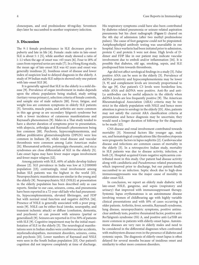
His respiratory symptoms could have also been contributedby diabetes-related pneumonia or seizure-related aspirationpneumonia but his chest radiograph (Figure 1) cleared onthe 4th day of admission (after two methyl prednisolonepulses). The cause of foot gangrene could not be pinpointed.Antiphospholipid antibody testing was unavailable in ourhospital. Sincewarfarin had been initiated prior to admission,protein C and protein S were not done. High levels of D-dimer and FDP like in our patient may indicate vascularinvolvement due to emboli and/or inflammation [14]. It ispossible that diabetes, old age, smoking, sepsis, and SLEpredisposed him towards thrombosis.
Age did not affect serological findings in a study [5]. Falsepositive ANA can be seen in the elderly [5]. Prevalence ofdsDNA positivity and hypocomplementemia may be lower[5, 9] and complement levels are inversely proportional tothe age [9]. Our patient’s C3 levels were borderline low,while ANA and dsDNA were positive. Anti-Ro and anti-La antibodies can be useful adjuncts in the elderly whendsDNA levels are less frequently positive [9]. The AmericanRheumatological Association (ARA) criteria may be toostrict in the elderly population with NSLE and hence moreattention is given to serology in the elderly [15]. Also, patientsmay not satisfy the current ARA classification criteria atpresentation and hence diagnosis may be uncertain; theywould need a longer duration of followup for the diagnosisto be made [12].
CNS disease and renal involvement contributed towardsmortality [3]. Nonrenal factors like younger age, malesex, and hematological complications like thrombocytopeniawere prognostic factors in lupus nephritis [2]. Cardiovasculardisease and infections are common causes of mortality inthe elderly [5]. In a retrospective Indian study, mortalityin SLE patients was due to disease activity, infection, orboth [3]. Hospital-acquired Gram-negative septicaemia con-tributed most in this study. Our patient had disease activityalong with candiduria and Pseudomonas-related pneumoniawhich improved prior to discharge, but our patient finallysuccumbed to an infection. Septic shock due to high-doseimmunosuppressants was the major cause of morality inolder-onset SLE.
In conclusion, we report an elderly male diabetic withlate-onset NSLE, gangrene, and sepsis (respiratory andurinary) that improved with immunosuppressant therapy.Systemic lupus erythematosus is an autoimmune diseaseinvolving women of childbearing age with highly variableclinical presentations and with 10% of cases occurring inolder patients. Arthritis, fever, serositis, Raynaud’s syndrome,lung disease, neuropsychiatric symptoms, positive antinu-clear antibody tests, positive rheumatoid factor, positive anti-Ro/Sjogren’s syndrome (SS) A, and positive anti-La/SSB aremore common in patients with elderly-onset lupus. Autoim-mune diseases are very rare in elderly males and need tobe considered in the differential diagnoses when confrontedwithmultisystem disease even in the presence of diabetes andsystemic sepsis. The diagnosis of elderly-onset lupus is oftendelayed for several months because of insidious onset andsimilarity to other more common disorders.

Case Reports in Immunology 5
Conflict of interests
The authors declare that they have no conflict of interests.
References
[1] S. K. Mak, E. K. M. Lam, and A. K. M. Wong, “Clinical profileof patients with late-onset SLE: not a benign subgroup,” Lupus,vol. 7, no. 1, pp. 23–28, 1998.
[2] C. Molino, F. Fabbian, and C. Longhini, “Clinical approach tolupus nephritis: recent advances,” European Journal of InternalMedicine, vol. 20, no. 5, pp. 447–453, 2009.
[3] A. Sharma, S. B. Shamanna, S. Kumar et al., “Causes ofmortalityamong inpatients with systemic lupus erythematosus in atertiary care hospital in North India over a 10-year period,”Lupus, vol. 22, no. 2, pp. 216–222, 2013.
[4] C. C. Mok, C. S. Lau, T. M. Chan, and R. W. S. Wong, “Clinicalcharacteristics and outcome of southern Chinese males withsystemic lupus erythematosus,” Lupus, vol. 8, no. 3, pp. 188–196,1999.
[5] D. Lazaro, “Elderly-onset systemic lupus erythematosus: preva-lence, clinical course and treatment,” Drugs and Aging, vol. 24,no. 9, pp. 701–715, 2007.
[6] S. J. Pu, S.-F. Luo, Y. J. J. Wu, H. S. Cheng, and H. H. Ho, “Theclinical features and prognosis of lupus with disease onset at age65 and older,” Lupus, vol. 9, no. 2, pp. 96–100, 2000.
[7] G. Medina, O. Vera-Lastra, L. Barile, M. Salas, and L. J. Jara,“Clinical spectrum of males with primary antiphospholipidsyndrome and systemic lupus erythematosus: a comparativestudy of 73 patients,” Lupus, vol. 13, no. 1, pp. 11–16, 2004.
[8] I. Pande, A. N.Malaviya, N. G. Sekharan, S. Kailash, S. S. Uppal,and A. Kumar, “SLE in indian men: analysis of the clinical andlaboratory features with a review of the literature,” Lupus, vol. 3,no. 3, pp. 181–186, 1994.
[9] M.Dennis, “Neuropsychiatric lupus erythematosus and the eld-erly,” International Journal of Geriatric Psychiatry, vol. 9, no. 2,pp. 97–106, 1994.
[10] J. Mongkoltanatus, S. Wangkaew, N. Kasitanon, and W. Louth-renoo, “Clinical features of Thai male lupus: an age-matchedcontrolled study,” Rheumatology International, vol. 28, no. 4, pp.339–344, 2008.
[11] M. A. Garcia, J. C. Marcos, A. I. Marcos et al., “Male systemiclupus erythematosus in a Latin-American inception cohort of1214 patients,” Lupus, vol. 14, no. 12, pp. 938–946, 2005.
[12] C. T. K. Ho, C. C.Mok, C. S. Lau, and R.W. S.Wong, “Late onsetsystemic lupus erythematosus in southern Chinese,” Annals ofthe Rheumatic Diseases, vol. 57, no. 7, pp. 437–440, 1998.
[13] A. N. Malaviya, A. N. Chandrasekaran, A. Kumar, and P. N.Shamar, “Occasional series—lupus around the world systemiclupus erythematosus in India,” Lupus, vol. 6, no. 9, pp. 690–700,1997.
[14] M. Yamaya, M. Yoshida, M. Yamasaki, H. Kubo, K. Furukawa,and H. Arai, “Seizure and pneumonia in an elderly patientwith systemic lupus erythematosus,” Journal of the AmericanGeriatrics Society, vol. 57, no. 9, pp. 1709–1711, 2009.
[15] M. S. Dennis, E. J. Byrne, N. Hopkinson, and P. Bendall, “Neu-ropsychiatric systemic lupus erythematosus in elderly people: acase series,” Journal of Neurology Neurosurgery and Psychiatry,vol. 55, no. 12, pp. 1157–1161, 1992.

Submit your manuscripts athttp://www.hindawi.com
Stem CellsInternational
Hindawi Publishing Corporationhttp://www.hindawi.com Volume 2014
Hindawi Publishing Corporationhttp://www.hindawi.com Volume 2014
MEDIATORSINFLAMMATION
of
Hindawi Publishing Corporationhttp://www.hindawi.com Volume 2014
Behavioural Neurology
EndocrinologyInternational Journal of
Hindawi Publishing Corporationhttp://www.hindawi.com Volume 2014
Hindawi Publishing Corporationhttp://www.hindawi.com Volume 2014
Disease Markers
Hindawi Publishing Corporationhttp://www.hindawi.com Volume 2014
BioMed Research International
OncologyJournal of
Hindawi Publishing Corporationhttp://www.hindawi.com Volume 2014
Hindawi Publishing Corporationhttp://www.hindawi.com Volume 2014
Oxidative Medicine and Cellular Longevity
Hindawi Publishing Corporationhttp://www.hindawi.com Volume 2014
PPAR Research
The Scientific World JournalHindawi Publishing Corporation http://www.hindawi.com Volume 2014
Immunology ResearchHindawi Publishing Corporationhttp://www.hindawi.com Volume 2014
Journal of
ObesityJournal of
Hindawi Publishing Corporationhttp://www.hindawi.com Volume 2014
Hindawi Publishing Corporationhttp://www.hindawi.com Volume 2014
Computational and Mathematical Methods in Medicine
OphthalmologyJournal of
Hindawi Publishing Corporationhttp://www.hindawi.com Volume 2014
Diabetes ResearchJournal of
Hindawi Publishing Corporationhttp://www.hindawi.com Volume 2014
Hindawi Publishing Corporationhttp://www.hindawi.com Volume 2014
Research and TreatmentAIDS
Hindawi Publishing Corporationhttp://www.hindawi.com Volume 2014
Gastroenterology Research and Practice
Hindawi Publishing Corporationhttp://www.hindawi.com Volume 2014
Parkinson’s Disease
Evidence-Based Complementary and Alternative Medicine
Volume 2014Hindawi Publishing Corporationhttp://www.hindawi.com