case report spontaneous dissection of the renal artery in
TRANSCRIPT

Case ReportSpontaneous Dissection of the Renal Artery inVascular Ehlers-Danlos Syndrome
Filipa Pereira,1 Teresa Cardoso,2 and Paula Sá1
1Servico de Anestesiologia, Centro Hospitalar do Porto, Largo Professor Abel Salazar, 4099-001 Porto, Portugal2Unidade de Cuidados Intensivos Polivalente, Centro Hospitalar do Porto, Largo Professor Abel Salazar, 4099-001 Porto, Portugal
Correspondence should be addressed to Filipa Pereira; [email protected]
Received 6 April 2015; Revised 1 June 2015; Accepted 3 June 2015
Academic Editor: Matej Podbregar
Copyright © 2015 Filipa Pereira et al. This is an open access article distributed under the Creative Commons Attribution License,which permits unrestricted use, distribution, and reproduction in any medium, provided the original work is properly cited.
Ehlers-Danlos syndrome (EDS) is a rare heterogeneous group of connective tissue disorders. The vascular type (vEDS) is anautosomal dominant disorder caused by heterozygousmutations in theCOL3A1 gene predisposing to premature arterial, intestinal,or uterine rupture.We report a case of a 38-year-oldwomanwith a recent diagnosis of vEDS admitted in the EmergencyDepartmentwith a suspicion of a pyelonephritis that evolved to a cardiopulmonary arrest. A fatal retroperitoneal hematoma related with ahaemorrhagic dissection of the right renal artery was found after emergency surgery. This case highlights the need to be aware ofthe particular characteristics of vEDS, such as a severe vascular complication that can lead to a fatal outcome.
1. Introduction
Ehlers-Danlos syndrome (EDS) is a rare heterogeneous groupof connective tissue disorders [1]. The estimated prevalencevaries between 1/5000 and 1/25000 [1, 2].
The vascular type (vEDS), formerly known as EDStype IV [3], is an autosomal dominant disorder caused byheterozygous mutations in the COL3A1 [4]. Vascular EDScompromises 5–10% of all cases of EDS [4, 5]. The clinicalhallmark of vEDS is excessive tissue fragility predisposingto premature arterial, intestinal, or uterine rupture, rarelyobserved in other forms of EDS [4, 6]. vEDS is also markedclinically by a characteristic facial appearance with decreasedsubcutaneous fat, a narrow nose, and thin lips and thinskin with visible superficial veins and prominent cutaneousbruising. Patients can also present premature aging of dis-tal extremities (acrogeria) and joint hypermobility [5, 7].However, these phenotype characteristics can be subtle andmay not be diagnosed until postmortem examination [4, 5].Furthermore, the natural course of vEDS and the clinicalphenotype of patients are influenced by the type of COL3A1variant [8].
A full differential diagnosis must be considered whenthe presenting signs and symptoms suggest vEDS. This
includesMarfan syndrome, Loeys-Dietz syndrome, kyphosco-liotic types of EDS, idiopathic isolated arterial aneurysms,and autosomal dominant polycystic kidney disease. Clinicalfeatures, radiologic findings, and genetic characterizationallow the clinical diagnosis and management [9–11].
Approximately 25% of patients with the vEDS experiencethe first complication by the age of 20, while 80% of patientshave at least one complication by the age of 40 [6]. Themedian survival described in literature varies between 48 and50 years [4, 6].
Treatment of complications associated with vEDS favoursminimal noninvasive interventions. Invasivemethods shouldbe used only when necessary, primarily to save the patients’life [12].
We report a case of a 38-year-old woman with vEDS,who died during emergency surgery due to a severe vascularcomplication.
2. Case Presentation
A 38-year-old woman was admitted in the EmergencyDepartment (ED) with a sudden onset of severe lower backpain, fever, nausea, and vomiting.The patient had a history of
Hindawi Publishing CorporationCase Reports in Critical CareVolume 2015, Article ID 804252, 4 pageshttp://dx.doi.org/10.1155/2015/804252

2 Case Reports in Critical Care
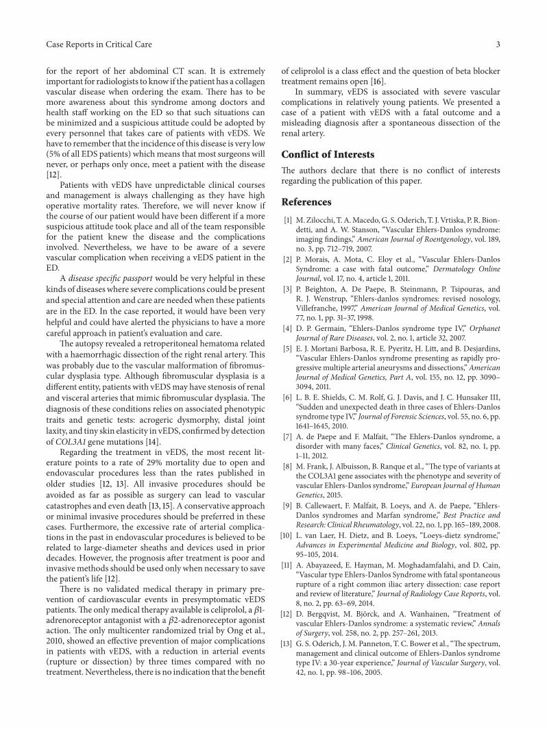
Figure 1: Abdominal Computer Tomography (CT) scan showingdiffuse reduction in the parenchymal thickness of the right kidneyand multiloculated fluid collection of about 10 centimetres (cm),involving the anterior part and the upper pole of the right kidneyand the ipsilateral renal artery.
repeated urinary infections and renal lithiasis. She was beingstudied in the genetic department because of her history ofinfertility and spontaneous abortions and also because of thehistory of sudden deaths in her family (including her father).Therefore, she was diagnosed recently as having vEDS witha mutation in COLA31 gene, but the specific defect was notmentioned in her genetic report.
On admission, her physical examination revealed translu-cid skin, thinned nose, and joint hypermobility of the lowerlimbs.
She had haemoglobin (Hg) of 13 g/dL, leukocytosis (16× 109/L), an elevated C-reactive protein (54mg/dL), andinactive urinary sediment.
The renal ultrasound showed perirenal fluid and signs ofnephritis but without obstruction.
The Abdominal Computer Tomography (CT) scan(Figure 1) showed a diffuse reduction in the parenchymalthickness of the right kidney and multiple areas of striatumnephrogram, suggesting an inflammatory/infectious process(pyelonephritis). It also showed a multiloculated fluidcollection of about 10 centimetres (cm), involving theanterior part and the upper pole of the right kidney and theipsilateral renal artery, which appeared tapered and with areduced calibre.
In between several exams, she was found unconsciousin the clinical observation area of the ED and was taken tothe Emergency Room (ER) in pulmonary arrest that rapidlyevolved to cardiac arrest, from which she recovered withadvanced life support in 10 minutes.
After initial stabilization in the ER, it was assumed thatthis clinical situation was due to a renal abscess, as shown inCT scan. A renal abscess drainage in the operating room wasimmediately proposed for her.
On arrival to the OR, she presented again in cardiopul-monary arrest in a nonshockable rhythm (pulseless electricalactivity). Advanced life support (ALS) was started immedi-ately.
The decision to perform an exploratory laparotomy, whilein ALS manoeuvres, was taken and a team of vascular andurologic surgeons started together the procedure.
During surgery, a massive retroperitoneal hematomawas found, being largest at the left side. The infrarenal
aorta was isolated but was found pulseless and empty. Withthe clamping of the supra- and infrarenal aorta no activehaemorrhage was identified.
ALS manoeuvres were stopped 30 minutes after thebeginning of surgery in the light of these findings and lackof any clinical response to massive transfusion measures.
Request for a clinical autopsy was obtained.
Autopsy Findings. The victim was a normally developed 38-year-old woman with apparent age superior to her real age.The skin of her knees was flaccid and pigmented and she hadan articular hypermobility.
Internal findings included a hematoma in the top andposterior face of the right kidney which extended until theexterior of the renal capsule.Thereweremultiple thrombosedrenal vessels of medium and large calibre in the renalhilum, some of which presented a haemorrhagic dissectionbetween the intimal andmedia arterial walls, sometimes withthrombosed pseudo lumens.
In all vascular territories the arterial walls presentedmorphological alterations of the intimal and media layers,namely, duplication and disruption of the internal elasticlamina, intimal nodular proliferation, and a media layer withthickening and thinning areas.
In These characteristics were suggestive of intimal andmedia fibromuscular dysplasia.
3. Discussion
Vascular EDS is a very challenging disorder. Therefore,it is important to suspect a severe vascular complicationin a patient with a history of vEDS that presents in theED with unspecific symptoms. These patients should beinvestigated promptly to rule out life-threatening situationsdue to vascular complications involving the vascular system,gastrointestinal tract, or gravid uterus [2, 6]. The vasculartype of EDS describes a defect in the synthesis of typeIII procollagen and is the most severe form. Rupture ofa previously nondilated artery is a common occurrencethat results in considerable mortality and morbidity. Lesscommonly, an artery can dissect or dilate in either focal ordiffuse form.
Therefore, patients with vEDS have a reduced lifeexpectancy due to severe vascular complications. In a seriesof 31 patients, 61% present with at least one life-threateningvascular complication before the age of 40 [13].
An acute low back/flank pain is a common complaintin young adult presented to the ED and is usually due to amusculoskeletal or genitourinary tract cause. In our patientthe exams showed an inflammatory/infectious process whichwas a likely diagnosis according to her past medical historyof renal lithiasis. However, her recent diagnosis of vEDS wasinitially neglected and only because of a rapid hemodynamicdeterioration was a surgical approach decided. There wasa lack of communication among the medical staff, as thedoctors responsible for the patient in the ED did not findher past medical history of vEDS important for this situationand this was not transmitted to the doctor responsible forthe Emergency Room (ER) or to the radiologist responsible
Case Reports in Critical Care 3
for the report of her abdominal CT scan. It is extremelyimportant for radiologists to know if the patient has a collagenvascular disease when ordering the exam. There has to bemore awareness about this syndrome among doctors andhealth staff working on the ED so that such situations canbe minimized and a suspicious attitude could be adopted byevery personnel that takes care of patients with vEDS. Wehave to remember that the incidence of this disease is very low(5% of all EDS patients) whichmeans that most surgeons willnever, or perhaps only once, meet a patient with the disease[12].
Patients with vEDS have unpredictable clinical coursesand management is always challenging as they have highoperative mortality rates. Therefore, we will never know ifthe course of our patient would have been different if a moresuspicious attitude took place and all of the team responsiblefor the patient knew the disease and the complicationsinvolved. Nevertheless, we have to be aware of a severevascular complication when receiving a vEDS patient in theED.
A disease specific passport would be very helpful in thesekinds of diseases where severe complications could be presentand special attention and care are needed when these patientsare in the ED. In the case reported, it would have been veryhelpful and could have alerted the physicians to have a morecareful approach in patient’s evaluation and care.
The autopsy revealed a retroperitoneal hematoma relatedwith a haemorrhagic dissection of the right renal artery. Thiswas probably due to the vascular malformation of fibromus-cular dysplasia type. Although fibromuscular dysplasia is adifferent entity, patients with vEDSmay have stenosis of renaland visceral arteries that mimic fibromuscular dysplasia. Thediagnosis of these conditions relies on associated phenotypictraits and genetic tests: acrogeric dysmorphy, distal jointlaxity, and tiny skin elasticity in vEDS, confirmedby detectionof COL3A1 gene mutations [14].
Regarding the treatment in vEDS, the most recent lit-erature points to a rate of 29% mortality due to open andendovascular procedures less than the rates published inolder studies [12, 13]. All invasive procedures should beavoided as far as possible as surgery can lead to vascularcatastrophes and even death [13, 15]. A conservative approachor minimal invasive procedures should be preferred in thesecases. Furthermore, the excessive rate of arterial complica-tions in the past in endovascular procedures is believed to berelated to large-diameter sheaths and devices used in priordecades. However, the prognosis after treatment is poor andinvasivemethods should be used only when necessary to savethe patient’s life [12].
There is no validated medical therapy in primary pre-vention of cardiovascular events in presymptomatic vEDSpatients.The onlymedical therapy available is celiprolol, a 𝛽1-adrenoreceptor antagonist with a 𝛽2-adrenoreceptor agonistaction. The only multicenter randomized trial by Ong et al.,2010, showed an effective prevention of major complicationsin patients with vEDS, with a reduction in arterial events(rupture or dissection) by three times compared with notreatment.Nevertheless, there is no indication that the benefit
of celiprolol is a class effect and the question of beta blockertreatment remains open [16].
In summary, vEDS is associated with severe vascularcomplications in relatively young patients. We presented acase of a patient with vEDS with a fatal outcome and amisleading diagnosis after a spontaneous dissection of therenal artery.
Conflict of Interests
The authors declare that there is no conflict of interestsregarding the publication of this paper.
References
[1] M.Zilocchi, T.A.Macedo,G. S.Oderich, T. J. Vrtiska, P. R. Bion-detti, and A. W. Stanson, “Vascular Ehlers-Danlos syndrome:imaging findings,” American Journal of Roentgenology, vol. 189,no. 3, pp. 712–719, 2007.
[2] P. Morais, A. Mota, C. Eloy et al., “Vascular Ehlers-DanlosSyndrome: a case with fatal outcome,” Dermatology OnlineJournal, vol. 17, no. 4, article 1, 2011.
[3] P. Beighton, A. De Paepe, B. Steinmann, P. Tsipouras, andR. J. Wenstrup, “Ehlers-danlos syndromes: revised nosology,Villefranche, 1997,” American Journal of Medical Genetics, vol.77, no. 1, pp. 31–37, 1998.
[4] D. P. Germain, “Ehlers-Danlos syndrome type IV,” OrphanetJournal of Rare Diseases, vol. 2, no. 1, article 32, 2007.
[5] E. J. Mortani Barbosa, R. E. Pyeritz, H. Litt, and B. Desjardins,“Vascular Ehlers-Danlos syndrome presenting as rapidly pro-gressive multiple arterial aneurysms and dissections,”AmericanJournal of Medical Genetics, Part A, vol. 155, no. 12, pp. 3090–3094, 2011.
[6] L. B. E. Shields, C. M. Rolf, G. J. Davis, and J. C. Hunsaker III,“Sudden and unexpected death in three cases of Ehlers-Danlossyndrome type IV,” Journal of Forensic Sciences, vol. 55, no. 6, pp.1641–1645, 2010.
[7] A. de Paepe and F. Malfait, “The Ehlers-Danlos syndrome, adisorder with many faces,” Clinical Genetics, vol. 82, no. 1, pp.1–11, 2012.
[8] M. Frank, J. Albuisson, B. Ranque et al., “The type of variants atthe COL3A1 gene associates with the phenotype and severity ofvascular Ehlers-Danlos syndrome,” European Journal of HumanGenetics, 2015.
[9] B. Callewaert, F. Malfait, B. Loeys, and A. de Paepe, “Ehlers-Danlos syndromes and Marfan syndrome,” Best Practice andResearch: Clinical Rheumatology, vol. 22, no. 1, pp. 165–189, 2008.
[10] L. van Laer, H. Dietz, and B. Loeys, “Loeys-dietz syndrome,”Advances in Experimental Medicine and Biology, vol. 802, pp.95–105, 2014.
[11] A. Abayazeed, E. Hayman, M. Moghadamfalahi, and D. Cain,“Vascular type Ehlers-Danlos Syndromewith fatal spontaneousrupture of a right common iliac artery dissection: case reportand review of literature,” Journal of Radiology Case Reports, vol.8, no. 2, pp. 63–69, 2014.
[12] D. Bergqvist, M. Bjorck, and A. Wanhainen, “Treatment ofvascular Ehlers-Danlos syndrome: a systematic review,” Annalsof Surgery, vol. 258, no. 2, pp. 257–261, 2013.
[13] G. S. Oderich, J.M. Panneton, T. C. Bower et al., “The spectrum,management and clinical outcome of Ehlers-Danlos syndrometype IV: a 30-year experience,” Journal of Vascular Surgery, vol.42, no. 1, pp. 98–106, 2005.

4 Case Reports in Critical Care
[14] P.-F. Plouin, J. Perdu, A. la Batide-Alanore, P. Boutouyrie,A.-P. Gimenez-Roqueplo, and X. Jeunemaitre, “Fibromusculardysplasia,” Orphanet Journal of Rare Diseases, vol. 2, article 28,2007.
[15] Y. Iida, Y. Obitsu, H. Komai, and H. Shigematsu, “Successfulcoil embolization for rupture of the subclavian artery associatedwith Ehlers-Danlos syndrome type IV,” Journal of VascularSurgery, vol. 50, no. 5, pp. 1191–1195, 2009.
[16] K.-T. Ong, J. Perdu, J. de Backer et al., “Effect of celiprolol onprevention of cardiovascular events in vascular Ehlers-Danlossyndrome: a prospective randomised, open, blinded-endpointstrial,”The Lancet, vol. 376, no. 9751, pp. 1476–1484, 2010.

Submit your manuscripts athttp://www.hindawi.com
Stem CellsInternational
Hindawi Publishing Corporationhttp://www.hindawi.com Volume 2014
Hindawi Publishing Corporationhttp://www.hindawi.com Volume 2014
MEDIATORSINFLAMMATION
of
Hindawi Publishing Corporationhttp://www.hindawi.com Volume 2014
Behavioural Neurology
EndocrinologyInternational Journal of
Hindawi Publishing Corporationhttp://www.hindawi.com Volume 2014
Hindawi Publishing Corporationhttp://www.hindawi.com Volume 2014
Disease Markers
Hindawi Publishing Corporationhttp://www.hindawi.com Volume 2014
BioMed Research International
OncologyJournal of
Hindawi Publishing Corporationhttp://www.hindawi.com Volume 2014
Hindawi Publishing Corporationhttp://www.hindawi.com Volume 2014
Oxidative Medicine and Cellular Longevity
Hindawi Publishing Corporationhttp://www.hindawi.com Volume 2014
PPAR Research
The Scientific World JournalHindawi Publishing Corporation http://www.hindawi.com Volume 2014
Immunology ResearchHindawi Publishing Corporationhttp://www.hindawi.com Volume 2014
Journal of
ObesityJournal of
Hindawi Publishing Corporationhttp://www.hindawi.com Volume 2014
Hindawi Publishing Corporationhttp://www.hindawi.com Volume 2014
Computational and Mathematical Methods in Medicine
OphthalmologyJournal of
Hindawi Publishing Corporationhttp://www.hindawi.com Volume 2014
Diabetes ResearchJournal of
Hindawi Publishing Corporationhttp://www.hindawi.com Volume 2014
Hindawi Publishing Corporationhttp://www.hindawi.com Volume 2014
Research and TreatmentAIDS
Hindawi Publishing Corporationhttp://www.hindawi.com Volume 2014
Gastroenterology Research and Practice
Hindawi Publishing Corporationhttp://www.hindawi.com Volume 2014
Parkinson’s Disease
Evidence-Based Complementary and Alternative Medicine
Volume 2014Hindawi Publishing Corporationhttp://www.hindawi.com