case report successful treatment of a case with pancreatic neuroendocrine carcinoma ... ·...
TRANSCRIPT
Int J Clin Exp Med 2014;7(10):3588-3594www.ijcem.com /ISSN:1940-5901/IJCEM0002016
Case Report Successful treatment of a case with pancreatic neuroendocrine carcinoma with focal hepatoid differentiation: a case report and literature review
Bao-Bao Xin1, Jian-Ang Li1, Xu Han1, Jing Zhao2, Yuan Ji2, Wen-Hui Lou1, Xue-Feng Xu1
Departments of 1General Surgery, 2Pathology, Zhongshan Hospital, Fudan University, Shanghai, China
Received August 21, 2014; Accepted September 20, 2014; Epub October 15, 2014; Published October 30, 2014
Abstract: A 33-year-old Chinese woman was admitted to our hospital because of an elevated serum alpha-fetopro-tein (AFP) level (300 ng/mL) found in a regular medical checkup. Computed tomography imaging of the abdomen revealed a 1.6 × 2.2 cm low-attenuation mass in the head of the pancreas, with no enlarged lymph nodes and no metastatic liver nodules, and a pancreaticoduodenectomy was performed and the tumor was completely removed. The tumor was solid, unencapsulated and poorly demarcated, measuring 2 × 1.4 × 1.8 cm, and the cut surface was grey-yellowish. Histologically, most of the areas of the tumor were composed of small monotonous and round shaped neuroendocrine cells, and approximately 20% of the areas were cells with indistinct cytoplasmic borders, large oval nuclei, prominent nucleoli and abundant eosinophilic cytoplasm, resembling the appearance of HCC. Immunohistochemical stains revealed that the neuroendocrine areas were diffusely positive for chromogranin, and the hepatoid areas showed diffuse and strong positive reaction to AFP. After surgery the AFP level reduced to nor-mal. She received six cycles of postoperative chemotherapy and three years after the surgery was found to have an elevated serum AFP level again which gave rise to the suspicion of tumor recurrence, and a positron emission tomography-computed tomography confirmed the speculation by showing a hypermetabolic lymph node behind the body of the pancreas. She then underwent radiotherapy and the AFP level reduced to normal. Up till now she has survived 46 months since the initial diagnosis. This case and previous cases suggest that the serum AFP could be a useful marker for early detection of the disease, but careful differential diagnosis should be performed, and AFP could also be a marker for evaluation of therapeutic response and recurrence of the AFP-producing hepatoid carcinomas of pancreas.
Keywords: Pancreas, hepatoid carcinoma, endocrine carcinoma, alpha-fetoprotein
Introduction
After Ishikura et al. [1] first reported a HCC-like differentiation in a primary gastric tumor in 1985, this unique feature has been presented in multiple other extrahepatic sites, including ovary, esophagus, duodenum, pancreas, colon and rectum, gallbladder, endometrium, uterine cervix, fallopian tube, lung, kidney and urinary bladder [2]. Hepatoid carcinoma refers to a rare tumor entity that resembles hepatocellular car-cinoma (HCC) in terms of cytological and archi-tectural features, often mixed with areas of a more common tumor differentiation. It is most-ly described in the stomach, and seldom in pancreas (16 cases reported until now). Here we report a case of Alpha-fetoprotein-producing
neuroendocrine carcinoma of the pancreas with focal hepatoid differentiation.
Clinical course
A 33-year-old Chinese woman was admitted to our hospital in April 2009 because of an elevat-ed serum alpha-fetoprotein (AFP) level (300 ng/mL) found in a regular medical checkup. The patient displayed no signs or symptoms. The serum AFP level at that time was 407.5 ng/mL, and the cancer embryo antigen (CEA) and CA19-9 level were normal. The serum albumin, alanine aminotransferase (ALT) and aspartate aminotransferase (AST) levels were within nor-mal ranges. The coagulation function tests and serologic tests for hepatitis B and hepatitis C

pNEC with focal hepatoid differentiation
3589 Int J Clin Exp Med 2014;7(10):3588-3594
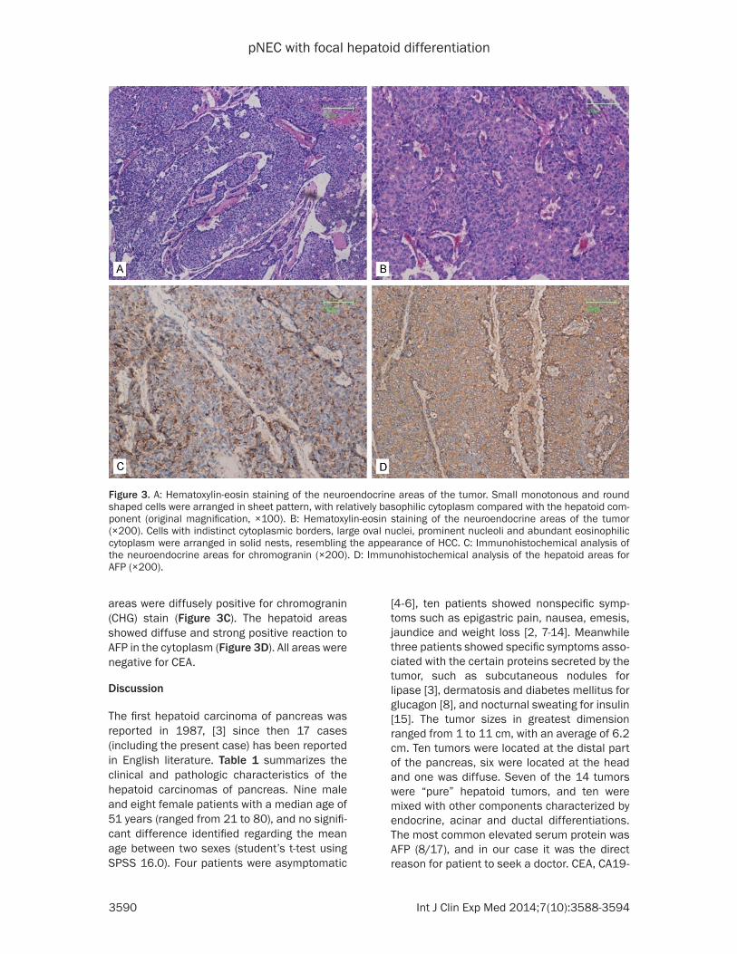
virus were negative. Computed tomography (CT) of the abdomen revealed a 1.6 × 2.2 cm low-attenuation mass in the head of the pan-creas, with no enlarged lymph nodes and no metastatic liver nodules (Figure 1). The patient underwent a pancreaticoduodenectomy and had an uncomplicated recovery. The primary pathologic diagnosis was undifferentiated car-cinoma of pancreas, with partly neuroendo-crine differentiation and partly hepatoid differ-entiation (revised pathologic diagnosis in the following paragraph). Her AFP level reduced to 232.3 ng/mL the day after the surgery, and about one month later, returned to normal range. Then the patient completed six cycles of chemotherapy (gemcitabine 1,000 mg/m2
intravenously on days 1, 8 and 15 of a 28-day cycle), and no severe complications were observed. A scheduled follow-up was main-tained, and the CT images and AFP levels were used to monitor the tumor status.
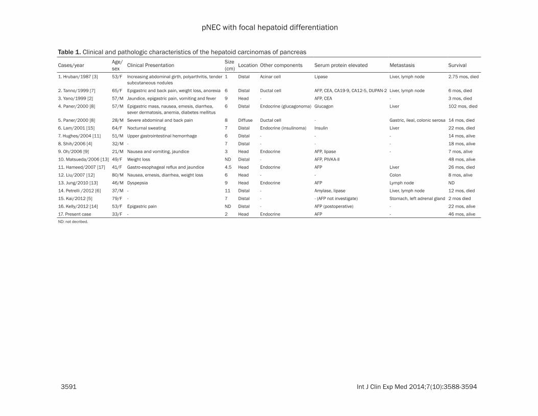
In July 2012, the patient’s AFP level went up to 93.1 ng/mL (CEA and CA19-9 being normal), meanwhile the CT scan showed no signs of local relapse or metastasis. Thus a positron emission tomography-computed tomography (PET-CT) was performed, which showed a hypermetabolic lymph node behind the body of the pancreas (Figure 2). The patient firstly received radiation therapy (45 Gy/8 fractions), and the AFP level reduced to normal (9.1 ng/mL) when the seventh fraction ended, then the patient began chemotherapy with the same regimen as before. Up till now she has finished two cycles of chemotherapy with the AFP level within normal range and has survived 46 months since the initial diagnosis. In December 2012, we were able to test her serum neuron specific enolase (NSE) and chromogranin A level, which was 0.2 ng/mL and 39.91 ng/mL respectively, both in normal range.
Pathological findings
In December 2012, during a multidisciplinary conference, this case was thoroughly reviewed and the slides were re-examined. Following the latest guidelines and consensuses, the revised pathologic diagnosis was neuroendocrine car-cinoma (G3) of the pancreas with focal hepa-toid differentiation.
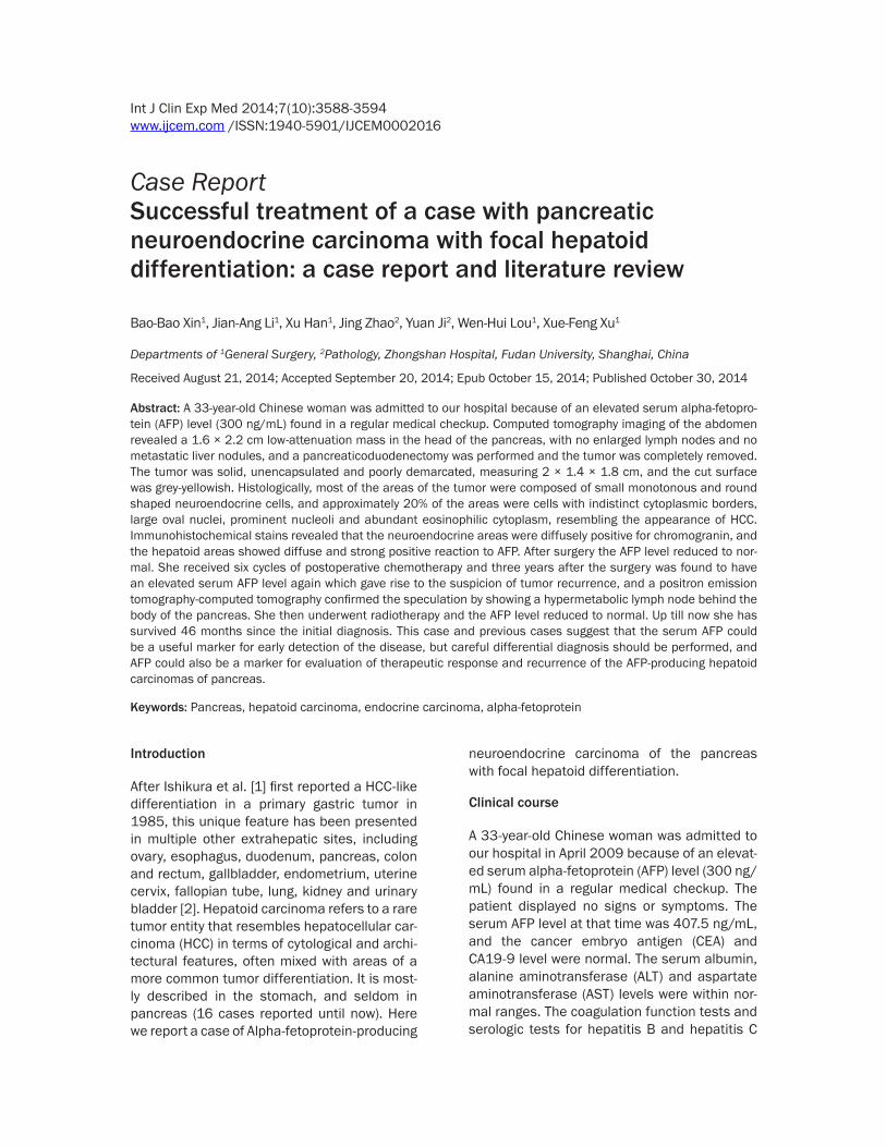
Macroscopic examination showed a solid tumor unencapsulated and poorly demarcated, mea-suring 2 × 1.4 × 1.8 cm, and the cut surface was grey-yellowish. Histologically, most of the areas of the tumor were composed of small monotonous and round shaped neuroendo-crine cells, and were rich in sinusoids. The cells were arranged in sheet pattern, with relatively basophilic cytoplasm compared with the hepa-toid component (Figure 3A). Approximately 20% of the areas of the tumor were cells arranged in solid nests with indistinct cytoplas-mic borders, large oval nuclei, prominent nucle-oli and abundant eosinophilic cytoplasm, resembling the appearance of HCC (Figure 3B). No bile production was identified.
Immunohistochemical stains were performed to confirm the speculation. The neuroendocrine
Figure 1. CT scan of the abdomen showing the low-attenuation mass within the head of the pancreas (arrow).
Figure 2. PET-CT showing a hypermetabolic lymph node behind the body of the pancreas (center of the crosshair).
pNEC with focal hepatoid differentiation
3590 Int J Clin Exp Med 2014;7(10):3588-3594
areas were diffusely positive for chromogranin (CHG) stain (Figure 3C). The hepatoid areas showed diffuse and strong positive reaction to AFP in the cytoplasm (Figure 3D). All areas were negative for CEA.
Discussion
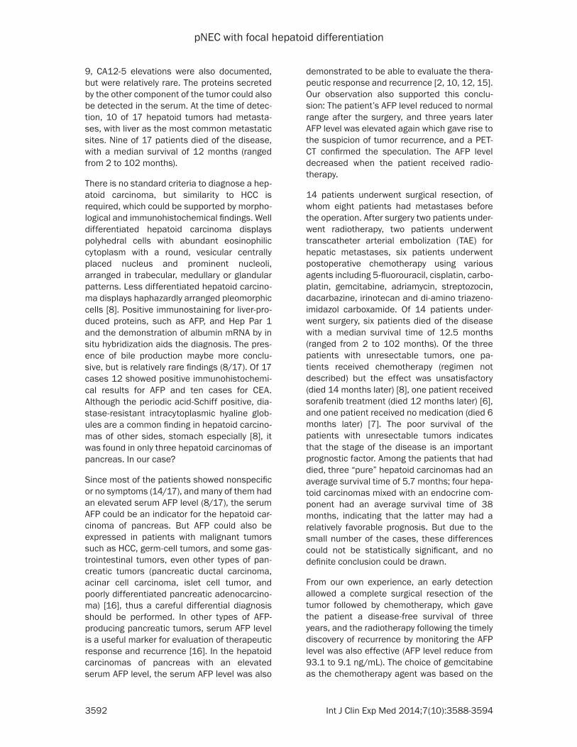
The first hepatoid carcinoma of pancreas was reported in 1987, [3] since then 17 cases (including the present case) has been reported in English literature. Table 1 summarizes the clinical and pathologic characteristics of the hepatoid carcinomas of pancreas. Nine male and eight female patients with a median age of 51 years (ranged from 21 to 80), and no signifi-cant difference identified regarding the mean age between two sexes (student’s t-test using SPSS 16.0). Four patients were asymptomatic
[4-6], ten patients showed nonspecific symp-toms such as epigastric pain, nausea, emesis, jaundice and weight loss [2, 7-14]. Meanwhile three patients showed specific symptoms asso-ciated with the certain proteins secreted by the tumor, such as subcutaneous nodules for lipase [3], dermatosis and diabetes mellitus for glucagon [8], and nocturnal sweating for insulin [15]. The tumor sizes in greatest dimension ranged from 1 to 11 cm, with an average of 6.2 cm. Ten tumors were located at the distal part of the pancreas, six were located at the head and one was diffuse. Seven of the 14 tumors were “pure” hepatoid tumors, and ten were mixed with other components characterized by endocrine, acinar and ductal differentiations. The most common elevated serum protein was AFP (8/17), and in our case it was the direct reason for patient to seek a doctor. CEA, CA19-
Figure 3. A: Hematoxylin-eosin staining of the neuroendocrine areas of the tumor. Small monotonous and round shaped cells were arranged in sheet pattern, with relatively basophilic cytoplasm compared with the hepatoid com-ponent (original magnification, ×100). B: Hematoxylin-eosin staining of the neuroendocrine areas of the tumor (×200). Cells with indistinct cytoplasmic borders, large oval nuclei, prominent nucleoli and abundant eosinophilic cytoplasm were arranged in solid nests, resembling the appearance of HCC. C: Immunohistochemical analysis of the neuroendocrine areas for chromogranin (×200). D: Immunohistochemical analysis of the hepatoid areas for AFP (×200).
pNEC with focal hepatoid differentiation
3591 Int J Clin Exp Med 2014;7(10):3588-3594
Table 1. Clinical and pathologic characteristics of the hepatoid carcinomas of pancreas
Cases/year Age/sex Clinical Presentation Size
(cm) Location Other components Serum protein elevated Metastasis Survival
1. Hruban/1987 [3] 53/F Increasing abdominal girth, polyarthritis, tender subcutaneous nodules
1 Distal Acinar cell Lipase Liver, lymph node 2.75 mos, died
2. Tanno/1999 [7] 65/F Epigastric and back pain, weight loss, anorexia 6 Distal Ductal cell AFP, CEA, CA19-9, CA12-5, DUPAN-2 Liver, lymph node 6 mos, died
3. Yano/1999 [2] 57/M Jaundice, epigastric pain, vomiting and fever 9 Head - AFP, CEA - 3 mos, died
4. Paner/2000 [8] 57/M Epigastric mass, nausea, emesis, diarrhea, sever dermatosis, anemia, diabetes mellitus
6 Distal Endocrine (glucagonoma) Glucagon Liver 102 mos, died
5. Paner/2000 [8] 28/M Severe abdominal and back pain 8 Diffuse Ductal cell - Gastric, ileal, colonic serosa 14 mos, died
6. Lam/2001 [15] 64/F Nocturnal sweating 7 Distal Endocrine (insulinoma) Insulin Liver 22 mos, died
7. Hughes/2004 [11] 51/M Upper gastrointestinal hemorrhage 6 Distal - - - 14 mos, alive
8. Shih/2006 [4] 32/M - 7 Distal - - - 18 mos, alive
9. Oh/2006 [9] 21/M Nausea and vomiting, jaundice 3 Head Endocrine AFP, lipase - 7 mos, alive
10. Matsueda/2006 [13] 49/F Weight loss ND Distal - AFP, PIVKA-II 48 mos, alive
11. Hameed/2007 [17] 41/F Gastro-esophageal reflux and jaundice 4.5 Head Endocrine AFP Liver 26 mos, died
12. Liu/2007 [12] 80/M Nausea, emesis, diarrhea, weight loss 6 Head - - Colon 8 mos, alive
13. Jung/2010 [13] 46/M Dyspepsia 9 Head Endocrine AFP Lymph node ND
14. Petrelli /2012 [6] 37/M - 11 Distal - Amylase, lipase Liver, lymph node 12 mos, died
15. Kai/2012 [5] 79/F - 7 Distal - - (AFP not investigate) Stomach, left adrenal gland 2 mos died
16. Kelly/2012 [14] 53/F Epigastric pain ND Distal - AFP (postoperative) - 22 mos, alive
17. Present case 33/F - 2 Head Endocrine AFP - 46 mos, aliveND: not decribed.
pNEC with focal hepatoid differentiation
3592 Int J Clin Exp Med 2014;7(10):3588-3594
9, CA12-5 elevations were also documented, but were relatively rare. The proteins secreted by the other component of the tumor could also be detected in the serum. At the time of detec-tion, 10 of 17 hepatoid tumors had metasta-ses, with liver as the most common metastatic sites. Nine of 17 patients died of the disease, with a median survival of 12 months (ranged from 2 to 102 months).
There is no standard criteria to diagnose a hep-atoid carcinoma, but similarity to HCC is required, which could be supported by morpho-logical and immunohistochemical findings. Well differentiated hepatoid carcinoma displays polyhedral cells with abundant eosinophilic cytoplasm with a round, vesicular centrally placed nucleus and prominent nucleoli, arranged in trabecular, medullary or glandular patterns. Less differentiated hepatoid carcino-ma displays haphazardly arranged pleomorphic cells [8]. Positive immunostaining for liver-pro-duced proteins, such as AFP, and Hep Par 1 and the demonstration of albumin mRNA by in situ hybridization aids the diagnosis. The pres-ence of bile production maybe more conclu-sive, but is relatively rare findings (8/17). Of 17 cases 12 showed positive immunohistochemi-cal results for AFP and ten cases for CEA. Although the periodic acid-Schiff positive, dia-stase-resistant intracytoplasmic hyaline glob-ules are a common finding in hepatoid carcino-mas of other sides, stomach especially [8], it was found in only three hepatoid carcinomas of pancreas. In our case?
Since most of the patients showed nonspecific or no symptoms (14/17), and many of them had an elevated serum AFP level (8/17), the serum AFP could be an indicator for the hepatoid car-cinoma of pancreas. But AFP could also be expressed in patients with malignant tumors such as HCC, germ-cell tumors, and some gas-trointestinal tumors, even other types of pan-creatic tumors (pancreatic ductal carcinoma, acinar cell carcinoma, islet cell tumor, and poorly differentiated pancreatic adenocarcino-ma) [16], thus a careful differential diagnosis should be performed. In other types of AFP-producing pancreatic tumors, serum AFP level is a useful marker for evaluation of therapeutic response and recurrence [16]. In the hepatoid carcinomas of pancreas with an elevated serum AFP level, the serum AFP level was also
demonstrated to be able to evaluate the thera-peutic response and recurrence [2, 10, 12, 15]. Our observation also supported this conclu-sion: The patient’s AFP level reduced to normal range after the surgery, and three years later AFP level was elevated again which gave rise to the suspicion of tumor recurrence, and a PET-CT confirmed the speculation. The AFP level decreased when the patient received radio- therapy.
14 patients underwent surgical resection, of whom eight patients had metastases before the operation. After surgery two patients under-went radiotherapy, two patients underwent transcatheter arterial embolization (TAE) for hepatic metastases, six patients underwent postoperative chemotherapy using various agents including 5-fluorouracil, cisplatin, carbo-platin, gemcitabine, adriamycin, streptozocin, dacarbazine, irinotecan and di-amino triazeno-imidazol carboxamide. Of 14 patients under-went surgery, six patients died of the disease with a median survival time of 12.5 months (ranged from 2 to 102 months). Of the three patients with unresectable tumors, one pa- tients received chemotherapy (regimen not described) but the effect was unsatisfactory (died 14 months later) [8], one patient received sorafenib treatment (died 12 months later) [6], and one patient received no medication (died 6 months later) [7]. The poor survival of the patients with unresectable tumors indicates that the stage of the disease is an important prognostic factor. Among the patients that had died, three “pure” hepatoid carcinomas had an average survival time of 5.7 months; four hepa-toid carcinomas mixed with an endocrine com-ponent had an average survival time of 38 months, indicating that the latter may had a relatively favorable prognosis. But due to the small number of the cases, these differences could not be statistically significant, and no definite conclusion could be drawn.
From our own experience, an early detection allowed a complete surgical resection of the tumor followed by chemotherapy, which gave the patient a disease-free survival of three years, and the radiotherapy following the timely discovery of recurrence by monitoring the AFP level was also effective (AFP level reduce from 93.1 to 9.1 ng/mL). The choice of gemcitabine as the chemotherapy agent was based on the
pNEC with focal hepatoid differentiation
3593 Int J Clin Exp Med 2014;7(10):3588-3594
primary pathologic diagnosis of undifferentiat-ed carcinoma of pancreas, with partly neuroen-docrine differentiation and partly hepatoid dif-ferentiation. The diagnosis was made in 2009 when our understanding of neuroendocrine tumor of pancreas was limited. But since there was no agreed agent and the result was accept-able, we decided to continue to use the present regimen.
In summary, we have presented a case of AFP-producing neuroendocrine carcinoma of the pancreas with focal hepatoid differentiation, which was discovered solely because of an ele-vated serum AFP level. A review of literature shows that patients with unresectable hepa-toid tumors of pancreas tend to have poor sur-vival, and the serum AFP could be a useful marker for early detection of the disease, but careful differential diagnosis should be per-formed. It could also be a marker for evaluation of therapeutic response and recurrence of the AFP-producing hepatoid carcinomas of pancr- eas.
Acknowledgements
We are grateful to the patient who kindly gave permission for her case to be reported. We thank all doctors and nurses during the diagno-sis, treatment, and nursing of the patient, espe-cially Dayong Jin, MD, Dansong Wang, MD, Tiantao Kuang, MD, Xiaoling Ni, Wenchuan Wu, MD, and Yefei Rong, MD.
Disclosure of conflict of interest
None.
Address correspondence to: Dr. Xue-Feng Xu, De- partment of General Surgery, Zhongshan Hospital, Fudan University, 180 Fenglin Road, Shanghai 200032, People’s Republic of China. Tel: (+86) 13061626568; E-mail: [email protected]
References
[1] Ishikura H, Fukasawa Y, Ogasawara K, Natori T, Tsukada Y, Aizawa M. An AFP-producing gastric carcinoma with features of hepatic differentia-tion. A case report. Cancer 1985; 56: 840-8.
[2] Yano T, Ishikura H, Wada T, Kishimoto T, Kondo S, Katoh H, Yoshiki T. Hepatoid adenocarcino-ma of the pancreas. Histopathology 1999; 35: 90-2.
[3] Hruban RH, Molina JM, Reddy MN, Boitnott JK. A neoplasm with pancreatic and hepatocellu-lar differentiation presenting with subcutane-ous fat necrosis. Am J Clin Pathol 1987; 88: 639-45.
[4] Shih NN, Tsung JS, Yang AH, Tsou MH, Cheng TY. A unique pancreatic tumor with exclusive hepatocytic differentiation. Ann Clin Lab Sci 2006; 36: 216-21.
[5] Kai K, Nakamura J, Ide T, Masuda M, Kitahara K, Miyoshi A, Noshiro H, Tokunaga O. Hepatoid carcinoma of the pancreas penetrating into the gastric cavity: A case report and literature review. Pathol Int 2012; 62: 485-90.
[6] Petrelli F, Ghilardi M, Colombo S, Stringhi E, Barbara C, Cabiddu M, Elia S, Corti D, Barni S. A rare case of metastatic pancreatic hepatoid carcinoma treated with sorafenib. J Gastroin-test Cancer 2012; 43: 97-102.
[7] Tanno S, Obara T, Fujii T, Izawa T, Mizukami Y, Saitoh Y, Ura H, Kohgo Y. α-Fetoprotein-producing adenocarcinoma of the pancreas presenting focal hepatoid differentiation. Int J Pancreatol 1999; 26: 43-7.
[8] Paner GP, Thompson KS, Reyes CV. Hepatoid carcinoma of the pancreas. Cancer 2000; 88: 1582-9.
[9] Oh HJ, Cheung DY, Kim TH, Kim SS, Kim MS, Kim JI, Park SH, Han JY, Han NI, Kim JK, Lee YS, Kim EK, Jung ES. A case of hepatoid carci-noma of the pancreas. Korean J Gastroenterol 2006; 47: 389-93.
[10] Matsueda K, Yamamoto H, Yoshida Y, Notoha-ra K. Hepatoid carcinoma of the pancreas pro-ducing protein induced by vitamin K absence or antagonist II (PIVKA-II) and alpha-fetoprotein (AFP). J Gastroenterol 2006; 41: 1011-9.
[11] Hughes K, Kelty S, Martin R. Hepatoid carci-noma of the pancreas. American Surg 2004; 70: 1030-3.
[12] Liu CZ, Hu SY, Wang L, Zhi XT, Jin B, Zhu M, Wachtel MS, Frezza EE. Hepatoid carcinoma of the pancreas: a case report. Chin Med J (Engl) 2007; 120: 1850-2.
[13] Jung JY, Kim YJ, Kim HM, Kim HJ, Park SW, Song SY, Chung JB, Kang CM, Pyo JY, Yang WI, Bang S. Hepatoid carcinoma of the pancreas combined with neuroendocrine carcinoma. Gut liver 2010; 4: 98-102.
[14] Kelly PJ, Spence R, Dasari BV, Burt AD, Taylor M, Loughrey MB. Primary hepatocellular carci-noma of the pancreas: a case report and re-view of the heterogeneous group of pancreatic hepatoid carcinomas. Histopathology 2012; 60: 1012-5.
[15] Lam K, Lo C, Wat M, Fan ST. Malignant insuli-noma with hepatoid differentiation: a unique case with alpha-fetoprotein production. Endocr Pathol 2001; 12: 351-4.
pNEC with focal hepatoid differentiation
3594 Int J Clin Exp Med 2014;7(10):3588-3594
[17] Hameed O, Xu H, Saddeghi S, Maluf H. Hepa-toid carcinoma of the pancreas: a case report and literature review of a heterogeneous group of tumors. Am J Surg Pathol 2007; 31: 146-52.
[16] Kawamoto S, Hiraoka T, Kanemitsu K, Kimura M, Miyauchi Y, Takeya M. Alpha-fetoprotein-producing pancreatic cancer--a case report and review of 28 cases. Hepatogastroenterol-ogy 1992; 39: 282-6.