case studies in movement disorders - alneurology.com
TRANSCRIPT

8/21/2019
1
Case Studies in Movement Disorders
Marissa N. Dean, MDAssistant Professor of Neurology
Victor W. Sung, MDAssociate Professor of Neurology
Disclosures
• Marissa Dean has nothing to disclose.
• Victor Sung has nothing to disclose.
Objectives
• Discuss phenomenology of movement disorders through case presentations.
• Provide a differential diagnosis for movement disorder cases.
8/21/2019
2
Acknowledgments
• Patients and families for allowing the sharing of videos
• Juliana Coleman, MD for creating videos
Case 1:Wiggly toes
Case 1
• 53 yo RH woman with toe movements
• 3 years ago
• Restlessness in bilateral feet
• Began after starting trazodone
• Involuntary movement of toes
• Burning, tingling, and numbness in toes
• 2 years ago
• Stopped trazodone – symptoms worsened
• Worst at night, but also present during day
• Can feel toes moving in shoes

8/21/2019
3
Case 1 – additional history
• Meds tried
• Ropinirole 0.5 mg BID ‐> minimal change in movements, no help with pain
• Medical history
• Hypertension, hypothyroidism
• Family history
• No known neurological disease or movement disorders.
• Social history
• Smokes tobacco ½ ppd x 20 yrs
Case 1 ‐ exam
• Reflexes
• Grade 1 at knees, absent at ankles
• Sensation
• Normal pinprick
• Decreased vibration up to medial malleolus bilaterally
• Decreased proprioception at toes bilaterally
• Muscle tone
• Normal
Case 1 Video

8/21/2019
4
Athetosis vs Pseudoathetosis
• Area of debate among MD specialists
• In theory, ‘athetosis’ is from BG lesions (almost solely associated with CP), and ‘pseudoathetosis’ is from proprioception loss
• However, ’athetosis’ in CP = ‘chorea’ and ‘dystonia’
• Pseudoathetosis
• Distal limbs
• Writhing, slow, continuous involuntary movements
• Patterned
Fahn et al, Principles and Practice of Movement Disorders, 2011; Abdo et al, Nature Reviews 2010
Work‐up
• EMG/NCS BLE
• BLE sensory polyneuropathy
Painful legs moving toes (PLMT)
• Uncommon disorder
• Mainly described in case reports and case series
• Pain in one or more limbs
• Repetitive, non‐rhythmic movement of toes
• Also,
• Painful arms and moving fingers
• Painful mouth and moving tongue ‐ rare
• Painless variants ‐ rare
Hassan et al, Neurology 2012
8/21/2019
5
PLMT
• Mean age at onset in 50s
• Mean age at presentation in 60s
• Possible women predominance
• Most commonly in legs
• Unilateral or bilateral
• Etiology
• Mechanism unclear – most suspect some central involvement (central processing of peripheral nerve dysfunction)
• Peripheral neuropathy, limb trauma, radiculopathy, cryptogenic
Hassan et al, Neurology 2012; Tocco et al, MDCP 2014
PLMT ‐ treatment
• Pain is most bothersome symptom
• Difficult to treat – no evidence‐based guidelines
• Poor response to medications
• Gabapentin, pregabalin, clonazepam, dopamine agonists
• Botulinum toxin injections
• Spinal cord stimulator
Hassan et al, Neurology 2012; Tocco et al, MDCP 2014; Takahashi et al, Pain 2002
Case 1 ‐ take home points
• Pseudoathetosis
• Distal limbs
• Writhing, slow, continuous involuntary movements
• Patterned
• PLMT
• Usually with peripheral etiology
• Pain has poor response to treatments

8/21/2019
6
Case 2:Arm spasms
Case 2
• 62 yo RH woman
• 7 years ago – tremor/spasm in left arm
• Worsened over time
• Now, can’t type at work
• Left hand found in weird positions
• Symptoms worst in AM
• Cannot tie shoes, play piano, button clothes
• Stumbling more, left foot tends to drag/catch on objects
Case 2
• Speech
• More stuttering, hesitations
• Memory
• Worse with names, name recall
• Leaves stove on and forgets – no longer using stove
• Confusion in un‐familiar environments, but quickly figures out where she is
• Independent in basic ADLs

8/21/2019
7
Case 2 – more history
• Prior treatments
• Valproic acid 1000 mg daily – no change
• Cervical spine surgery for cervical stenosis – no change
• PT – some improvement
• Medical history
• C5‐C6 spine surgery for cervical stenosis, asthma
• Family history
• Dementia in maternal grandmother.
Case 2 ‐ exam
• Muscle tone – normal
• Reflexes – normal
• Sensation – normal pinprick, vibration, proprioception
• Left arm extinction
• Agraphesthesia bilaterally
Case 2 Video
8/21/2019
8
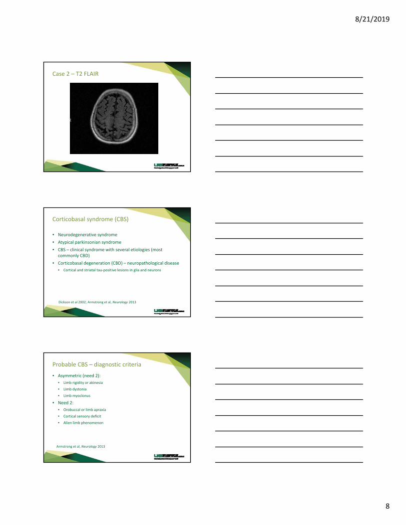
Case 2 – T2 FLAIR
Corticobasal syndrome (CBS)
• Neurodegenerative syndrome
• Atypical parkinsonian syndrome
• CBS – clinical syndrome with several etiologies (most commonly CBD)
• Corticobasal degeneration (CBD) – neuropathological disease
• Cortical and striatal tau‐positive lesions in glia and neurons
Dickson et al 2002; Armstrong et al, Neurology 2013
Probable CBS – diagnostic criteria
• Asymmetric (need 2):
• Limb rigidity or akinesia
• Limb dystonia
• Limb myoclonus
• Need 2:
• Orobuccal or limb apraxia
• Cortical sensory deficit
• Alien limb phenomenon
Armstrong et al, Neurology 2013
8/21/2019
9
Probable CBD – diagnostic criteria
• Insidious onset with gradual progression
• Symptoms 1 yr
• Age 50 yo
• No family history of similar symptoms (2 or more)
• Probable CBS
• **Dementia frequently seen, but not included in diagnostic criteria.**
Armstrong et al, Neurology 2013
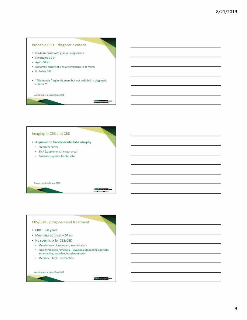
Imaging in CBS and CBD
• Asymmetric frontoparietal lobe atrophy
• Premotor cortex
• SMA (supplemental motor area)
• Posterior superior frontal lobe
Boxer et al, Arch Neurol 2006
CBS/CBD ‐ prognosis and treatment
• CBD – 6‐8 years
• Mean age at onset – 64 yo
• No specific tx for CBS/CBD
• Myoclonus – clonazepam, levetiracetam
• Rigidity/akinesia/dystonia – levodopa, dopamine agonists, amantadine, baclofen, botulinum toxin
• Memory – AchEI, memantine
Armstrong et al, Neurology 2013

8/21/2019
10
Case 2 ‐ take home points
• CBS
• Atypical parkinsonism with more rapid progression
• Prognosis 6‐8 years
• Asymmetric rigidity/akinesias, dystonia, and/or myoclonus + cortical sensory loss/alien limb
Case 3:Imbalance
Case 3
• 50 yo man – gait difficulty
• 4 months ago – change in personality
• Loud and talkative ‐> quiet in conversation
• Lack of insight into this change
• 3 months ago – balance problems began
• Stumbling more
• Double vision
• Short term memory problems – forgetting prior events in the day (ex: brushing teeth)

8/21/2019
11
Case 3
• 2 months ago – change in speech
• Fragmented, only speaking in short phrases
• Awkward while holding objects
• Clumsy
• Using cane for ambulation
• Within last month – mute and using a walker
• Significant difficulty with all motor tasks
• Family reports fluctuating symptoms from day‐to‐day
Case 3 – more history
• Family history
• Father died at 50 yo – alcohol abuse, liver cirrhosis, unable to walk prior to death
• Medical history
• Appendectomy
• Meds – none
• Social history
• High school graduate
• Father abusive to mother, divorced, and left him when he was 6 yo
• Used to drink ETOH heavily; none in >15 years
Case 3 ‐ exam
• Non‐verbal
• Appears to follow some commands on the right, mostly consistent
• Muscle tone – severe rigidity in neck and extremities (R>L)

8/21/2019
12
Case 3 ‐ Video
Case 3 – workup
• Labs
• Thiamine – undetectable
• CSF
• Glucose – 76 (116 serum)
• Protein – 44
• WBC – 1
• RBC ‐ 10
• EEG – mild slowing
• Imaging
Case 3 ‐ DWI
8/21/2019
13
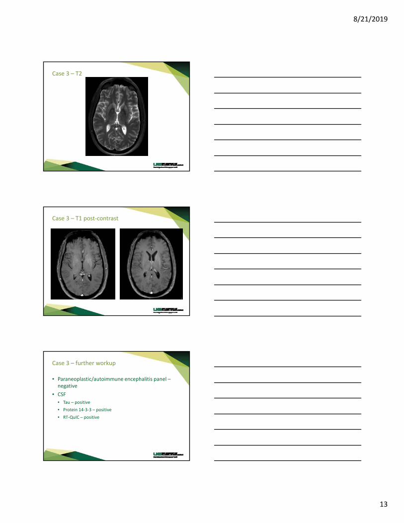
Case 3 – T2
Case 3 – T1 post‐contrast
Case 3 – further workup
• Paraneoplastic/autoimmune encephalitis panel –negative
• CSF
• Tau – positive
• Protein 14‐3‐3 – positive
• RT‐QuIC – positive
8/21/2019
14
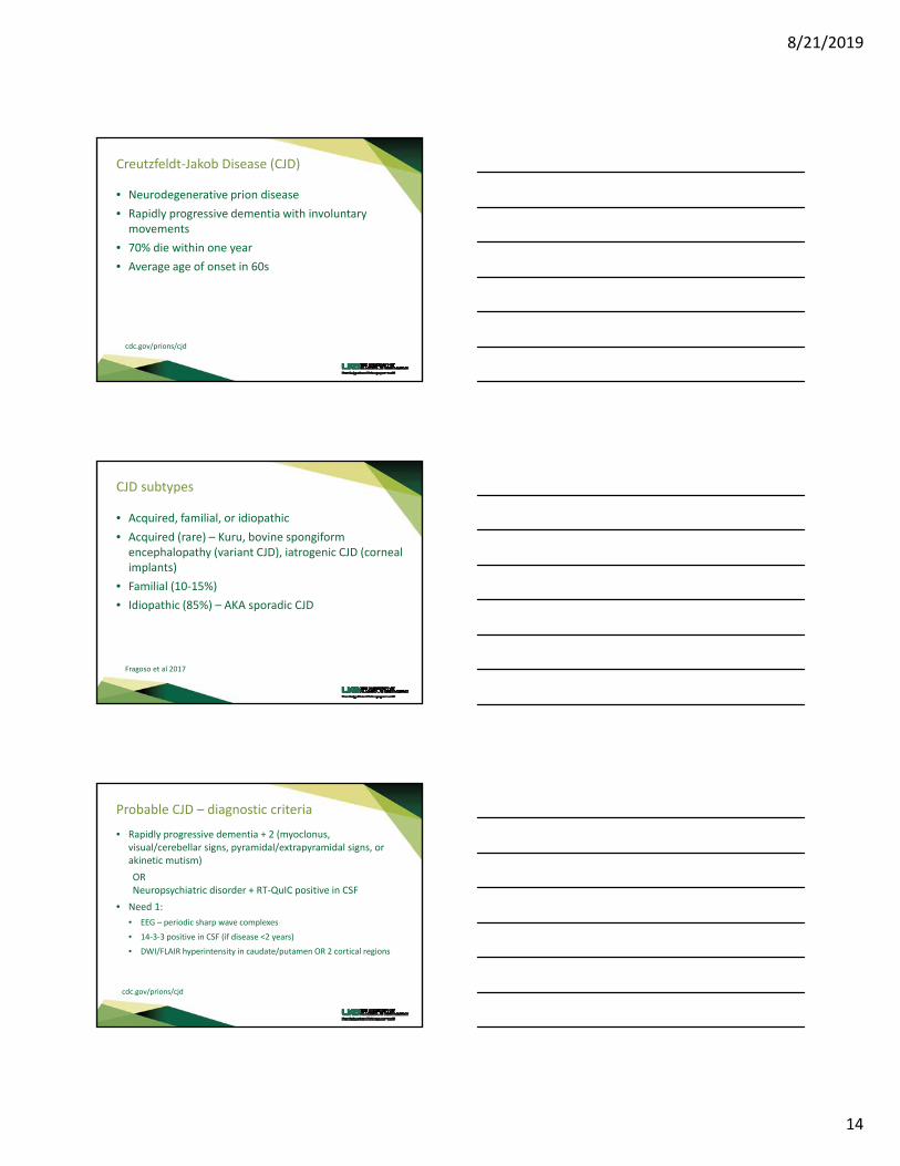
Creutzfeldt‐Jakob Disease (CJD)
• Neurodegenerative prion disease
• Rapidly progressive dementia with involuntary movements
• 70% die within one year
• Average age of onset in 60s
cdc.gov/prions/cjd
CJD subtypes
• Acquired, familial, or idiopathic
• Acquired (rare) – Kuru, bovine spongiform encephalopathy (variant CJD), iatrogenic CJD (corneal implants)
• Familial (10‐15%)
• Idiopathic (85%) – AKA sporadic CJD
Fragoso et al 2017
Probable CJD – diagnostic criteria
• Rapidly progressive dementia + 2 (myoclonus, visual/cerebellar signs, pyramidal/extrapyramidal signs, or akinetic mutism)
ORNeuropsychiatric disorder + RT‐QuIC positive in CSF
• Need 1:
• EEG – periodic sharp wave complexes
• 14‐3‐3 positive in CSF (if disease <2 years)
• DWI/FLAIR hyperintensity in caudate/putamen OR 2 cortical regions
cdc.gov/prions/cjd

8/21/2019
15
Imaging in CJD
• Classic sCJD
• MR – BG + cingulate/frontal/parietal lobes (cortical ribboning)
• Atypical sCJD (less frequent)
• MR – BG + thalamus
• Familial CJD (PRNP gene mutations)
• MR – similar to sCJD
• vCJD
• MR – Pulvinar and hockey stick sign
Fragoso et al 2017
Our patient – probable CJD
• Rapidly progressive dementia + 2 (myoclonus, visual/cerebellar signs, pyramidal/extrapyramidal signs, or akinetic mutism)
ORNeuropsychiatric disorder + RT‐QuIC positive in CSF
• Need 1:
• EEG – periodic sharp wave complexes
• 14‐3‐3 positive in CSF (if disease <2 years)
• DWI/FLAIR hyperintensity in caudate/putamen OR 2 cortical regions
Our patient – imaging
• Classic sCJD
• MR – BG + cingulate/frontal/parietal lobes (cortical ribboning)
• Atypical sCJD (less frequent)
• MR – BG + thalamus
• Familial CJD (PRNP gene mutations) – possible
• MR – similar to sCJD
• vCJD
• MR – Pulvinar and hockey stick sign

8/21/2019
16
Take home points
• CJD – rapidly progressive dementia within <12 mo
• Sporadic CJD most common
• 2 imaging patterns
• CSF – RT‐QuIC and 14‐3‐3
• Recognition of phenomenology can assist in diagnosis (akinetic mutism + myoclonus)
Case 4:Left knee pain from fall
Case 4
• 56 yo RH woman with frequent falls
• 3 months ago – had fall and fell onto left knee
• Went to ED for severe pain
• SBP>180 and HgbA1c=12.6 (BG 321)
• CT head done – interpreted as normal
• Diagnosed with HTN and DMII
• Discharged home

8/21/2019
17
Case 4
• 2.5 weeks later, presented to ED with involuntary movements x1 week
• Left leg and arm involuntary movements that began 2 days after a steroid injection into left knee
• Progressed in severity over past week
• Continuous movements – no alleviating factors
Case 4 – Video
Chorea vs ballism
• Chorea – random, non‐rhythmic, abrupt, rapid, un‐sustained movements that flow from one body part to another
• Ballism – Chorea that affects a proximal joint (such as hip or shoulder) and produces large amplitude movements

8/21/2019
18
Case 4 – CTH first ED visit
Case 4 – T1
Case 4 – T2 FLAIR

8/21/2019
19
Case 4 – follow up
• Labs during admission
• BG – 97
• Heavy metal screen, autoimmune encephalopathy panel, UDS – negative
• Vit b12, copper, ceruloplasmin – wnl
• Diagnosis?
Nonketotic hyperglycemia‐induced hemichorea/hemiballism
• Seen in uncontrolled DM or as initial symptom of DM
• Slightly more common in women
• Mean age at onset 70 yo
• Age is greatest risk factor
• Average BG 300 at symptom onset
• Imaging – striatum hyperdense (CT) and hyperintense (MRI T1); T2 findings vary
Cosentino et al 2016
Nonketotic hyperglycemia‐induced hemichorea/hemiballism
• Prognosis – usually good
• Most will have complete resolution of symptoms over time
• Treatments
• dopamine antagonists (risperidone, olanzapine, haloperidol, etc.)
• Clonazepam
• Tetrabenazine, deutetrabenazine
Cosentino et al 2016

8/21/2019
20
Take home points
• Hemichorea/hemiballism may be first presentation of DM
• Striatum – hyperdense on CT and hyperintense on MRI T1
• Movements usually improve with normalizing BG, but some may require treatment
Case 5a and 5b
• 2 videos of parkinsonism
• Idiopathic PD
• Functional parkinsonism (FMD)
Case 5a and 5b Videos

8/21/2019
21
Functional parkinsonism
• Marked slowness with examined tasks, but normal casual tasks
• No cogwheel rigidity
• Pincer function preserved and lack of decrement
• ‘huffing and puffing’ sign
• Tremor – non‐rhythmic, varying frequencies, same severity with action and at rest, entrainment, distractible
LaFaver and Espay, Semin Neurol 2017
Thank you!