case study: satellite tobacco mosaic virus study: satellite tobacco mosaic virus ... in this case...
TRANSCRIPT
Case Study: Satellite Tobacco Mosaic Virus
Boon Chong Goh and Yaroslav Daniel Bodnar
A colored version of this Case Study is available athttp://www.ks.uiuc.edu/Training/CaseStudies/pdfs/stmv.pdf.
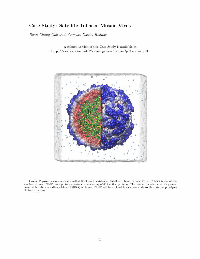
Cover Figure: Viruses are the smallest life form in existence. Satellite Tobacco Mosaic Virus (STMV) is one of thesimplest viruses. STMV has a protective outer coat consisting of 60 identical proteins. The coat surrounds the virus’s geneticmaterial, in this case a ribonucleic acid (RNA) molecule. STMV will be explored in this case study to illustrate the principlesof virus structure.
1
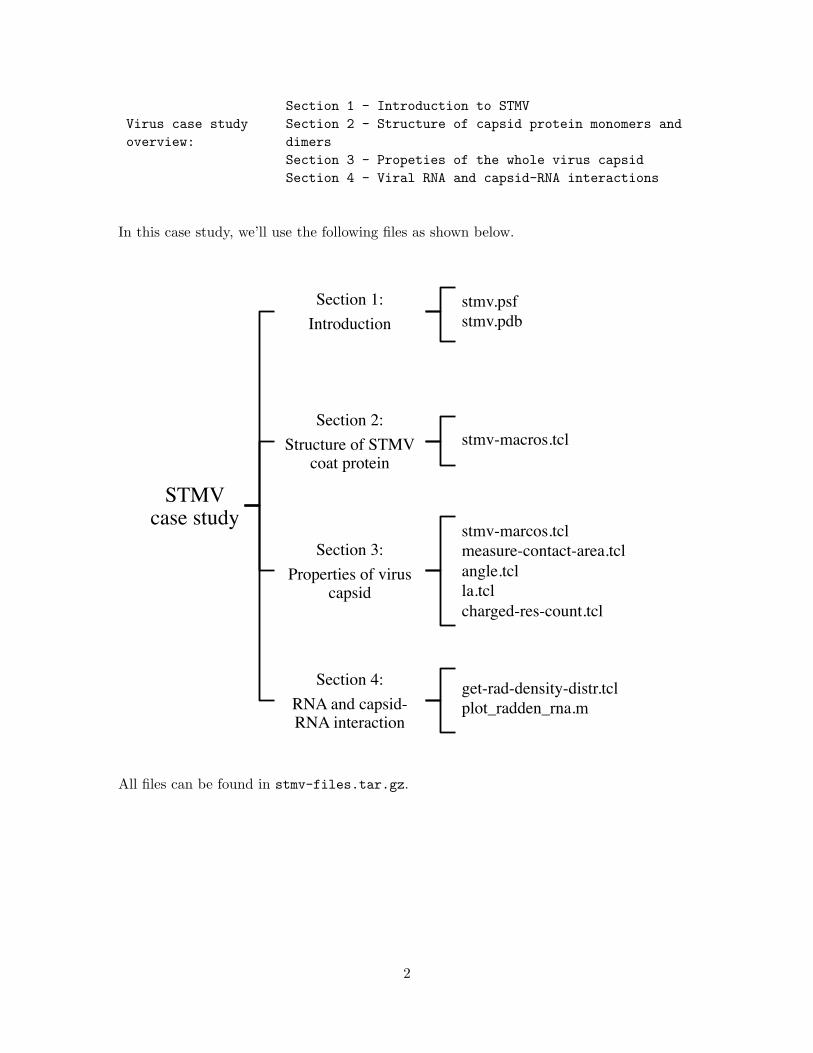
Section 1 - Introduction to STMVVirus case studyoverview:
Section 2 - Structure of capsid protein monomers anddimersSection 3 - Propeties of the whole virus capsidSection 4 - Viral RNA and capsid-RNA interactions
In this case study, we’ll use the following files as shown below.
STMV case study
Section 1: Introduction
Section 2: Structure of STMV
coat protein
Section 3: Properties of virus
capsid
Section 4: RNA and capsid-RNA interaction
stmv.psf stmv.pdb
stmv-macros.tcl
stmv-marcos.tcl measure-contact-area.tcl angle.tcl la.tcl charged-res-count.tcl
get-rad-density-distr.tcl plot_radden_rna.m
All files can be found in stmv-files.tar.gz.
2

1 Introduction
A virus is an infectious agent that can replicate only inside a living cell. Viruses were first discov-ered by Martinus Beijerinck, a Dutch microbiologist, in 1898. Beijerinck filtered the sap of diseasedplants through porcelain, a microporous material known to block the passage of cellular organismssuch as bacteria. Through a series of filtration experiments, Beijerinck showed that tobacco mosaicdisease is caused by an infectious agent smaller than any known bacterium. Beijerinck describedthe infectious agent, which was too small to be seen using a light microscope, as a “contagium vivimfluidum” (contagious living fluid) [1]. It was not until development of the electron microscope in the1930s that the morphology of virus particles was first observed [2]. The successful crystallizationof tobacco mosaic virus (TMV) by Wendell Stanley in 1935, for which he earned the 1946 NobelPrize in Chemistry, eventually lead to visualization of the virus structure at atomic resolution byX-ray crystallography[3].
Figure 1: (A) An electron micrograph showing a long tubular Tobacco Mosaic Virus (TMV) surrounded bymany small, spherical Satellite tobacco mosaic virus (STMV) particles. Bar marker represents 50nm. (B)Photograph of a healthy and TMV-infected tobacco plant.
Satellite tobacco mosaic virus (STMV) has a spherical shape and, with a diameter of approx-imately 17 nm, is much smaller than TMV. STMV was initially discovered on tobacco trees insouthern California in association with the rod-shaped TMV. In Fig.1, panel A shows a rod-shapedTMV particle, which has a length of approximately 300 nm, surrounded by spherical STMV par-ticles[4]. The infected leaf in panel B forms a mottled pattern of light and dark green areas in theleaf region, better known as the “mosaic” pattern [5]. STMV is a satellite virus since it is entirelyreliant on co-infection of a cell with TMV to replicate. Like all non-enveloped viruses, which arenot surrounded by a lipid membrane, STMV is composed of a closed protein shell, or the viralcapsid, and genetic material (single-stranded RNA in the case of STMV).
Due to its small size, STMV could be studied early on computationally by molecular dynamicssimulation [6]. In this case study, we will use STMV as a model system to explore the propertiesof small RNA viruses, including the structure and key interactions of capsid proteins, the virus
3

capsid, the RNA genome, and capsid-RNA complexes.
Exercise 1: General structural features of STMV
In this exercise you will use VMD to explore the structural properties of STMV described in thissection. Load the provided coordinate files stmv.psf and stmv.pdb into VMD.
A. Create a representation for the protein component of STMV. You can do so by opening theGraphical Representations window via Graphics → Representations in the VMD Main window,and creating a new representation with protein as the Selected Atoms. Try using Quick Surffor the Drawing Method and Segname for the Coloring Method. You should be able to see the 60proteins that make up the virus capsid.
Create a new representation with nucleic as the Selected Atoms. Use Tube for the DrawingMethod, ColorID for the Coloring method and color the RNA in green. Hide the protein to viewthe RNA that is encapsulated by the protein capsid.
B. Reproduce the cover figure as best as you can, and save an image of your view via File →Render... on the VMD main menu.*Hint: To color the surface of the virus capsid by the radial distance from the center, use Radialvia Coloring Method → Position in the VMD Graphical Representations window.
C. The complete nucleotide sequence of STMV RNA deduced by experiments is approximately 1059nucleotides in length [7]. Estimate the length of RNA needed to encode all of the capsid proteins.Based on your estimate, argue that the STMV capsid must be composed of identical proteins.Hint: Three nucleotides encode an amino acid. Note that not all nucleotides are necessarily trans-lated into amino acids. Please refer to [7] for more information.
4
2 Structure of STMV Coat Protein Monomer and Dimer
2.1 The coat protein is the building block of the STMV capsid
As is demonstrated in Exercise 1.D, genetic efficiency is a requirement of viruses, since the viralgenome must be able to encode all of the coat proteins that comprise the capsid. This observation,along with the self-assembly properties of viruses, led Watson and Crick to hypothesize in 1956that viruses must be highly symmetric assemblies of a structural subunit [8], which has since beenconfirmed by experimental studies. In the case of STMV, for which one of the highest resolutioncrystallographic structures of a virus has been solved, the capsid is composed of 60 identical copiesof the coat protein (CP), which is shown in Fig. 2. As with most other virus capsid proteins,the STMV coat protein has a distinctive structure known as the jellyroll fold, which has a uniquearrangement of eight β-strands that form two anti-parallel sheets, known as the BIDG and CHEFsheets [9]. The CPs are arranged in STMV such that the CHEF sheet is on the outer surface of thecapsid and the BIDG sheet is on the inner surface. The eight β-strands that make up the structuralcore of the CP are highlighted in Fig. 2. The structure of the jellyroll fold has evolved to enablevirus capsid proteins to efficiently self-assemble into closed shells.
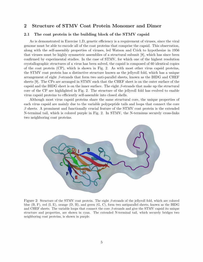
Although most virus capsid proteins share the same structural core, the unique properties ofeach virus capsid are mainly due to the variable polypeptide tails and loops that connect the coreβ sheets. A prominent and functionally crucial feature of the STMV coat protein is the extendedN-terminal tail, which is colored purple in Fig. 2. In STMV, the N-terminus securely cross-linkstwo neighboring coat proteins.
Figure 2: Structure of the STMV coat protein. The eight β-strands of the jellyroll fold, which are coloredblue (B, F), red (I, E), orange (D, H), and green (G, C), form two antiparallel sheets, known as the BIDGand CHEF sheets. The variable loops that connect the core β-strands and give the STMV capsid its uniquestructure and properties, are shown in cyan. The extended N-terminal tail, which securely bridges twoneighboring coat proteins, is shown in purple.
5
2.2 Coat protein dimers are important capsid assembly intermediates
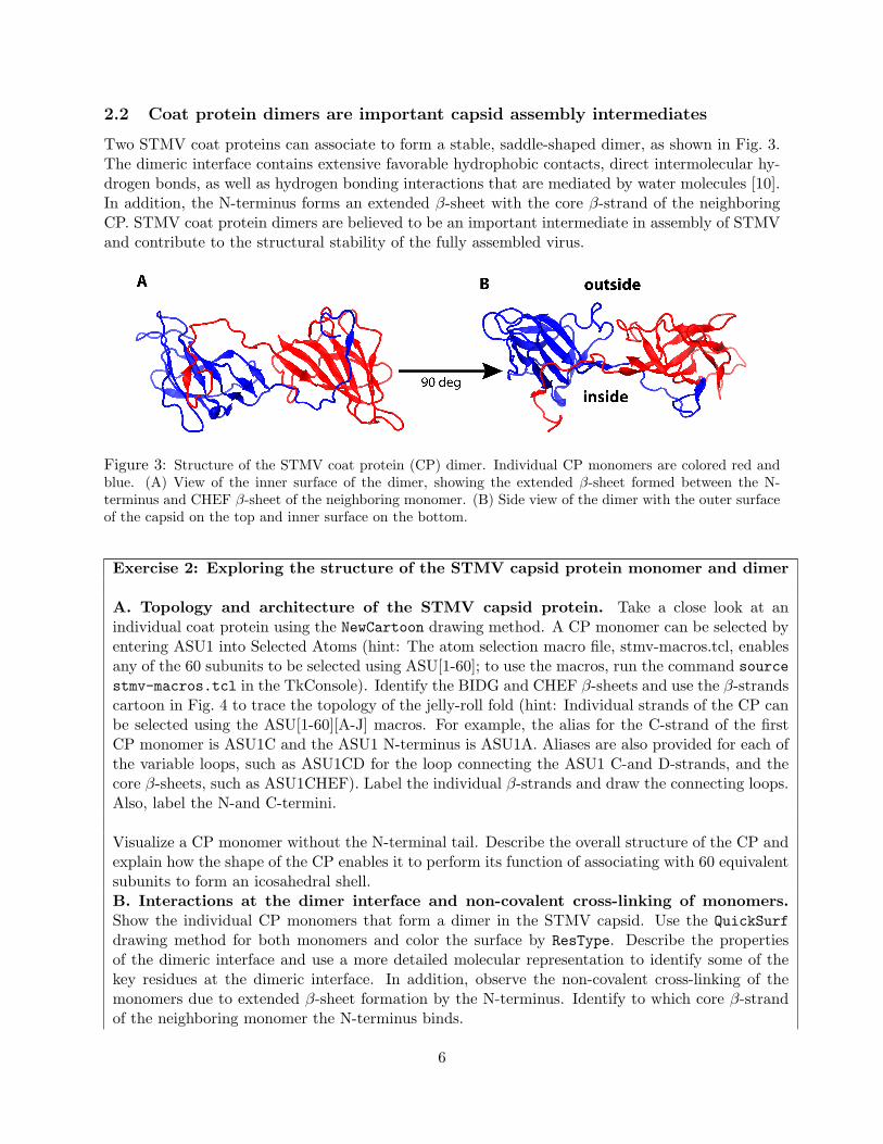
Two STMV coat proteins can associate to form a stable, saddle-shaped dimer, as shown in Fig. 3.The dimeric interface contains extensive favorable hydrophobic contacts, direct intermolecular hy-drogen bonds, as well as hydrogen bonding interactions that are mediated by water molecules [10].In addition, the N-terminus forms an extended β-sheet with the core β-strand of the neighboringCP. STMV coat protein dimers are believed to be an important intermediate in assembly of STMVand contribute to the structural stability of the fully assembled virus.
Figure 3: Structure of the STMV coat protein (CP) dimer. Individual CP monomers are colored red andblue. (A) View of the inner surface of the dimer, showing the extended β-sheet formed between the N-terminus and CHEF β-sheet of the neighboring monomer. (B) Side view of the dimer with the outer surfaceof the capsid on the top and inner surface on the bottom.
Exercise 2: Exploring the structure of the STMV capsid protein monomer and dimer
A. Topology and architecture of the STMV capsid protein. Take a close look at anindividual coat protein using the NewCartoon drawing method. A CP monomer can be selected byentering ASU1 into Selected Atoms (hint: The atom selection macro file, stmv-macros.tcl, enablesany of the 60 subunits to be selected using ASU[1-60]; to use the macros, run the command sourcestmv-macros.tcl in the TkConsole). Identify the BIDG and CHEF β-sheets and use the β-strandscartoon in Fig. 4 to trace the topology of the jelly-roll fold (hint: Individual strands of the CP canbe selected using the ASU[1-60][A-J] macros. For example, the alias for the C-strand of the firstCP monomer is ASU1C and the ASU1 N-terminus is ASU1A. Aliases are also provided for each ofthe variable loops, such as ASU1CD for the loop connecting the ASU1 C-and D-strands, and thecore β-sheets, such as ASU1CHEF). Label the individual β-strands and draw the connecting loops.Also, label the N-and C-termini.
Visualize a CP monomer without the N-terminal tail. Describe the overall structure of the CP andexplain how the shape of the CP enables it to perform its function of associating with 60 equivalentsubunits to form an icosahedral shell.B. Interactions at the dimer interface and non-covalent cross-linking of monomers.Show the individual CP monomers that form a dimer in the STMV capsid. Use the QuickSurfdrawing method for both monomers and color the surface by ResType. Describe the propertiesof the dimeric interface and use a more detailed molecular representation to identify some of thekey residues at the dimeric interface. In addition, observe the non-covalent cross-linking of themonomers due to extended β-sheet formation by the N-terminus. Identify to which core β-strandof the neighboring monomer the N-terminus binds.
6

Figure 4: Cartoon representation of the β-strands in the jellyroll fold of viral capsid proteins.
7
3 Properties of the Virus Capsid
3.1 The Whole STMV capsid
In the previous section, we looked at structural features of CP monomers and dimers, the basicbuilding blocks of STMV. In this section, we will discuss the fundamental properties of the wholeSTMV capsid.
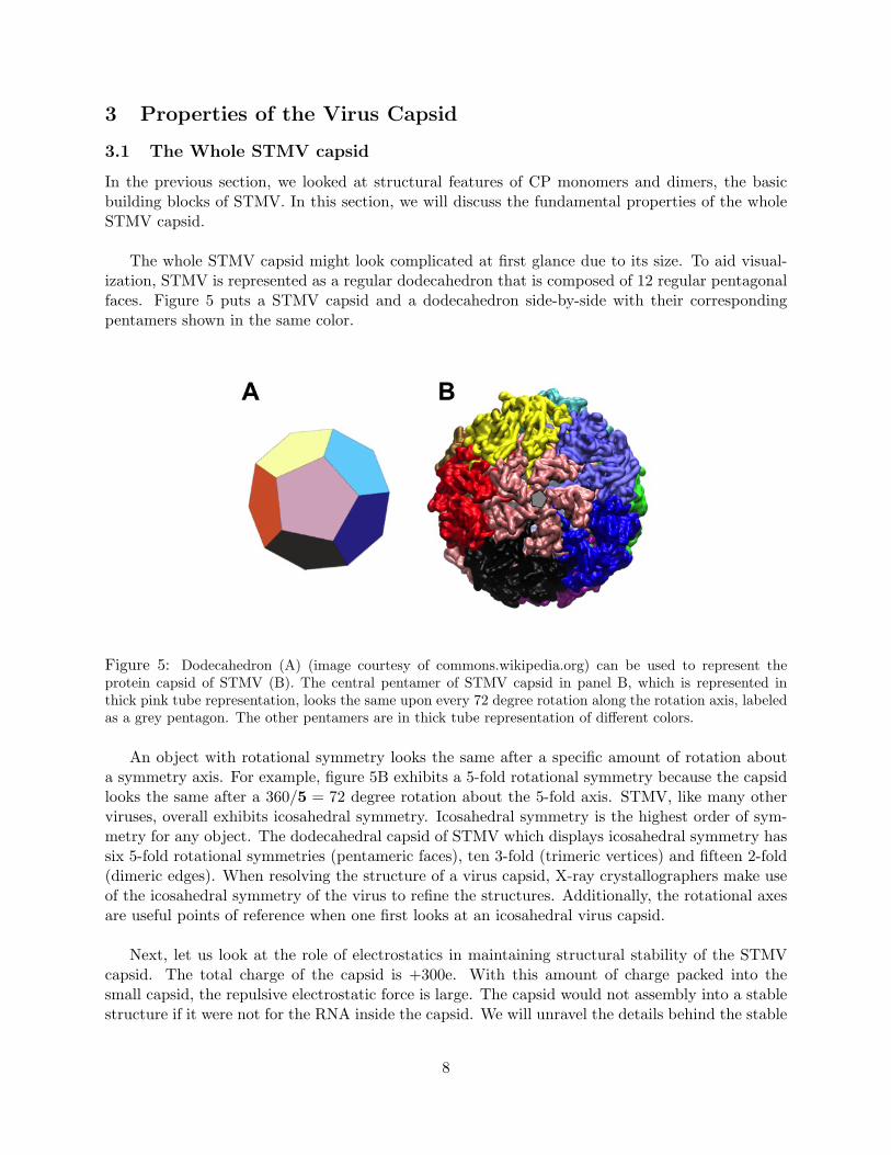
The whole STMV capsid might look complicated at first glance due to its size. To aid visual-ization, STMV is represented as a regular dodecahedron that is composed of 12 regular pentagonalfaces. Figure 5 puts a STMV capsid and a dodecahedron side-by-side with their correspondingpentamers shown in the same color.
Figure 5: Dodecahedron (A) (image courtesy of commons.wikipedia.org) can be used to represent theprotein capsid of STMV (B). The central pentamer of STMV capsid in panel B, which is represented inthick pink tube representation, looks the same upon every 72 degree rotation along the rotation axis, labeledas a grey pentagon. The other pentamers are in thick tube representation of different colors.
An object with rotational symmetry looks the same after a specific amount of rotation abouta symmetry axis. For example, figure 5B exhibits a 5-fold rotational symmetry because the capsidlooks the same after a 360/5 = 72 degree rotation about the 5-fold axis. STMV, like many otherviruses, overall exhibits icosahedral symmetry. Icosahedral symmetry is the highest order of sym-metry for any object. The dodecahedral capsid of STMV which displays icosahedral symmetry hassix 5-fold rotational symmetries (pentameric faces), ten 3-fold (trimeric vertices) and fifteen 2-fold(dimeric edges). When resolving the structure of a virus capsid, X-ray crystallographers make useof the icosahedral symmetry of the virus to refine the structures. Additionally, the rotational axesare useful points of reference when one first looks at an icosahedral virus capsid.
Next, let us look at the role of electrostatics in maintaining structural stability of the STMVcapsid. The total charge of the capsid is +300e. With this amount of charge packed into thesmall capsid, the repulsive electrostatic force is large. The capsid would not assembly into a stablestructure if it were not for the RNA inside the capsid. We will unravel the details behind the stable
8
capsid of STMV in the following exercise.
Exercise 3: Icosahedral Symmetry and electrostatics of the STMV capsid
In problems A, B and C, we will explore the symmetry of STMV; in problem D, we will investigatethe electrostatic properties of the capsid.
A. In a new VMD session, open the visualization state file symmetrical-axes.vmd to load thecoordinates of the proteins. An alias file stmv-macros.tcl contains the macros for selecting thepentameric protein complexes in STMV. Instead of typing segname C0 C1 C2 C3 C4 in SelectedAtoms to display one of the pentamers, you could type penta1 in the Selections tab of theGraphical Representations window. The pentamers are named penta1, penta2, ..., penta12.Can you find the 3-fold and 2-fold rotational axes of STMV? Save the respective views of STMVdisplaying 3-fold and 2-fold rotational symmetry via File → Render... on the VMD main menu.After you have printed out the images, draw a triangle and an ellipse to highlight the positions ofthe 3-fold and 2-fold rotational axes.
B. We can gain useful insight into the assembly process of the capsid by measuring the contactsurface area between the CPs at the 5-fold, 3-fold, and 2-fold rotational axes. Using the firstCP (segname C0) as the reference, find the corresponding CPs that are involved in 5-fold, 3-fold,and 2-fold rotational symmetries relative to segname C0. Next, measure the contact surface areabetween the involved CPs for the respective rotational symmetries. For example, the pentamerthat exhibits 5-fold rotational symmetry in figure 5B is composed of 5 CPs, namely segname C0C1 C2 C3 C4. The contact surface area among the 5 CPs could be calculated as follows:
1. Run source measure-contact-area.tcl and measure contact area 0 {segname C0}{segname C1} contact c0-c1.txt in the VMD TkConsole. The output file contains thecontact surface area between segname C0 and segname C1.
2. Repeat the calculation for segname C2, C3, and C4 respectively to obtain the total contactsurface area for the pentamer.
3. Divide that total contact area value by 5 to get the contact surface area contributed by oneCP in the 5-fold symmetrical plane.
Repeat step 1 to 3 for the CPs that display the 3-fold and 2-fold symmetrical axes. Now with thetotal contact area per CP for different symmetries, propose a model of the assembly process of theSTMV capsid.
C. Next we will measure the angles between two nearby pentamers. With the same VMD session,open the VMD TkConsole and type source angle.tcl. Make sure the files angle.tcl, la.tcl,and orient.tcl are in the working directory. The output file angle stmv.txt contains the dis-tances between the centers of mass of the nearby pentamers and their angles. Plot a histogramwith bin size of 1 degree to show the angle distribution. Did you expect the angle distribution ofthe STMV capsid to look like the histogram?
9

D. To have a better understanding of the electrostatics of the capsid, we will calculate the radial dis-tribution of the charged residues. In the same VMD session, type source charged-res-count.tclin VMD TkConsole to calculate the number of positively (basic) and negatively-charged (acidic)residues at different radial distances from the nucleic acid. Two text files will be generated:basic-residues.txt and acidic-residues.txt. Plot the radial distribution for basic and acidicresidues. Comment on the distribution and suggest what role the RNA inside the capsid mightplay in the stability of capsid.
3.2 More complex viruses
While STMV can be represented as a regular dodecahedron, many viruses are larger and more com-plex than STMV. Larger capsids are typically constructed of both hexameric and pentameric capsidsubunits. One of Euler’s theorems states that exactly 12 pentagons are needed to form a closedsurface from a 2-dimensional hexagonal network. Thus, to build larger capsids, more hexamers areincorporated into the capsid structure, while the number of pentamers remains exactly 12. Forexample, Dengue virus and Cowpea Chlorotic Mottle Virus take on a truncated icosahedral struc-ture, with 20 hexagons and 12 pentagons (the same shape, incidentally, as Buckminsterfullerene).However, not all virus capsids have a symmetric shape. An example of an irregular virus capsid isthe human immunodeficiency virus (HIV) capsid. Figure 6A shows the fullerene cone model of anHIV capsid that has 216 hexagons and 12 pentamers [11].
Figure 6: The human immunodeficiency virus (HIV) has an irregular capsid. (A) A fullerene cone modelof an HIV capsid with the pentamers colored in green (image courtesy of Juan R. Perilla). (B) An all-atomstructure of HIV capsid comprising 216 hexamers (amino-terminal domain in blue; carboxy-terminal domainin red) and 12 pentamers (green).
10

4 Structure of encapsidated viral RNA and capsid-RNA interac-tions
Due to limitations in experimental methods, the atomic structures of most encapsidated viralgenomes are not known. STMV is one of the few viruses for which a high resolution atomic structureof a significant portion of the genome, approximately 59%, has been solved [10]. The STMV genome,which is shown in Fig. 7, consists of a single-stranded RNA molecule that folds inside the capsidto form 30 icosahedrally arranged double helical stemloops. The 30 helical segments are assignedin the X-ray atomic model of STMV. However, due to their flexibility, the linkers that connectthe stemloops were not resolved. The structures of the linkers joining the stemloops were modeledcomputationally in order to construct a complete model of the STMV encapsidated genome [6].
Figure 7: Structure of the STMV RNA genome. Segments of the RNA that correspond to double helicalstemloops, of which there are 30, are shown in green and linkers between the helical segments are shown inblue. The red box highlights a single stemloop in the RNA structure.
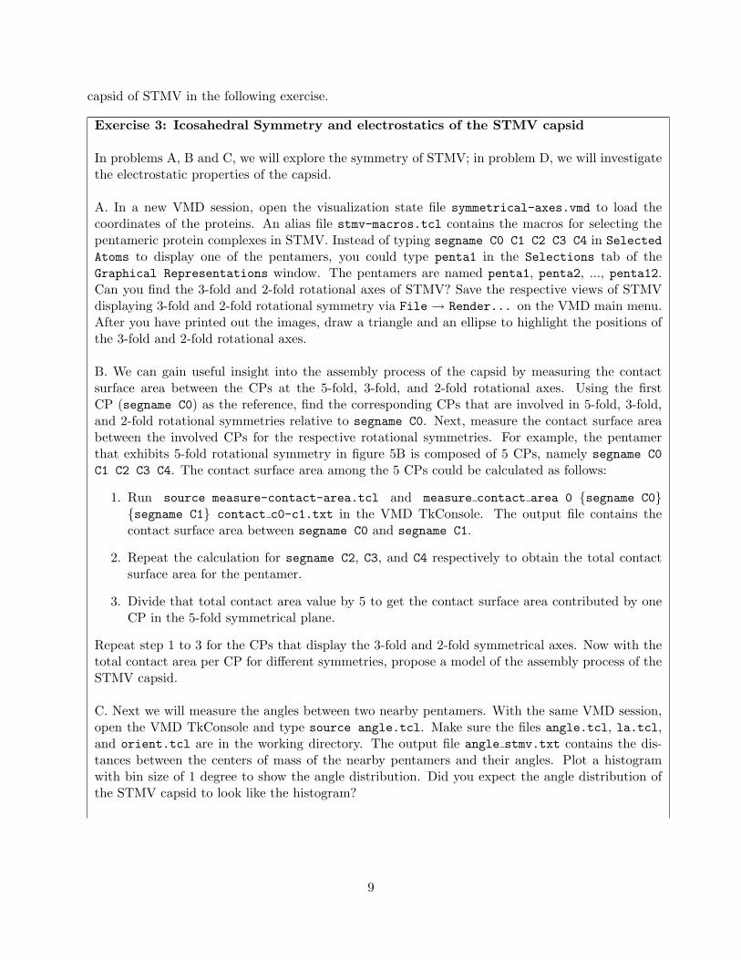
Since the structure of STMV has been solved at 1.8 A resolution, it permits a detailed look atinteractions between a virus capsid and the genetic material that it contains. An important featureof STMV is that each of the helical stemloops is intimately associated with an RNA binding siteon the inner surface of a coat protein dimer and is oriented perpendicular to a 2-fold symmetryaxis of the capsid. As can be seen in Fig. 8, the saddle-shape of the inner surface of the CP dimerenables it to wrap around the double helical RNA segment.
A detailed examination of the interactions at the CP dimer-RNA stemloop interface showsthat the coat proteins primarily interact with the RNA via salt bridges and hydrogen bonds tothe sugar-phosphate backbone of the RNA. Since the capsid-RNA interactions mainly involve theRNA backbone, association of the viral genome with coat proteins is independent of its sequenceof nucleotides. Due to the high variation in length and nucleotide sequence of the genome betweendifferent STMV virus particles, the ability of the coat proteins to encapsidate a variety of RNAmolecules is crucial to the successful replication of STMV [10].
The intimate association of STMV coat proteins with the RNA genome suggests that the coatprotein-RNA interactions play an essential role during assembly of the virus [10]. An assembly
11
Figure 8: A STMV coat protein dimer (individual CP monomers are colored red and blue) interactingwith an RNA double helical stemloop (backbone shown in green and nucleotide bases in orange) of theencapsidated viral genome. The orientation of the helical axis of the RNA stemloop is perpendicular to the2-fold symmetry axis of the capsid.
mechanism for STMV has been proposed in which CP dimers bind to double helical segments ofthe viral genome and, in a concerted process, condense the RNA by forming higher orders of capsidsymmetry elements, specifically associating into the trimeric and pentameric CP structures at the3-fold and 5-fold symmetry axes, respectively. Hence, association of CP dimers bound to helicalsegments of viral RNA may drive STMV assembly. In addition to playing a critical role duringSTMV assembly, the viral RNA may also be essential for the mechanical stability of the STMVparticle. Molecular dynamics simulations of empty STMV capsids with the RNA removed haveshown that the capsid collapses, as shown in Fig. 9 [6]. The essential role of the RNA in STMVassembly and, possibly, stability is also supported by the fact that empty STMV capsids have notbeen observed by electron microcopy studies.
Exercise 4: Secondary structure of the STMV genome, ionization of the viral RNA,and capsid-RNA interactions.
A. Secondary structure of the STMV genome.
12
Load the provided structure and coordinate files, stmv.psf and stmv.pdb, into VMD. Calculatethe maximum number of hydrogen bonds that can be formed between the bases of the viral RNA(Hint: The hydrogen-bond donor atoms are ("(resname ADE and name N6) or (resname URAand name N3)") and the acceptor atoms are ("(resname ADE and name N1) or (resname URAand name O4)") in the RNA bases). Now, measure the actual number of hydrogen bonds betweenthe bases of the RNA (Hint: Use "resname ADE and not backbone" for selection 1 and "resnameURA and not backbone" for selection 2) using the Hydrogen Bonds analysis extension (Extensions→ Analysis → Hydrogen Bonds). Check the write output to files? option and run Findhydrogen bonds! using a Donor-Acceptor distance of 3.5A and Angle cutoff of 30 degrees. Theoutput file hbonds.dat will be produced in your current working directory. Open hbonds.datusing a text editor, which will contain the number of measured hydrogen bonds for frame 0 ofthe selected molecule. What fraction of the possible base pairing hydrogen bonds are formed inthe encapsidated genome and what does the extent of base pairing indicate about the secondarystructure of the viral RNA? Select the RNA bases inside the capsid and visualize the base pairinghydrogen bonds using the HBonds drawing method with a Distance Cutoff of 3.5A and AngleCutoff of 30 degrees. You will need to increase the Line Thickness to easily see the hydrogenbonds. Comment on the hydrogen bonding pattern and secondary structure of the encapsidatedRNA.
B. Ionization of the STMV genome.Measure the total charge of the viral RNA. In the VMD TkConsole, use atomselect to selectthe RNA and run the command measure sumweights $your-RNA-selection weight charge toobtain the total charge. As you should expect, the RNA is highly negatively charged. Howdoes the viral RNA remain stable despite having such a high negative charge (Hint: Take aclose look at other components of the STMV system surrounding the RNA)? Next, measurethe radial density distribution of the protein, RNA, and ions in the STMV particle by run-ning the get-rad-density-distr.tcl script, which produces the output files radden prot.dat,radden rna.dat, radden mg.dat and radden cl.dat in the current working directory. Plot theradial density distribution of protein, RNA, magnesium ions, and chloride ions (Hint: a Matlabscript plot radden.m is provided. If you do not use Matlab, you can plot the graph with yourpreferred graphing software). Do you notice anything in the radial density distribution profiles thatsupports your hypothesis on the stability of the viral RNA?
C. Capsid-RNA interactions.Look at the interactions between the RNA and the inner surface of the capsid, paying specific at-tention to interactions at the two-fold, three-fold, and five-fold symmetry axes. At which symmetryaxis of the capsid does the RNA make the most contact with the capsid? Take a close look at theRNA and capsid proteins at the main points of contact and describe the structure of the RNA atthese contact points. Identify the type of interactions between the RNA and the capsid proteins.What regions of the RNA do the capsid proteins interact with and what implications might thishave on assembly of the STMV particle?
13
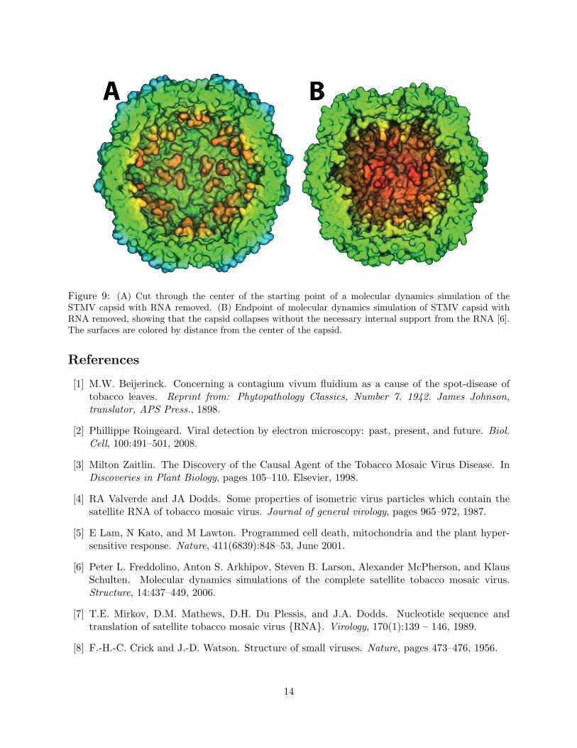
Figure 9: (A) Cut through the center of the starting point of a molecular dynamics simulation of theSTMV capsid with RNA removed. (B) Endpoint of molecular dynamics simulation of STMV capsid withRNA removed, showing that the capsid collapses without the necessary internal support from the RNA [6].The surfaces are colored by distance from the center of the capsid.
References
[1] M.W. Beijerinck. Concerning a contagium vivum fluidium as a cause of the spot-disease oftobacco leaves. Reprint from: Phytopathology Classics, Number 7. 1942. James Johnson,translator, APS Press., 1898.
[2] Phillippe Roingeard. Viral detection by electron microscopy: past, present, and future. Biol.Cell, 100:491–501, 2008.
[3] Milton Zaitlin. The Discovery of the Causal Agent of the Tobacco Mosaic Virus Disease. InDiscoveries in Plant Biology, pages 105–110. Elsevier, 1998.
[4] RA Valverde and JA Dodds. Some properties of isometric virus particles which contain thesatellite RNA of tobacco mosaic virus. Journal of general virology, pages 965–972, 1987.
[5] E Lam, N Kato, and M Lawton. Programmed cell death, mitochondria and the plant hyper-sensitive response. Nature, 411(6839):848–53, June 2001.
[6] Peter L. Freddolino, Anton S. Arkhipov, Steven B. Larson, Alexander McPherson, and KlausSchulten. Molecular dynamics simulations of the complete satellite tobacco mosaic virus.Structure, 14:437–449, 2006.
[7] T.E. Mirkov, D.M. Mathews, D.H. Du Plessis, and J.A. Dodds. Nucleotide sequence andtranslation of satellite tobacco mosaic virus {RNA}. Virology, 170(1):139 – 146, 1989.
[8] F.-H.-C. Crick and J.-D. Watson. Structure of small viruses. Nature, pages 473–476, 1956.
14
[9] Shanshan Cheng and Charles L.-Brooks. Viral capsid proteins are segregated in structural foldspace. PLoS Comput. Biol., 9(2):e1002905, 2013.
[10] S. B. Larson, J. Day, A. Greenwood, and A. McPherson. Refined structure of satellite tobaccomosaic virus at 1.8 A resolution. J. Mol. Biol., 277:37–59, 1998.
[11] Gongpu Zhao, Juan R. Perilla, Ernest L. Yufenyuy, Xin Meng, Bo Chen, Jiying Ning, JinwooAhn, Angela M. Gronenborn, Klaus Schulten, Christopher Aiken, and Peijun Zhang. MatureHIV-1 capsid structure by cryo-electron microscopy and all-atom molecular dynamics. Nature,497:643–646, 2013. doi:10.1038/nature12162.
15