casein production by human breast cancer · female with metastatic breast cancer (16). ... it has...
TRANSCRIPT

[CANCER RESEARCH 37, 749-754, March 1977]
we have additionally examined the capacity of these cell
lines to synthesize casein by measuring incorporation of[3H]proline into material that was precipitable by antibodyusing a direct precipitation method. Our results indicate
that serum samples from breast cancer patients and breastcancer cells in culture seldom contain significant casein
levels, while 17% of the breast tumor cytosols tested arepositive for casein. Breast cancer cells in culture do notappear to synthesize a protein that is specifically precipitable by anticasein antibody.
MATERIALS AND METHODS
Patient Samples. Blood was collected from patients andnormals, and the serum was separated and stored at —70°.
Preparation of Tumor Cytosols. Tumor samples wereobtained from surgical specimens on biopsy material andimmediately frozen in liquid nitrogen and stoned at —70°until used. The frozen tissue was weighed, pulverized and
homogenized using a Bninkmann Polytron PT 10 ST (Brinkmann Instruments, Inc. , Westbury, N.Y.) in a buffer contain
ing 0.01 MTris-HCI, 0.0015 M EDTA, and 0.005 Mdithiothreitol, pH 7.4. One mlofbufferwasadded pen 100 mg of tissue.The resulting homogenate was centrifuged at 105,000 x gfor 1 hr at 4°.The pellet and fat layer were discarded, andthe cytosol was used for assay. Many assays were performed on cytosols which had been stored frozen for up to 6months. Protein concentrations were determined by themethod ofLowmyetal.(11).
Techniques of Cell Culture. The MCF-7 cell line wasestablished from the pleural effusion of a postmenopausalfemale with metastatic breast cancer (16). Starter cultureswere provided by Marvin Rich of the Michigan Cancer Foundation. The Evsa-T cell line was established in this labonatory as previously described (9). The ZR-75-1 cells wereprovided by Nathaniel Young and Linda Engel of the National Cancer Institute. All other breast cell lines (MDA-231,G-11, HT-39, and BT-20) were provided by Ronald Herbemman of the National Cancer Institute. Cells were grown inmonolayer culture in MEM1 supplemented with 10% fetalcalf serum (North American Biological Co. , Miami, Fla.). Forhormonal stimulation experiments, the medium waschanged to MEM without serum before hormone addition.Cells were harvested in Dulbecco's phosphate-buffered saline, pH 7.4, without calcium or magnesium, by scrapingwith a rubber policeman and collected by centnifugation athigh speed in a Sero-fuge (Clay-Adams, Pansi@pany, N. J.).Samples of the media were saved for casein determination.
, The abbreviations used are: MEM, Eagle's minimal essential media plus
2 x glutamine, penicillin, and streptomycin; SDS, sodium dodecyl sulfate.
MARCH 1977 749
Casein Production by Human Breast Cancer
Marie E. Monaco, Diane A. Bronzert, Douglass C. Tormey, Phillip Waalkes, and Marc E. Lippman
Medicine Branch, National Cancer Institute, NIH, Bethesda, Maryland 20014
SUMMARY
Casein was measured in the sera of breast cancer patients, in breast cancer tumors, and in breast cancer cells inlong-term tissue culture using a sensitive and specific madioimmunoassay. Levels present in breast cancer sera werenot elevated above control values. Eight of forty-seven(17%) of the tumor samples tested were positive for casein,the highest level representing 0.003% ofthe soluble protein.When seven human breast cancer cell lines were assayedfor casein, the results were uniformly negative even under
conditions of stimulation by lactogenic hormones. In addition, direct immunoprecipitation of labeled cellular proteinsupported the negative result of the radioimmunoassay.Thus it appears that casein production is not a commoncharacteristic of most human breast cancers.
INTRODUCTION
It has been previously reported that the milk protein,casein, is present in large amounts in the sena of patientswith carcinoma of the breast (5). These data imply thatneoplastic mammary epithelial cells have the capacity tosynthesize a product normally secreted by the mammarygland in the presence of lactogenic hormones. On the otherhand, the milk protein, a-lactalbumin, found in rodent systems to be coordinately regulated with casein (14), was notsignificantly elevated in the semaof breast cancer patients,although it was present in some mammary tumor samples(8). Furthermore, MCF-7, a human breast cancer cell line incontinuous tissue culture, has been reported to accumulatea-lactalbumin (15). Determination of the capacity of humanbreast cancer to synthesize discrete milk proteins in vivoand in vitro would be desirable for a number of reasons: (a)these proteins might be valuable as serum markers in thedetection and monitoring of carcinoma ofthe breast; (b) therelative ability of individual tumors to synthesize these proteins might provide clinical data relevant to therapy; forexample, the presence or absence of casein might be correlated with the endocrine responsiveness of the tumor; and(c) the existence of breast cancer cells in culture whichwere capable of synthesizing milk proteins could provide anexcellent model for the study of the regulation of lactogenicfunctions.
We have developed a sensitive and specific radioimmunoassay for human casein and examined a variety of serumsamples, tumor cytosols, and breast cancer cell tissue cultune lines for the presence of this milk protein. Furthermore,
Received August 9, 1976; accepted December 3, 1976.
on March 20, 2020. © 1977 American Association for Cancer Research.cancerres.aacrjournals.org Downloaded from

M. E. Monaco et a!.
The cell pellets were resuspended in 0.05 M sodium phosphate buffer, pH 7.5. Cells were sonically disrupted for 5sec at the lowest setting (Branson Sonicator; Heat SystemsUltrasonics, Plainview, Long Island, N. Y.), and the resulting sonicate was centrifuged for 30 mm at 105,000 x g in aBeckman L5-65 ultracentnifuge. Both the cytosol and pelletwere used for casein determinations.
Preparation of the Antibody. Casein used for immunization was prepared from human milk by a combination ofacid precipitation and polyacrylamide disc gel electrophoresis. The techniques for acid precipitation have been descnibed elsewhere (13). Approximately 50 @gof acid-precipitated material were applied to a 10% polyacrylamide gelcontaining 0.1% SDS and subjected to electrophomesis using a Tris-glycine-SDS buffer, as described elsewhere (12).Fig. la illustrates the electrophometic pattern of the acidprecipitated material. Fig. lb shows the migration of humancasein after purification by d iethylaminoethyl chromatography, according to the method described by Groves andGordon (4). This 25,000-molecular weight species, comesponding to caseins I through VI of Groves and Gordon,was used for immunization (approximately 150 pg/animal).Unstained gels were scanned at 280 nm using a Beckmangel scanner, and the protein band migrating in the positionof the 25,000-molecular weight species was excised andhomogenized in complete Freund's adjuvant using a Brinkmann Polytron. The resulting emulsion was injected intradenmally into the back of a castrated male sheep in 20injections of 0.1 ml each (17, 18). A booster injection wasgiven 4 weeks later, and the sheep was bled during the 5thweek.
Preparation of Radiolabeled Casein. Acid-precipitatedcasein was chromatographed on diethylaminoethyl, and casein Fractions I through VI and Kappa casein were isolated.The identity of Fractions I through VI was verified by electmophoresis on 10% polyacrylamide gels containing 6 M urea.Fig. 2 illustrates that the casein fraction used for iodinationcorresponds to casein IV described by Groves and Gordon(4). Labeling was effected by a modification of the procedune of Hunter and Greenwood (6). One mCi of Na'251(Amensham/Seanle,Arlington Heights, III.) was reacted with5 lLg of casein IV in 0.05 M sodium phosphate buffer, pH7.5. Radioactive casein was purified by chromatography onSephadex G-200 (Pharmacia, Uppsala, Sweden) in 0.05 Mphosphate buffer containing 0.5% bovine serum albumin,pH 7.5. The specific activity of the preparation was generally150 @Ci/@g.The same casein (Fraction IV) was used as thecompeting antigen in determination of the standard curve.
Radloimmunoassay Conditions. A double antibody equilibnium system was used in the radioimmunoassay. Theincubation mixture consisted of 0.05 ng of labeled casein(approximately 10,000 cpm), 0.1 ml of sample, and a1:50,000 dilution of antiserum, in a final volume of 0.4 ml of0.05 M phosphate buffer with 0.5% bovine serum albumin,pH 7.5. When serum samples were assayed, the antiserumwas first adsorbed against a 10-fold excess of normal maleserum (1 ml antiserum plus 9 ml normal male serum) beforefinal dilution in the assay mixture; an additional 0.1 ml of themale serum was added to each tube of the standard curve.The mixture was allowed to stand overnight at 4°.Then 0.1
ml of 1:100 dilution of normal sheep serum was added toeach tube. After vortexing, 0.1 ml (0.1 mg) of goat antisheep gamma globulin (Miles Yeda, Ltd., Rehovot, Israel)was added, and the mixture was allowed to stand for 18 hrat 4°.One ml of cold phosphate buffer was then added toeach tube, and the precipitate was collected by centrifugation at 1000 X g for 30 mm. The supernatant was decanted,and the pellet was counted directly in a Micromedics ModelMS588 gamma counter.
PrecipItation of Labeled Cellular Proteins. MCF-7 cellswere grown in T-75 flasks in MEM supplemented with 10%fetal calf serum. When subconfluent, the medium waschanged to serum-free MEM supplemented with humanplacental lactogen (1 .tg/ml), insulin (106 M), and dexamethasone (10@ M). Following 48 hr of incubation with thehormones, cells were labeled overnight with 100 x 106dpmL-[3,4-3H]pnoline, specific activity, 40 Ci/mmole (Amensham/Searle). Cells were harvested and cytosols were prepared as previously described. Approximately 1 x 10⁶cellswere homogenized in 1 ml of buffer. Part of the cytosol wasused directly for SDS-10% polyacrylamide gel electnophoresis. These gels were identical to those used to prepare thehuman casein for immunization. Another portion of thecytosol was subjected to immunopnecipitation. Fifty @gofhuman casein carrier were added to 0.1 ml (approximately300 j.@gof protein) of the cytosol. Sodium phosphate bufferwas added to a final volume of 1.0 ml, and 0.5 ml of undiluted antiserum was added. The mixture was incubated at37°for 1 hr and permitted to stand at 4°overnight. Theresulting precipitate was collected by centnifugation at 800x g for 30 mm and washed 3 times with phosphate buffer.
The precipitate was dissolved in Tnis-glycine electrophonesis buffer plus 1% SDS and heated at 100°for 30 sec beforebeing applied to the gel. At the end of the electrophoresisperiod, the gels were sliced into 1-mm sections, digestedfor 2 hr at 50°in NCS (Amersham/Seamle), and radioactivitywas determined in 8 ml of Aquasol (New England Nuclear,Boston, Mass.) with a Packard Tn-Garb Model 3390 liquidscintillation spectrometer.
RESULTS
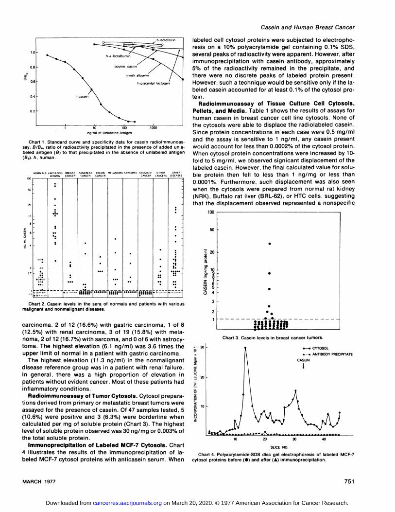
Sensitivity and Specificity of Radioimmunoassay. Thesensitivity and specificity of the madioimmunoassay are illustrated in Chant 1. The assay as described is capable ofmeasuringhuman caseinat1 ng/mland does notappeartocross-react significantly with other milk proteins, bovinecasein, on human placental lactogen.
Radiolmmunoassay of Serum Samples. The values obtamed with normal and patient semaare shown in Chart 2.Since 107 of 113 (95%) normal samples had values 1.7ng/mI, this value was chosen as the upper limit of normal.The highest value obtained with normal serum was 2.6 ng/ml. In samples obtained from lactating women, the lowestvalue was 2.5 ng/ml with a mangeextending to 76 ng/mland a mean of15.4ng/ml.
Among patients with cancer, there were elevations in 3 of31 (9.7%) with metastatic breast carcinoma, 2 of 20 (10%)with pancreatic carcinoma, 10 of 19 (52.6%) with colon
750 CANCER RESEARCH VOL. 37
on March 20, 2020. © 1977 American Association for Cancer Research.cancerres.aacrjournals.org Downloaded from
20
10
10 20 30 40
Casein and Human Breast Cancer
labeled cell cytosol proteins were subjected to electrophomesison a 10% polyacrylamide gel containing 0.1% SDS,several peaks of radioactivity were apparent. However, afterimmunoprecipitation with casein antibody, approximately5% of the radioactivity remained in the precipitate, andtheme were no discrete peaks of labeled protein present.However, such a technique would be sensitive only if the Iabeled casein accounted for at least 0.1% of the cytosol protein.
Radiolmmunoassay of Tissue Culture Cell Cytosols,Pellets, and Media. Table 1 shows the results of assaysforhuman casein in breast cancer cell line cytosols. None ofthe cytosols were able to displace the madiolabeled casein.Since protein concentrations in each case were 0.5 mg/mIand the assay is sensitive to 1 ng/mI, any casein presentwould account for less than 0.0002% of the cytosol protein.When cytosol protein concentrations were increased by 10-fold to 5 mg/mI, we observed signicant displacement of thelabeled casein. However, the final calculated value for soluble protein then fell to less than 1 ng/mg on less than0.0001%. Furthermore, such displacement was also seenwhen the cytosols were prepared from normal rat kidney(NRK), Buffalo rat liven (BRL-62), or HTC cells, suggestingthat the displacement observed represented a nonspecific
0
E
zU3
3:
03:0
20Uz
ng/ml at Unlabeled Anligen
Chart 1. Standard curve and specificity data for casein radioimmunoassay. B/B0, ratio of radioactivity precipitated in the presence of added unlabeled antigen (B) to that precipitated in the absence of unlabeled antigen(B0).h,human.
NORMALS LACTATING BREAST PANCREAS COLON MELANOMA SARCOMA STOMACH OTHER OTHERWOMEN CANCER CANCER CANCER CANCER CANCERS DISEASES
so
20
10
17
Chart 2. Casein levels in the sera of normals and patients with variousmalignant and nonmalignant diseases.
carcinoma, 2 of 12 (16.6%) with gastric carcinoma, 1 of 8(12.5%) with renal carcinoma, 3 of 19 (15.8%) with melanoma, 2 of 12 (16.7%) with sarcoma, and 0 of 6 with astrocytoma. The highest elevation (6.1 ng/ml) was 3.6 times theupper limit of normal in a patient with gastric carcinoma.
The highest elevation (11.3 ng/mI) in the nonmalignantdiseasereferencegroup was ina patientwithrenalfailure.In general, there was a high proportion of elevation inpatients without evident cancer. Most of these patients hadinflammatory conditions.
Radioimmunoassay of Tumor Cytosols. Cytosol prepamations derived from primary or metastatic breast tumors wereassayed for the presence of casein. Of 47 samples tested, 5(10.6%) were positive and 3 (6.3%) were borderline whencalculated per mg of soluble protein (Chart 3). The highestlevel of soluble protein observed was 30 ng/mg or 0.003% ofthetotalsolubleprotein.
Immunoprecipitation of Labeled MCF-7 Cytosols. Chart4 illustratesthe resultsof the immunoprecipitationof Iabeled MCF-7 cytosol proteins with anticasein serum. When
100
50
20
S
S .
.
.
C
C.--C_C- -
UIi!IIjI@
.5
Ca,
0aa).@1
zLU(05U
3
2
Chart 3. Casein levels in breast cancer tumors.
Chart 4. Polyacrylamide-SDS disc gel electrophoresis of labeled MCF-7cytosol proteins before (O) and after (A) immunoprecipitation.
.—. CYTOSOL‘.-.@ ANTIBODY PRECIPITATE
CASEIN
SLICE NO.
MARCH 1977 751
I
on March 20, 2020. © 1977 American Association for Cancer Research.cancerres.aacrjournals.org Downloaded from

Casein accumulation by human breast cancer cells in tissuecultureCell
lineCasein (ng/mg solubleprotein)MCF-7<2ZR75-1<2Evsa-T<2MDA-231<2BT-20<2HT-39<2G-11<2
AdditionCasein(ng/mg soluble pro
tein)None<2HPLa
(1ng!mI)<2l(106M)<2DEX(107M)<2E2
(108M)<2P0(107M)<2HPL+l+DEX<2HPL+I+DEX+E2+P0
<2
M. E. Monaco et a!.
Table 1 be useful in detecting disease and monitoring changes indisease status. We developed a sensitive and specific madioimmunoassay in order to measure casein levels in se
rum, tumors, and breast cancer cells in long-term culture.Our results do not support the observations of Hendrick andFranchimont. We did not find significant elevations in casein levels in patients with breast on other cancers. Similarresults in breast cancer sera have been observed by Dr.David Kleinberg, New York University (personal communication). The incidence of elevated casein levels in nonmalignant disease states was generally higher than that foundin cancer patients. The reason for this is not obvious. Furthemmome,the elevated casein levels detected by our assaywould have been below the limits of detectability (100 ng/ml) in the assay described by Hendnck and Franchimont.The highest elevation we detected, 76 ng/ml in the serum ofa lactating woman, was at least 30 times lower than thatreported by Hendrick and Fnanchimont for a similar serumsample.
We also assayed tumor samples directly and found a totalof 17% (8 of 47) to be positive. The highest value obtainedrepresented 0.003% of the tumor's soluble protein. Theseresults correlate well with those reported by Hunlimann etal. (7), who found 5 of 43 (11%) breast cancer tumors to bepositiveforcaseinby immunofluomescencetechniques.Bussolati et a!. (2) found a higher incidence of casein in moredifferentiated breast cancer tumors. Whether on not thisgroup with tumors positive for casein represents a clinicallyor pathologically significant subpopulation of breast cancerpatients remains to be determined.
Lastly, we examined breast cancer cell lines in tissueculture. We were unable to demonstrate the presence ofcasein either by direct immunoprecipitation on by a com petitive protein-binding assay with this antibody in 7 humanbreast cancer lines in continuous culture. In addition, nocombination of lactogenic hormones used in this studyresulted in detectable casein production.
Our data are consistent with the idea that neoplasticmammary cells in culture no longer retain the hormonalresponsiveness necessary for initiation of casein synthesis.The fact that both breast cancer cell lines (10) as well asmany mammary tumors (8) fail to respond to prolactin mayexplain the failure of the cell lines to accumulate casein.However, we cannot rule out the possibility that the conditions we used were not optimal for induction of caseinsynthesis. Perhaps hormones found to be sufficient forlactation in rodents are inadequate in humans.
In summary, we have developed a sensitive and specificantibody to human casein and used it to measure caseinlevels in the semaof patients with breast cancer, in mammany tumors, and in breast cancer cells in tissue culture.
None of the serum samples for individuals with breast cancemhad clinically significant casein levels; 17% of the breasttumors assayed were positive. No human breast cancer cellline tested appeared to accumulate casein. From these datawe conclude that significant casein synthesis is not a general characteristic of human breast cancer, when measuredwith our antibody. The absence of detailed specificity datain the report of Hendrick and Franchimont (5) prevents usfrom commenting on the validity of their assay.
Table 2
Effect of hormones on casein accumulation in MCF-7 human breastcancer cells
a The abbreviations used are: HPL, human placental lactogen; I,
insulin; DEX,dexamethasone;E2,estradiol; P0,progesterone.
protein effect or cross-reactivity with some componentcommon to these cells. The 105,000 x g pellets and themedium in which the cells were grown were also assayedfor casein. Pellets were first nedissolved in the assay bufferwith the aid of sonic disruption. Both were negative withinthe limits of the assay. To muleout the possibility that thehomogenization procedure was destroying any casein present, carrier casein was added to a cell preparation beforehomogenization. The casein was recovered in the cytosol.
Lastly, an attempt was made to stimulate casein synthesisin the MCF-7 cell line with lactogenic hormones. Table 2shows that none of the hormones or hormone combinationstested had any observable effect on the levels of casein inthe cells. Estradiol , 5a-d ihydrotestosterone, insulin , anddexamethasone have previously been shown to elicit a vanety of responses in these cells at the concentrations used (9,10). Furthermore, the triple hormone combination of placental lactogen, insulin, and dexamethasone has been determined to be maximally effective in stimulating caseinsynthesis in rodent mammary glands in organ culture (14).
DISCUSSION
Casein biosynthesis is normally activated only in the fullydeveloped lactating mammary gland under conditions ofstimulation by a number of hormones (19). One might nottherefore expect to observe casein production under conditions of malignant growth. However, the production of differentiated products by malignant tissue is not uncommon.For example, adrenocorticotropin production by lung carcinomas (3) and human chonionic gonadotropin productionin patients with gastric carcinomas have been reported (1).
Hendrick and Franchimont (5) have reported that caseincan be detected in the sena of patients with breast cancerand other cancers and suggested that its measurement may
752 CANCER RESEARCH VOL. 37
on March 20, 2020. © 1977 American Association for Cancer Research.cancerres.aacrjournals.org Downloaded from

Casein and Human Breast Cancer
Model Systems in the Study of Hormone Dependent Human BreastCancer. New EngI. J. Med., in press.
11. Lowry. 0. H., Rosebrough, N. J.. Farr, A. L. , and Randall. A. J. ProteinMeasurement with the Folin Phenol Reagent. J. Biol. , Chem. , 193: 265-275, 1951.
12. Maizel, J. V. Polyacrylamide Gel Electrophoresis of Viral Proteins. In: K.Maramorosch and N. Koprowski (eds.), Methods in Virology. Vol. 5, pp.179-246. New York: Academic Press, Inc., 1971.
13. Malpress. F. H., and Hytten, F. E. Studies on Human Casein Preparationsfrom SingleMilk Samples.Biochem.J., 91: 130-136,1964.
14. Oka, T., and Perry, J. W. Spermidine as a Possible Mediator of Glucocorticoid Effect on Milk Protein Synthesis in Mouse Mammary Epithelium inVitro. J. Biol. Chem., 249: 7647-7652, 1974.
15. Rose, H., and McGrath, C. a-Lactalbumin Production in Human Mammary Carcinoma. Science, 190: 673-675, 1975.
16. Soule, H. 0., Vayquez, J., Long. A., Alberts, S., and Brennan, M. AHuman Cell Line from a Pleural Effusion Derived from a Breast Carcinoma. J. NatI. Cancer Inst., 51: 1409-1414, 1973.
17. Tjian, A., Stinchcomb, 0., and Losick, A. Antibody Directed AgainstBacillus Subtilis a Factor Purified by Sodium Dodecyl Sulfate Slab GelElectrophoresis. Effect on Transcription by ANA Polymerase in CrudeExtracts of Vegetative and Sporulating Cells. J. Biol. Chem., 250: 8824-8828, 1975.
18. Vaitukaitis, J., Robbins, J., Nieschlag, E., and Ross, G. A Method forProducing Specific Antisera with Small Doses of Immunogen. J. Clin.Endocrinol. Metab., 33: 988-991, 1971.
19. Vorherr, H. Galactoporesis, Galactosecretion,and Onset of Lactation. In:The Breast: Morphology, Physiology, and Lactation, pp. 71-124. NewYork: Academic Press, Inc., 1974.
MARCH 1977 753
REFERENCES
1. Braunstein, G.. Vaitukaitis. J., carbone. P., and Ross, G. Ectopic Production of Human Chorionic Gonadotrophin by Neoplasms. Ann. Internat Med., 78: 39-45, 1973.
2. Bussolati, G. . Pich, A. , and Alfani, V. Immunofluorescence Detection ofCasein in Human Mammary Dysplastic and Neoplastic Tissues. Virchows. Arch. Abt. A Pathol. Anat., 365: 15-21 , 1975.
3. Charles, A.. Claypool. A., Schaal, M., Rosen, S.. and Weintraub, B. LungCarcinoma Associated with Production of Three Placental Proteins.Arch. Internal Med., 132: 427-431 , 1973.
4. Groves, M. L. , and Gordon, W. G. The Major Component of HumanCasein: A Protein Phosphorylated at Different Levels. Arch. Biochem.Biophys., 140: 47-51 , 1970.
5. Hendrick, J. C. , and Franchimont, P. Radioimmunoassay of Casein inthe Serum of Normal Subjects and Patients with Various Malignancies.European J. Cancer 10: 725-730, 1974.
6. Hunter, W., and Greenwood, F. Preparation of lodine-131 Labeled Human Growth Hormone of High Specific Activity. Nature, 194: 495-496,1962.
7. Hurlimann, J. . Lichaa, M. , and Ozzello, L. In Vitro Synthesis of Immunoglobulins and Other Proteins by Dysplastic and Neoplastic Human Mammary Tissues. Cancer Aes.. 36: 1284-1292, 1976.
8. Kleinberg. D. L. Human a-Lactalbumin: Measurement in Serum and inBreast Cancer Organ Cultures by Radioimmunoassay. Science, 190:276-278, 1975.
9. Lippman. M. E. , and Bolan, G. Oestrogen-Aesponsive Human BreastCancer in Long Term Tissue Culture. Nature, 256: 592-593, 1975.
10. Lippman. M. E.. Osborne,@ K., Knazek, A., and Young. N. In Vitro
on March 20, 2020. © 1977 American Association for Cancer Research.cancerres.aacrjournals.org Downloaded from

M. E. Monaco et a!.
r@@-.
-@-
VIV@IV@
‘:7- IV
I I
a bFig. 1. Polyacrylamide-SDS disc gel electrophoresis of acid precipitated
casein before (a) and after (b) diethylaminoethyl chromatography.
a bFig. 2. Polyacrylamide-urea gel electrophoresis of acid-precipitated ca
sein before (a) and after (b) diethylaminoethyl chromatography.
754 Casein and Human Breast Cancer
on March 20, 2020. © 1977 American Association for Cancer Research.cancerres.aacrjournals.org Downloaded from

1977;37:749-754. Cancer Res Marie E. Monaco, Diane A. Bronzert, Douglass C. Tormey, et al. Casein Production by Human Breast Cancer
Updated version
http://cancerres.aacrjournals.org/content/37/3/749
Access the most recent version of this article at:
E-mail alerts related to this article or journal.Sign up to receive free email-alerts
Subscriptions
Reprints and
To order reprints of this article or to subscribe to the journal, contact the AACR Publications
Permissions
Rightslink site. Click on "Request Permissions" which will take you to the Copyright Clearance Center's (CCC)
.http://cancerres.aacrjournals.org/content/37/3/749To request permission to re-use all or part of this article, use this link
on March 20, 2020. © 1977 American Association for Cancer Research.cancerres.aacrjournals.org Downloaded from