casereport - downloads.hindawi.comdownloads.hindawi.com/journals/crig/2018/6968395.pdf ·...
TRANSCRIPT
Case ReportWhole Exome Sequencing and Molecular Modeling ofa Missense Variant in TNFAIP3 That Segregates with Disease ina Family with Chronic Urticaria and Angioedema
Antoneicka L. Harris,1 Patrick R. Blackburn,2,3 John E. Richter Jr.,2,4 Jennifer M. Gass,2
Thomas R. Caulfield ,5 Ahmed N. Mohammad,2,4 and Paldeep S. Atwal 2,4
1Department of Cancer Biology, Mayo Clinic, Jacksonville, FL, USA2Center for Individualized Medicine, Mayo Clinic, Jacksonville, FL, USA3Department of Health Sciences Research, Mayo Clinic, Jacksonville, FL, USA4Department of Clinical Genomics, Mayo Clinic, Jacksonville, FL, USA5Department of Neuroscience, Mayo Clinic, Jacksonville, FL, USA
Correspondence should be addressed to Paldeep S. Atwal; [email protected]
Received 3 October 2017; Accepted 18 January 2018; Published 22 February 2018
Academic Editor: Shoji Ichikawa
Copyright © 2018 Antoneicka L. Harris et al. This is an open access article distributed under the Creative Commons AttributionLicense, which permits unrestricted use, distribution, and reproduction in any medium, provided the original work is properlycited.
Chronic urticaria is a common condition characterized by recurrent hives lasting several weeks ormonths and is usually idiopathic.Approximately half of the individuals with chronic urticaria will present with episodes of angioedema that can be severe anddebilitating. In this report, we describe a 47-year-old Hispanic male who presented initially for an evaluation of chronic hivesfollowing hospitalization due to hive-induced anaphylaxis. The individual had a history significant for urticaria and angioedemabeginning in his early 30s. Interestingly, both the individual’s 41-year-old sister and 12-year-old daughter were also affected withchronic urticaria and severe angioedema. Whole exome sequencing of the proband and several family members revealed aheterozygous variant of uncertain significance in exon 2 of TNFAIP3, denoted as c.65G>A (p.R22Q), in all affected members.Variants in TNFAIP3 have been associated with multiple autoimmune diseases, susceptibility to allergy and asthma, and periodicfever syndromes, suggesting that this variant could potentially play a role in disease.
1. Introduction
Chronic urticaria is a common skin disorder characterizedby recurrent hives lasting more than six weeks or hives thatrecur over several months or years [1, 2]. It is mainly a diseaseof adults with women being more affected than men [3].Chronic urticaria is usually idiopathic, arising spontaneouslywithout a stimulus. Other common types include physicalurticarias that are caused by physical stimuli such as cold,heat, or pressure. Various types of urticaria may coexist to-gether, which is not uncommon [3]. Angioedema is a swellingof the deeper epidermal and dermal layers of the skin. Theassociation between chronic urticaria and angioedema is wellestablished [4, 5]. Genetic factors may play a role in chronicidiopathic urticaria, but the mechanism of disease is largely
unknown [6]. In this report, we describe a patient with aseveral-year history of chronic hives, angioedema, skin flush-ing, and swelling. Whole exome sequencing (WES) did notdetect any causative variants in known disease genes butdid identify a variant in tumor necrosis factor-alpha-inducedprotein 3 (TNFAIP3, known commonly as A20) that is possi-bly associated with the reported phenotype.
2. Case History
A 47-year-old Hispanic male was initially referred to theMayo Clinic at 34 years of age due to severe episodes ofangioedema (occurring every three to four months) andhistory of chronic urticaria. The initial onset of urticariaoccurred in 2002 while the proband was living in Italy. He
HindawiCase Reports in GeneticsVolume 2018, Article ID 6968395, 6 pageshttps://doi.org/10.1155/2018/6968395

2 Case Reports in Genetics
has since relocated to the United States, where he soughttreatment for his urticaria flares. The proband reported thatepisodes of urticaria occur when his skin is exposed to hot orcold temperatures or significant pressure.
The proband tried numerous treatment modalities tosuppress his urticaria, including the following antihistamines:levocetirizine, cetirizine, fexofenadine, and loratadine. Pred-nisone and steroid injections were the only treatments thatcleared the patient’s hives; however, treatment with predni-sone was eventually stopped due to side effects. His elevatedlevels of IgE suggested that the hives were autoimmune innature, though the accompanying tryptase elevation led to theconsideration ofmastocytosis.The patient had a normal bonemarrow biopsy (differential and aspirate), a complete bloodcount (CBC) with differential within normal limits, and anormal mast cell panel (CD2, CD25, CD69, and CD117). Hisanti-IgE receptor antibody was elevated at 18.5% (referencerange: <13%) and his C1q complement was elevated at 43(12–22mg/dL). He also had a normal TSH of 2.0 (>20 years:0.3–4.2mIU/L) and serum protein electrophoresis that waswithin normal limits. His tryptase levels were consistentlyelevated and ranged from 13.9 and 19.2 (<11.5 ng/mL), indi-catingmast cell activation. Due to the absence of mast cells inthe bone marrow biopsy, the patient did not meet diagnosticcriteria for mastocytosis and was ultimately diagnosed withchronic urticaria. At 37 years of age, the patient began expe-riencing “attacks” approximately every two weeks, which hedescribed as involving hives, fever, weakness, diaphoresis,and diarrhea. The patient also experienced episodes of dizzi-ness when standing after walking only a few feet.
Upon ascertainment of family history, we found that oneof the proband’s sisters and his daughter both shared hissymptoms of hives and angioedema (Figure 1).The proband’sdaughter, who was 12 years of age at the time of evaluation,had episodes of hives and swelling of her hands and feet. Sheparticipated in gymnastics but had to discontinue this activitydue to swelling of her feet after practice. She has also devel-oped hand swelling after playing the cello.
3. Methods
3.1. Whole Exome Sequencing. Using genomic DNA extract-ed from the proband, his mother, sister, and daughter, theAgilent Clinical Research Exome kit was used to target exonicregions and flanking splice junctions of the genome. Theseregions were sequenced by massively parallel sequencingon an Illumina HiSeq with 100 bp paired-end reads (meandepth of coverage: 119x; quality threshold: 94.9%). Bidirec-tional sequence was assembled, aligned to reference genesequences based on human genome build GRCh37/UCSChg19, and analyzed for sequence variants using a custom-developed analysis tool (Xome Analyzer, GeneDx). Capillarysequencing was used to confirm the presence or absence of allpotentially pathogenic variants identified in both the probandand relative samples.
3.2. Structural Modeling. TNFAIP3, also known as A20, is anubiquitin-editing enzyme, which has dual roles in ubiquitinligase and deubiquitinase activity. The N-terminal domain of
3
47
12
41
6526
∗
∗ ∗
∗
Chronic urticaria, angioedema
Figure 1: Family pedigree. Standard pedigree symbols are used.An arrow indicates the proband. The numbers inside the symbolsrepresent the number of family members. The numbers at the rightlower side of the symbols represent current age or age at death ofthe individuals. Asterisks indicate individuals that underwent wholeexome sequencing.
TNFAIP3 contains an ovarian tumor (OTU) domain, involv-ed in deubiquitinating Lys-63-polyubiquitin chains in a num-ber of substrates.TheC-terminal domain of TNFAIP3 is com-posed of seven zinc finger domains that coordinate ubiquitinligase activity, including the addition of lysine-48-linkedubiquitin chains that are necessary for targeting substrates forproteasomal degradation [7, 8].
HumanTNFAIP3 sequencewas taken from theNCBIRef-erence Accession number NG 032761 version NG 032761.1and was used for computer-assisted modeling. Monte Carlosimulations were performed on the full-length wild typeprotein (791 amino acids) and the p.R22Q variant. The X-ray refinement for Monte Carlo was built using YASARASSP/PSSM method [9–14]. The structure was relaxed to theYASARA/Amber force field using knowledge-based poten-tials within YASARA. The side chains and rotamers wereadjusted with knowledge-based potentials, simulated anneal-ing with explicit solvent, and small equilibration simulationsusing YASARA’s refinement protocol [15]. The entire full-length structure was modeled, filling in any gaps or unre-solved portions from the X-ray.
Refinement of the finalized models was completed usingeither Schrodinger’s LC-MODMonte Carlo-basedmodule orNAMD2 protocols. These refinements started with YASARAgenerated initial refinement [9–11, 13].The superposition andsubsequent refinement of the overlapping regions yieldeda complete model for TNFAIP3. The final structures weresubjected to energy optimization with PR conjugate gradientwith an R-dependent dielectric.
Atom consistency was checked for all 791 amino acids(13,028 atoms) of the full-length wild type model and 791amino acids (13,028 atoms) for the p.R22Q variant, verify-ing correctness of chain name, dihedrals, angles, torsions,
Case Reports in Genetics 3
nonbonds, electrostatics, atom typing, and parameters. Eachmodelwas exported to the following formats:Maestro (MAE)and YASARA (PDB). Model manipulation was done withMaestro (Macromodel, version 9.8, Schrodinger, LLC, NewYork, NY, 2010) or Visual Molecular Dynamics (VMD) [16].Analyses were restricted to theN-terminus region containingthe first 350 amino acids given the length andC-termdistancefrom the site of mutation.
Monte Carlo dynamics searching (LCMOD-MC) wascompleted on each model for conformational sampling,using methods previously described in the literature [17–20].Briefly, each TNFAIP3 variant system was minimized withrelaxed restraints using either Steepest Descent or ConjugateGradient PR and then allowed to undergo theMC search cri-teria, as shown in the literature [17–20].The primary purposeof MC, in this scenario, is to examine any conformationalvariability that may occur with different mutations in theregion near the mutation and the possible effect on TNFAIP3structure and function.
4. Results
4.1. Exome Sequencing Results. WES of the probanduncovered a novel variant in TNFAIP3 (Chr6(GRCh38):g.137871292G>A; NM 001270507.1: c.65G>A; p.(Arg22Gln)),which could possibly correlate with the proband’s diseasephenotype.The p.R22Q variant is absent from ExAC (ExomeAggregation Consortium) and has only been observed in1/30936 whole genomes sequenced in gnomAD (GenomeAggregation Database) [21]. Of note, this variant was seen in1/838 Latino genomeswith a predictedminor allele frequency(MAF) of 0.001193%, suggesting that it may bemore commonin Latino populations. The Arg22 residue is conserved acrossspecies down to zebrafish (Danio rerio), and in silicoprediction algorithms predict the variant to be deleterious(PredictSNP2 deleterious score: 1.0000; CADD deleteriousscore: 22.6000; FATHMM deleterious score: 0.9806) [22].This variant results in a semiconservative amino acid substi-tution that may impact secondary protein structure, andmore detailed studies including in silicomolecular modelinganddynamics simulationswerepursued. Based on theACMG2015 guidelines, the c.65G>A (p.R22Q) variant was classifiedas a variant of uncertain significance (VUS) [23]. The pro-band’s mother was found to be negative for the p.R22Q vari-ant (the proband’s father was deceased), whereas both hisdaughter and sister (both affected) were heterozygous forthe VUS. Another pathogenic variant was detected in GJB2(Chr13(GRCh38): g.20189547del; NM 004004.5: c.35del;p.Gly12Valfs∗2; exon 2) in the proband and his mother only,which may be associated with his mild hearing loss.
4.2. Structure-Function Results. When comparing the wildtype and the p.R22Q variant, we found the energetic sta-bility of the object based on ΔG per amino acid calcula-tions to be comparable. The wild type object stability was53.12 kcal/mol∗A2, while the p.R22Q variant results in a netincrease in free energy of 0.755 kcal/aa∗mol∗A2, which couldbe locally destabilizing [17–19, 24–27]. This object stability
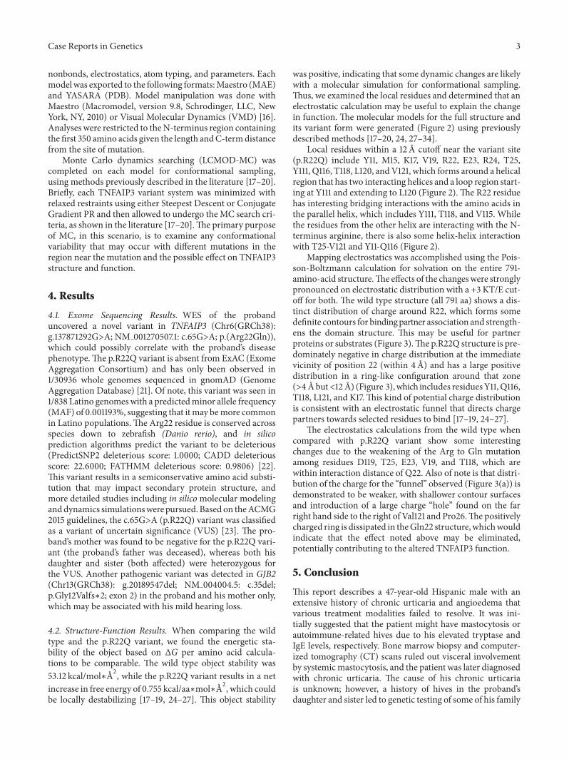
was positive, indicating that some dynamic changes are likelywith a molecular simulation for conformational sampling.Thus, we examined the local residues and determined that anelectrostatic calculation may be useful to explain the changein function. The molecular models for the full structure andits variant form were generated (Figure 2) using previouslydescribed methods [17–20, 24, 27–34].
Local residues within a 12 A cutoff near the variant site(p.R22Q) include Y11, M15, K17, V19, R22, E23, R24, T25,Y111, Q116, T118, L120, andV121, which forms around a helicalregion that has two interacting helices and a loop region start-ing at Y111 and extending to L120 (Figure 2). The R22 residuehas interesting bridging interactions with the amino acids inthe parallel helix, which includes Y111, T118, and V115. Whilethe residues from the other helix are interacting with the N-terminus arginine, there is also some helix-helix interactionwith T25-V121 and Y11-Q116 (Figure 2).
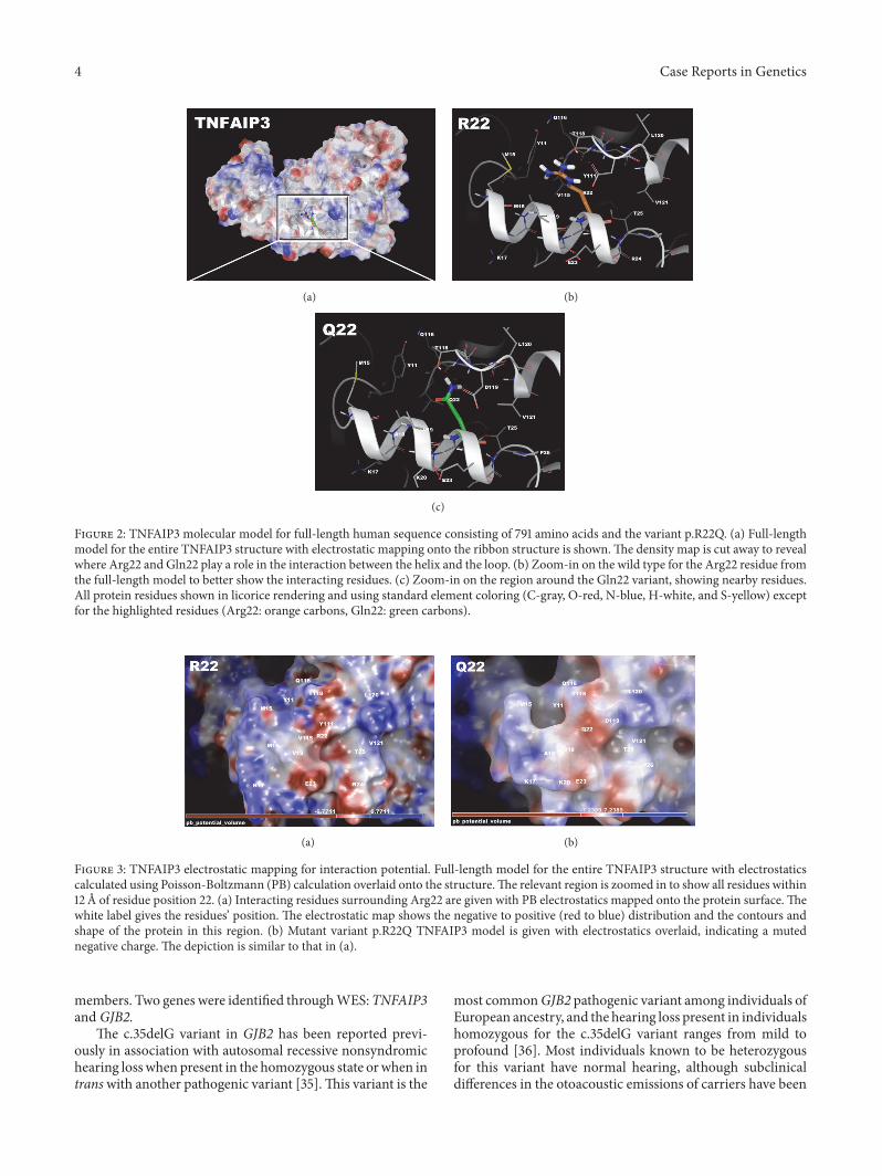
Mapping electrostatics was accomplished using the Pois-son-Boltzmann calculation for solvation on the entire 791-amino-acid structure.The effects of the changeswere stronglypronounced on electrostatic distribution with a +3 KT/E cut-off for both. The wild type structure (all 791 aa) shows a dis-tinct distribution of charge around R22, which forms somedefinite contours for bindingpartner association and strength-ens the domain structure. This may be useful for partnerproteins or substrates (Figure 3).The p.R22Q structure is pre-dominately negative in charge distribution at the immediatevicinity of position 22 (within 4 A) and has a large positivedistribution in a ring-like configuration around that zone(>4 A but<12 A) (Figure 3),which includes residuesY11,Q116,T118, L121, and K17.This kind of potential charge distributionis consistent with an electrostatic funnel that directs chargepartners towards selected residues to bind [17–19, 24–27].
The electrostatics calculations from the wild type whencompared with p.R22Q variant show some interestingchanges due to the weakening of the Arg to Gln mutationamong residues D119, T25, E23, V19, and T118, which arewithin interaction distance of Q22. Also of note is that distri-bution of the charge for the “funnel” observed (Figure 3(a)) isdemonstrated to be weaker, with shallower contour surfacesand introduction of a large charge “hole” found on the farright hand side to the right of Val121 and Pro26.The positivelycharged ring is dissipated in theGln22 structure, whichwouldindicate that the effect noted above may be eliminated,potentially contributing to the altered TNFAIP3 function.
5. Conclusion
This report describes a 47-year-old Hispanic male with anextensive history of chronic urticaria and angioedema thatvarious treatment modalities failed to resolve. It was ini-tially suggested that the patient might have mastocytosis orautoimmune-related hives due to his elevated tryptase andIgE levels, respectively. Bone marrow biopsy and computer-ized tomography (CT) scans ruled out visceral involvementby systemicmastocytosis, and the patient was later diagnosedwith chronic urticaria. The cause of his chronic urticariais unknown; however, a history of hives in the proband’sdaughter and sister led to genetic testing of some of his family
4 Case Reports in Genetics
(a) (b)
(c)
Figure 2: TNFAIP3 molecular model for full-length human sequence consisting of 791 amino acids and the variant p.R22Q. (a) Full-lengthmodel for the entire TNFAIP3 structure with electrostatic mapping onto the ribbon structure is shown.The density map is cut away to revealwhere Arg22 and Gln22 play a role in the interaction between the helix and the loop. (b) Zoom-in on the wild type for the Arg22 residue fromthe full-length model to better show the interacting residues. (c) Zoom-in on the region around the Gln22 variant, showing nearby residues.All protein residues shown in licorice rendering and using standard element coloring (C-gray, O-red, N-blue, H-white, and S-yellow) exceptfor the highlighted residues (Arg22: orange carbons, Gln22: green carbons).
(a) (b)
Figure 3: TNFAIP3 electrostatic mapping for interaction potential. Full-length model for the entire TNFAIP3 structure with electrostaticscalculated using Poisson-Boltzmann (PB) calculation overlaid onto the structure.The relevant region is zoomed in to show all residues within12 A of residue position 22. (a) Interacting residues surrounding Arg22 are given with PB electrostatics mapped onto the protein surface. Thewhite label gives the residues’ position. The electrostatic map shows the negative to positive (red to blue) distribution and the contours andshape of the protein in this region. (b) Mutant variant p.R22Q TNFAIP3 model is given with electrostatics overlaid, indicating a mutednegative charge. The depiction is similar to that in (a).
members. Two genes were identified throughWES:TNFAIP3and GJB2.
The c.35delG variant in GJB2 has been reported previ-ously in association with autosomal recessive nonsyndromichearing losswhen present in the homozygous state orwhen intranswith another pathogenic variant [35].This variant is the
most commonGJB2 pathogenic variant among individuals ofEuropean ancestry, and the hearing loss present in individualshomozygous for the c.35delG variant ranges from mild toprofound [36]. Most individuals known to be heterozygousfor this variant have normal hearing, although subclinicaldifferences in the otoacoustic emissions of carriers have been

Case Reports in Genetics 5
noted upon audiologic examination [37].Theproband’smoth-er was heterozygous for the c.35delG variant inGJB2while hissister and daughter did not carry this variant.
The proband, his daughter, and his sister who are allaffected with chronic urticaria and angioedema each carrythe c.65G>A (p.R22Q) variant in TNFAIP3. The variant isabsent in the proband’smotherwho is unaffected.This variantin TNFAIP3 has not been reported previously as pathogenicor benign to our knowledge and was classified as a VUS.However, TNFAIP3 is known to be involved in immune andinflammatory responses signaled by cytokines, such as tumornecrosis factor-alpha and interleukin-1 beta [38]. Addition-ally, multiple variants within the gene have been associatedwith autoimmune diseases, susceptibility to allergy and asth-ma, and periodic fever syndromes [39].
TNFAIP3 is a ubiquitin-editing enzyme that participatesin ubiquitin ligase and deubiquitinase activities. It is alsoknown to have multiple overlapping functions between nu-clear factor kappa B (NF𝜅B) and interferon regulatory factors(IRF) [8, 40]. There is evidence of cross talk between thesetwo signaling pathways and their roles in the inflammatoryand antiviral responses, which positions TNFAIP3 as a keyeffector of the innate immune response [41]. Recently, Zhouet al. (2016) identified individuals from six different fami-lies affected with a Behcet-like autoinflammatory syndrome(MIM: 616744) that has a variable presentation includinguveitis, chorioretinal scarring, oral, gastrointestinal, and gen-ital ulcers, gastrointestinal inflammation, polyarthritis, rash,periodic fevers, and autoinflammation with the productionof autoantibodies and increased circulating proinflammatorycytokines [42]. The age of onset for this disorder is usuallyin the first or second decade; however, only a few cases havebeen reported to date.
Currently, there is no known association between mis-sense variants inTNFAIP3 and familial chronic urticaria withangioedema. Many patients with chronic urticaria improvewith anti-IgE therapy (omalizumab), suggesting that inap-propriate activation of mast cells and basophils may play arole in disease pathogenesis. It has also been proposed thatsome individuals with chronic urticaria may produce IgEdirected against self-antigens, but evidence for this has beenlacking. The proband in this study had elevated tryptase andIgE levels, suggesting that he may benefit from a trial of thistherapy in the future. Interestingly, TNFAIP3 has been shownto regulate inflammation downstream of themast cell antigenreceptor module as well as the alarmin receptor, IL-33R [43].Heger et al. (2014) showed that mast cell-specific ablationof Tnfaip3 in mice exacerbated disease in mouse models forrheumatoid arthritis and asthma, suggesting that dysregula-tion of mast cell inflammatory responses via Tnfaip3 loss cancontribute to disease pathology [43].
In summary, we report a novel variant in TNFAIP3 inthree family members, which potentially correlates with aphenotype of chronic urticaria. Additional studies, includingfunctional analyses, will be necessary to determine the role ofTNFAIP3 variation in susceptibility to chronic urticaria.
Additional Points
Limitations.Although there were no other disease-associatedvariants identified by WES, it is possible that this individual
may harbor additional sequence variant(s) that are unde-tectable by this test. Chronic urticaria is also a relatively com-mon disorder and may have a nongenetic etiology. However,cases with a strong family historymay beworthy of additionalstudy in order to identify underlying geneticmechanisms thatmay be dysregulated in this condition.
Conflicts of Interest
There are no conflicts of interest reported by the authors.
Acknowledgments
The authors would like to thank the patient and his familyfor participating in this study and the Mayo Clinic Center forIndividualized Medicine for supporting this work.
References
[1] T. Zuberbier, W. Aberer, and R. Asero, “The EAACI/GA(2)LEN/EDF/WAO guideline for the definition, classification,diagnosis, and management of urticaria: the 2013 revision andupdate,” Allergy, vol. 69, no. 7, pp. 868–887, 2014.
[2] J. A. Bernstein, D. M. Lang, D. A. Khan et al., “The diagnosisand management of acute and chronic urticaria: 2014 update,”The Journal of Allergy and Clinical Immunology, vol. 133, no. 5,pp. 1270–1277, 2014.
[3] M.W. Greaves, “Chronic Urticaria,”TheNew England Journal ofMedicine, vol. 332, no. 26, pp. 1767–1772, 1995.
[4] R. H. Champion, S. O. Roberts, R. G. Carpenter, and J. H. Roger,“Urticaria and angio-oedema. A review of 554 patients,” BritishJournal of Dermatology, vol. 81, no. 8, pp. 588–597, 1969.
[5] A. P. Kaplan,Urticaria and Angioedema: synopsis: World allergyorganization, 2004, http://www.worldallergy.org/professional/allergic diseases center/urticaria/urticariasynopsis.php.
[6] P. Losol,HS. Yoo, andHS. Park, “Molecular geneticmechanismsof chronic urticaria,” Allergy, asthma & immunology research,vol. 6, no. 1, pp. 13–21, 2014.
[7] T. E. T. Mevissen, M. K. Hospenthal, P. P. Geurink et al., “OTUdeubiquitinases reveal mechanisms of linkage specificity andenable ubiquitin chain restriction analysis,” Cell, vol. 154, no. 1,pp. X169–184, 2013.
[8] D. Komander and D. Barford, “Structure of the A20 OTU do-main andmechanistic insights into deubiquitination,” Biochem-ical Journal, vol. 409, no. 1, pp. 77–85, 2008.
[9] S. F. Altschul, T. L. Madden, A. A. Schaffer et al., “GappedBLAST and PSI-BLAST: a new generation of protein databasesearch programs,” Nucleic Acids Research, vol. 25, no. 17, pp.3389–3402, 1997.
[10] R. W. W. Hooft, C. Sander, M. Scharf, and G. Vriend, “ThePDBFINDER database: A summary of PDB, DSSP and HSSPinformation with added value,” Computer Applications in theBiosciences, vol. 12, no. 6, pp. 525–529, 1996.
[11] R. W. W. Hooft, G. Vriend, C. Sander, and E. E. Abola, “Errorsin protein structures,” Nature, vol. 381, no. 6580, p. 272, 1996.
[12] R. D. King and M. J. Sternberg, “Identification and applicationof the concepts important for accurate and reliable proteinsecondary structure prediction,” Protein Science, vol. 5, no. 11,pp. 2298–2310, 1996.

6 Case Reports in Genetics
[13] E. Krieger, K. Joo, J. Lee et al., “Improving physical realism,stereochemistry, and side-chain accuracy in homology model-ing: four approaches that performed well in CASP8,” Proteins:Structure, Function, and Bioinformatics, vol. 9, supplement 9, pp.114–122, 2009.
[14] J. Qiu and R. Elber, “SSALN: An alignment algorithm usingstructure-dependent substitution matrices and gap penaltieslearned from structurally aligned protein pairs,” Proteins: Struc-ture, Function, and Genetics, vol. 62, no. 4, pp. 881–891, 2006.
[15] R. A. Laskowski,M.W.MacArthur, D. S.Moss, and J.M.Thorn-ton, “PROCHECK: a program to check the stereochemicalquality of protein structures,” Journal of Applied Crystallogra-phy, vol. 26, no. 2, pp. 283–291, 1993.
[16] W. Humphrey, A. Dalke, and K. Schulten, “VMD: visual mole-cular dynamics,” Journal of Molecular Graphics, vol. 14, no. 1, pp.33–38, 1996.
[17] T. Caulfield and B. Devkota, “Motion of transfer RNA fromthe A/T state into the A-site using docking and simulations,”Proteins: Structure, Function, and Bioinformatics, vol. 80, no. 11,pp. 2489–2500, 2012.
[18] T. Caulfield and J. L. Medina-Franco, “Molecular dynamicssimulations of humanDNAmethyltransferase 3B with selectiveinhibitor nanaomycin A,” Journal of Structural Biology, vol. 176,no. 2, pp. 185–191, 2011.
[19] T. R. Caulfield, “Inter-ring rotation of apolipoprotein A-I pro-tein monomers for the double-belt model using biased molecu-lar dynamics,” Journal of Molecular Graphics andModelling, vol.29, no. 8, pp. 1006–1014, 2011.
[20] T. R. Caulfield, B. Devkota, and G. C. Rollins, “Examinations oftRNA Range of Motion Using Simulations of Cryo-EM Micro-scopy and X-Ray Data,” Journal of Biophysics, vol. 2011, pp. 1–11,2011.
[21] M. Lek, K. J. Karczewski, E. V. Minikel et al., “Analysis of pro-tein-coding genetic variation in 60,706 humans,” Nature, vol.536, no. 7616, pp. 285–291, 2016.
[22] J. Bendl, M. Musil, J. Stourac et al., “PredictSNP2: a unifiedplatform for accurately evaluating SNP effects by exploiting thedifferent characteristics of variants in distinct genomic regions,”PLoS Computational Biology, vol. 12, no. 5, Article ID e1004962,2016.
[23] S. Richards, N. Aziz, and S. Bale, “Standards and guidelines forthe interpretation of sequence variants: a joint consensus re-commendation of the American College of Medical Geneticsand Genomics and the Association for Molecular Pathology,”Genetics in Medicine, vol. 17, no. 5, pp. 405–423, 2015.
[24] F. Lopez-Vallejo, T. Caulfield, K. Martınez-Mayorga et al., “Inte-grating virtual screening and combinatorial chemistry for acce-lerated drug discovery,” Combinatorial Chemistry & HighThroughput Screening, vol. 14, no. 6, pp. 475–487, 2011.
[25] J. Reumers, J. Schymkowitz, J. Ferkinghoff-Borg, F. Stricher, L.Serrano, and F. Rousseau, “SNPeffect: a databasemappingmole-cular phenotypic effects of human non-synonymous codingSNPs,” Nucleic Acids Research, vol. 33, pp. D527–D532, 2005.
[26] J. W. Schymkowitz, F. Rousseau, I. C. Martins, J. Ferkinghoff-Borg, F. Stricher, and L. Serrano, “Prediction of water andmetalbinding sites and their affinities by using the Fold-X force field,”Proceedings of the National Acadamy of Sciences of the UnitedStates of America, vol. 102, no. 29, pp. 10147–10152, 2005.
[27] Y.-J. Zhang, T. Caulfield, Y.-F. Xu et al., “The dual functions ofthe extreme N-terminus of TDP-43 in regulating its biologicalactivity and inclusion formation,” Human Molecular Genetics,vol. 22, no. 15, pp. 3112–3122, 2013.
[28] S. O. Abdul-Hay, A. L. Lane, T. R. Caulfield et al., “Optimiza-tion of peptide hydroxamate inhibitors of insulin-degradingenzyme reveals marked substrate-selectivity,” Journal of Medic-inal Chemistry, vol. 56, no. 6, pp. 2246–2255, 2013.
[29] M. Ando, F. Fiesel, R. Hudec et al., “The PINK1 p.I368N muta-tion affects protein stability and disrupts kinase activity,” Journalof the Neurological Sciences, vol. 381, p. 214, 2017.
[30] T. R. Caulfield, F. C. Fiesel, E. L. Moussaud-Lamodiere, D. F.A. R. Dourado, S. C. Flores, andW. Springer, “Phosphorylationby PINK1 Releases the UBL Domain and Initializes the Con-formational Opening of the E3 Ubiquitin Ligase Parkin,” PLoSComputational Biology, vol. 10, no. 11, 2014.
[31] T. Caulfield, F. Fiesel, and W. Springer, “Activation of the E3ubiquitin ligase Parkin,” Biochemical Society Transactions, vol.43, no. 2, pp. 269–274, 2015.
[32] F. C. Fiesel, M. Ando, R. Hudec et al., “(Patho-)physiologicalrelevance of PINK1-dependent ubiquitin phosphorylation,”EMBO Reports, vol. 16, no. 9, pp. 1114–1130, 2015.
[33] F. C. Fiesel, T. R. Caulfield, E. L. Moussaud-Lamodiere et al.,“Structural and Functional Impact of Parkinson Disease-Asso-ciated Mutations in the E3 Ubiquitin Ligase Parkin,” HumanMutation, vol. 36, no. 8, pp. 774–786, 2015.
[34] A. Puschmann, F. C. Fiesel, T. R. Caulfield et al., “HeterozygousPINK1 p.G411S increases risk of Parkinson’s disease via adominant-negative mechanism,” Brain : a journal of neurology,vol. 140, pp. 98–117, 2017.
[35] A. Kenneson, K. V.N. Braun, andC. Boyle, “GJB2 (connexin 26)variants and nonsyndromic sensorineural hearing loss: a HuGEreview,” Genetics in Medicine, vol. 4, no. 4, pp. 258–274, 2002.
[36] P. Gasparini, R. Rabionet, G. Barbujani et al., “High carrierfrequency of the 35delG deafness mutation in European pop-ulations,” European Journal of Human Genetics, vol. 8, no. 1, pp.19–23, 2000.
[37] R. J. Morell, H. J. Kim, and L. J. Hood, “Mutations in the con-nexin 26 gene (GJB2) among Ashkenazi jews with nonsyndro-mic recessive deafness,” The New England Journal of Medicine,vol. 339, no. 21, pp. 1500–1505, 1998.
[38] “Tumor necrosis factor alpha-induced protein 3. UniProt Con-sortium,” http://www.uniprot.org/uniprot/P21580.
[39] “Tumor necrosis factor receptor superfamily, member 1A;TNFRSF1A,” Johns Hopkins University, 2017, https://www.omim.org/entry/191190#references.
[40] I. E. Wartz, K. M. O’Rourke, H. Zhou et al., “De-ubiquitinationand ubiquitin ligase domains of A20 downregulate NF-𝜅Bsignalling,” Nature, vol. 430, no. 7000, pp. 694–699, 2004.
[41] M. Iwanaszko and M. Kimmel, “NF-𝜅B and IRF pathways:cross-regulation on target genes promoter level,” BMC Genom-ics, vol. 16, no. 1, 2015.
[42] Q. Zhou, H. Wang, D. M. Schwartz et al., “Loss-of-functionmutations in TNFAIP3 leading to A20 haploinsufficiency causean early-onset autoinflammatory disease,” Nature Genetics, vol.48, no. 1, pp. 67–73, 2016.
[43] K. Heger, K. Fierens, J. C. Vahl et al., “A20-Deficient Mast CellsExacerbate Inflammatory Responses InVivo,”PLoS Biology, vol.12, no. 1, Article ID e1001762, 2014.

Stem Cells International
Hindawiwww.hindawi.com Volume 2018
Hindawiwww.hindawi.com Volume 2018
MEDIATORSINFLAMMATION
of
EndocrinologyInternational Journal of
Hindawiwww.hindawi.com Volume 2018
Hindawiwww.hindawi.com Volume 2018
Disease Markers
Hindawiwww.hindawi.com Volume 2018
BioMed Research International
OncologyJournal of
Hindawiwww.hindawi.com Volume 2013
Hindawiwww.hindawi.com Volume 2018
Oxidative Medicine and Cellular Longevity
Hindawiwww.hindawi.com Volume 2018
PPAR Research
Hindawi Publishing Corporation http://www.hindawi.com Volume 2013Hindawiwww.hindawi.com
The Scientific World Journal
Volume 2018
Immunology ResearchHindawiwww.hindawi.com Volume 2018
Journal of
ObesityJournal of
Hindawiwww.hindawi.com Volume 2018
Hindawiwww.hindawi.com Volume 2018
Computational and Mathematical Methods in Medicine
Hindawiwww.hindawi.com Volume 2018
Behavioural Neurology
OphthalmologyJournal of
Hindawiwww.hindawi.com Volume 2018
Diabetes ResearchJournal of
Hindawiwww.hindawi.com Volume 2018
Hindawiwww.hindawi.com Volume 2018
Research and TreatmentAIDS
Hindawiwww.hindawi.com Volume 2018
Gastroenterology Research and Practice
Hindawiwww.hindawi.com Volume 2018
Parkinson’s Disease
Evidence-Based Complementary andAlternative Medicine
Volume 2018Hindawiwww.hindawi.com
Submit your manuscripts atwww.hindawi.com