casereport igg4-related disease presenting as isolated...
TRANSCRIPT
Case ReportIgG4-Related Disease Presenting as Isolated Scleritis
Eran Berkowitz, Ella Arnon, Alona Yaakobi, Yuval Cohen, and Beatrice Tiosano
Department of Ophthalmology, Hillel Yaffe Medical Center Affiliated with The Bruce Rappaport School of Medicine,The Technion, Haifa, Israel
Correspondence should be addressed to Eran Berkowitz; [email protected]
Received 27 July 2016; Accepted 29 November 2016; Published 9 January 2017
Academic Editor: Alexander A. Bialasiewicz
Copyright © 2017 Eran Berkowitz et al.This is an open access article distributed under the Creative Commons Attribution License,which permits unrestricted use, distribution, and reproduction in any medium, provided the original work is properly cited.
A rare case of IgG4-related disease (IgG4-RD) manifesting as nodular scleritis is presented in a 20-year-old female. Patientcomplained of left eye pain and redness for one week. Ocular examination together with ancillary testing led to the diagnosisof nodular scleritis. Since the patient did not show apparent improvement after one week of systemic steroidal treatment,she underwent a biopsy of the affected area revealing histopathological characteristics of IgG4-RD. Long-term treatment withcorticosteroids and a steroid-sparing agent (methotrexate) led to significant improvement in signs and symptoms. This casehighlights the significance of IgG4-RD in the differential diagnosis of scleritis and raises the question as to whether various organsaffected by IgG4-RD may have different underlying pathophysiological mechanisms in which pathogenic T cells play a role.
1. Introduction
Immunoglobulin G4-related disease (IgG4-RD) is animmune-mediated condition that can affect almost anyorgan.The typical ophthalmicmanifestation is lacrimal glandenlargement (dacryoadenitis). The diagnosis can be con-firmed by blood tests and histology during the acute phase.We present a unique case of nodular scleritis due to isolatedIgG4-RD, which responded well to immunosuppressive andimmunomodulatory treatment.
2. Report of a Case
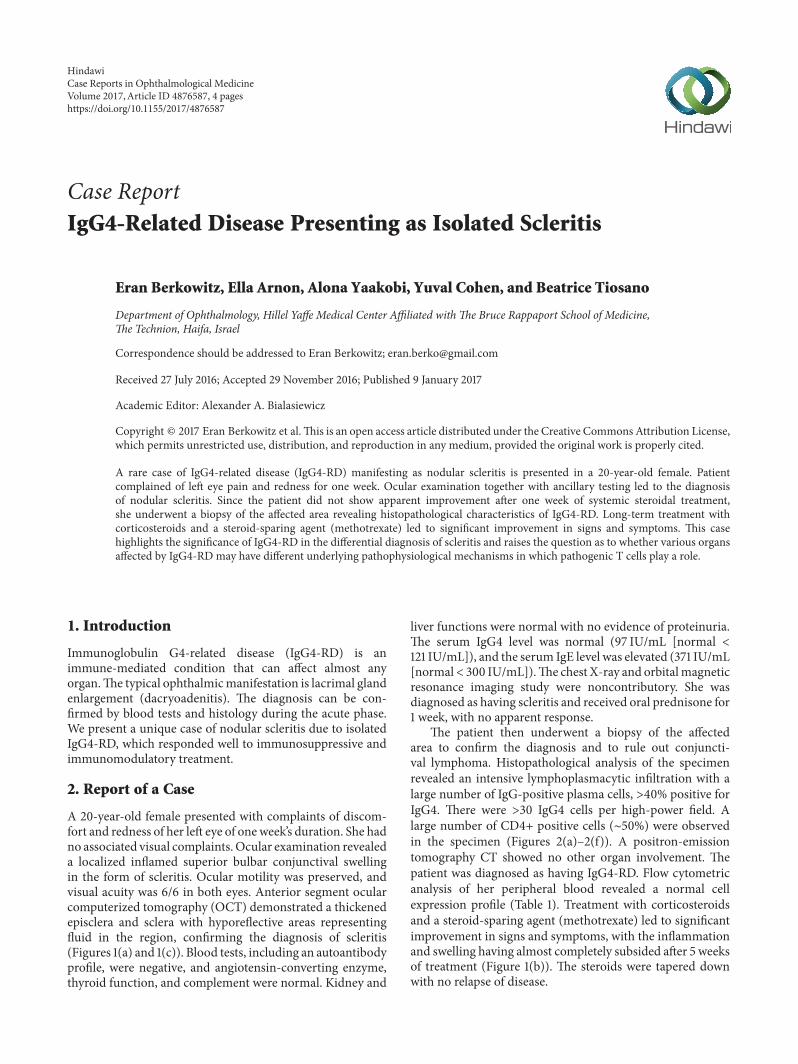
A 20-year-old female presented with complaints of discom-fort and redness of her left eye of oneweek’s duration. She hadno associated visual complaints.Ocular examination revealeda localized inflamed superior bulbar conjunctival swellingin the form of scleritis. Ocular motility was preserved, andvisual acuity was 6/6 in both eyes. Anterior segment ocularcomputerized tomography (OCT) demonstrated a thickenedepisclera and sclera with hyporeflective areas representingfluid in the region, confirming the diagnosis of scleritis(Figures 1(a) and 1(c)). Blood tests, including an autoantibodyprofile, were negative, and angiotensin-converting enzyme,thyroid function, and complement were normal. Kidney and
liver functions were normal with no evidence of proteinuria.The serum IgG4 level was normal (97 IU/mL [normal <121 IU/mL]), and the serum IgE level was elevated (371 IU/mL[normal< 300 IU/mL]).The chest X-ray and orbitalmagneticresonance imaging study were noncontributory. She wasdiagnosed as having scleritis and received oral prednisone for1 week, with no apparent response.
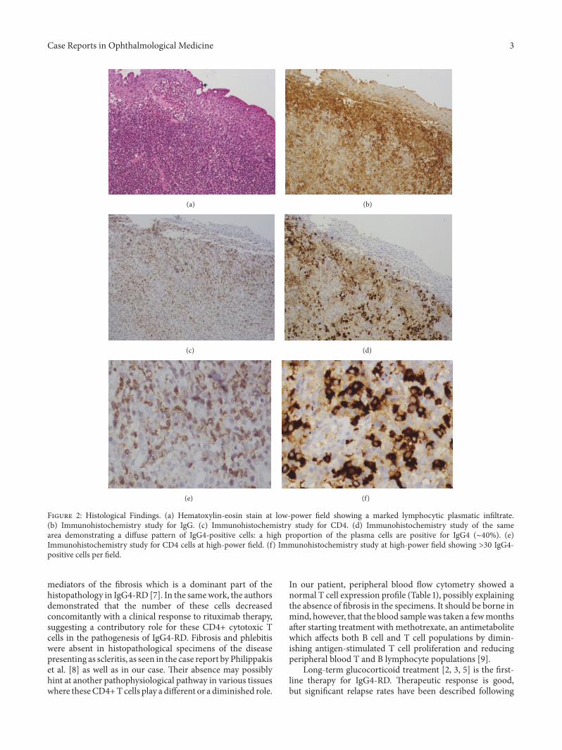
The patient then underwent a biopsy of the affectedarea to confirm the diagnosis and to rule out conjuncti-val lymphoma. Histopathological analysis of the specimenrevealed an intensive lymphoplasmacytic infiltration with alarge number of IgG-positive plasma cells, >40% positive forIgG4. There were >30 IgG4 cells per high-power field. Alarge number of CD4+ positive cells (∼50%) were observedin the specimen (Figures 2(a)–2(f)). A positron-emissiontomography CT showed no other organ involvement. Thepatient was diagnosed as having IgG4-RD. Flow cytometricanalysis of her peripheral blood revealed a normal cellexpression profile (Table 1). Treatment with corticosteroidsand a steroid-sparing agent (methotrexate) led to significantimprovement in signs and symptoms, with the inflammationand swelling having almost completely subsided after 5 weeksof treatment (Figure 1(b)). The steroids were tapered downwith no relapse of disease.
HindawiCase Reports in Ophthalmological MedicineVolume 2017, Article ID 4876587, 4 pageshttps://doi.org/10.1155/2017/4876587
2 Case Reports in Ophthalmological Medicine
(a) (b)
(c)
Figure 1: Color photograph of the superior bulbar area showing the area of inflammation before (a) and after (b) 5 weeks of treatment.Anterior segment ocular computerized tomography showing a thickened sclera and episclera with hyporeflective areas (asterisks) beforetreatment (c).
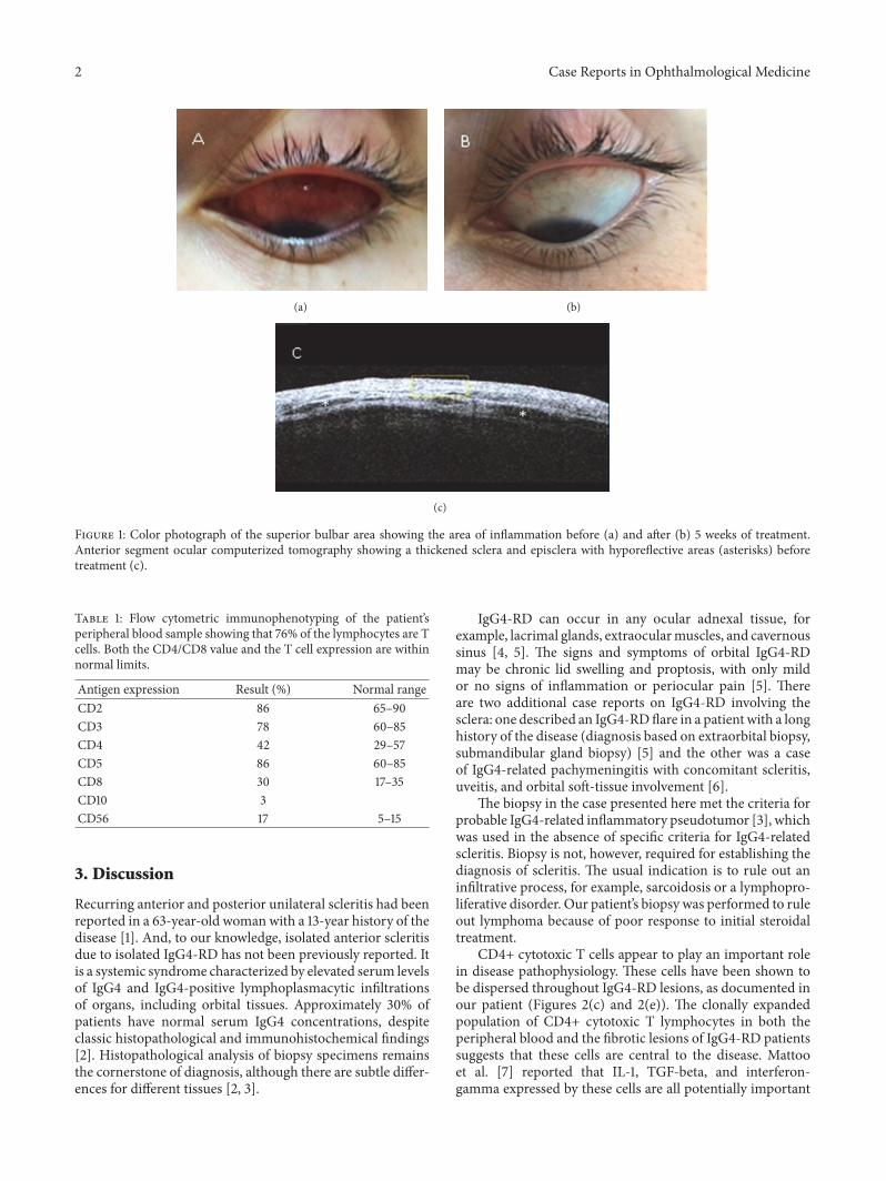
Table 1: Flow cytometric immunophenotyping of the patient’speripheral blood sample showing that 76% of the lymphocytes are Tcells. Both the CD4/CD8 value and the T cell expression are withinnormal limits.
Antigen expression Result (%) Normal rangeCD2 86 65–90CD3 78 60–85CD4 42 29–57CD5 86 60–85CD8 30 17–35CD10 3CD56 17 5–15
3. Discussion
Recurring anterior and posterior unilateral scleritis had beenreported in a 63-year-old woman with a 13-year history of thedisease [1]. And, to our knowledge, isolated anterior scleritisdue to isolated IgG4-RD has not been previously reported. Itis a systemic syndrome characterized by elevated serum levelsof IgG4 and IgG4-positive lymphoplasmacytic infiltrationsof organs, including orbital tissues. Approximately 30% ofpatients have normal serum IgG4 concentrations, despiteclassic histopathological and immunohistochemical findings[2]. Histopathological analysis of biopsy specimens remainsthe cornerstone of diagnosis, although there are subtle differ-ences for different tissues [2, 3].
IgG4-RD can occur in any ocular adnexal tissue, forexample, lacrimal glands, extraocularmuscles, and cavernoussinus [4, 5]. The signs and symptoms of orbital IgG4-RDmay be chronic lid swelling and proptosis, with only mildor no signs of inflammation or periocular pain [5]. Thereare two additional case reports on IgG4-RD involving thesclera: one described an IgG4-RDflare in a patient with a longhistory of the disease (diagnosis based on extraorbital biopsy,submandibular gland biopsy) [5] and the other was a caseof IgG4-related pachymeningitis with concomitant scleritis,uveitis, and orbital soft-tissue involvement [6].
The biopsy in the case presented here met the criteria forprobable IgG4-related inflammatory pseudotumor [3], whichwas used in the absence of specific criteria for IgG4-relatedscleritis. Biopsy is not, however, required for establishing thediagnosis of scleritis. The usual indication is to rule out aninfiltrative process, for example, sarcoidosis or a lymphopro-liferative disorder. Our patient’s biopsy was performed to ruleout lymphoma because of poor response to initial steroidaltreatment.
CD4+ cytotoxic T cells appear to play an important rolein disease pathophysiology. These cells have been shown tobe dispersed throughout IgG4-RD lesions, as documented inour patient (Figures 2(c) and 2(e)). The clonally expandedpopulation of CD4+ cytotoxic T lymphocytes in both theperipheral blood and the fibrotic lesions of IgG4-RD patientssuggests that these cells are central to the disease. Mattooet al. [7] reported that IL-1, TGF-beta, and interferon-gamma expressed by these cells are all potentially important
Case Reports in Ophthalmological Medicine 3
(a) (b)
(c) (d)
(e) (f)
Figure 2: Histological Findings. (a) Hematoxylin-eosin stain at low-power field showing a marked lymphocytic plasmatic infiltrate.(b) Immunohistochemistry study for IgG. (c) Immunohistochemistry study for CD4. (d) Immunohistochemistry study of the samearea demonstrating a diffuse pattern of IgG4-positive cells: a high proportion of the plasma cells are positive for IgG4 (∼40%). (e)Immunohistochemistry study for CD4 cells at high-power field. (f) Immunohistochemistry study at high-power field showing >30 IgG4-positive cells per field.
mediators of the fibrosis which is a dominant part of thehistopathology in IgG4-RD [7]. In the samework, the authorsdemonstrated that the number of these cells decreasedconcomitantly with a clinical response to rituximab therapy,suggesting a contributory role for these CD4+ cytotoxic Tcells in the pathogenesis of IgG4-RD. Fibrosis and phlebitiswere absent in histopathological specimens of the diseasepresenting as scleritis, as seen in the case report by Philippakiset al. [8] as well as in our case. Their absence may possiblyhint at another pathophysiological pathway in various tissueswhere theseCD4+T cells play a different or a diminished role.
In our patient, peripheral blood flow cytometry showed anormal T cell expression profile (Table 1), possibly explainingthe absence of fibrosis in the specimens. It should be borne inmind, however, that the blood samplewas taken a fewmonthsafter starting treatment with methotrexate, an antimetabolitewhich affects both B cell and T cell populations by dimin-ishing antigen-stimulated T cell proliferation and reducingperipheral blood T and B lymphocyte populations [9].
Long-term glucocorticoid treatment [2, 3, 5] is the first-line therapy for IgG4-RD. Therapeutic response is good,but significant relapse rates have been described following
4 Case Reports in Ophthalmological Medicine
steroid discontinuation with a rise in the serum IgG4 levels,which necessitates a lowmaintenance dose of corticosteroids,possibly in combination with steroid-sparing agent, as waseffective in the present case.
Based on these findings, we believe that the variousorgans affected by IgG4-RD may have different underlyingpathophysiological mechanisms in which pathogenic T cellmay play a different role.
Competing Interests
No conflicting relationship exists for any author.
Acknowledgments
The authors thank Dr. Vicktoria (Vicky) Vishnevskia Dai,the Goldschleger Eye Institute, Tel HaShomer Hospital, forsharing her pearls of wisdom and insights in the care of thispatient.
References
[1] J. H. Stone, Y. Zen, and V. Deshpande, “IgG4-related disease,”New England Journal of Medicine, vol. 366, no. 6, pp. 539–551,2012.
[2] H. Umehara, K. Okazaki, Y. Masaki et al., “Comprehensivediagnostic criteria for IgG4-related disease (IgG4-RD), 2011,”Modern Rheumatology, vol. 22, no. 1, pp. 21–30, 2012.
[3] J. A. Plaza, J. A. Garrity, A. Dogan, A. Ananthamurthy, T. E.Witzig, and D. R. Salomao, “Orbital inflammation with IgG4-positive plasma cells: manifestation of IgG4 systemic disease,”Archives of Ophthalmology, vol. 129, no. 4, pp. 421–428, 2011.
[4] T. Kubota and S. Moritani, “Orbital IgG4-related disease: clini-cal features and diagnosis,” ISRN Rheumatology, vol. 2012,Article ID 412896, 5 pages, 2012.
[5] Z. S.Wallace, V.Deshpande, and J.H. Stone, “Ophthalmicmani-festations of IgG4-related disease: single-center experience andliterature review,” Seminars inArthritis andRheumatism, vol. 43,no. 6, pp. 806–817, 2014.
[6] A. Kosakai, D. Ito, S. Yamada, S. Ideta, Y. Ota, and N. Suzuki, “Acase of definite IgG4-related pachymeningitis,” Neurology, vol.75, no. 15, pp. 1390–1392, 2010.
[7] H. Mattoo, V. S. Mahajan, T. Maehara et al., “Clonal expansionof CD4+ cytotoxic T lymphocytes in patients with IgG4-relateddisease,” Journal of Allergy and Clinical Immunology, vol. 138,no. 3, pp. 825–838, 2016.
[8] E. Philippakis, N. Cassoux, F. Charlotte et al., “IgG4-relateddisease masquerading as recurrent scleritis and chronic con-junctivitis,” Ocular Immunology and Inflammation, vol. 23, no.2, pp. 168–172, 2015.
[9] E. S. L. Chan and B. N. Cronstein, “Molecular action of metho-trexate in inflammatory diseases,” Arthritis Research, vol. 4, no.4, pp. 266–273, 2002.

Submit your manuscripts athttps://www.hindawi.com
Stem CellsInternational
Hindawi Publishing Corporationhttp://www.hindawi.com Volume 2014
Hindawi Publishing Corporationhttp://www.hindawi.com Volume 2014
MEDIATORSINFLAMMATION
of
Hindawi Publishing Corporationhttp://www.hindawi.com Volume 2014
Behavioural Neurology
EndocrinologyInternational Journal of
Hindawi Publishing Corporationhttp://www.hindawi.com Volume 2014
Hindawi Publishing Corporationhttp://www.hindawi.com Volume 2014
Disease Markers
Hindawi Publishing Corporationhttp://www.hindawi.com Volume 2014
BioMed Research International
OncologyJournal of
Hindawi Publishing Corporationhttp://www.hindawi.com Volume 2014
Hindawi Publishing Corporationhttp://www.hindawi.com Volume 2014
Oxidative Medicine and Cellular Longevity
Hindawi Publishing Corporationhttp://www.hindawi.com Volume 2014
PPAR Research
The Scientific World JournalHindawi Publishing Corporation http://www.hindawi.com Volume 2014
Immunology ResearchHindawi Publishing Corporationhttp://www.hindawi.com Volume 2014
Journal of
ObesityJournal of
Hindawi Publishing Corporationhttp://www.hindawi.com Volume 2014
Hindawi Publishing Corporationhttp://www.hindawi.com Volume 2014
Computational and Mathematical Methods in Medicine
OphthalmologyJournal of
Hindawi Publishing Corporationhttp://www.hindawi.com Volume 2014
Diabetes ResearchJournal of
Hindawi Publishing Corporationhttp://www.hindawi.com Volume 2014
Hindawi Publishing Corporationhttp://www.hindawi.com Volume 2014
Research and TreatmentAIDS
Hindawi Publishing Corporationhttp://www.hindawi.com Volume 2014
Gastroenterology Research and Practice
Hindawi Publishing Corporationhttp://www.hindawi.com Volume 2014
Parkinson’s Disease
Evidence-Based Complementary and Alternative Medicine
Volume 2014Hindawi Publishing Corporationhttp://www.hindawi.com