cases submitted for postmortem examination to universiti...
TRANSCRIPT
Pertanika 3(2), 155-158 (1980)
SHORT COMMUNICATION (I)
Cases submitted for Postmortem Examination toUniversiti Pertanian Malaysia in 1977
RINGKASAN
Satu kajian telah dibuat di atas kes-kes yang diterima untuk pemeriksaan posm t l h '¥ b. .,. . " . or em 0 e J a atanP~th?logz dan. !VIzkr?bzologz r:eter~nar) Unzversztz Pertanian d~lam tah~n 1977. Sebanyak 1413 kes telahd~t~rzma terdzrz da~z 13 speszs hazwan. Data tentang penyakzt-penyakzt yang diketemui dibentan k ddzbzncang dengan rzngkas. 'If an an
INTRODUCTION
The Department of Veterinary Pathologyand Microbiology, Faculty of Veterinary Medicineand Animal Sciences, Universiti Pertanian Malaysia (UPM), started receiving animal carcassesand tissues for teaching and diagnostic purposesin 1975. This marked the start of the teachingof the Veterinary Pathology course for the Doctorof Veterinary Medicine (DVM) programme inthe 1975/1976 session. This is a report on casesreceived in the calendar year 1977.
MATERIALS AND METHODS
All cases received for postmortem examination by the Department of Veterinary Pathology and Microbiology in 1977 were studied.The livestock carcasses came from UPM,MARDI and several neighbouring private farms,while dog and cat carcasses were submitted bythe SPCA and private owners. Tissues submittedcomprised either materials condemned atslaughter from the Shah Alam abattoir or biopsiesperformed routinely by DVM students.
Most of the diagnoses made on the caseswere based on gross pathological findings only.Routine microbiological and histological studieswere limited to a small proportion of cases onlysince the respective laboratories were not fullyequipped for diagnostic purposes. In view ofthis, the study only attempts to arrive at conclusions on the common diseases and conditionsseen in the various species of animals received.No attempts were made to enumerate the actualnumber of animals involved in the variousdiseases.
155
RESULTS AND DISCUSSION
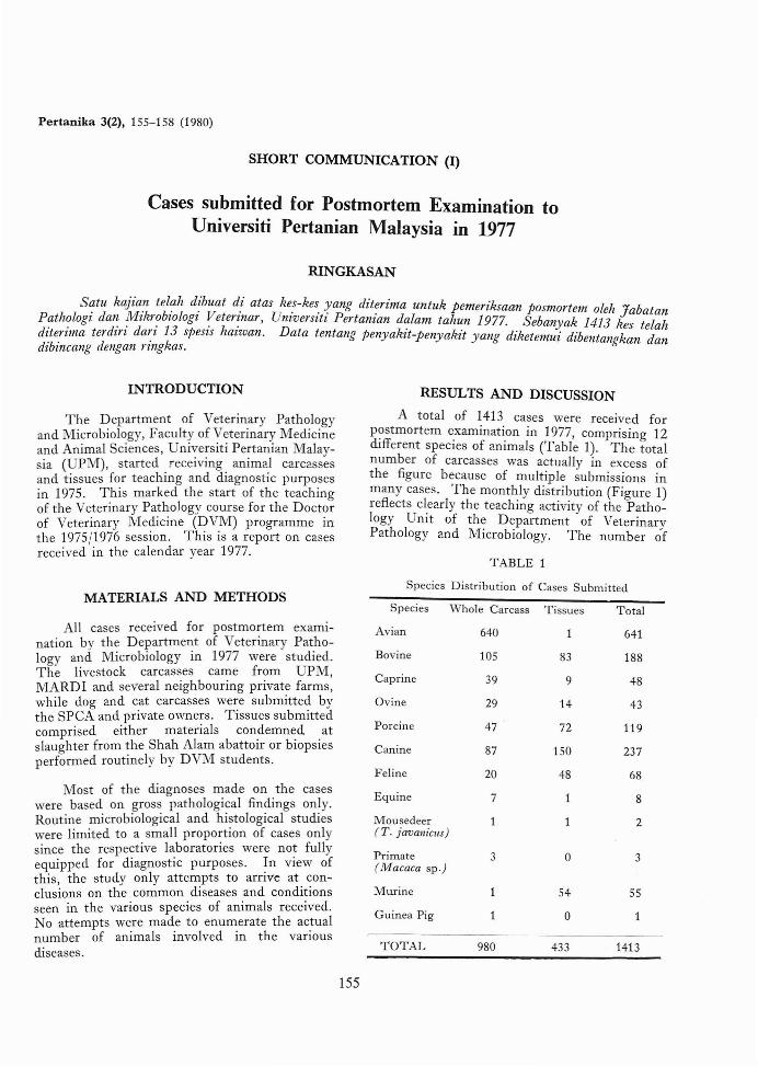
A total of 1413 cases were received forP?stmortem examination in 1977, comprising 12dIfferent species of animals (Table 1). The totalnumber of carcasses was actually in excess ofthe figure because of multiple submissions inmany cases. The monthly distribution (Figure 1)reflects cl.early the teaching activity of the Pathology Uillt of the Department of VeterinaryPathology and Microbiology. The number of
TABLE 1
Species Distribution of Cases Submitted
Species Whole Carcass Tissues Total
Avian 640 641
Bovine 105 83 188
Caprine 39 9 48
Ovine 29 14 43
Porcine 47 72 119
Canine 87 150 237
Feline 20 48 68
Equine 7 8
Mousedeer 2(T. javal1icus)
Primate 3 0 3(Tv!acaca sp.)
Mmine 54 55
Guinea Pig 0 1
TOTAL 980 433 1413

200
A. R. SHEIKH·OMAR AND U. CHULAN
195192
Number
of
Submissions
160
120
80
40
J F M A M JMONTHS
J A S o N D
~ Whole Carcass
o Tissues
Fig. 1. Monthly Distribution of Cases Submitted.
cases was low during the long vacation in themonths of April and May and during the intersemester break in October. The submissions ofbovine, ovine and porcine tissues from the ShahAlam abattoir as well as canine and feline biopsied tissues during these periods, were eitherentirely nil or very low. The total number ofsubmissions in March and November was high.The high count in March was due to assigning apathology number to each individual bird ratherthan to a batch of birds submitted from thesame flock; in November there was a largenumber of murine tissues submitted for histopathological studies.
Avian cases
The common diseases seen are shown inTable 2. Leucocytozoonosis was the mostcommon disease. It affected birds between theages of 6-8 weeks causing a mortality rate ofbetween 5-10% per flock. Newcastle Diseaseoccurs in sporadic outbreaks in certain flocksperhaps due to vaccination br~akdowns.
156
TABLE 2
Avian: The common diagnoses listed accordingto estimated frequency
1. Leucocytozoonosis2. Coccidiosis3 . Air sacculitis4. Colibacillosis5. Leucosis6. Marek's Disease7. Nephrosis8. Starvation9. Visceral gout
10. Newcastle Disease11. Cannibalism
Bovine cases
Table 3 shows the common diagnoses madein ruminant carcasses including bovine. Mohd.Anwar (1977) gives a more comprehensive listof cattle diseases found in Malaysia. With theexception of Bovine Malignant Catarrh (BMC)most of the other diseases involved single animals

CASES FOR POSTMORTEM AT UPM IN 1977
and were observed either once or at the mostonly in several cases. BMC was seen in 29 deadcattle submitted from a research station and h2.sbeen reported elsewhere (Vanselow, 1977).Oxalate nephrosis occurred in several cattle thathad been grazing on Setaria splendida for a fewdays. Of the abattoir specimens, the majorityconsisted of livers with Fasciola gigantica andGigantocotyle explanatum infections, hepaticabscessations, pulmonary abscessations, chronicproliferative pericarditis and aortas with theparasite Eleophora poeli.
Caprine and ovine casesThe common diagnoses made on carcasses
are also listed in Table 3. Starvation/malnutrition and internal parasitism were the mostcommon findings. Caprine tissues comprisedmainly biopsies of normal uteruses and ovarieswhile ovine tissues were mainly abattoir specimens, the majority of which were lungs andmediastinal lymph nodes with caseous abscessations caused by Corynebacterium ovis.
Porcine casesThe tissues consisted mainly of abattoir
specimens. The common diseases and conditions seen are shown in Table 4. The occurrence
of melioidosis was of significance because of itszoonotic behaviour.
Canine and feline casesThe common diseases and conditions seen
are listed in Table 5. Dirofilariasis was a commonproblem in dogs consistent with the finding of arecent survey which showed an incidence of25.8% among 764 dogs (Retnasabapathy andKhoo, 1976). Among cats, panleukopenia is acommon problem. The tissues submitted inboth species were normal uteruses ovaries testeskidney, spleen and intestines removed b; DVMstudents at surgical exercise.
Equine casesSix of the 7 carcasses came from the UPM
Horse Unit, most of which showed inguinaledema and meningoencephalitis suggestive oftrypanosomiasis (Surra) and corroborated bythe clinicopathologic studies (Ng and Vanselow,1979; Seiler et ai, 1980). One of these horsesalso had salmonellosis. The horse submittedfrom outside UPM had focal encephalomalaciaof the brain stem.
TABLE 3
Ruminants: The common diagnoses in carcasses
Bovine
1. Bovine malignant catarrh 1.2. Starvation/malnutrition 2.
3. Internal parasitism 3.(mainly hemonchosis)
4. Oxalate nephrosis 4.S. Babesiosis S.6. Pneumonia (mainly aspiration) 6.7. Abortion
(unestablished aetiology)8. Stillbirth9. Dystocia
10. Brucellosis11. Haemorrhagic septicaemia
Caprine
Starvation/malnutritionInternal parasitism(hemonchosis & coccidiosis)External parasitism(lice and mites)BronchopneumoniaEnterotoxaemiaContagious ecthyma
TABLE 4
Ovine
1. Hemonchosis2. Starvation/malnutrition
3. Pregnancy toxaemia
4. EnterotoxaemiaS. Suspected plant poisonings
Whole Carcass
Porcine: The common diagnoses
Abattoir Specimen
1. Abscessation (various organs)2. Enteritis (salmonellosis & colibacillosis)3. Pneumonia4. Septicemia (E. coli)5. Aujesky's Disease6. Erysipelas7. Melioidosis
157
1. Chronic Interstitial Nephritis2. Hydronephrosis3. Renal cysts4. Abscessation (mainly liver)5. Pneumonia6. Embryonal nephroblastoma

A. R. SHEIKH-OMAR AND U. CHULAN
TABLE 5
Canine and Feline: The common diagnoses in carcasses
Canine Feline
A. R. Sheikh-OmarU. Chulan
1. Dirofilariasis
2. Parasitism - spirocercosis- hookworms
3. Distemper - Bronchopneumonia- Encephalitis
4. Parasitic dermatitis - demodectic mange
5. Tumours - Transmissible Venereal tumour- Lymphosarcoma- Hemangiosarcoma
6. Tropical pancytopenia(Ehrlichosis)
CONCLUSION
The data presented indicate the animaldiseases present in Malaysia or, at least, inSelangor. However, it is too biased and sdectiveto allow for a detailed epidemiological study onthe various diseases observed. Submission ofcases was determined by various factors such asthe level of teaching activity, geography, transportation and also the availability of sources.
ACKNOWLEDGEMENT
The authors would like to thank Cik Jamilahbinti Haji Abdul Rahman for typing the manuscript.
Department of Veterinary Pathology andMicrobiology,
Faculty of Veterinary Medicine andAnimal Science,
Universiti Pertanian Malaysia,Serdang.
•
158
1. Infectious Feline Enteritis(Panleukopenia)
2. Chronic Interstitial Nephritis
3. Cystitis
4. Diaphragmatic hernia
5. Squamous cell carcinoma
6. Necrotising dermatitis
REFERENCES
MOHD. ANWAR, H. (1977): Common Diseases ofCattle and Bufialoes in Peninsular Malaysia.Proceedings of the First Joint Conference of theAssociation of Cattle Veterinarians, May 16-20,1977. Bulletin. 146: 90-102. Ministry of Agriculture, Kuala Lumpur.
1 G, B.K.Y. and VANSELOW, B. (1978): Outbreak ofSurra in Horses and the Pathogenesis of theAnaemia. Kajian Veterinar. 10 (2): 88-98.
RETNASABAPATHY, A. and KHoo, T.S. (1976): Incidence of Canine Heartworm (Dirofilaria immitis)in Malaysia. Vet. Rec. 98: 68-69.
SEILER, R.J., OMAR, S. and JACKSON, A.R.B. (1980):Meningo-encephalitis in naturally occurringTrypanosoma evansi Infection (Surra) of Horses.Vet. Path. (In press).
VANSELOW, B.A. (1977): An outbreak of BovineMalignant Catarrh in Selangor, Malaysia. Proceedings of the First Joint Conference of theAssociation of Cattle Veterinarians, May 16-20,1977. Bulletin. 146: 108-121. Ministry ofAgriculture, Kuala Lumpur.
(Received 4 October 1979)