casting and traction treatment methods for scoliosis and traction_ sanders.pdf · fig. 1. a proper...
TRANSCRIPT

Orthop Clin N Am 38 (2007) 477–484
Casting and Traction TreatmentMethods for Scoliosis
Jacques L. D’Astous, MD, FRCS(C)a,b,*, James O. Sanders, MDc
aDepartment of Orthopaedic Surgery, University of Utah Orthopaedic Center, 590 Wakara Way,
Salt Lake City, UT 84108, USAbShriners Hospitals for Children – Intermountain, Fairfax Road & Virginia Street,
Salt Lake City, UT 84103, USAcShriners Hospitals for Children, 1645 West 8th Street, Erie, PA 16505, USA
Casting for infantile or early-onset scoliosis
Casting for scoliosis was common until PaulHarrington introduced effective spinal instrumen-tation. Since then, casting largely has been rele-gated to history books along with racks, corsets,
and other medieval implements. As its role di-minished, knowledge of casting techniques dis-appeared, experienced surgeons passed on, and
the remaining historic relicsdcasting tablesdfellinto disuse and were discarded.
To a large degree, the abandonment of casting
is justified. Instrumentation is now solid andsecure, provides excellent curve correction, andallows rapid mobilization and return to activity. A
rigid cast can create pressure sores, significant ribor mandibular deformities, and constrict thechest. The historical term for superior mesentericartery syndrome is ‘‘cast syndrome.’’ Many of
these problems seem to be the result of indiscrim-inate casting of all types of scoliosis and impropertechnique combined with a limited understanding
of spinal, and, particularly, chest wall deformities.We have used serial casting in a small group of
our patients for several years, and our early results
indicate that in selected patients and with propertechnique, casting plays a useful role in ourtreatment armamentarium for early-onset scoliosis.
* Corresponding author. Shriners Hospitals for
Children – Intermountain, Fairfax Road & Virginia
Street, Salt Lake City, UT 84103.
E-mail address: [email protected]
(J.L. D’Astous).
0030-5898/07/$ - see front matter � 2007 Elsevier Inc. All r
doi:10.1016/j.ocl.2007.03.006
With few exceptions, we have confined scolio-
sis casting to patients who had infantile scoliosisand do not believe it has a role in adolescent,neuromuscular, or congenital scoliosis with therare indications for postoperative stabilization,
temporary correction, or immobilization to assesspain relief.
Scoliosis casting comes in several varieties. The
most commonly used method in the United States isthat of Risser [1]. Indeed, body casting in this coun-try often is called Risser casting. Risser developed
two types of casts. The initial method used turn-buckles, whereas the latter, termed a localizer cast,used a three-point mold and a pusher. Although it
is possible to obtain significant curve correctionwith this technique, it does not account sufficientlyfor rotational abnormality, and, especially in youn-ger children with flexible bones, it can cause signifi-
cant rib deformities and chest constriction.Scoliosis in young children has two classifica-
tions. The traditional classification used by the
Scoliosis Research Society divides these curves intoinfantile and juvenile, based on whether the curvewas diagnosedbefore or after the ageof 3 years. The
classification of Dickson [2] divides curves intoearly onset (diagnosed before age 5 years) and lateonset (diagnosed after age 5 years). Dickson’s logicwas that curves occurring after age 5 years were less
likely to result in pulmonary failure. We prefer theformer classification because of Pehrsson and col-leagues’ [3] work showing increased mortality in
the infantile and juvenile curves and because youn-ger age of treatment, in our experience, seems toimprove the results.
ights reserved.
orthopedic.theclinics.com

478 D’ASTOUS & SANDERS
Infantile idiopathic scoliosis occurs in twobasic types: resolving and progressive. Mehta [4]was able to distinguish resolving scoliosis from
progressive scoliosis by using the rib vertebral an-gle difference (RVAD) on an early supine radio-graph. The RVAD nearly follows the 80/20 rule:80% of curves with an RVAD of 20 or more con-
tinue to progress, whereas only 20% of those withlesser angles progress. This was confirmed byother investigators [5–7].
More recently,Mehta [8] described her results ofcasting in 136 patients who had infantile scoliosisusing the technique of Cotrel and Morel [9] with
the philosophy that early rapid growth, if guidedby the cast, would assist an initially curved spineto straighten. She grouped her patients into fourphysiologic patterns: a ‘‘sturdy phenotype’’ with
goodmuscle mass and tone; a ‘‘slender phenotype’’withmore delicate features, ligamentous laxity, andmore rapidly progressive curves; those with known
syndromes; and those with unknown syndromes.Her program consisted of cast changes under anes-thesia in younger patients every 8 to 16 weeks until
the curvewas nearly resolved followed by an under-arm brace that may be weaned if the patient’s curvecorrection continues. Treatment was related to the
patient’s phenotype; the sturdy patients respondedthebest, followedby the slender phenotype, andpa-tients with unknown syndromes responded theworst. Even more important was the age at treat-
ment onset. Casting resulted in full correction in94 patients, whereas 42 had only partial correction.Treatment was much more likely to be successful if
started before the age of 2 years.Dr. James Sanders learned the basic Cotrel
technique fromDr.Albert Sanders andbeganusing
it on several patients presenting with progressiveinfantile scoliosis. The technique was developedfurther in conjunction with Dr. Jacques D’Astousat the Salt Lake City Shriners Hospital through
study and personal instruction by Min Mehta. Atthe Erie Shriners Hospital, working with Dr. JoeKhoury and an excellent casting team, the tech-
nique has become effective and reproducible.The first requirement is a proper casting table.
Some hospitals still have vintage Risser or Cotrel
tables, although they are a rarity. Although weinitially worked on aRisser frame, the Risser frameis large for a small child. We are indebted to Min
Mehta for her design of a smaller, practical framethat leaves the head, arms, and legs supported butthe body free.
We have identified a few principles that we
believe help to create a better cast (Box 1).
We have a routine of doing the initial casts at 2-month intervals with a schedule based upon howfast the child is growing. Typically, casts are
changed every 2 months for those aged 2 yearsand younger, every 3months for those aged 3 years,and every 4 months for those aged 4 years andolder. It is particularly helpful to be accommodat-
ing for the families’s busy schedules when bookinga date for the casting.We do not knowwhen to stopcasting, but have aimed for curves nearing 10� andthen proceed with brace fitting. Occasionally, chil-dren are given a brace during the summer monthswith resumption of casting in the fall.
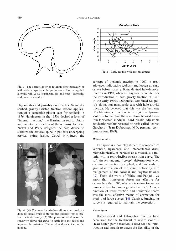
Our early results show significant promise withthe typical left thoracic infantile curve being themost likely to respond; however, we lack compar-ison with other treatment modalities (eg, bracing,
growing rods, or the Vertical Expandable Pros-thetic Titanium Rib [VEPTR, SYNTHES Spine,Paoli, PA]), and still requires longer follow-upprior
to arriving at any definitive conclusions. As Fig. 5shows, most patients have a significant response,particularly younger ones. The most significant
failure is a girl who has Prader-Willi syndromeand a stiff double thoracic curve. Until we have fur-ther follow-up, we cannot distinguish those pa-
tients who are most likely to respond from thosewho are likely to fail serial casting, although Meh-ta’s finding that younger children respond bettercorresponds with our experience. (Fig. 6)
Traction in early-onset scoliosis
‘‘Despite the fact that traction is rarely usedtoday, it does remain an important option for thetreatment of children with spinal disorders’’ [10].
Halo-gravity traction can restore coronal andsagittal balance in severely decompensated curvesand may decrease the neurologic risks associatedwith the surgical correction of these severe de-
formities, be it by casting, subcutaneous rods,VEPTR, or definitive instrumentation and fusion.Patients who have severe spinal deformities and
secondary cor pulmonalemay benefit fromaperiodof preoperative traction to allow aggressive pul-monary toilet. Improvements in vital capacity,
arterial blood gases, and right heart failure maydecrease postoperative complications and can helpwith patient selection in difficult cases [11–13].
Historical background
Traction is one of the oldest methods forcorrection of spinal deformity, going back to

479CASTING AND TRACTION TREATMENT FOR SCOLIOSIS
Box 1. Guidelines for applying an effective cast
One must have a casting table that stabilizes the body for the anesthesiologist whilepositioning the trunk and extremities for effective casting.
Light head halter and pelvic traction assist in stabilizing the patient and in narrowing thebody (Fig. 1).
A mirror slanted under the table is useful for visualizing the gibbus, the posterior cast, andthe molds (Fig. 2).
Traction should not be large. Although traction can correct the curve while applied, theposition cannot be retained in the cast once traction is released and the body recoils. Thepurpose of traction is to align the trunk and narrow the torso slightly for good cast fitting.
Proper casting requires an excellent purchase on the pelvis. Without this basic foundation,the cast will slip, create sores, and fail to support the curve.
Only a small amount of padding is necessary with a well-fitting cast. Mehta uses directplaster over crepe paper on the skin with occasional felt pads for significant bonyprominences, but we use a thin layer of webril over a silver-impregnated body shirt inaddition to thin layers of felt on significant bony prominences.
If there is a lumbar curve, we believe that flexion of the hips to decrease lumbar lordosisfacilitates curve correction.
The cast must not push the ribs toward the spine and narrow the space available for the lung.Rather, the posteriorly rotated ribs must be moved anteriorly to create a more normalchest configuration (Fig. 3). Counterrotation is applied through the pelvic mold and uppertorso or shoulders.
The original Cotrel/Morel technique uses rotational straps to obtain correction. In mostyoung children, using one’s hands works well, but we use the straps in more difficultcurves, particularly of the lumbar spine.
Although the Cotrel/Morel technique and Mehta’s modifications use an over-the-shouldercast, we have had excellent success staying below the shoulders because most infantilecurves have low apices, typically at T10 to T11.
Finally, well-positioned anterior and posterior windows are helpful in dynamic curvecorrection and cast comfort (Fig. 4).
Fig. 1. A proper casting table provides good patient sta-
bility with support of the head and lower extremities, full
access to the torso, and the ability to provide light
traction.
Fig. 2. Rotational correction occurs by rotating the rib
or lumbar prominence anteriorly while providing coun-
terrotation through the pelvis and upper torso or shoul-
ders. A slanted mirror is helpful in applying correct
molds.
480 D’ASTOUS & SANDERS
Hippocrates and possibly even earlier. Sayre de-
scribed gravity-assisted traction before applica-tion of a corrective plaster cast for scoliosis in1876. Harrington, in the 1950s, devised a form of
‘‘internal traction,’’ the Harrington rod to obtainand maintain correction of the scoliosis. In 1959,Nickel and Perry designed the halo device tostabilize the cervical spine in patients undergoing
cervical spine fusion. Cotrel introduced the
Fig. 3. The correct anterior rotation done manually or
with wide straps over the prominence. Forces applied
laterally will cause significant rib and chest deformity
and must be avoided.
Fig. 4. (A) The anterior window allows chest and ab-
dominal space while capturing the anterior ribs to pre-
vent their deformity. (B) The posterior window on the
concavity allows the curve to settle into the defect and
improve the rotation. The window does not cross the
midline.
concept of dynamic traction in 1960 to treatadolescent idiopathic scoliosis and loosen up rigid
curves before surgery. Kane devised halo-femoraltraction in 1967, whereas Stagnara is credited forthe introduction of halo-gravity traction in 1969.
In the early 1990s, Dubousset combined Stagna-ra’s elongation turnbuckle cast with halo-gravitytraction. He believed that this was the best way
of obtaining correction in a rigid early-onsetscoliosis; to maintain the correction, he used a cus-tom-fabricated modular, hard plastic adjustable
cervicothoracolumbosacral orthosis called ‘‘corsetGarchois’’ (Jean Dubousset, MD, personal com-munication, 1999).
Biomechanics
The spine is a complex structure composed of
vertebrae, ligaments, and intervertebral discs;biomechanically, it behaves as a viscoelastic ma-terial with a reproducible stress/strain curve. The
soft tissues undergo ‘‘creep’’ deformation whencontinuous traction is applied, and this leads togradual correction of the spinal deformity with
realignment of the coronal and sagittal balance[12]. From the work of White and Panjabi, weknow that transverses forces are effective forcurves less than 50�, whereas traction forces are
more effective for curves greater than 50�. A com-bination of axial traction and transverse forceswas the most effective means of correction for
small and large curves [14]. Casting, bracing, orsurgery is required to maintain the correction.
Techniques
Halo-femoral and halo-pelvic traction have
been used for the treatment of severe scoliosis.Head halter–pelvic traction is used for the initialtraction radiograph to assess the flexibility of the
Fig. 5. Early results with cast treatment.

481CASTING AND TRACTION TREATMENT FOR SCOLIOSIS
Fig. 6. Younger patients seem to have a better response than older children. (A) Eight-month-old girl who has infantile
idiopathic scoliosis just before the start of casting, T6–L1 51�, rib phase 2, RVAD 40�. (B) Radiograph showing the cor-
rection in the second cast. (C) The same patient at age 3 years with scoliosis completely corrected; careful follow-up is
still required.
scoliosis/kyphosis and again during the applica-
tion of the Risser cast. The discussion is limited tohalo-gravity traction because this is the mostcommon type of traction used in our patients
who have early-onset scoliosis.Axial skeletal traction to the spine may be
applied by a halo. In children younger than 2years of age, because of their thin calvarium, 10 to
12 pins should be used, and the torque should notexceed 2 inch-pounds or finger tightness [15].Multiple pins (6–8 pins) should be used in older
children, and 6 to 8 inch-pounds of torque are ap-plied. The pins are placed under general anesthe-sia if the child’s overall condition allows. It is not
necessary to shave the scalp or make a skin inci-sion. The skin and subcutaneous tissues are infil-trated with 1% xylocaine with epinephrine. The
anterior pin is placed 1 cm above the eyebrowsor between the supraciliary ridge and the frontalprominence. It is important to avoid the anteriorfrontal sinus with the most medial pin. Placement
too medially also can cause supraorbital or sup-ratrochlear nerve damage. Placement too farlaterally (behind the hairline) impinges on the
temporalis and masseter muscles, leading to possi-ble skull penetration and difficulty with mastica-tion. The posterior pin is placed 1 cm above and
posterior to the pinna, below the equator of theskull. The proper ring size allows 1 to 2 cm ofclearance between the skin and halo. This
alleviates problems caused by edema and facili-
tates proper pin care. The screws should be 180�
opposite from each other whenever possible.The pins should be retightened once at 24 hours
after halo application. Further tightening is dan-gerous, possibly leading to skull penetration.The exact method of pin care is less importantthan meticulous daily attention, taking care to re-
move all encrusted material and to examine forerythema or drainage. The pins are cleansedonce a day with half-strength hydrogen peroxide
or plain soap and water. Pins should be replacedif erythema and drainage do not improve withmeticulous pin care or oral antibiotics. The hair
and scalp should be washed at least once a week.Stagnara [16] is credited with the development
of halo-gravity traction; it is the mostly widely
used method of halo traction. This method usesthe patient’s own body weight as countertraction.Depending on the child’s size, 3 to 5 pounds is ap-plied, with daily additions of 1 to 2 pounds to
a maximum of 30% to 50% of body weight. Dur-ing the day, countertraction may be obtained bythe use of a wheelchair or walker. At night, we
use a bed in maximum reverse Trendelenburg po-sition, with blocks to elevate the head of the bed,which provides a 45� incline (Fig. 7). Alterna-
tively, a pelvic sling or a Circoelectric bed canbe used. This method allows the child to be up-right during the day, preventing decubiti and

482 D’ASTOUS & SANDERS
osteopenia and promoting renal drainage. It has
been used safely in children younger than 2 yearsof age. If a wheelchair is used, it may be necessaryto place counterweights on the footrests to pre-
vent tipping backward (Fig. 8). Halo-walker trac-tion is used to allow ambulation in traction(Fig. 9). We have not found it necessary to usea spring scale for traction as described by Sink
and colleagues [12], although it may act as a safetymechanism against excessive traction forces.
Fig. 8. Halo wheelchair.
Fig. 7. Bed in maximum reverse Trendelenburg
position.
Indications
Our indications for the use of halo-gravitytraction in early-onset scoliosis are early-onsetscoliosis greater than 80�, scoliosis associated with
kyphosis, and to ‘‘loosen up’’ the scoliosis andkyphosis before VEPTR or subcutaneous rodimplantation, because both of these methods arekyphogenic and are mechanically disadvantaged
in the presence of kyphosis.
Contraindications
Relative contraindications to halo-gravity trac-tion include short sharp rigid kyphosis, C-spine
abnormalities or instability because of their obvi-ous neurologic implications, cranial defects orthin skull, and age younger than 18 months
because of pin penetration problems.
Complications
Several complications have been associatedwith the use of halo-gravity traction; the mostcommon one in our series is pain and weakness of
neck musculature. Other complications includepin tract infections, pin loosening, and cranialnerve palsy (abducens, oculomotor, glossophar-
yngeal, hypoglossal). It is important to ask aboutdouble vision, difficulty swallowing, voice hoarse-ness, and tongue weakness. The cause of cranial
nerve symptoms is believed to be the result ofstretching or kinking of the nerve. Alternatively,an interference with the blood supply to the nerves
Fig. 9. Halo-walker traction is used to allow ambulation
in traction.

483CASTING AND TRACTION TREATMENT FOR SCOLIOSIS
Fig. 10. A 3-year-old girl who had severe early-onset kyphoscoliosis. Halo-gravity traction improved the coronal and
sagittal balance and decreased the kyphosis, allowing the use of a VEPTR device to maintain the correction.
secondary to traction may lead to cranial nerve
palsy. Other reported complications include skullpenetration and brain or epidural abscess, para-plegia, paraparesis, brachial plexus palsy, superior
mesenteric artery syndrome, and avascular necro-sis of the odontoid [17–20].
Practical advice
A thorough hair wash with shampoo is done thenight before surgery. If the patient has long hair, itshould be braided. Keep a chart at the bedside todocument the patient’s weight; chart the amount
of weight on bed traction and chair traction, andnote all increases in weight. Perform a quick neuro-logic examination and repeat at 2, 4, and 8 hours af-
ter increasing the traction weights.
Case example
A 3-year-old girl presented with severe early-
onset kyphoscoliosis. Halo-gravity traction im-proved the coronal and sagittal balance anddecreased the kyphosis, allowing the use ofa VEPTR device to maintain the correction
(Fig. 10) until the definitive fusion and instrumen-tation are undertaken.
Summary
The presence of a significant spinal deformity
in an infant or toddler with several years ofgrowth remaining has the potential to producea severe deformity with significant cardiopulmo-
nary consequences. We present alternative, albeitlabor-intensive, modalities to bracing, subcutane-ous rods, and VEPTR instrumentation. In milder
cases and if started before 2 years of age, casting
may correct the deformity completely, whereas inmore severe cases, it allows the child to grow,minimizing spinal and chest wall deformities, until
he/she is old enough to undergo definitive treat-ment of the spinal deformity.
Acknowledgments
In addition to Dr Sanders, casting in Erie hasbeen done by Drs. Joseph Khoury and Shyam
Kishan. We are indebted to them for theirassistance and insights. Marcie Fitzgerald PA,has been instrumental in helping us track our
early results in these patients. For the most part,the casting at Intermountain was done by Dr.D’Astous and Michael Pond, PA. We would like
to recognize Michael Pond, without whose enthu-siasm and multiple talents this work would havebeen near impossible. Finally, we thank RobertEldridge, CPO, who designed and built our
casting table and Matt Lowell, PT and theorthotic team who adapted the chairs, walkers,and beds for halo-traction.
References
[1] Risser JC. Scoliosis treated by cast correction and
spine fusion. Clin Orthop Relat Res 1976;116:86–94.
[2] Dickson RA. Early-onset idiopathic scoliosis. In:
Weinstein SL, editor. 1st edition. The pediatric
spine: principles and practice, vol 1. New York:
Raven Press, Ltd; 1994. p. 421–9.
[3] Pehrsson K, Larsson S, Oden A, et al. Long-term
follow-up of patients with untreated scoliosis. A
study of mortality, causes of death, and symptoms.
Spine 1992;17(9):1091–6.

484 D’ASTOUS & SANDERS
[4] MehtaMH. The rib-vertebral angle in the early diag-
nosis between resolving and progressive infantile
idiopathic scoliosis. J Bone Surg Br 1972;54:230–42.
[5] Ceballos T, Ferrer-Torrelles M, Castillo F, et al.
Prognosis in infantile idiopathic scoliosis. J Bone
Joint Surg Am 1980;62(6):863–75.
[6] Ferreira JH, Janeiro R, de James JI. Progressive and
resolving infantile idiopathic scoliosis. The differen-
tial diagnosis. J Bone Joint Surg Br 1972;54(4):
648–55.
[7] Thompson SK, Bentley G. Prognosis in infantile id-
iopathic scoliosis. J Bone Joint Surg Br 1980;62(2):
151–4.
[8] MehtaMH.Growth as a corrective force in the early
treatment of progressive infantile scoliosis. J Bone
Joint Surg Br 2005;87(9):1237–47.
[9] Cotrel Y, Morel G. [The elongation-derotation-flex-
ion technic in the correction of scoliosis]. Rev
Chir OrthopReparatrice ApparMot 1964;50:59–75.
[10] Crawford H. Traction. In: Weinstein SL, editor.
Pediatric spine surgery. Philadelphia: Lippincott,
Williams & Wilkins; 2001. p. 69–81.
[11] Arlet V, Papin P,Marchesi D. Halo femoral traction
and sliding rods in the treatment of a neurologically
compromised congenital scoliosis: technique. Eur
Spine J 1999;8(4):329–31.
[12] Sink EL, Karol LA, Sanders J, et al. Efficacy of peri-
operative halo-gravity traction in the treatment of
severe scoliosis in children. J Pediatr Orthop 2001;
21(4):519–24.
[13] Swank SM, Winter RB, Moe JH. Scoliosis and cor
pulmonale. Spine 1982;7(4):343–54.
[14] White AAPM. Clinical biomechanics of the spine.
2nd edition. Philadelphia: JB Lippincott; 1990.
[15] Mubarak SJ, Camp JF, Vuletich W, et al. Halo
application in the infant. J Pediatr Orthop 1989;
9(5):612–4.
[16] Stagnara P. [Cranial traction using the ‘‘Halo’’ of
Rancho Los Amigos]. Rev Chir Orthop Reparatrice
Appar Mot 1971;57(4):287–300.
[17] Garfin SR, Botte MJ, Nickel VL. Complications in
the use of the halo fixation device. J Bone Joint
Surg Am 1987;69(6):954.
[18] Rozario RA, Stein BM. Complications of halo-
pelvic traction. Case report. J Neurosurg 1976;
45(6):716–8.
[19] Tredwell SJ, O’Brien JP. Avascular necrosis of the
proximal end of the dens. A complication of halo-
pelvic distraction. J Bone Joint Surg Am 1975;
57(3):332–6.
[20] Wilkins C, MacEwen GD. Cranial nerve injury from
halo traction.ClinOrthopRelatRes1977;126:106–10.