catalytic site prediction of azoreductase enzyme of e
TRANSCRIPT

Catalytic Site Prediction of Azoreductase Enzyme of E. coli with Potentially Important Industrial Dyes Using
Molecular Docking Tools
Bikash Thakuria, Nangkyntiewbor Jungai, Samrat Adhikari*
Bioinformatics Centre, Department of Biotechnology, St Edmund’s College, Shillong, Pin-793003, Meghalaya, India. * Corresponding author. Tel.: 91-0364-2220808; email: [email protected] Manuscript submitted January 5, 2015; accepted March 10, 2015. doi: 10.17706/ijbbb.2015.5.2.91-99
Abstract: Azoreductase is an FMN-dependent and NADH-dependent enzyme of Escherichia coli. This
enzyme is responsible for the degradation of azo dyes. In this study, we retrieved the crystal structure of the
enzyme from PDB and 18 azo dyes from NCBI PubChem compound. These azo dyes were then docked with
the FMN- dependent NADH-azoreductase enzyme to analyze the binding affinity of the azo dyes with the
enzymes and predict the catalytic sites. In this approach, we identified the catalytic residues of
FMN-dependent and NADH dependent enzyme of Escherichia coli which were then evaluated in terms of
properties including function, conservation, hydrogen bonding, B-factor and flexibility. The results indicated
that Phe-172, Glu-174, Lys-145, Asp-146 and Lys-169 played an important role as catalytic site residues in
the enzyme. It is hoped that this information will provide a better understanding of enzyme mechanisms
and also be used to improve the designing strategies for dyes detoxification. In this study, the approach
emphasizes on a better understanding of the biodegradation of some of the commercially important
azodyes mediated by azoreductase from E. coli. Furthermore, the catalytic site residues information is
essential for understanding and altering the substrate specificity and for the design of a harmless azodye.
Key words: Azoreductase, azo dyes, EDO, FMN, IPA, NADH.
1. Introduction
Azo dyes are widely used for industrial, printing, clinical purposes as well as textile dyeing because of its
chemical stability, ease of synthesis, and versatility. In addition, azo dye compounds are also the most
commonly used drugs in the treatment of inflammatory bowel disease. Their durability, however, causes
pollution when they are released into the environment as the effluent [1]. Moreover, the release of these
dyes into the environment is undesirable, not only because of their colour, but also of their by-products
acting as agents of toxicity and mutagenicity. Biological treatment of azo dyes by the use of bacteria has
been widely reported. Enzymes that catalyse the reduction of these azodyes are termed as azoreductases
(EC 1.7.1.6). This enzyme basically utilizes NADH/FMN and/or NADPH as an electron donor to
decolorize azo dyes into corresponding aromatic amines by reductive cleavage of azo bonds. The
decolourization is a rate- limiting step, which is followed by the aerobic mineralization of the colourless
aromatic amines. Furthermore, azoreductase is also involved in the site-specific delivery of azo prodrugs,
which are therapeutically inactive in their intact form and rely on azo reduction by azoreductases of
International Journal of Bioscience, Biochemistry and Bioinformatics
91 Volume 5, Number 2, March 2015

intestinal microflora for activation. Proteins with azoreductase activity have been identified and
characterized from a wide variety of bacteria [2], [3].
There are at least two different types of bacterial azoreductases: Flavin dependent and Flavin
independent [4]. Flavin-dependent azoreductases can be further classified into two families according to
their amino acid sequences. Azoreductases from E. coli and Bacillus sp. strain OY1-2 are representative of
the two flavin- dependent azoreductase families, respectively. Although they are effective for an in vitro
enzyme assay, overexpression of these azoreductases in vivo results in little or no increase of bacterial
decolourization activity. Thus, the physiological role of azoreductase has recently been a subject of debate.
As the introduction of azo dyes into the environment is due mainly to human activities, reduction of azo
dyes may not be the primary role of these enzymes [5].
Azo dyes such as Sudan dyes are not legal for use as colorants in food; however, recently these dyes
have been detected as contaminants in the food supply [6]. The human health impact of exposure to azo
dyes used in certain food products has caused concerns since they may have genotoxic properties. The
environmental fate and subsequent health effects of the azo dyes released in textile and paper industry
wastewater are increasing being studied by the scientific community [7].
While azo dyes are generally considered to be persistent pollutants because they are typically
recalcitrant to aerobic biotransformation [8], they might be metabolized by azoreductases from
commensal microorganisms, mammalian liver cells, and soil microorganisms [9]. A variety of
microorganisms, including bacteria and fungi, are capable of decolorizing a diverse range of azo dyes.
Some bacteria have the ability to degrade azo dyes both aerobically and anaerobically [6], [10]. Bacterial
degradation of azo dyes is often initiated with the cleavage of azo bonds by azoreductases which are
followed by the aerobic degradation of the resulting amines [4].
To understand the biodegradation of the azo dyes mediated by the enzyme and to design harmless
azo dyes, it is essential to discover the catalytic site residues within the azoreductase enzyme and to
perform flexibility analysis of the catalytic residues [11].
In the present study, docking study of the FMN-dependent NADH-azoreductase of E. coli and the
industrially important dyes was carried out to understand the mechanism of azoreductase catalyzing
enzymatic reactions, the catalytic site residues and the binding affinities of the azodyes and the enzyme.
Therefore in context to the present study, an FMN-dependent NADH-azoreductase enzyme of E. coli
had been selected as the target protein or protein of interest for the theoretical studies of
biodegradation of azodyes compounds. This enzyme was chosen to view the interaction with azodyes,
which are pollutants and toxic to the environment. This study would pave various strategies for
understanding and altering the substrate specificity for effective design of a potential harmless azodyes.
2. Materials and Methods
Data Set 2.1.
The enzyme molecule, FMN-dependent NADH-azoreductase of E. coli was obtained from Protein Data
Bank (PDB) [12], and the three-dimensional (3D) structures of this protein had been solved by X-ray
crystallographic techniques with 1.80 Ao resolution. For substrate 18 commercially important azodyes
were retrieved from the NCBI PubChem Compound database [13].
Computation of Docking Score between the Ligands and the Enzyme 2.2.
Protein or ligand modification was carried out by removing water molecules from the protein and
energy minimization was carried out using Swiss PDB Viewer (SPDBV) tools [14]. Autodock Vina [15] was
used for docking studies of azoreductase (protein) and azodyes (ligands). The interactions between the
International Journal of Bioscience, Biochemistry and Bioinformatics
92 Volume 5, Number 2, March 2015

different ligands with the protein of interest were viewed in PyMOL [16] and the analysis of the H-bond
and its positions were also evaluated with PyMOL.
Functional Site Location 2.3.
The catalytic or functionally important residues of a protein are known to exist in evolutionary
constrained regions. However, the patterns of residue conservation alone are sometimes not very
informative, depending on the homologous sequences available for a given query protein. Hence, the
prediction of functional sites in newly solved protein structures is a challenge for computational structural
biology. Most methods for functional site identification utilize measures of amino acid sequence
conservation in homologous sequences, based on the assumption that functional sites are relatively
conserved during evolution. Protein structural information has also been used to help identify protein
functional sites. Ligand binding site prediction of the azoreductase enzyme was carried out using
Q-SiteFinder [17], by binding hydrophobic (CH3) probes to the protein, and finding clusters of probes with
the most favourable binding energy. These clusters are placed in rank order of the likelihood of being a
binding site according to the sum total binding energies for each cluster.
Hydrogen Bonding and B Factor 2.4.
The hydrogen bonding and the B-factor are the two vital parameters in the catalytic residue prediction.
Catalytic residue hydrogen bonding was investigated using the result obtained from the docking study. Catalytic
residue B-factors is a measure of residue flexibility. Catalytic residues tend to have lower B-factors than all
residues. Catalytic residues are very precisely positioned and held in place, as predicted by their low B-factors
and Hydrogen bonding. The hydrogen bonding information is obtained from the docking studies. The B-factors of
the residues interacting with the ligands and the active sites were taken from PDB file for each atom in a residue,
and then averaged over the whole residue.
3. Results and Discussion
PDB Structure 3.1.
The enzyme, FMN-dependent NADH-azoreductase of E. coli has a crystallized structure characterized by
three ligands FMN, EDO and IPA. The structure was obtained from PDB with the PDB id, 1V4B (Fig 1(a)).
Fig. 1 (a) The three dimensional structure of the target protein (1V4B) obtained from PDB; (b) The active
sites displayed in the three dimensional structure of the target protein predicted with the aid of
Q-SiteFinder and viewed under Chimera.
Multiple Sequence Alignment 3.2.
(a) (b)
International Journal of Bioscience, Biochemistry and Bioinformatics
93 Volume 5, Number 2, March 2015

Catalytic residues are more conserved than the average residues [18]. Hence the conserved residues
were investigated to have a relationship with the catalytic residues. A BLAST search along with a multiple
sequence alignment with the BLAST result showed the conservation of almost all the residues.
Computation of Docking Score between the Ligands and the Enzyme 3.3.
Proteins are the basis of life processes of all enzymes at the molecular level. The protein interaction is
either with another protein or with small molecules. Many biological studies will benefit from interactive
predictions. Docking study showed the binding affinity, number of hydrogen bonds and the binding
residues. It is interesting to note that the binding affinities have negative values as shown in Table 1. This
revealed the high feasibility of this reaction. The docked complexes were further analysed with the
molecular visualization tools, PyMOL as shown in Fig. 2. The docking analysis showed that three dyes viz.
Sudan black B (61336), Kayaku acid blueblack 10B (5473482) and FD & C Yellow no.5 (25245842) formed
H-bonds with the enzyme residues Asp-109, Arg-59 and Ala-112, and Arg-59 of Site 7 respectively.
Moreover, six dyes showed docking conformation with two of the natural ligands of the target protein;
3.3’dichlorobenzidine (7070) with IPA, Methyl red (10303) with IPA and Glu-174, HABA (5357439) IPA
and Lys-145, Amaranth (FD & C red no. 2) (5473445) with IPA, Glu-174 and Phe-172, Grasal orange
(5858445) with IPA and Antipyrylazo III (9573878) with FMN and His-144 (Table 2). The variation in the
docking score indirectly depicted the rate of decolourization [19]. The rate of colour removal for trypan
blue was higher than any other azo dye. The amino acids Phe-172; Glu-174; Asp-189; Asp-146; Lys-145;
Asp-109; Arg-59; Ala-112; Ala-200; Asp-146; Asp-150; Gly-174; Lys-169; Phe-170; Asp-167; Thr-166;
Gly-131; Gly-164; Lys-132; His-144; Arg-59 interacted with different ligands and were taken into account
for comparison with the active sites obtained from active site prediction.
Fig. 2. Docked conformations of (a) Sudan black B (61336), (b) Kayaku acid blueblack 10B (5473482), (c)
FD & C Yellow no.5 (25245842) in the active site; (d) 3.3’Dichlorobenzidine with IPA; (e) Grasal orange with
IPA and (f) Antipyrylazo III with FMN and His-144.
Functional Site Location 3.4.
Active sites of the target protein were predicted using Q-SiteFinder and the output file was viewed
under Chimera. 10 active sites were obtained from the study along with the corresponding amino acid
residues presented in each active site (Fig. 1(b)) and the corresponding aminoacids evaluated are depicted
in Table 3. Each of the sites was analysed and compared with the amino acids interacting with the ligands
(a) (b) (c)
(d) (e) (f)
International Journal of Bioscience, Biochemistry and Bioinformatics
94 Volume 5, Number 2, March 2015
in the docking study. The amino acid positions of the sites are also listed in Table 3. The amino acid residues
were found to have lower B-factor than the other residues and the site 6 showed the absence of aminoacids.
The 7th active site consists of amino acids which are interacting with ligands in the docking study. The
docking result shows that the amino acids such as Phe-172, Glu-174, Lys-145, Asp-146 and Lys-169 are
very much repeated in the interaction with more than one ligand. This reveals that these amino acids are
catalytic residues. The active site variations suggest that the enzyme could probably decolourize a wide
range of azo dyes [20].
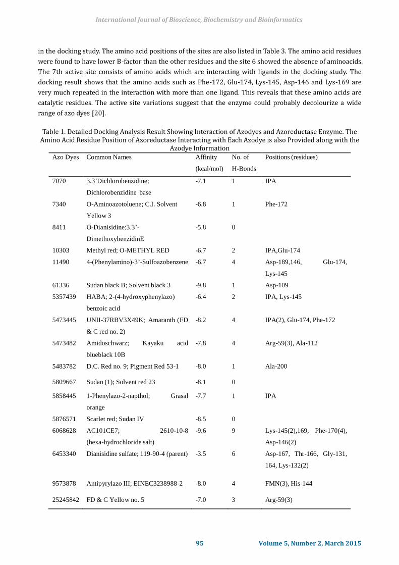
Table 1. Detailed Docking Analysis Result Showing Interaction of Azodyes and Azoreductase Enzyme. The
Amino Acid Residue Position of Azoreductase Interacting with Each Azodye is also Provided along with the Azodye Information
Azo Dyes Common Names Affinity
(kcal/mol)
No. of
H-Bonds
Positions (residues)
7070 3.3’Dichlorobenzidine;
Dichlorobenzidine base
-7.1 1 IPA
7340 O-Aminoazotoluene; C.I. Solvent
Yellow 3
-6.8 1 Phe-172
8411 O-Dianisidine;3.3’-
DimethoxybenzidinE
-5.8 0
10303 Methyl red; O-METHYL RED -6.7 2 IPA,Glu-174
11490 4-(Phenylamino)-3’-Sulfoazobenzene -6.7 4 Asp-189,146, Glu-174,
Lys-145
61336 Sudan black B; Solvent black 3 -9.8 1 Asp-109
5357439 HABA; 2-(4-hydroxyphenylazo)
benzoic acid
-6.4 2 IPA, Lys-145
5473445 UNII-37RBV3X49K; Amaranth (FD
& C red no. 2)
-8.2 4 IPA(2), Glu-174, Phe-172
5473482 Amidoschwarz; Kayaku acid
blueblack 10B
-7.8 4 Arg-59(3), Ala-112
5483782 D.C. Red no. 9; Pigment Red 53-1 -8.0 1 Ala-200
5809667 Sudan (1); Solvent red 23 -8.1 0
5858445 1-Phenylazo-2-napthol; Grasal
orange
-7.7 1 IPA
5876571 Scarlet red; Sudan IV -8.5 0
6068628 AC101CE7; 2610-10-8
(hexa-hydrochloride salt)
-9.6 9 Lys-145(2),169, Phe-170(4),
Asp-146(2)
6453340 Dianisidine sulfate; 119-90-4 (parent) -3.5 6 Asp-167, Thr-166, Gly-131,
164, Lys-132(2)
9573878 Antipyrylazo III; EINEC3238988-2 -8.0 4 FMN(3), His-144
25245842 FD & C Yellow no. 5 -7.0 3 Arg-59(3)
International Journal of Bioscience, Biochemistry and Bioinformatics
95 Volume 5, Number 2, March 2015

Hydrogen Bonding and B-factor 3.5.
Both the Hydrogen bonding and the B-factors in the crystal structures were used as a measure of residue
flexibility. Analysis shows that the amino acids interacting with the ligands are involved in hydrogen
interactions as a donor or as an acceptor [21]. This shows that catalytic residues have a limited
conformational freedom. The docking result shows that the ligands have hydrogen bonding with amino
acids and it is illustrated in Table 1. Catalytic residues tend to have lower B-factors than all residues,
suggesting that they have to be more rigidly held in place than the averaged over the whole residue [21].
B-factors were taken from the PDB file for each atom in a residue and then averaged over the whole residue
[22]. The B-factor for the residues, Phe-172 is 16.45, Glu-174 is 16.53, Lys-145 is 25.16, Asp-146 is 25.77
and Lys-169 is 31.8 [23].
Table 2. Target Protein Docked with Natural Ligands
Ligands
Affinity(kcal/mol)
H-Bonds
Positions(residues)
EDO -3.3 6 Ser-15,17(2),139(2), Gln-16
FMN
-10.5
15
His-144, Gly-142,141, Phe-98, Asn-97,
Tyr-90, Met-95, Ser-9,15,17(2),139(2),
Gln-16(2) IPA -3.2 5
Ser-15,17(2),139, Gln-16
Table 3. The Amino Acid Composition of the Different Active Sites Predicted Active Sites Residues
Site 1 Ile 143
Site 2
Ser 9, Leu 11, Tyr 14, Ser 15,
Gln 16, Ser 17, Pro 94, Met 95, Tyr 96, Ser 139, Arg 140, Gly 141
Site 3
Phe 24, Trp 28, Val 88, Lys 133,
Ala 134, Ile 135
Site 4
Met 95, Tyr 96, Asn 97, Phe 98,
Ser 139, Arg 140, Gly 141, Gly 142
Site 5 Pro 48, Leu 54, Arg 68,Glu 71,
Ala 72, Leu 75
Site 6 -
Site 7 Leu 50, Val 55, Leu 58, Arg 59,
Asp 109, Ala 112, Arg 113, Ala 114, Phe 118
Site 8
Gln 19, Leu 20, Asp 22, Tyr 23,
Glu 26
Site 9 Ile 10, Tyr 96, Asn 97, Asn 99, Ile
100, Ser 101,Thr 102, Gln 103 Site 10 Phe 98, Asn 99, Ile 100
International Journal of Bioscience, Biochemistry and Bioinformatics
96 Volume 5, Number 2, March 2015
4. Conclusion
A major challenge in azodye degradation and a harmless azodye design is the elucidation of biochemical
and biological properties of enzymes, including the determination of catalytic residues that belong to the
ligand-substrate binding site. Comparing the results from Q-Site Finder and docking studies, it indicates
that the amino acid residues Phe-172, Glu-174, Lys-145, Asp-146 and Lys-169 play an important role as
catalytic site residues in the azoreductase enzyme of E. coli. This docking study also provides information
on the binding affinity of the ligands with azoreductase enzyme. The rate of colour removal for trypan blue
is higher than any other azo dye. It is hoped that this information would provide a better understanding
of the molecular mechanisms involved in catalysis and a heuristic basis for predicting the catalytic
residues in enzymes of unknown function. The natural ligands (FMN, EDO and IPA) were also found to
interact with some of the ligands. In this work the catalytic residues are reported as well as the binding
affinities for some commercially important azodyes. The study made in this project would facilitate
researchers a better understanding of enzyme mechanisms and also used to improve the designing
strategies of less harmful azodyes.
Acknowledgment
The authors take this opportunity to acknowledge the funding received from Department of
Biotechnology, Govt. of India for setting up Bioinformatics Centre under BTISNET programme in
Department of Biotechnology, St. Edmund’s College, Shillong. The authors also express their heartfelt
gratitude to Dr Sylvanus Lamare, Principal, St. Edmund’s College, Shillong for his support throughout the
work.
References
[1] Nakanishi, M., Yatome, C., Ishida, N., & Kitade, Y. ( 2001). Putative ACP phosphodiesterase gene (acpD)
encodes an azoreductase. Journal of Biological Chemistry, 276(49), 46394-46399.
[2] Hanauer, S. B. (1996). Inflammatory bowel disease. New England Journal of Medicine, 334, 841-848.
[3] Wang, C. J., Hagemeier, C., Rahman, N., Lowe, E., Noble, M., Coughtrie, M., et al. (2007). Molecular
cloning, characterization and ligand-bound structure of an azoreductase from Pseudomonas
aeruginosa. Journal of Molecular Biology, 373(5), 1213-1228.
[4] Chen, H. ( 2006). Recent advances in azo dye degrading enzyme research. Current Protein
Precipitation Science, 7(2), 101-111.
[5] Liu, G., Zhou, J., Fu, Q. S., & Wang, J. (2009). The E. coli azoreductase azoR is involved in resistance to
t hiol-specific stress caused by electrophilic quinones. Journal of Bacteriology, 191(20), 6394-6300.
[6] Xu, H., Heinze, T. M., Chen, S., Cerniglia, C. E., & Chen, H. (2007). Anaerobic metabolism of 1-amino-2-
naphthol-based azo dyes (Sudan dyes) by human intestinal microflora. Applied Environmental
Microbiology, 73(23), 7759-7762.
[7] McMullan, G., Meehan, C., Conneely, A., Kirby, N., Robinson, T., Nigam, P., et al. (2001). Microbial
decolourisation and degradation of textile dyes. Applied Microbiology and Biotechnology, 56(1-2),
81-87.
[8] Stolz, A. (2001). Basic and applied aspects in the microbial degradation of azo dyes. Applied
Microbiology and Biotechnology, 56(1-2), 69-80.
[9] Levine, W. G. (1991). Metabolism of azo dyes: Implication for detoxication and activation. Drug
Metabolism Reviews, 23(3-4), 253-309.
[10] Prasad, N. K., Vindal, V., Narayana, S. L., Kunal, S. P., et al. (2012). In silico analysis of Pycnoporus
cinnabarinus laccase active site with toxic industrial dyes. Journal of Molecular Modeling, 18(5),
International Journal of Bioscience, Biochemistry and Bioinformatics
97 Volume 5, Number 2, March 2015

2013-2019.
[11] Ramanthan, K., Shanthi, V., & Sethumadhavan, R. (2009). In silico identification of catalytic residues in
azobenzene reductase from Bacillus subtilis and its docking studies with azo dyes. Interdisciplinary
Science, 1(4), 290-297.
[12] Bernstein, F. C., Koetzle, T. F., Williams, G. J. B., Meyer, E. F. Jr., Brice, M. D., Rogers, J. R., et al. (1977).
The Protein Data Bank: A computer-based archival file for macromolecular structures. Journal of
Molecular Biology, 112, 535-542.
[13] Wang, Y., Xiao, J., Suzek, T. O., Zhang, J., Wang, J., & Bryant, S. H. (2009). Pubchem: A public information
system for analysing bioactivities of small molecules. Nucleic Acids Res., 37, W623-W633.
[14] Guex, N., & Peitsch, M. C. (1997). Swiss-Model and the Swiss-Pdb Viewer: An environment for
comparative protein modeling. Electrophoresis, 18, 2714-2723.
[15] Trott, O., & Olson, A. (2010). AutoDock Vina: Improving the speed and accuracy of docking with a new
scoring function, efficient optimization, and multithreading. Journal of Computational Chemistry, 31,
455-461.
[16] DeLano, W. L. (2013). The PyMOL molecular graphics system. DeLano Scientific San Carlos, California
USA. from http://www.pymol.org
[17] Laurie, A. T. R., & Jackson, R. M. (2005). Q-SiteFinder: An energy-based method for the prediction of
protein-ligand binding sites. Bioinformatics, 21, 1908-1916.
[18] Bartlett, G. J., Porter, C. T., Borkakoti, N., & Thornton, J. M. (2002). Analysis of catalytic residues in
enzyme active sites. Journal of Moecular Biology, 324(1), 105-121.
[19] Abo-Farha, S. A. (2010). Photocatalytic degradation of monoazo and diazo dyes in wastewater on
nanometer-sized TiO2. Journal of American Science, 6(11), 130-142.
[20] Philem, P. D., & Adhikari, S. (2012). Homology modeling, docking studies and functional analysis of
various azoreductase accessory interacting proteins of Nostoc. sp. PCC7120. Bioinformation, 8(7),
296-300.
[21] Sacquin-Mora, S., & Lavery, R. (2006). Investigating the local flexibility of functional residues in
hemoproteins. Biophysical Journal, 90(8), 2706-2717.
[22] Yuan, Z., Zhao, J., & Wang, Z. (2003). Flexibility analysis of enzyme active sites by crystallographic
temperature factors. Protein engineering design and selection, 16(2), 109-114.
[23] Sridhar, S., & Chandra, J. H. (2014). Involvement of computational tools towards in silico remediation―
Synthetic textile dyes interacting with azoreductase. International Journal of Chemical Technology
Research, 6(9), 4412-4441.
Bikash Thakuria was born in Guwahati, Assam (India) in 1988. He has obtained a
master’s degree in the field of biotechnology from Bangalore University (India) in 2012.
Currently he is working as a research associate under Bioinformatics Infrastructure
Facility (BIF), funded by the Department of Biotechnology, Govt. of India, in St. Edmund’s
College Shillong. His field of research is proteomics and computational biology. He has also
participated in different national level workshops in the field of Biotechnology. He is also
associated with the docking and interaction studies of different azo dyes and their environmental impact.
Currently he is working on a medicinal plant named Smilax aspera and its proteins for cancer studies.
International Journal of Bioscience, Biochemistry and Bioinformatics
98 Volume 5, Number 2, March 2015

Nangkyntiewbor Jungai was born in Shillong, India in 1988. He has obtained a master
degree in biotechnology at North Eastern Hill University, Shillong, Department of
Biotechnology and Bioinformatics (2011). His areas of research are in the fields of genetic
engineering, cyanobacterial biotechnology, cancer and cell cultures and traditional
medicinal plants. At present, he is working as a senior research fellow (SRF) in the
Institutional Biotech Hub, St. Edmund’s College, Shillong under the sponsorship of
Department of Biotechnology (DBT), Government of India (GoI). He has also participated in various
seminars and workshops at the national level.
Samrat Adhikari was born in Shillong, Meghalaya in 1979. He has obtained the doctoral
degree from North Eastern Hill University, Shillong (2011) in the field of environmental
biotechnology and the masters with specialization in molecular microbiology (2002) from
Bangalore University, Bangalore. He has received the Biotech Industrial Training
Programme fellowship (2003) and also has a vast teaching and research experience for 12
years. Currently he is an assistant professor & the head of the Biotechnology Department,
St. Edmund’s College, Shillong. He has published 5 papers in peer reviewed journals and 3 in national &
international conferences. He has undergone many trainings and also has organized many workshops for
young researchers. He is also the reviewer of many academic journals. He has supervised for 3 M.Tech
thesis and is currently handling 3 projects sponsored by the different funding agencies of Govt. of India. He
is presently a member of many academic bodies in universities. At present, he is working on environmental
biotechnology & bioinformatics.
International Journal of Bioscience, Biochemistry and Bioinformatics
99 Volume 5, Number 2, March 2015