catechol-o-methyltransferase val158met polymorphism … · catechol-o-methyltransferase val158met...
TRANSCRIPT

Catechol-O-Methyltransferase Val158Met Polymorphismon Striatum Structural Covariance Networksin Alzheimer’s Disease
Chiung-Chih Chang1 & Shih-Jen Tsai2,3 & Nai-Ching Chen1& Chi-Wei Huang1 &
Shih-Wei Hsu4& Ya-Ting Chang1 & Mu-En Liu2
& Wen-Neng Chang1 & Wan-Chen Tsai1 &
Chen-Chang Lee4
Received: 21 January 2017 /Accepted: 20 June 2017 /Published online: 13 July 2017# The Author(s) 2017. This article is an open access publication
Abstract The catechol-O-methyltransferase enzyme metabo-lizes dopamine in the prefrontal axis, and its genetic polymor-phism (rs4680; Val158Met) is a known determinant of dopa-mine signaling. In this study, we investigated the possible struc-tural covariance networks that may be modulated by this func-tional polymorphism in patients with Alzheimer’s disease.Structural covariance networkswere constructed by 3DT1mag-netic resonance imaging. The patients were divided into twogroups: Met-carriers (n = 91) and Val-homozygotes (n = 101).Seed-based analysis was performed focusing on triple-networkmodels and six striatal networks. Neurobehavioral scores servedas the major outcome factors. The role of seed or peak clustervolumes, or a covariance strength showing Met-carriers > Val-homozygotes were tested for the effect on dopamine. Clinically,the Met-carriers had higher mental manipulation and hallucina-tion scores than the Val-homozygotes. The volume-score corre-lations suggested the significance of the putaminal seed in the
Met-carriers and caudate seed in the Val-homozygotes. Only thedorsal-rostral and dorsal-caudal putamen interconnected peakclusters showed covariance strength interactions (Met-carriers > Val-homozygotes), and the peak clusters also correlat-ed with the neurobehavioral scores. Although the triple-networkmodel is important for a diagnosis of Alzheimer’s disease, ourresults validated the role of the dorsal-putaminal-anchored net-work by the catechol-O-methyltransferase Val158Met polymor-phism in predicting the severity of cognitive and behavior insubjects with Alzheimer’s disease.
Keywords Alzheimer’s disease . Anatomical structuralcovariance . Default mode network . Genetic effect . Striatalnetwork . Posterior cingulate cortex
Introduction
Dopamine pathways modulate learning, memory, and neuro-psychiatric presentations, and the catechol-O-methyltransferase(COMT) gene has been implicated in the enzymatic degrada-tion of dopamine. In humans, a functional single nucleotidepolymorphism (rs4680 G to A), consisting of a change in thecoding exon at position 158 (Val158Met), has been reported toresult in a two- to fourfold decrease in the activity of the COMTenzyme. Consequently, the low enzyme activity in Met/Methomozygotes results in increased dopamine levels that maypreferentially affect prefrontal-related tasks [1].
Although the hypothesis of dopamine dysfunction in theearly stage of Alzheimer’s disease (AD) is still under debate,several experimental models support the role of Aβ oligomersand dopamine dysfunction [2]. In addition, a recent meta-analysis suggested that COMT Val158Met Val/Val alleleswere associated with an increased risk of AD in Asians [3].
Electronic supplementary material The online version of this article(doi:10.1007/s12035-017-0668-2) contains supplementary material,which is available to authorized users.
* Chiung-Chih [email protected]
1 Department of Neurology, Cognition and Aging Center, KaohsiungChang GungMemorial Hospital, Chang Gung University College ofMedicine, #123, Ta-Pei Road, Niaosung, Kaohsiung 833, Taiwan
2 Psychiatric Department, Taipei Veterans General Hospital,Taipei, Taiwan
3 Psychiatric Division, School of Medicine, National Yang-MingUniversity, Taipei, Taiwan
4 Department of Radiology, Kaohsiung Chang Gung MemorialHospital, Chang Gung University College of Medicine,Kaohsiung, Taiwan
Mol Neurobiol (2018) 55:4637–4649DOI 10.1007/s12035-017-0668-2

In Taiwan,Wang et al. [4] found that the Val/Val genotype andapolipoprotein E4 (ApoE4) allele exert a synergistic effect onthe risk of AD. An association of the rs4680 polymorphismwith susceptibility to AD through a synergistic effect withApoE4 alleles has also been reported in Caucasian [5] andBasque populations [6]. In AD, the COMT genotype has beenshown to play a major role in the presentation of psychosis [7]and cognitive profiles [8]. Other studies, however, suggest nodirect link between Val158Met and susceptibility to AD in thegeneral population [3, 5, 9]. In Akil et al.’s study, pathologicalspecimens of normal human brains [10] from individuals withthe Val/Val genotype may have had higher levels ofthyroxylase messenger RNA (mRNA) in mesencephalic do-pamine neuronal populations projecting to the striatum.However, whether the higher levels of striatal dopamine cancompensate for the lower dopamine concentration in the pre-frontal cortex is unclear, and the mechanism by which thepresentations are related to distinct network alterations re-mains to be explored.
The dopamine pathway represents one of the major bio-chemical signals in the striatum. Classically regarded as amotor structure, the striatum subserves a wide range of func-tions including cognitive, motivational, and emotional pro-cesses. In recent years, researchers have started to conceptu-alize the functional connectivity of distinct neural circuits as-sociated with different sub-regions of the striatum. For in-stance, the reward-related function has been attributed to theventral versus superior striatum [11]. Similarly, executive dys-function has been reported in patients with putamen and cau-date damage [12, 13]. Changes in the anatomical connectionswithin the fronto-striatal circuits are related to syndrome com-plexes in the neurodegenerative spectrum. In AD, there maybe lesser striatal atrophy, but the AD neuroimaging initiategroup reported that the striatum may be an adjunctive bio-marker [14]. Therefore, it is important to understand whetherthe psychiatric presentation [7] and cognitive profile [8] arerelated to COMT genotype-driven striatal pathways.
In 2006, Postuma and Dagher [15] proposed a seed-basedmodel defining six striatal sub-regions and related corticalconnections in the Talairach space. The striatal model dividesthe striatum into motor, associative, and limbic divisions. Forthe caudate nucleus, the most ventral to dorsal gradient spiralsmodulate the emotional/motivational aspects, followed by de-cision-making/executive control, and motor control functions[16, 17]. For the putamen, the functional connectivity has alsobeen reported to show a rostral/caudal distinction that is pri-marily connected to the primary and secondary cortical motorareas and executive control [18]. Dopamine is necessary forprefrontal-dependent tasks, and the interest in COMT activityin neurodegenerative diseases has been based on its role indopamine degradation [19]. Given that dopamine is a crucialmediator of neuronal function in AD, the striatal model [15]may be a good choice to understand the relationships between
COMT Val158Met genotypes and neurobehavioralpresentations.
Recent research has suggested that highly related regionsshow covariance in morphometric characteristics, the so-calledstructural covariance. With careful control of confounding fac-tors, structural covariance networks (SCNs) have been used totest the influence of genotypes [20]. The triple-network modelproposed byMenon [21] has been reported to be of great clinicalrelevance in AD and includes the default mode network (DMN)[22–24], salience network [25], and executive control network[26, 27]. Within the DMN, two subsystems are particularly rel-evant [28]: the Bmedial temporal lobe subsystem^ and theBdorsal medial prefrontal cortex subsystem^ (or themidline coresubsystem).
Dopamine may affect cognition by facilitating neuronalsynchrony; however, a direct correlation between COMT ge-notypes and prefrontal-striatal dopamine levels or gene-cognitive profiles remains controversial [10, 29]. The aimsof the current study were to explore the network effects ofCOMT genotypes and to assess in vivo whether different ge-notype groups may modulate the SCN patterns and therebydetermine neurobehavioral outcomes in AD. Based on a liter-ature review, we selected the striatal model [15] to assessdopamine activity and the triple-network model [21] to assesswhich networks are characteristically affected in AD. Basedon the biological properties of COMTon the prefrontal cortex,we hypothesized that the COMT Met158Val functional poly-morphism may modulate selective striatal SCNs that deter-mined the neurobehavioral scores in AD.
Results
Demographic Data, Cognitive Data, and NPI
The distribution of the genotypes of the Val158Met polymor-phism in the patients with AD was in Hardy-Weinberg equi-librium. There were no significant differences in gender, age,and educational level between the two genotype groups(Table 1). In addition, there was no difference in ApoE4 statusbetween the two groups. The Met-carriers had significantlyhigher scores in mental manipulation subdomains comparedwith the Val-homozygotes. TheMet-carriers had higher scoresin the hallucination domains.
Patterns of SCN and Genetic Variants
Adirect comparison between the graymatter (GM) volume of theMet-carriers and Val-homozygotes using voxel-based morphom-etry [30] showed no significant differences (with the threshold setat p < 0.05, corrected for a false discovery rate (FDR) with acluster size >100 voxels). In the striatal model, the dorsal caudalputamen (DCP) seed had a significantly higher volume in the
4638 Mol Neurobiol (2018) 55:4637–4649

Val-homozygotes (Fig. 1a), whereas in the triple-network model,the posterior cingulate cortex (PCC) seed volume was higher inthe Met-carriers (Supplementary Figure 1B). The SCN patternsand related clusters in each genotype are shown in Fig. 1c,Supplementary 1C, and Supplementary Tables 1, 2, 3, 4, 5, 6,7, 8, 9, 10, 11, 12, 13, 14, 15, 16, 17, 18, 19, 20.
Peak Clusters Showing Significant InteractionsBetween Genotype Groups
For each seed, we explored the genotypic interactions with re-gard to the topography showing differences in structural covari-ance strength between seed and peak clusters (Fig. 2;supplementary Table 21; Supplementary Figure 1D). The left
superior medial frontal region anchored to the entorhinal seedwas the only significant cluster within the triple-network modelshowingMet-carriers > Val-homozygotes in covariance strength(Supplementary Figure 1D). In contrast, two seeds (Fig. 2a,DCP; Fig. 2b, dorsal rostral putamen (DRP)) within the striatalmodel exhibited 11 clusters showing Met-carriers > Val-homo-zygotes in covariance strength. However, there were nosignificant differences in direct comparisons of the volumes ofthe peak clusters between the two genotype groups.
Relationships Between Seed Volume and Cognitive Score
We also explored whether the seed volumes were correlatedwith the cognitive test scores in each group (supplementary
Table 1 Demographicalcharacteristics andneuropsychiatric tests in theCOMT genotype groups in 192Alzheimer’s disease
Genotype group Met-carriers (n = 91) Val/Val (n = 101) p value
Age 73.3 ± 8.6 73.9 ± 7.2 0.62
Education (year) 7.3 ± 4.9 7.4 ± 4.9 0.91
Apolipoprotein E4 carrier (positive case, %) 32, 35.16% 34, 33.66% 0.88
Sex (male/female) 43/48 52/49 0.56
MMSE 19.9 ± 6.36 19.6 ± 6.81 0.772
Clinical dementia rating sum of box 3.6 ± 3.02 4.0 ± 3.22 0.375
Mental manipulation 6.0 ± 3.30 4.7 ± 3.18 0.008
Attention 6.3 ± 1.44 6.0 ± 1.63 0.202
Orientation 12.6 ± 5.23 12.5 ± 5.31 0.821
Long-term memory 8.2 ± 2.96 7.9 ± 2.72 0.497
Short-term memory 4.9 ± 3.87 5.6 ± 3.92 0.199
Abstract thinking 8.2 ± 2.91 7.9 ± 2.77 0.398
Drawing 7.7 ± 2.91 7.6 ± 3.06 0.797
Verbal fluency 5.1 ± 2.69 4.9 ± 2.87 0.663
Language 8.1 ± 2.20 7.9 ± 2.44 0.528
Executive function test 25.5 ± 8.17 23.5 ± 8.41 0.507
Total scores of CASI 67.2 ± 21.85 65.1 ± 22.26 0.510
Neuropsychiatric inventory scores total 8.49 ± 13.45 7.89 ± 9.99 0.723
Delusion 0.84 ± 2.32 0.54 ± 1.81 0.332
Hallucination 0.31 ± 1.18 0.01 ± 0.10 0.012
Aggression 0.53 ± 2.03 0.25 ± 1.03 0.223
Depression 1.15 ± 2.70 1.23 ± 2.59 0.847
Anxiety 0.35 ± 1.48 0.44 ± 1.48 0.695
Elation 0.08 ± 0.52 0.01 ± 0.10 0.207
Apathy 0.77 ± 2.50 0.88 ± 2.74 0.769
Disinhibition 0.11 ± 0.75 0.36 ± 1.75 0.216
Irritability 1.12 ± 2.41 0.98 ± 2.02 0.661
Aberrant motor behavior 0.42 ± 1.75 0.39 ± 1.73 0.901
Sleep disorders 2.45 ± 4.42 2.10 ± 4.12 0.569
Eating behavior 0.37 ± 1.28 0.71 ± 2.02 0.171
Data are presented as mean (standard deviation) or number (percentage; %); attention, verbal fluency, abstractthinking, and mental manipulation sub-domain scores of the CASI were added to assess executive function;APOE4 carriers were defined as the presence of one or two APOE4 alleles. The italicized word represents scoresshowing significance
CASI cognitive ability screening instrument, COMT catechol-O-methyltransferase
Mol Neurobiol (2018) 55:4637–4649 4639

Table 22: triple-network model; Table 2: striatal model). Forthe triple-network model, only the posterior cingulate seedvolume was significantly correlated with cognitive test scoresin both genotypes, while more cognitive domains reachedstatistical significance in theMet-carriers. For the striatal mod-el, the seed volumes showed variable correlations with thecognitive test scores and neuropsychiatric inventory (NPI)subdomains. As the DCP and DRP seed-connected striatalnetwork showed greater Met-carriers > Val-homozygotes in-teractions, both seed volumes were related to the Mini-MentalState Examination (MMSE) scores in the Met-carriers andattention scores in the Val-homozygotes.
Clinical Significance of Peak Clusters Showing GenotypeDifferences
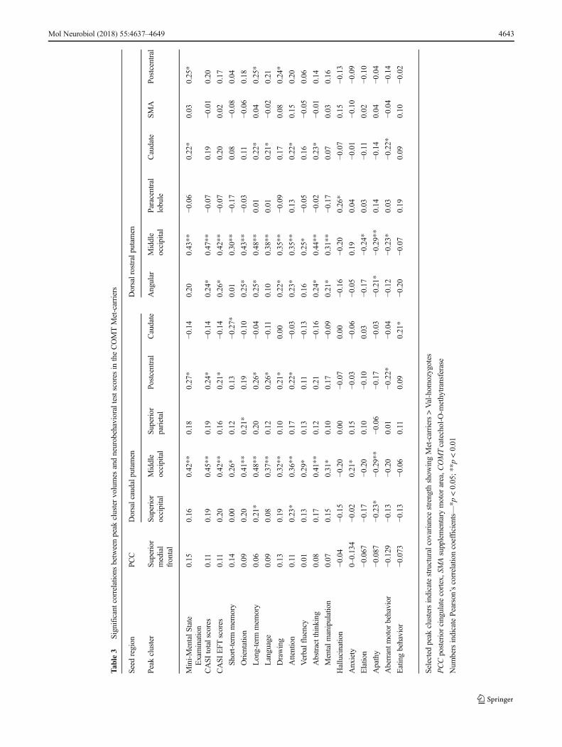
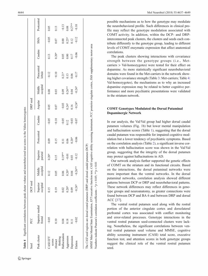
The clinical significance of the aforementioned 12 peak clus-ters (Supplementary Figure 1D, Fig. 2) showing genotypeinteractions was evaluated by correlation analysis with cogni-tive tests (Table 3 for the Met-carriers, Table 4 for the Val-homozygotes). The results suggested more significant
correlations with behavioral domains in the Met-carriers(Table 3) compared with the Val-homozygotes (Table 4).
Discussion
This study provides data on the neurobehavioral and networkinfluence of COMT Val158Met in patients with AD. Thefindings can be considered in three levels: clinical, corticalregional, and network level. From the clinical level, the neu-robehavioral comparisons between the Met-carriers and Val-homozygotes showed that the lower COMTactivity group hadhigher scores in mental manipulation scores and hallucinationdomains. From the regional aspect, the significant correlationsbetween test scores and the seed or peak cluster volumes dem-onstrated the clinical significance of PCC in DMN and all sixstriatum regions. Lastly, although the triple-network modelhas been well studied in AD, our network analysis resultssupport a higher weighting of striatum-related circuits accord-ing to the COMT genotype, of which the DCP- or DRP-
Fig. 1 Statistical maps depicting brain areas in which the gray matterintensity covaried with a six target seeds, b seed volume comparisons,and c structural covariance networks (Z-statistic maps [p < 0.01, correctedwith a false discovery rate with extended cluster voxels >100]) in allpatients with Alzheimer’s disease with the catechol-O-methyltransferase
Val158Met polymorphism (Met-carriers, n = 91; Val-homozygote car-riers, n = 101). A significantly lower dorsal caudal putamen gray matterseed volume was found in the Met-carriers (p < 0.05). The images weredisplayed on a standard brain render
4640 Mol Neurobiol (2018) 55:4637–4649

interconnected networks that contributed differently to theprediction of clinical outcome were most pronounced.
COMT Genotypes in AD Symptomatology Modulation
Although the disease-causing genetic profiles for AD havebeen identified in genome-wide association studies, these ge-netic markers have not been fully investigated with regard tooutcome correlations. This formed the basis of the currentstudy. Met-carriers can be considered as a group with long-term lower COMT activity compared to Val-homozygotes.Our Met-carriers presented with higher mental manipulationtest and hallucination scores, supporting the biological linkbetween the COMT Val158Met polymorphism and prefrontaldopamine metabolism [31]. Our results also validate thosereported in normal elderly [32] and in patients with dementia[8] in that those with low COMT enzyme activity performbetter in prefrontal-directed tasks or that it is related to psy-chiatric manifestations [7, 33]. Of note, COMTactivity can beconfounded by physiological factors such as gender, age, sex
hormones, and ApoE4 status [4, 34]. However, the associationbetween COMT polymorphisms and decline in executive con-trol with aging is controversial [35, 36].
The clinical correlation suggests that the PCC volume canbe used to predict cognitive but not NPI performance(Supplementary Table 22). The peak clusters anchored bythe PCC and the clinical correlations were also not significantin the Met-carriers (Table 3) or Val/Val group (Table 4).Therefore, the COMT Val158Met polymorphism showedgreater weighting in the striatal network than in the triplenetwork to predict cognitive symptoms.
COMT Genotype Effects on the Clinical PresentationsModulated by Large-Scale Striatum Networks
The DCP and DRP seed volumes were related to the MMSEand long-term memory scores in our Met-carriers, whereassignificant correlations between the DCP and DRP seed vol-umes were found in the attention scores, hallucination, andeating behavior in the Val-homozygotes. There are several
Fig. 2 Peak clusters showing significant interactions of Met-carriers > Val-homozygotes from the a dorsal caudal putamen (DCP) and b dorsal rostralputamen (DRP) seed. There were five DCP-related peak clusters and six DRP-related clusters. x, y, z = Montreal Neurological Institute coordinates
Mol Neurobiol (2018) 55:4637–4649 4641

Tab
le2
Correlatio
nsbetweenneurobehavioraltestscoresandseed
volumes
inthestriatalmodelin
each
catechol-O
-methyltransferasegenotype
groups
Caudate
Putamen
Seed
no.
Ventralinferior
Ventralsuperior
Dorsal
Dorsalcaudal
Dorsalrostral
Ventralrostral
Genotypegroups
Met-carriers
Val/Val
Met-carriers
Val/Val
Met-carriers
Val/Val
Met-carriers
Val/Val
Met-carriers
Val/Val
Met-carriers
Val/Val
MMSE
0.11
0.21*
0.20
0.23*
0.03
0.19
0.23*
0.14
0.23*
0.17
0.28*
0.25*
CASI
totalscores
0.11
0.20*
0.19
0.21*
0.02
0.18
0.21*
0.11
0.19
0.13
0.23*
0.22*
Executiv
efunctio
ntest
0.11
0.21*
0.19
0.21*
0.02
0.18
0.20
0.12
0.19
0.14
0.23*
0.21*
MentalM
anipulation
−0.07
0.12
0.06
0.24*
−0.02
0.22*
0.15
0.14
0.15
0.16
0.13
0.22*
Attention
0.19
0.23*
0.27**
0.26**
0.12
0.14
0.17
0.23*
0.19
0.22*
0.23*
0.29**
Orientatio
n0.10
0.18
0.12
0.10
−0.04
0.05
0.09
0.10
0.08
0.09
0.15
0.14
Long-term
mem
ory
0.22*
−0.01
0.26*
0.13
0.14
0.12
0.29**
0.03
0.23*
0.07
0.24*
0.17
Short-term
mem
ory
0.11
0.29**
0.10
0.18
−0.14
0.15
0.08
0.10
0.03
0.09
0.11
0.15
Abstractthinking
0.09
0.18
0.15
0.10
−0.01
0.11
0.15
0.03
0.14
0.03
0.17
0.03
Drawing
−0.03
0.13
0.13
0.15
0.10
0.18
0.20
0.04
0.20
0.12
0.25*
0.16
Verbalfluency
0.11
0.14
0.22*
0.20*
0.06
0.12
0.22
0.08
0.24*
0.09
0.23*
0.17
Language
0.12
0.18
0.16
0.25*
0.11
0.23*
0.20
0.13
0.19
0.17
0.20
0.25*
NPI
totalscores
−0.04
0.06
0.02
−0.05
0.08
−0.01
−0.05
−0.03
0.00
−0.04
−0.03
0.02
Delusion
−0.14
−0.06
0.00
0.00
0.00
0.08
−0.08
0.06
−0.06
0.07
−0.07
0.04
Hallucinatio
n−0
.05
−0.14
−0.07
−0.21*
−0.12
−0.20*
−0.16
−0.27**
−0.16
−0.28**
−0.13
−0.22*
Aggression
−0.16
−0.04
−0.10
−0.02
−0.02
0.01
−0.09
−0.15
−0.03
−0.16
−0.12
−0.08
Depression
0.10
0.16
0.07
0.00
0.06
0.13
0.08
0.14
0.05
0.14
0.01
0.22*
Anxiety
0.04
0.21*
0.05
−0.01
0.00
0.08
0.05
0.02
0.00
0.09
−0.03
0.10
Elatio
n−0
.09
−0.12
−0.17
−0.15
−0.04
−0.04
−0.16
−0.04
−0.04
−0.01
−0.11
−0.02
Apathy
−0.06
−0.22*
−0.10
−0.02
0.01
−0.07
−0.16
0.10
−0.09
0.03
−0.12
0.07
Disinhibitio
n−0
.20
−0.02
−0.02
−0.12
−0.01
−0.03
−0.06
−0.05
−0.01
−0.08
0.01
−0.05
Irritability
−0.07
0.23*
0.02
0.11
0.00
0.07
−0.05
−0.09
−0.04
0.02
−0.06
0.13
Aberrantm
otor
behavior
−0.14
−0.11
−0.07
−0.10
−0.05
−0.08
−0.20
−0.02
−0.15
−0.04
−0.15
−0.02
Sleepdisorder
0.12
0.15
0.13
0.00
0.23*
0.02
0.16
−0.01
0.21*
−0.02
0.22*
−0.07
Eatingbehavior
0.04
−0.07
0.09
−0.09
0.06
−0.24*
−0.03
−0.27**
−0.06
−0.31**
0.03
−0.24*
MMSE
Mini-MentalS
tateExamination,NPIneuropsychiatricinventory,CASI
cognitive
ability
screeninginstrument
Num
bersindicatePearson’scorrelationcoefficients—*p
<0.05;*
*p<0.01
4642 Mol Neurobiol (2018) 55:4637–4649
Tab
le3
Significantcorrelatio
nsbetweenpeak
clustervolumes
andneurobehavioraltestscoresin
theCOMTMet-carriers
Seed
region
PCC
Dorsalcaudalp
utam
enDorsalrostralputamen
Peakcluster
Superior
medial
frontal
Superior
occipital
Middle
occipital
Superior
parietal
Postcentral
Caudate
Angular
Middle
occipital
Paracentral
lobule
Caudate
SMA
Postcentral
Mini-MentalS
tate
Examination
0.15
0.16
0.42**
0.18
0.27*
−0.14
0.20
0.43**
−0.06
0.22*
0.03
0.25*
CASI
totalscores
0.11
0.19
0.45**
0.19
0.24*
−0.14
0.24*
0.47**
−0.07
0.19
−0.01
0.20
CASI
EFT
scores
0.11
0.20
0.42**
0.16
0.21*
−0.14
0.26*
0.42**
−0.07
0.20
0.02
0.17
Short-term
mem
ory
0.14
0.00
0.26*
0.12
0.13
−0.27*
0.01
0.30**
−0.17
0.08
−0.08
0.04
Orientatio
n0.09
0.20
0.41**
0.21*
0.19
−0.10
0.25*
0.43**
−0.03
0.11
−0.06
0.18
Long-term
mem
ory
0.06
0.21*
0.48**
0.20
0.26*
−0.04
0.25*
0.48**
0.01
0.22*
0.04
0.25*
Language
0.09
0.08
0.37**
0.12
0.26*
−0.11
0.10
0.38**
0.01
0.21*
−0.02
0.21
Drawing
0.13
0.19
0.32**
0.10
0.21*
0.00
0.22*
0.35**
−0.09
0.17
0.08
0.24*
Attention
0.11
0.23*
0.36**
0.17
0.22*
−0.03
0.23*
0.35**
0.13
0.22*
0.15
0.20
Verbalfluency
0.01
0.13
0.29*
0.13
0.11
−0.13
0.16
0.25*
−0.05
0.16
−0.05
0.06
Abstractthinking
0.08
0.17
0.41**
0.12
0.21
−0.16
0.24*
0.44**
−0.02
0.23*
−0.01
0.14
Mentalm
anipulation
0.07
0.15
0.31*
0.10
0.17
−0.09
0.21*
0.31**
−0.17
0.07
0.03
0.16
Hallucinatio
n−0
.04
−0.15
−0.20
0.00
−0.07
0.00
−0.16
−0.20
0.26*
−0.07
0.15
−0.13
Anxiety
0–0.134
−0.02
0.21*
0.15
−0.03
−0.06
−0.05
0.19
0.04
−0.01
−0.10
−0.09
Elatio
n−0
.067
−0.17
−0.20
0.10
−0.10
0.03
−0.17
−0.24*
0.03
−0.11
0.02
−0.10
Apathy
−0.087
−0.23*
−0.29**
−0.06
−0.17
−0.03
−0.21*
−0.29**
0.14
−0.14
0.04
−0.04
Aberrantm
otor
behavior
−0.129
−0.13
−0.20
0.01
−0.22*
−0.04
−0.12
−0.23*
0.03
−0.22*
−0.04
−0.14
Eatingbehavior
−0.073
−0.13
−0.06
0.11
0.09
0.21*
−0.20
−0.07
0.19
0.09
0.10
−0.02
Selected
peak
clustersindicatestructuralcovariance
strength
show
ingMet-carriers>Val-hom
ozygotes
PCCposteriorcingulatecortex,SMAsupplementary
motor
area,C
OMTcatechol-O
-methytransferase
Num
bersindicatePearson’scorrelationcoefficients—*p
<0.05;*
*p<0.01
Mol Neurobiol (2018) 55:4637–4649 4643
possible mechanisms as to how the genotype may modulatethe neurobehavioral profile. Such differences in clinical pro-file may reflect the genotype modulation associated withCOMT activity. In addition, within the DCP- and DRP-interconnected peak clusters, the clusters and seeds each con-tribute differently to the genotype group, leading to differentlevels of COMT enzymatic expression that affect anatomicalcorrelations.
The peak clusters showing interactions with covariancestrength between the genotype groups (i.e., Met-carriers > Val-homozygotes) were tested for their effect ondopamine. As more statistically significant neurobehavioraldomains were found in the Met-carriers in the network show-ing higher covariance strength (Table 3: Met-carriers; Table 4:Val-homozygotes), the mechanisms as to why an increaseddopamine expression may be related to better cognitive per-formance and more psychiatric presentations were validatedin the striatum network.
COMT Genotypes Modulated the Dorsal PutaminalDopaminergic Network
In our analysis, the Val/Val group had higher dorsal caudalputamen volumes (Fig. 1b) but lower mental manipulationand hallucination scores (Table 1), suggesting that the dorsalcaudal putamen was responsible for impaired cognitive mod-ulation but a lower tendency of psychiatric symptoms. Basedon the correlation analysis (Table 2), a significant inverse cor-relation with hallucination score was shown in the Val/Valgroup, suggesting that the integrity of the dorsal putamenmay protect against hallucinations in AD.
Our network analysis further supported the genetic effectsof COMT on the striatum and its functional circuits. Basedon the interactions, the dorsal putaminal networks weremore important than the ventral networks. In the dorsalputaminal networks, correlation analysis showed differentpatterns between DCP or DRP and neurobehavioral patterns.These network differences may reflect differences in geno-type groups and neuroanatomy, as greater connections werefound between DCP and BA 6 and between DRP and dorsalACC [37].
The ventral rostral putamen seed along with the rostralportion of the anterior cingulate cortex and dorsolateralprefrontal cortex was associated with conflict monitoringand error-related processes. Genotype interactions in theventral rostral putamen seed-connected clusters were lack-ing. Nonetheless, the significant correlations between ven-tral rostral putamen seed volume and MMSE, cognitiveability screening instrument (CASI) total score, executivefunction test, and attention scores in both genotype groupssuggest the clinical role of the ventral rostral putamenseed.T
able4
Significantcorrelatio
nsbetweenpeak
clustervolumeandneurobehavioraltestscoresin
theValinehomozygotes
Seed
PCC
DCPseed
DRPseed
Peakcluster
Superior
medial
frontal
Superior
occipital
Middle
occipital
Superior
parietal
Postcentral
Caudate
Angular
Middle
occipital
Paracentral
lobule
Caudate
SMA
Postcentral
CASIEFT
scores
−0.05
0.15
0.12
0.20*
0.09
−0.05
0.05
0.08
−0.12
0.13
−0.05
0.05
Abstract
thinking
0.10
0.19
0.05
0.15
0.15
−0.10
0.07
0.03
−0.10
0.20*
0.07
0.13
Hallucinatio
n0.04
0.02
0.06
0.19**
0.11
−0.05
−0.03
0.09
0.21*
0.05
0.00
−0.13
Aggression
0.18
0.20*
0.20*
−0.01
0.06
0.12
0.24*
0.26**
0.13
0.00
0.25*
0.08
Depression
0.11
0.09
0.04
0.03
0.20*
−0.03
0.16
0.04
0.06
0.17
0.12
0.27**
Elatio
n−0
.02
−0.24*
−0.26**
−0.12
−0.06
−0.07
−0.24*
−0.24*
−0.17
−0.06
−0.12
−0.10
The
CASI
seed
regionsaredorsalrostralp
utam
en(D
RP)
anddorsalcaudalputamen
(DCP)
MMSE
Mini-MentalS
tateExamination,EFTexecutivefunctio
ntest,C
ASI
cognitive
ability
screeninginstrument,PCCposteriorcingulatecortex,S
MAsupplementary
motor
area
Num
bersindicatePearson’scorrelationcoefficients—*p
<0.05;*
*p<0.01
4644 Mol Neurobiol (2018) 55:4637–4649

Caudate Seed and Clinical Featuresin the Val-Homozygotes
None of the peak clusters connected to the ventral striatumseed showed greater structural covariance strength in the Met-carriers. This may be due to a minor genotype modulationeffect on the ventral striatum-interconnected clusters. The cor-relations between ventral striatum seed and neurobehavioralsymptoms were still significant and displayed a parallel cor-relation pattern in the superior ventral striatum or inferior ven-tral striatum, especially in the Val-homozygotes. These resultsmay be related to the identical structural projection zone of thesuperior ventral striatum [38–41] and inferior ventral striatumseed [42–45].
For the dorsal caudate nucleus, the anatomical connectionhas been associated with the dorsolateral prefrontal cortex andexecutive control regions [46–48]. The dorsal caudate nucleusseed volume was related to the mental manipulation and lan-guage ability in the Val-homozygotes. Of note, in the Val-homozygotes, the COMT genotype effect that significantlymodulated the caudate seed determined the clinical features.
Inconsistent Effect of COMTon Cortices and PossibleExplanations
Individuals with the Val/Val genotype have been reported tohave higher levels of thyroxylase mRNA in mesencephalicdopamine neuronal populations that project to the striatum[10], which may explain why the COMT valine allele leadsto susceptibility to psychosis. However, as the pathologicalspecimens in Akil et al.’s study excluded those from patientswith AD, the direct application of their results is not possible.In our analysis, the Val/Val group had higher dorsal caudalputamen volumes (Fig. 1b) but lower mental manipulationand hallucination scores (Table 1), suggesting that the dorsalcaudal putamen is responsible for impaired cognitive modu-lation but a lower tendency toward psychiatric symptoms. Inour correlation analysis (Table 2), a significant inverse corre-lation with hallucination score was shown in the Val/Valgroup, suggesting that the integrity of the dorsal putamenmay protect against hallucinations in AD.
In addition to the physiological role of striatal networks,several factors in AD may also contribute to the inconsistenteffect of COMT. For example, the genetic expression ofCOMT and the effect of the dopamine system can be influ-enced by aging, amyloid load, and disease severity. During thephysiological aging process, decreased dopamine release, de-creased receptor expression (especially D2), and reducedtransporter expression are found in the caudate, putamen, hip-pocampus, and prefrontal cortex of human brains [49, 50]. InAD, atrophy of the caudate [14] and putamen [51] has alsobeen reported, and decreases in volume have also been corre-lated with cognitive deficits. In addition, the integrity of
nicotinic acetylcholine receptors can be affected by amyloid-related pathologies in AD [52, 53] such as neuronal homeo-stasis, synaptic plasticity, learning, and memory.
The striatum network in AD is mediated by different path-ways and neurotransmitters, and dopaminergic pathways suchas nigrostriatal pathways (substantia nigra and striatum) mod-ulate voluntary movement and mesocorticolimbic pathways(ventral tegmental area, hippocampus, nucleus accumbens)modulate cognitive-behavior-reward function. Although do-pamine levels have been reported to be higher in the striatumof individuals with the Val/Val genotype than in those with theVal/Met genotype [10], the areas showing most significantdifferences were in the ventral tier of the substantia nigra.Animal studies have shown that diminished prefrontal dopa-mine neurotransmission leads to upregulation of striatal dopa-mine activity, while higher dopamine levels in Val/Val maydownregulate the activity at the level of the prefrontal cortexand also the mesolimbic system. The increased mesencephalicdopamine activity with the Val allele may regulate corticalglutaminergic projections (prefrontal, hippocampus, andamygdala) back to the mesolimbic pathways. The dynamicchanges in network alterations along with the pathologicalcascades may have confounded the data with regard to theeffects of the COMT genotype.
Study Limitations
An important limitation of this study is that we did not includea control group. The enrolment of controls may have helped toelucidate whether the COMT polymorphism has a similareffect on the normative brain network. Our results supportpublished data on elderly healthy subjects that genetic varia-tions of the COMT polymorphism may mediate pre-frontal-related tasks [31]. However, direct analysis of SCN patterns inthe striatal or triple network with changes in structural covari-ance strength in controls was not available. The results of thissuggest how the COMT genetic polymorphism may interferewith structural networks and may be correlated with the neu-robehavioral symptoms in AD. Another potential limitation isthat we reported the peak clusters which showed greater co-variance in the Met-carriers compared with the Val-homozy-gotes. Such group stratification only explores the intra-cerebral long-term effects of dopamine on the neurobehavioraloutcomes. The expression of the COMT genotype has beenreported to be affected by gene-environment interactions [54]which could not be fully included in this study model.Nonetheless, our results may suggest that the underlying sen-sitivity of genotype groups or the dopamine transmitter systemis due to an environmental impact. Third, it has also beenreported that cross-sectional findings of genetic effects couldnot be replicated in longitudinal observations [35, 36]. Furtherlongitudinal studies including more extensive cognitive testitems are warranted.
Mol Neurobiol (2018) 55:4637–4649 4645

Conclusion
In AD, the COMT Val158Met polymorphism modulates thestriatal network rather than the triple network with regard topredicting symptoms. The genotype group itself, seed volume,or striatal network provided variable predictions of the clinicalfeatures. In the striatal network, greater covariance strength inthe Met-carriers was found in the DCP- and DRP-interconnected networks that were suggestive of a long-termdopamine-related effect. Along with the significant clinicalcorrelations, the DCP- and DRP-interconnected networksmay be considered to be the major networks modulated bythe COMT genotype.
Materials and Methods
This study was conducted in accordance with theDeclaration of Helsinki and was approved by theInstitutional Review Board of Chang Gung MemorialHospital. The study participants were treated at theCognition and Aging Center, Department of GeneralNeurology, Kaohsiung Chang Gung Memorial Hospital. Atotal of 192 subjects (95 males, 97 females) were includedafter the consensus of a panel composed of neurologists,neuropsychologists, neuroradiologists, and experts in nuclearmedicine [55]. AD was diagnosed according to theInternational Working Group criteria [45] with a clinicaldiagnosis of typical AD. All of the patients were in a stablecondition under acetylcholine esterase inhibitor treatmentfrom the time of diagnosis. The exclusion criteria were apast history of clinical stroke, a modified Hachinski ische-mic score >4, and depression.
Study Working Scheme
Because of the limited number of subjects in the Met/Metgroup, we grouped the Val/Met and Met/Met subjects intothe Met-carrier group in all subsequent analysis: Met-carriers(Met/Met = 20, Met/Val = 71, n = 91) and Val-homozygotes(n = 101). The working scheme was as follows. First, theSCNs were established by seed-based correlation analysis.Differences in each seed regional volume were comparedbetween two genotype groups and correlated with the neu-robehavioral scores. In order to evaluate the dopaminergicnetwork effects, only the peak clusters showing Met-carriers > Val/Val in covariance strength were consideredas statistically significant. The volumes of the significantpeak clusters were selected and correlated with cognitive testscores to evaluate the clinical relevance in each genotypegroup.
Clinical and Neurobehavioral Assessments
After enrolment, demographic data of each patient were re-corded. A trained neuropsychologist administered the neuro-behavioral tests. The MMSE scores and CASI total scoreswere used as a global assessment of cognitive function.Attention, verbal fluency, abstract thinking, and mental ma-nipulation sub-domain scores of the CASI were used to assessexecutive function test (EFT) [56], while the non-executivedomains included orientation, short- and long-term memory,language ability, and drawing. For the behavioral observa-tions, we used the 12-item version of the NPI [57], with scoresranging from 0 to 144.
Genotyping for COMT
Genotyping of COMT Val158Met was performed using thepolymerase chain reaction-restriction fragment length poly-morphism method. In brief, a DNA fragment containing theVal/Met polymorphism in COMTwas amplified by polymer-ase chain reaction with primers reported by Lachman et al.[58]. The Val/Met polymorphism was differentiated by theNlaIII restriction fragment length polymorphism analyzed on10% polyacrylamide gel. Partial digestion and contaminationamplification were ruled out by the complete digestion of anintrinsic restriction site and a blank sample in each batch ofexperiments, respectively. The ApoE genotype was deter-mined using a PCR-restriction fragment length polymorphismassay and restriction enzyme HhaI. ApoE4 carriers were de-fined as those with one or two E4 alleles.
Image Acquisition
MR images were acquired using a 3.0T MRI scanner (Excite,GE Medical Systems, Milwaukee, WI, USA). Structural im-ages were acquired for SCN constructions using the followingprotocols: a T1-weighted, inversion-recovery-prepared, three-dimensional, gradient-recalled acquisition in a steady-state se-quence with a repetition time/echo time/inversion time of8600 ms/minimal/450 ms, a 256 × 256 mm field of view,and a 1-mm slice sagittal thickness with a resolution of0.5 × 0.5 × 1 mm3.
Data Analysis for Neuroimaging Biomarkers
Image preprocessing and statistical analysis were performedusing SPM8 (SPM8, Wellcome Trust Centre of CognitiveNeurology, University College London, UK, http://www.fil.ion.ucl.ac.uk/spm/). The T1 images were reoriented,realigned, and normalized using the standard MontrealNeurological Institute space. The images were thensegmented into GM and white matter. Related tissuesegments were used to create a custom template using the
4646 Mol Neurobiol (2018) 55:4637–4649

diffeomorphic anatomical registration using exponentiated liealgebra (DARTEL) approach. The DARTEL approach is oneof the highest ranking registration methods in patients withAD [59]. The modulated and warped images were thensmoothed using a Gaussian kernel of 8 mm full width at halfmaximum.
Images Analysis
To investigate the SCNs, 10 regions of interest, representingseeds, were selected from the 192 preprocessed images. Thestriatal network [15] included the following seeds (Fig. 1a):inferior ventral striatum [coordinates: [9, 9, −8], superior ven-tral striatum [coordinates: 10, 15, 0]; dorsal caudate [coordi-nates: 13, 15, 9]; ventral rostral putamen [coordinates: 20, 12,−3]; dorsal caudal putamen [DCP; coordinates: 28, 1, 3]; dor-sal rostral putamen [DRP; coordinates: 25, 8, 6]. The coordi-nates of seed in the triple-network model included the rightentorhinal cortex [coordinates: 25, −9, −28] and left posteriorcingulate cortex [PCC; coordinates: −2, −36, 35] of the DMN,right frontoinsular cortex [coordinates: 38, 26, −10] of thesalience network, and right dorsolateral prefrontal cortex [co-ordinates: 44, 36, 20] of the executive control network [25](supplementary fig 1).
From the modified GM images, the GM volumes of a 4-mmradius sphere around the seed coordinates were also calculated,followed by 10 separate correlation analyses using the extract-ed GM volumes as the covariates of interest, to form the SCN.The two genotype groups were modeled separately. Based onthe equivalent sample sizes in each genotype group, Tcontrastswere used to identify voxels that showed positive correlationsfor each seed. The results reflected the SCNs anchored by eachseed. The threshold was set at p < 0.01, corrected for falsediscovery rate (FDR) with a cluster size >100 voxels.
In addition, to investigate how genetic variance may inter-fere with SCN covariance strength, voxels showing signifi-cant differences in the regression slopes in each seed-peakcluster correlations were compared that pointed to interactionsbetweenMet-carriers > Val-homozygotes. Specific Tcontrastswere established to map the voxels that expressed significantbetween-group associations.
For the peak clusters showing significant between-groupdifferences, a 4-mm radius sphere was placed on the peakvoxel, and the GM volumes were then calculated. To evaluatethe clinical significance of the seed or the identified peakvoxel, we used correlation analysis with the cognitive test orNPI scores as outcome measures. The threshold was set atp < 0.05 with multiple corrections.
Statistical Analysis
Clinical and laboratory data were expressed as mean ± standarddeviation. The Student t test was used to compare the
continuous variables and chi-square test for category variables.Pearson’s correlation was used to analyze the seed or clustervolume on predicting the cognitive or NPI scores. All statisticalanalyses were conducted using SPSS software (SPSS version22 for Windows®, SPSS Inc., Chicago, IL). Statistical signifi-cance was set at p < 0.05.
1. Serretti A, Olgiati P (2012) Catechol-o-methyltransferase andAlzheimer’s disease: a review of biological and genetic findings.CNS Neurol Disord Drug Targets 11(3):299–305
2. Martorana A, Koch G (2014) Is dopamine involved in Alzheimer’sdisease? Front Aging Neurosci 6:252. doi:10.3389/fnagi.2014.00252
3. Yan W, Zhao C, Sun L, Tang B (2016) Association between poly-morphism of COMT gene (Val158Met) with Alzheimer’s disease:an updated analysis. J Neurol Sci 361:250–255. doi:10.1016/j.jns.2016.01.014
4. Wang PN, Liu HC, Liu TY, Chu A, Hong CJ, Lin KN, Chi CW(2005) Estrogen-metabolizing gene COMT polymorphism syner-gistic APOE epsilon4 allele increases the risk of Alzheimer disease.Dement Geriatr Cogn Disord 19(2–3):120–125. doi:10.1159/000082663
5. Lanni C, Garbin G, Lisa A, Biundo F, Ranzenigo A, Sinforiani E,Cuzzoni G, Govoni S et al (2012) Influence of COMT Val158Metpolymorphism on Alzheimer’s disease and mild cognitive impair-ment in Italian patients. J Alzheimer’s Dis: JAD 32(4):919–926.doi:10.3233/jad-2012-120358
6. Martinez MF, Martin XE, Alcelay LG, Flores JC, Valiente JM,Juanbeltz BI, Beldarrain MA, Lopez JM et al (2009) The COMTVal158 Met polymorphism as an associated risk factor forAlzheimer disease and mild cognitive impairment in APOE 4 car-riers. BMC Neurosci 10:125. doi:10.1186/1471-2202-10-125
7. Sweet RA, Devlin B, Pollock BG, Sukonick DL, Kastango KB,Bacanu SA, Chowdari KV, DeKosky ST et al (2005) Catechol-O-methyltransferase haplotypes are associated with psychosis inAlzheimer disease. Mol Psychiatry 10(11):1026–1036. doi:10.1038/sj.mp.4001709
8. Nedic G, Borovecki F, Klepac N, Mubrin Z, Hajnsek S, NikolacM,Muck-Seler D, Pivac N (2011) Association study of a functionalcatechol-o-methyltransferase polymorphism and cognitive functionin patients with dementia. Coll Antropol 35(Suppl 1):79–84
Mol Neurobiol (2018) 55:4637–4649 4647
Acknowledgements The authors wish to thank the patients and theircaregivers for their time and commitment to this research.
Compliance with Ethical Standards
Funding This work was supported by grants CMRPG8C0571,CMRPG8D0771 and CMRPG8E0541 from the Chang Gung MemorialHospital and 104-2314-B-182A-026-MY2 from the National ScienceCouncil to CCC for MRI acquisition, genetic and clinical data collection,and analysis.
Open Access This article is distributed under the terms of the CreativeCommons At t r ibut ion 4 .0 In te rna t ional License (h t tp : / /creativecommons.org/licenses/by/4.0/), which permits unrestricted use,distribution, and reproduction in any medium, provided you giveappropriate credit to the original author(s) and the source, provide a linkto the Creative Commons license, and indicate if changes were made.
References

9. Pereira PA, Romano-Silva MA, Bicalho MA, de Moraes EN,Malloy-Diniz L, Pimenta GJ, Mello MP, Bozzi IC et al (2012)Catechol-O-methyltransferase genetic variant associated with therisk of Alzheimer’s disease in a Brazilian population. DementGeriatr Cogn Disord 34(2):90–95. doi:10.1159/000341578
10. Akil M, Kolachana BS, Rothmond DA, Hyde TM,Weinberger DR,Kleinman JE (2003) Catechol-O-methyltransferase genotype anddopamine regulation in the human brain. J Neurosci 23(6):2008–2013
11. O’Doherty J, Dayan P, Schultz J, Deichmann R, Friston K, DolanRJ (2004) Dissociable roles of ventral and dorsal striatum in instru-mental conditioning. Science (NewYork, NY) 304(5669):452–454.doi:10.1126/science.1094285
12. Chang CC, Hsu JL, ChangWN,Huang SH, HuangCW, ChangYT,Chen NC, Lui CC et al (2016) Metabolic covariant network inrelation to nigrostriatal degeneration in carbon monoxideintoxication-related parkinsonism. Front Neurosci 10:187. doi:10.3389/fnins.2016.00187
13. Chang CC, Hsiao IT, Huang SH, Lui CC, Yen TC, Chang WN,Huang CW, Hsieh CJ et al (2015) F-FP-(+)-DTBZ positron emis-sion tomography detection of monoaminergic deficient network inpatients with carbon monoxide related parkinsonism. Eur J Neurol22(5):845–852, e859-860. doi:10.1111/ene.12672
14. Madsen SK, Ho AJ, Hua X, Saharan PS, Toga AW, Jack CR Jr,Weiner MW, Thompson PM (2010) 3D maps localize caudate nu-cleus atrophy in 400 Alzheimer’s disease, mild cognitive impair-ment, and healthy elderly subjects. Neurobiol Aging 31(8):1312–1325. doi:10.1016/j.neurobiolaging.2010.05.002
15. Postuma RB, Dagher A (2006) Basal ganglia functional connectiv-ity based on a meta-analysis of 126 positron emission tomographyand functional magnetic resonance imaging publications. CerebCortex 16(10):1508–1521. doi:10.1093/cercor/bhj088
16. Haber SN, Fudge JL, McFarland NR (2000) Striatonigrostriatalpathways in primates form an ascending spiral from the shell tothe dorsolateral striatum. J Neurosci 20(6):2369–2382
17. Haber SN (2003) The primate basal ganglia: parallel and integrativenetworks. J Chem Neuroanat 26(4):317–330
18. Parent A, Hazrati LN (1995) Functional anatomy of the basal gan-glia. I. The cortico-basal ganglia-thalamo-cortical loop. Brain ResRev 20(1):91–127
19. Yavich L, Forsberg MM, Karayiorgou M, Gogos JA, Mannisto PT(2007) Site specific role of catechol-O-methyltransferase in dopa-mine overflow within prefrontal cortex and dorsal striatum. JNeurosci 27(38):10196-10209. doi:10.1523/jneurosci.0665-07.2007
20. Lin PH, Tsai SJ, Huang CW,Mu-En L, Hsu SW, Lee CC, Chen NC,Chang YT et al (2016) Dose-dependent genotype effects of BDNFVal66Met polymorphism on default mode network in early stageAlzheimer’s disease. Oncotarget. doi:10.18632/oncotarget.11027
21. Menon V (2011) Large-scale brain networks and psychopathology:a unifying triple network model. Trends Cogn Sci 15(10):483–506.doi:10.1016/j.tics.2011.08.003
22. Greicius MD, Srivastava G, Reiss AL, Menon V (2004) Default-mode network activity distinguishes Alzheimer’s disease fromhealthy aging: evidence from functional MRI. Proc Natl Acad SciU S A 101(13):4637–4642. doi:10.1073/pnas.0308627101
23. Greicius MD, Supekar K, Menon V, Dougherty RF (2009) Resting-state functional connectivity reflects structural connectivity in thedefault mode network. Cereb Cortex 19(1):72–78. doi:10.1093/cercor/bhn059
24. Seeley WW, Allman JM, Carlin DA, Crawford RK, Macedo MN,Greicius MD, Dearmond SJ, Miller BL (2007) Divergent socialfunctioning in behavioral variant frontotemporal dementia andAlzheimer disease: reciprocal networks and neuronal evolution.Alzheimer Dis Assoc Disord 21(4):S50–S57. doi:10.1097/WAD.0b013e31815c0f14
25. SeeleyWW,Menon V, Schatzberg AF, Keller J, Glover GH, KennaH, Reiss AL, GreiciusMD (2007) Dissociable intrinsic connectivitynetworks for salience processing and executive control. J Neurosci27(9):2349–2356. doi:10.1523/jneurosci.5587-06.2007
26. Agosta F, Pievani M, Geroldi C, Copetti M, Frisoni GB, Filippi M(2012) Resting state fMRI in Alzheimer’s disease: beyond the de-fault mode network. Neurobiol Aging 33(8):1564–1578. doi:10.1016/j.neurobiolaging.2011.06.007
27. Filippi M, Agosta F, Scola E, Canu E, Magnani G, Marcone A,Valsasina P, Caso F et al (2013) Functional network connectivityin the behavioral variant of frontotemporal dementia. Cortex 49(9):2389–2401. doi:10.1016/j.cortex.2012.09.017
28. Andrews-Hanna JR, Reidler JS, Sepulcre J, Poulin R, Buckner RL(2010) Functional-anatomic fractionation of the brain's default net-work. Neuron 65(4):550–562. doi:10.1016/j.neuron.2010.02.005
29. Dickinson D, Elvevag B (2009) Genes, cognition and brain througha COMT lens. Neuroscience 164(1):72–87. doi:10.1016/j.neuroscience.2009.05.014
30. Ashburner J, Friston KJ (2000) Voxel-based morphometry—themethods. NeuroImage 11(6 Pt 1):805–821
31. Liu ME, Hong CJ, Liou YJ, Tsai YL, Hsieh CH, Tsai SJ (2008)Association study of a functional catechol-O-methyltransferasepolymorphism and executive function in elderly males without de-mentia. Neurosci Lett 436(2):193–195. doi:10.1016/j.neulet.2008.03.018
32. Liu ME, Huang CC, Yang AC, Tu PC, Yeh HL, Hong CJ, Liou YJ,Chen JF et al (2014) Catechol-O-methyltransferase Val158Metpolymorphism on the relationship between white matterhyperintensity and cognition in healthy people. PLoS One 9(2):e88749. doi:10.1371/journal.pone.0088749
33. Mitaki S, Isomura M,Maniwa K, Yamasaki M, Nagai A, Nabika T,Yamaguchi S (2013) Apathy is associated with a single-nucleotidepolymorphism in a dopamine-related gene. Neurosci Lett 549:87–91. doi:10.1016/j.neulet.2013.05.075
34. Teng EL, Hasegawa K, Homma A, Imai Y, Larson E, Graves A,Sugimoto K, Yamaguchi T et al (1994) The Cognitive AbilitiesScreening Instrument (CASI): a practical test for cross-cultural ep-idemiological studies of dementia. Int Psychogeriatr/IPA 6(1):45–58 discussion 62
35. Fiocco AJ, Lindquist K, Ferrell R, Li R, Simonsick EM, Nalls M,Harris TB, Yaffe K (2010) COMT genotype and cognitive function:an 8-year longitudinal study in white and black elders. Neurology74(16):1296–1302. doi:10.1212/WNL.0b013e3181d9edba
36. Erickson KI, Kim JS, Suever BL, Voss MW, Francis BM, KramerAF (2008) Genetic contributions to age-related decline in executivefunction: a 10-year longitudinal study of COMT and BDNF poly-morphisms. Front Hum Neurosci 2:11. doi:10.3389/neuro.09.011.2008
37. Kunzle H (1975) Bilateral projections from precentral motor cortexto the putamen and other parts of the basal ganglia. An autoradio-graphic study inMacaca fascicularis. Brain Res 88(2):195–209
38. Ilinsky IA, Jouandet ML, Goldman-Rakic PS (1985) Organizationof the nigrothalamocortical system in the rhesus monkey. J CompNeurol 236(3):315–330. doi:10.1002/cne.902360304
39. Ruiz JR, Castillo R, Labayen I, Moreno LA, Fuentes MG, LamunoDG, Alvarez Granda JL, Lucia A et al (2010) Individual and com-bined effects of ApoE and MTHFR 677C/T polymorphisms on cog-nitive performance in Spanish adolescents: the AVENA study. JPediatr 156(6):978–984, 984 e971. doi:10.1016/j.jpeds.2009.12.018
40. Ferry AT, Ongur D, An X, Price JL (2000) Prefrontal cortical pro-jections to the striatum in macaque monkeys: evidence for an orga-nization related to prefrontal networks. J CompNeurol 425(3):447–470
41. Haber SN, Kim KS, Mailly P, Calzavara R (2006) Reward-relatedcortical inputs define a large striatal region in primates that interfacewith associative cortical connections, providing a substrate for
4648 Mol Neurobiol (2018) 55:4637–4649

incentive-based learning. J Neurosci 26(32):8368–8376. doi:10.1523/jneurosci.0271-06.2006
42. Powell EW, Leman RB (1976) Connections of the nucleus accum-bens. Brain Res 105(3):389–403
43. Yeterian EH, Van Hoesen GW (1978) Cortico-striate projections inthe rhesus monkey: the organization of certain cortico-caudate con-nections. Brain Res 139(1):43–63
44. Zhang MY, Miao L, Li YS, Hu GY (2010) Meta-analysis of themethylenetetrahydrofolate reductase C677T polymorphism andsusceptibility to Alzheimer’s disease. Neurosci Res 68(2):142–150. doi:10.1016/j.neures.2010.06.011
45. Dubois B, Feldman HH, Jacova C, Cummings JL, Dekosky ST,Barberger-Gateau P, Delacourte A, Frisoni G et al (2010)Revising the definition of Alzheimer’s disease: a new lexicon.Lancet Neurol 9(11):1118–1127. doi:10.1016/s1474-4422(10)70223-4
46. Selemon LD, Goldman-Rakic PS (1985) Longitudinal topographyand interdigitation of corticostriatal projections in the rhesus mon-key. J Neurosci 5(3):776–794
47. Middleton FA, Strick PL (2002) Basal-ganglia ‘projections’ to theprefrontal cortex of the primate. Cereb Cortex 12(9):926–935
48. Kelly RM, Strick PL (2004) Macro-architecture of basal ganglialoops with the cerebral cortex: use of rabies virus to revealmultisynaptic circuits. Prog Brain Res 143:449–459
49. Backman L, Lindenberger U, Li SC, Nyberg L (2010) Linkingcognitive aging to alterations in dopamine neurotransmitter func-tioning: recent data and future avenues. Neurosci Biobehav Rev34(5):670–677. doi:10.1016/j.neubiorev.2009.12.008
50. Volkow ND, Fowler JS, Wang GJ, Logan J, Schlyer D, MacGregorR, Hitzemann R,Wolf AP (1994) Decreased dopamine transporterswith age in health human subjects. Ann Neurol 36(2):237–239. doi:10.1002/ana.410360218
51. de Jong LW, van der Hiele K, Veer IM, Houwing JJ, WestendorpRG, Bollen EL, de Bruin PW, Middelkoop HA et al (2008)Strongly reduced volumes of putamen and thalamus in
Alzheimer’s disease: an MRI study. Brain 131(Pt 12):3277–3285.doi:10.1093/brain/awn278
52. Posadas I, Lopez-Hernandez B, Cena V (2013) Nicotinic receptorsin neurodegeneration. Curr Neuropharmacol 11(3):298–314. doi:10.2174/1570159x11311030005
53. Schliebs R, Arendt T (2011) The cholinergic system in aging andneuronal degeneration. Behav Brain Res 221(2):555–563. doi:10.1016/j.bbr.2010.11.058
54. Hoth KF, Paul RH, Williams LM, Dobson-Stone C, Todd E,Schofield PR, Gunstad J, Cohen RA et al (2006) Associations be-tween the COMT Val/Met polymorphism, early life stress, andpersonality among healthy adults. Neuropsychiatr Dis Treat 2(2):219–225
55. Huang CW, Tsai MH, Chen NC, ChenWH, Lu YT, Lui CC, ChangYT, Chang WN et al (2015) Clinical significance of circulatingvascular cell adhesion molecule-1 to white matter disintegrity inAlzheimer’s dementia. Thromb Haemost 114(6):1230–1240. doi:10.1160/th14-11-0938
56. Huang CW, ChangWN, Huang SH, Lui CC, Chen NC, Chang YT,Lee CC, Chang CC et al (2013) Impact of homocysteine on corticalperfusion and cognitive decline in mild Alzheimer’s dementia. EurJ Neurol 20(8):1191–1197. doi:10.1111/ene.12159
57. Cummings JL, Mega M, Gray K, Rosenberg-Thompson S, CarusiDA, Gornbein J (1994) The Neuropsychiatric Inventory: compre-hensive assessment of psychopathology in dementia. Neurology44(12):2308–2314
58. Lachman HM, Papolos DF, Saito T, Yu YM, Szumlanski CL,Weinshilboum RM (1996) Human catechol-O-methyltransferasepharmacogenetics: description of a functional polymorphism andits potential application to neuropsychiatric disorders.Pharmacogenetics 6(3):243–250
59. Cuingnet R, Gerardin E, Tessieras J, Auzias G, Lehericy S, HabertMO, Chupin M, Benali H et al (2011) Automatic classification ofpatients with Alzheimer’s disease from structural MRI: a compari-son of ten methods using the ADNI database. NeuroImage 56(2):766–781. doi:10.1016/j.neuroimage.2010.06.013
Mol Neurobiol (2018) 55:4637–4649 4649