cauliflower mosaic vírus gene vi controls translation from ... · by virus infection are...
TRANSCRIPT

The Plant Cell, Vol. 4, 1471-1484, December 1992 O 1992 American Society of Plant Physiologists
Cauliflower Mosaic Vírus Gene VI Controls Translation from Dicistronic Expression Units in Transgenic Arabidopsis Plants
Carolien Zijlstra and Thomas Hohn’ Friedrich Miescher-lnstitut, !?O. Box 2543, CH-4002 Basel, Switzerland
Transformed Arabidopsis plants were used to study the effect of the cauliflower mosaic virus (CaMV) inclusion body protein on translation of dicistronic RNA. Reporter plants contain a dicistronic transcription unit with CaMV open read- ing frame Vil (ORF VII) as the first and the B-glucuronidase (GUS) reporter ORF as the second cistron. “Transactivator p1ants”contain CaMV ORF VI under the control of the strong CaMV 35s promoter. The transactivator plants were difficult to regenerate and showed an abnormal phenotype. Expression of GUS activity in the reporter plants was very low, but high GUS activity could be induced by introduction of gene VI, either by crossing with plants containing gene VI as a transgene or by infection with CaMV. Histological GUS assays showed that transactivation occurred in all types of tissue and at all developmental stages. The practical implications of the induction of GUS expression from the dicistronic unit by virus infection are discussed.
INTRODUCTION
The genome of cauliflower mosaic virus (CaMV), a plant pararetrovirus, is shown in Figure 1. It accumulates in the form of -1000 minichromosomes per nucleus of infected cells dur- ing the infection cycle (Olszewski, 1982). These are transcribed by the host RNA polymerase II, resulting in the production of the 19s and the 35s RNAs. The 35s RNA is a particularly com- plex RNA that is thought to be involved in several basic steps of the v i r a life cycle: it is packaged into capsids (Fütterer and Hohn, 1987), reverse transcribed to yield circular DNA, and translated into six or seven (po1y)proteins. The 35s RNA con- tains a 600-nucleotide leader sequence with several small open reading frames (sORFs) followed by six tightly arranged larger ORFs that are expressed during infection (reviewed by Mason et al., 1987; Bonneville, 1992).
The translation mechanism from this unusual mRNA has been studied in CaMV and figwort mosaic virus (another mem- ber of the caulimovirus group) by isolating and testing polar mutants and their revertants (Sieg and Gronenborn, 1982; Dixon and Hohn, 1984) and by expressing CaMV derivatives in vitro (Gordon et al., 1988) and in plant protoplasts (Baughman and Howell, 1988; Fütterer et al., 1988, 1989, 1990a, 1990b; Bonneville et al., 1989; Gowda et al., 1989; Fütterer and Hohn, 1991; Scholthof et al., 1992). In summary, the results suggest that the translational machinery starts scanning at the cap site
To whom correspondence should be addressed.
in accordance with the central dogma of eukaryotic transla- tion (Kozak, 1989), but then bypasses (shunts) the central region of the leader sequence in a less defined process in- volving parts of the leader itself and host factor(s) (Fütterer et al., 1989, 199Ob). This results in translation of the first coding region (ORFVII). ThefollowingORFs(1, II, 111, IV, V, and probably VI) are translated by a reinitiation mechanism which depends on the presence of the CaMV ORF VI protein (Bonneville et al., 1989; Gowdaet al., 1989, 1991, 1992; Scholthof et al., 1992). Thus, the gene VI gene product appears to be a transactivator protein, whose initial appearance results from translation of the subgenomic 19s RNA.
The ORF VI protein is an interesting and probably multifunc- tional protein. It is the major component of the amorphous inclusion bodies accumulating in infected cells (Odell and Howell, 1980; Covey and Hull, 1981). lnclusion bodies have been suggested to be the site of translation of CaMV RNA, ac- cumulation of vira1 translation products (De Zoeten et al., 1989), reverse transcription (Pfeiffer and Hohn, 1983; Bonneville et al., 1984; Mazzolini et al., 1985; Thomas et al., 1985), and vi- rus assembly. In electron micrographs, polysomes are seen attached to the surface of inclusion bodies, whereas virus par- ticles are inside (Shepherd et al., 1979). The ORF VI protein might also alter the translation machinery by interaction with ribosomes and/or initiation factors.
These properties of ORF VI protein can be expected to af- fect the host plant, and in fact they determine the severity of

1472 The Plant Cell
Figure 1. Map of the CaMV Genome.
The black ring symbolizes the 8-kb double-stranded CaMV DNA; the stippled unnumbered blocks within the ring show the set of sORFs; the numbered stippled and white blocks designate the ORFs proper. VI1 is known to be translated but nonessential (Dixon and Hohn, 1984); I and II code for functions typical of plant viruses, i.e., systemic spreading (Linstead et al., 1988; Citovski et al., 1991) and aphid aquisition factor (Woolston et al., 1983); 111 codes for a DNA binding protein (Giband et al., 1986). The following ORFs have counterparts in retroviruses: IV (GAG) codes for a set of structural proteins related to retrovirus core proteins; V codes for a polyprotein with protease and polymerase ac- tivities (Fütterer and Hohn, 1987); VI encodes a protein that forms large cytoplasmic inclusion bodies (Covey and Hull, 1981) within which the virus particles accumulate; it is also the transactivator protein (Bonneville et al., 1989). The two major vira1 transcripts are shown as arrows along their respective coding regions. These 19s and 35s RNAs are transcribed clockwise from distinct promoters located just upstream of their respective 5' ends, but share the same 3' end (arrowheads).
the cauliflower mosaic disease, the types of symptoms, and, together with other factors, the host range (Daubert et al., 1984; Schoelz et al., 1986; Stratford and Covey, 1989; Daubert and Routh, 1990). Interestingly, expression from ORF VI transgenes also leads to viruslike symptoms (Bálasz, 1988; Baughman et al., 1988; Kiernan et al., 1989; Takahashi et al., 1989; Goldberg et al., 1991; Schoelz et al., 1991). Plants that are not systemic hosts for CaMV are especially affected, whereas cer- tain transformed host plants remain symptomless (Goldberg et al., 1991). It remains to be seen whether these phenotypic effects are caused by modulation of the cell's translation ma- chinery, resulting in overproduction of othewise poorly translated host proteins, or by other functions of the ORF VI protein.
We wanted to test whether transactivation by gene VI oc- curs in all types of plant tissues and at all developmental stages.
Because this problem cannot be studied using transient ex- pression in protoplasts, we produced Arabidopsis plants transformed with a dicistronic transcription unit containing CaMV ORF VI1 as the first and the P-glucuronidase (GUS) reporter gene as the second ORF. GUS expression was low in these plants, but high activity could be induced by crossing with plants containing the ORF VI transgene or by infection with CaMV. Transactivation occurred in all tissues and develop- mental stages tested and may constitute a useful tool for induced expression and induced resistance in plants.
RESULTS
Production and Testing of Arabidopsis Plants Transformed with Dicistronic Expression Units
Plasmids pMonoGUS and pBiGUS (Bonneville et al., 1989) contain monocistronic and dicistronic GUS expression cas- settes, respectively, consisting of the CaMV 35s promoter, the GUS reporter ORF fused to the AUG of CaMV ORF I , and the CaMV polyadenylator. The BiGUS cassette contains an addi- tional ORF, the CaMV ORF VII, between the promoter and the GUS ORF. The expression cassettes from these plasmids, sym- bolized in Figure 2, were cloned between the T-DNA borders
AgBiGUS (V613)
LB RB
AgMonoGUS (V614)
LB RB
AgTAV (V799) Promoters
nos
AgTAVTAV (VSOO)
Figure 2. Agrobacterium Lines and Shuttle Plasmids.
Only the T-DNAs are shown. Vectors are pBIN19 for V614 and V613 in AgMonoGUS and AgBiGUS, respectively, and pGSC1704 for V799 and V800 in AgTAV and AgTAVTAV, respectively (see Methods). These binary vectors were transferred from E. coli to Agrobacterium. Trans- genic plants produced by using these Agrobacterium lines were given an At designation with numbers identifying the individual primary trans- formants. LB and RB, left and right T-DNA borders; NPT, neomycin phosphotransferase I I ; HPT, hygromycin phosphotransferase; nos, nopaline synthase promoter; 35S, CaMV 35s promoter. V-numbers are collection numbers under which the plasmids are available.

Transact!vation of Translation in Plants 1473
B
BH/GUS
BH/NPT
to w8 8
kb
4.4 -
2.3 -2.0 -iii;
23
9.6 -6.6 -4.4 -
of the Agrobacterium binary shuttle vector pBIN19 (Sevan,1984) in addition to its resident neomycin phosphotransferaseII (NPTII) marker. The resulting plasmids, V614 and V613, wereintroduced into Agrobacterium by electroporation, and theresulting strains, AgMonoGUS and AgBiGUS, were used totransform Arabidopsis leaf discs.
Kanamycin-resistant calli were regenerated to several normal-appearing TWMonoGUS" and 'WfBiGUS" (T0) Arabidopsisplants over a 4-month period. Seeds from selfed transformedplants were sown on selective medium. Progeny plants givinga 3:1 segregation ratio for kanamycin-resistant seedlings wereselected for further analysis, on the assumption that only asingle locus had been affected in these transformants. DMAgel blot analysis revealed that most of the /AfMonoGUS plantscontained multiple insertions (data not shown), whereas mostof the XWBiGUS plants contained single insertions, as shownin Figure 3. Only plants with single-copy insertions were ana-lyzed further.
GUS activity in the transformed plants was determined ei-ther in plant extracts or in extracts from protoplasts. Figure 4shows that X\fMonoGUS transgenic plants, as expected, con-tained high levels of GUS activity, whereas no activity was foundin untransformed controls. These levels are ~150 times higherthan the ones we observed regularly in transfected protoplasts.The GUS activity in AfBiGUS transgenic plants and in theirprotoplasts was low but detectable, i.e., ~1% of the activityof AfMonoGUS transgenic plants. In contrast, the level of GUSactivity in wild-type protoplasts transfected with 5 u,g of pBi-GUS was less than 0.05% of the activity in protoplaststransfected with 5 ng of pMonoGUS (data not shown). The rel-atively high expression and background levels in transformed
HD/GUS
HD/NPT
2.3 -
23
9.6 -6.6 -
4.4 -
2.3 -
23
9.4 -6.6 -
4.4 -
2.3 -
Figure 3. DMA Gel Blot Analysis of Transgenic /AfBiGUS Plants.
(A) T-DNA from AgBiGUS. Arrangement of genes, restriction sites,probes used, and possible restriction fragments produced by diges-tion with BamHI when probed with GUS (BH/GUS), BamHI when
probed with NPT (BH/NPT), Hindlll when probed with GUS (HD/GUS),and Hindlll when probed with NPT (HD/NPT). LB, left border; RB, rightborder. The number of T-DNA insertions in an individual transformantwere determined by generating border fragments between T-DNA andplant DNA. Border fragments were obtained and characterized bydigesting DNA with BamHI or Hindlll and probing with the NPT probeor by digesting DNA with Hindlll and probing with the GUS probe.In both cases, a single fragment was expected if only one copy of theT-DNA had integrated into the plant DNA.(B) Autoradiograms of gel blots of DNA extracted from wild-type andXW3IGUS Arabidopsis plants. DNA was digested with BamHI andHindlll, respectively, and filter hybridized with the GUS and NPT probes,respectively. Lanes 1C and 5C contain DNA from untransformed plantsand one and five equivalents, respectively, of the /(gBiGUS T-DNA.Blot BH/GUS shows that all transformants had the complete BamHIfragment of ~2.2 kb, whereas no signal was obtained with untrans-formed tissue. Plants AfBiGUSI, X\fBiGUS3, and AfBiGUS4 containa single copy of XigBiGUS (appearance of single fragments in blotsBH/NPT, HD/GUS, HD/NPT). According to the strength of the signals,AfBiGUS2 contains approximately five copies of the integrated T-DNAsin a head-to-tail arrangement, explaining the appearance of the inter-nal 3.2-kb fragment in blot BH/NPT and the internal 5.3-kb fragmentin blots HD/GUS and HD/NPT. The banding pattern of AfBiGUSS inblots HD/GUS and HD/NPT cannot be explained by any obvious model.ArBiGUSS must, therefore, have rearranged its T-DNA. WT, wild type.

1474 The Plant Cell
GUS activity (pmol 4-MU/mg protein per min)
2000 i——————————————————————I———————i 20000
1500
1000
500
15000
10000
5000
wt AtB1 AtB3 AtB4 AtM
•1 Mock Mi +HELP4 I I +HELP7
Figure 4. GUS Expression in Wild-Type and ArBiGUS Protoplasts.
GUS activities measured in representative Arabidopsis protoplasts aftertransfection of 50 ng calf thymus DNA (mock), 50 ng calf thymus and10 ng pHELP4 DNA, and 50 ng calf thymus and 10 ng pHELP7 DNA.GUS activity was measured in samples containing 7 |jg of crude pro-tein and is expressed as picomoles of 4-methylumbelliferone (4-MU)per milligram of protein per minute, wt, wild type; AtB1, ̂ fBiGUSI; AtB3,X\fBiGUS3; AtB4, /AfBiGUS4; AtM, AfMonoGUS (for which GUS activ-ity is shown on a different scale).
plants compared to transfected protoplasts is probably due tothe accumulation of GUS enzyme produced by leaky expres-sion over a long period.
Super-transfection of /ArBiGUS protoplasts with transactivatorplasmids pHELP4 or pHELP7 coding for the same ORF VI un-der 19S and 35S promoter control, respectively, increased GUSactivity about eightfold and 10- to 20-fold, respectively (Figure4), showing that the GUS coding region in the dicistronic ex-pression unit was intact and confirming the results of Bonnevilleet al. (1989) on the activity of the pHELP plasmids.
Production and Testing of Arabidopsis PlantsTransformed with the CaMV Transactivator Gene
The CaMV transactivator (gene VI) cassette from pHELP? con-sisting of the 35S promoter, ORF VI, and the polyadenylatorwas cloned between the T-DNA borders of the binary shuttlevector pGSC1704 (kindly provided by Plant Genetic Systems,
Gent, Belgium) in addition to the resident hygromycin phos-photransferase (HPT) marker. Plasmids V799 in >4gTAV andV800 in AgTAVTAV contained one and two copies of gene VI,respectively, under the control of the 35S promoter (Figure 2).
After incubation of leaf discs with X\gTAV or AgTAVTAV,hygromycin-resistant calli were obtained with the same effi-ciency as the kanamycin-resistant calli after transformation withAgBiGUS or AgMonoGUS. However, the production of plantsexpressing ORF VI proved very difficult because only a fewof the calli survived and only a small percentage of the sur-vivors regenerated shoots. Many of these shoots died at a laterstage and the survivors produced very few or no seeds. Mostregenerated hygromycin-resistant plants had the wild-typephenotype, but some transformants produced abnormal ro-settes with an unusually large number of leaves. The leaves
WT
AfcTAVTAVS
AtTAV\
I
AtTAVTAV!
AtTAVTAV2
AfcTAVTAV3
AtTAVTAV4
AtTAVTAVG
500 ng pVI
Figure 5. Protein Gel Blotting of Transgenic Arabidopsis.
Extracts containing 50 ng of total protein from AffW and x^fTAVTAVplants were subjected to gel electrophoresis and electroblotting. A 500-ng sample of purified ORF VI protein (pVI) was loaded as a positivecontrol. An obviously unspecific weak band is observed at about thesame position as pVI in all samples including the untransformed con-trol (wild type, wt) and also with other types of antibodies. We consideronly the strong bands in 4rTAV2 and AfTAVTAV6 to be positive signals.
Transactivation of Translation in Plants 1475
GUS a c t i v i t y (pmol 4-MU/mg prote in p e r min)
70
60
50
40
30
20
10
O w t AtT AtTT At T T(-)
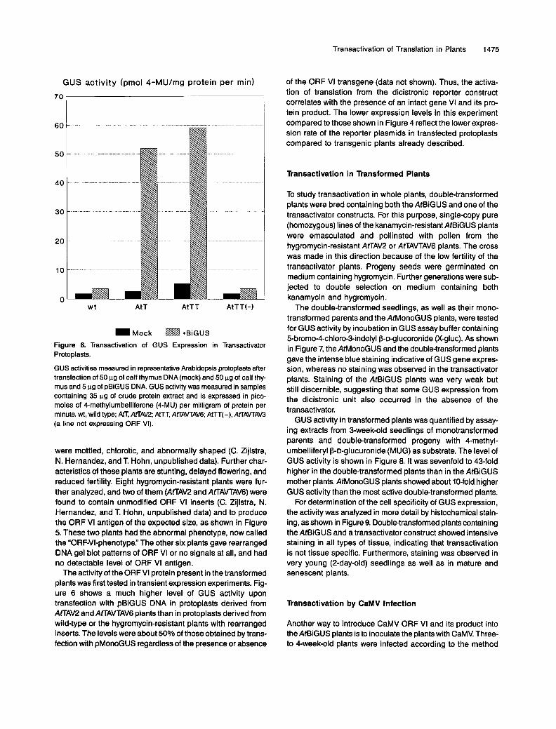
Mock ~ +BiGUS Figure 6. Transactivation of GUS Expression in Transactivator Protoplasts.
GUS activities measured in representative Arabidopsis protoplasts after transfection of 50 pg of calf thymus DNA (mock) and 50 pg of calf thy- mus and 5 pg of pBiGUS DNA. GUS activity was measured in samples containing 35 pg of crude protein extract and is expressed in pico- moles of 4-methylumbelliferone (4-MU) per milligram of protein per minute. wt, wild type; AtT, AtTAV2; AtTT, AffAVTAV6; AtTT(-), AfLAVTAW (a line not expressing ORF VI).
were mottled, chlorotic, and abnormally shaped (C. Zijlstra, N. Hernandez, and T Hohn, unpublished data). Further char- acteristics of these plants are stunting, delayed flowering, and reduced fertility. Eight hygromycin-resistant plants were fur- ther analyzed, and two of them (AfrAV2 and AfLAVTAV6) were found to contain unmodified ORF VI inserts (C. Zijlstra, N. Hernandez, and T. Hohn, unpublished data) and to produce the ORF VI antigen of the expected size, as shown in Figure 5. These two plants had the abnormal phenotype, now called the “ORFVI-phenotype.” The other six plants gave rearranged DNA gel blot patterns of ORF VI or no signals at all, and had no detectable level of ORF VI antigen.
The activity of the ORF VI protein present in the transformed plants was first tested in transient expression experiments. Fig- ure 6 shows a much higher level of GUS activity upon transfection with pBiGUS DNA in protoplasts derived from AtTAV2 and AfLAVTAV6 plants than in protoplasts derived from wild-type or the hygromycin-resistant plants with rearranged inserts. The levels were about 50% of those obtained by trans- fection with pMonoGUS regardless of the presence or absence
of the ORF VI transgene (data not shown). Thus, the activa- tion of translation from the dicistronic reporter construct correlates with the presence of an intact gene VI and its pro- tein product. The lower expression levels in this experiment compared to those shown in Figure 4 reflect the lower expres- sion rate of the reporter plasmids in transfected protoplasts compared to transgenic plants already described.
Transactivation in Transformed Plants
To study transactivation in whole plants, double-transformed plants were bred containing both the AtBiGUS and one of the transactivator constructs. For this purpose, single-copy pure (homozygous) lines of the kanamycin-resistant AtBiGUS plants were emasculated and pollinated with pollen from the hygromycin-resistant AtTAV2 or AtTAVTAV6 plants. The cross was made in this direction because of the low fertility of the transactivator plants. Progeny seeds were germinated on medium containing hygromycin. Further generations were sub- jected to double selection on medium containing both kanamycin and hygromycin.
The double-transformed seedlings, as well as their mono- transformed parents and the AtMonoGUS plants, were tested for GUS activity by incubation in GUS assay buffer containing 5-bromo-4-chloro-3-indolyl P-D-glucoronide (X-gluc). As shown in Figure 7, the AtMonoGUS and the double-transformed plants gave the intense blue staining indicative of GUS gene expres- sion, whereas no staining was observed in the transactivator plants. Staining of the AtBiGUS plants was very weak but still discernible, suggesting that some GUS expression from the dicistronic unit also occurred in the absence of the transactivator.
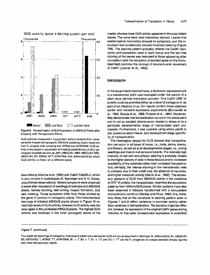
GUS activity in transformed plants was quantified by assay- ing extracts from 3-week-old seedlings of monotransformed parents and double-transformed progeny with 4-methyl- umbelliferyl P-o-glucuronide (MUG) as substrate. The level of GUS activity is shown in Figure 8. It was sevenfold to 43-fold higher in the double-transformed plants than in the AtBiGUS mother plants. AtMonoGUS plants showed about 10-fold higher GUS activity than the most active double-transformed plants.
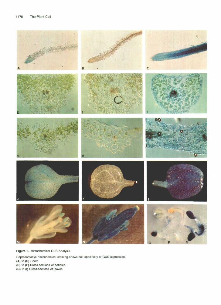
For determination of the cell specificity of GUS expression, the activity was analyzed in more detail by histochemical stain- ing, as shown in Figure 9. Double-transformed plants containing the AtBiGUS and a transactivator construct showed intensive staining in all types of tissue, indicating that transactivation is not tissue specific. Furthermore, staining was observed in very young (2-day-old) seedlings as well as in mature and senescent plants.
Transactivation by CaMV lnfection
Another way to introduce CaMV ORF VI and its product into the AtBiGUS plants is to inoculate the plants with CaMV. Three- to 4-week-old plants were infected according to the method

1476 The Plant Cell
M
<«•."'
B1 B3
B1XT
TT BixTT B3XTT
Figure 7. GUS Expression in Transgenic Arabidopsis Plants.
Transactivation of Translation in Plants 1477
GUS ac t iv i t y (pmol 4-MU/mg protein pe r min)
Thousands Thousands
4 -
2 ---
O
l l 60
40
20
n - AtBl AtB3 AtB4 AtT AtTT AtM
Basic ~ xAtTAV2 xAtTAVTAV6
Figure 8. Transactivation of GUS Expression in AWiGUS Plants after Crossing with Transactivator Plants.
GUS activities measured in 4-pg protein extract samples from repre- sentative 3-week-old transgenic AtBiGUS seedlings (basic level) and their F1 progeny after crossing with AtTAV2 and AfLAVTAV6. GUS ac- tivity is expressed in picomoles of 4-methylumbelliferone (4-MU) per milligram of protein per minute. AtB1, AtBiGUSl; AtB3, AmiGUS3; AtB4, AtBiGUS4; AtT, AfLAV2; AtTT, AfTAVTAV6; AtM, AtMonoGUS (for which GUS activity is shown on a different scale).
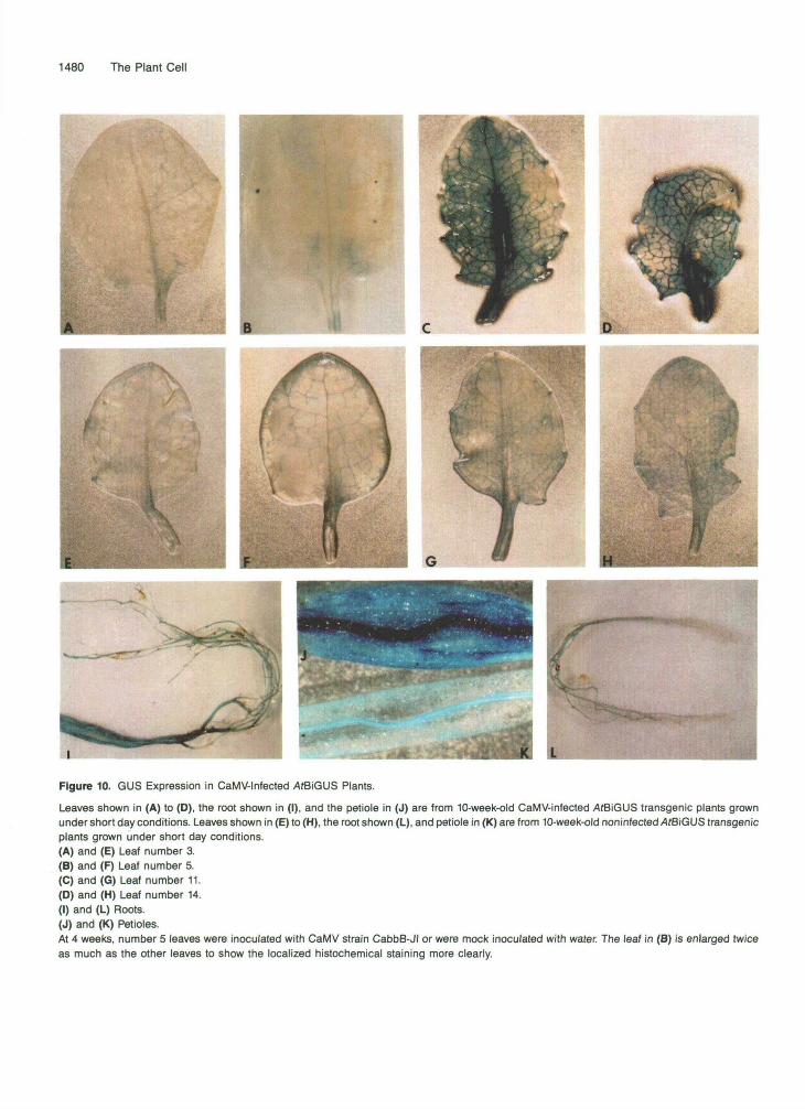
described by Melcher et al. (1989) with CaMV CabbB-JI, which is very virulent in Arabidopsis (A. Bannister and S. N. Covey, unpublished observations). Severe symptoms were observed 2 weeks after inoculation of seedlings of wild-type and AtBiGUS plants, namely stunting, leaf curling, mosaic formation, and vein clearing. These symptoms differ from those elicited by the gene VI product in transgenic plants. The histochemical stainings of infected AtBiGUS plants shown in Figure 10 re- veal high levels of GUS activity, whereas GUS activity was low once again in the uninfected AtBiGUS plants. The highest GUS activity was localized in the inner (youngest) leaves of the
rosette, whereas lower GUS activity appeared in the outer (older) leaves. The veins were most intensively stained. Leaves that existed before inoculation showed no symptoms, and the in- oculated leaf occasionally showed localized staining (Figure 106). The staining pattern probably reflects the CaMV repli- cation and expression rates in each tissue and the fact that staining of the leaves was restricted to those appearing after inoculation (with the exception of isolated spots on the inocu- lated leaf) confirms the concept of source-to-sink movement of CaMV (Leisner et al., 1992).
DISCUSSION
In the experiments reported here, a dicistronic expression unit in a transformed plant was translated under the control of a plant virus-derived translation activator. This CaMV ORF VI protein could be provided either as a second transgene or as part of an infecting virus. Our results confirm those obtained earlier with transient expression experiments (Bonneville et al., 1989; Gowda et al., 1989; Fütterer et al., 1991). Moreover, they demonstrate that transactivation occurs in the whole plant and is not an isolated phenomenon related to stress or to a particular developmental stage of manipulated plant pro- toplasts. Furthermore, it was possible using whole plants to ask questions about tissue- and development-stage specific- ity of transactivation.
The histological assays for GUS showed that transactiva- tion can occur in all types of tissue, i.e., roots, stems, leaves, and flowers, as well as at all developmental stages, i.e., young seedlings and mature and senescent plants. The relatively high intensity of leaf vein and phloem staining is probably related to the higher density of cells in these tissues and to increased availability of the substrate rather than increased transactiva- tion; similarly, the intense staining in the meristematic cells is probably due to their small size, the absence of vacuoles, and higher metabolic activity (Martin et al., 1992). The expres- sion patterns of GUS from AtBiGUS plants in the presence of ORF VI protein, the transactivator, resemble the expression patterns from AtMonoGUS plants. Similar patterns have also been observed in tobacco transformed with a comparable monocistronic construct (Benfey and Chua, 1990). It is, there- fore, likely that all the variations in staining pattern seen in Figures 7 and 9 reflect variations in promoter activity rather than variations in transactivation. The situation might be differ- ent, however, for expression of the original CaMV genes during infection. In that case, transactivator expression is controlled
Figure 7. (continued).
Four-week-old seedlings of transgenic Arabidopsis plants were stained for GUS activity as described in Methods. M, AtMonoGUS; B1, AtBiGUSl; 83, AtBiGUS3; T, AfLAV2; TT, AfTAVTAV6; B1 x T, 83 x T, B1 x TT, and 83 x TT are the Ft progenies of crosses between female reporter and male transactivator plants.
1478 The Plant Cell
O
1
-^*V«r?^S^
Figure 9. Histochemical GUS Analysis.
Representative histochemical staining shows cell specificity of GUS expression.(A) to (C) Roots.(D) to (F) Cross-sections of petioles.(G) to (I) Cross-sections of leaves.
Transactivation of Translation in Plants 1479
by the 19s rather than the 35s promoter, by CaMV DNA-copy number, possibly by positive feedback control (M. Driesen, T. Hohn, and J. Fütterer, manuscript in preparation), and by par- titioning of CaMV RNA into pools for packaging, reverse transcription, and translation.
Arabidopsis plants containing the CaMV (strain CM4-184) ORF VI as a transgene were difficult to obtain, and those that survived and expressed ORF VI had an abnormal phenotype, indicating that the ORF VI protein interferes with certain meta- bolic or developmental plant processes. Such difficulties were also encountered by Schoelz et al., (1991) with ORF Vi-trans- formed Nicotiana bigelovii, and abnormal phenotypes were observed in severa1 types of nonhost plants by BAlasz (1988), Baughman et al. (1988), Kiernan et al. (1989), Takahashi et al. (1989), and Goldberg et al. (1991). The basis of such pheno- typic alterations in plants transformed with gene VI will be discussed in detail elsewhere (C. Zijlstra, N. Hernandez, and T. Hohn, manuscript in preparation).
The fact that GUS expression from the dicistronic unit can be induced by virus infection has practical implications. For example, one could trace the route of systemic virus spread by analyzing transactivated expression of GUS following an initial infection event. One could also use transgenes to in- duce virus resistance: translation of a ribosome-inactivating endotoxin gene (Stirpe et al., 1992) or a special RNase gene, such as barnase (Hartley, 1991; Natsoulis and Boeke, 1991), located on a dicistronic expression unit and, therefore, depen- dent for its expression on the transactivator, would be induced upon CaMV infection. Production of the toxin would specifi- cally eliminate the infected cells, thereby inhibiting virus spread. This process would thus resemble the hypersensitive reac- tion, a natural plant defense mechanism.
We are aware that the relatively high background of GUS expression in our plants containing the dicistronic transgenic unit might be a problem for this type of application. This back- ground was higher in transformed plants than in transfected protoplasts, probatjly due to accumulation of the GUS enzyme during the life span of the transformed plants. The problem might be overcome by the use of dicistronic units with first cis- trons other than the sORF VI1 used. In preliminary experiments in our laboratory (J. VlasBk, unpublished data), dicistronic transgenes containing the NPTll gene as the first cistron did not express detectable levels of the second cistron until transactivated.
METHODS
Plasmids Used for Transient Expression in Plant Protoplasts and for Further Constructions
The cauliflower mosaic virus (CaMV) sequences used were derived from strain CM4-184. Plasmid pMono-P-glucuronidase (GUS) consists of a CaMV open reading frame (ORF) I-GUS translational fusion, pBiGUS of ORF VI1 and the ORF I-GUS translational fusion, and pHELF7 of ORF VI, all provided with the CaMV 355 promoter and poly- adenylator. pHELP4 consists of the CaMV 19s promoter, ORF VI, and polyadenylator (Bonneville et al., 1989).
Agrobacterlum Constructs for Plant Ransformation
AgBiGUS V613. This plasmid was used in AgBiGUS (Figure 2) and was obtained by cloning the expression unit from pBiGUS as a Pvull fragment into the Smal cloning site within the T-DNA of the binary shuttle vector pBIN19; pBIN19 confers kanamycin resistance to trans- formed plants (Bevan, 1984).
AgMonoGUS V614. This plasmid was used in AgMonoGUS and is an ORF VII-less derivative of AgBiGUS. It was obtained by ligating the 2227-bp Scal-Hindlll fragment from pBiGUS containing the ORF I-GUS fusion with the 3180-bp Smal-Hindlll fragment of the plant ex- pression plasmid pDH51 (Pietrzak et al., 1986), resulting in intermediate plasmid V594. From this plasmid, the monocistronic GUS expression cassette was isolated as a Pvull fragment and cloned into the T-region of pBIN19, as given above.
AgTAV. To construct V799, the plasmid present in AgTAV, first a trans- activator expression cassette suitable for integration into the binary Ppbacterium tumefaciens vector pGSC1704 was constructed by com- bining two existing ORF VI expression plasmids. The 35s promoter and the first part of ORF VI were isolated as an Xhol-Aval fragment from V709 (J. M. Bonneville, personal communication) containing ORF VI as an Xhol cassette in the multiple site cloning vector pMTL23 (Chambers et al., 1988). The second part of ORF VI and the polyadeny- lator were isolated as an Aval-Xhol fragment from pHELP7. 60th fragments were ligated into the T-region of pGSC1704 opened at Sal1 and Hindlll. pGSC1704 was kindly provided by Plant Genetic Systems and contains the HPTcassette, described by Van den Elzen et al. (1985),
Figure 9. (continued).
(J) to (L) Cotyledons, all of 4-week-old plantlets. (M) and (N) Flowers of 12-week-old plantlets. (O) to (Q) Whole 2-day-old seedlings. Representative AfBiGUS (A, D, G, J, M, and O) and AfLAV (B, E, H, K, and P) plants and their F1 progeny (C, F, I, L, N, and Q) are shown.
1480 The Plant Cell
Figure 10. GUS Expression in CaMV-lnfected ̂ fBiGUS Plants.
Leaves shown in (A) to (D), the root shown in (I), and the petiole in (J) are from 10-week-old CaMV-infected AtBiGUS transgenic plants grownunder short day conditions. Leaves shown in (E) to (H), the root shown (L), and petiole in (K) are from 10-week-old noninfected AfBiGUS transgenicplants grown under short day conditions.(A) and (E) Leaf number 3.(B) and (F) Leaf number 5.(C) and (G) Leaf number 11.(0) and (H) Leaf number 14.(I) and (L) Roots.(J) and (K) Petioles.At 4 weeks, number 5 leaves were inoculated with CaMV strain CabbB-JI or were mock inoculated with water. The leaf in (B) is enlarged twiceas much as the other leaves to show the localized histochemical staining more clearly.

Transactivation of Translation in Plants 1481
between its T-DNA borders, which confers hygromycin resistance to transformed plant tissue.
AgTAVTAV V800. This plasmid is present in MTAVTAV and was ob- tained by serendipity in the procedure used to obtain MTAV. It contains two copies of the Xhol ORF VI expression cassette. The binary vec- tors originally grown in Escherichia coli JMlOl (Yanisch-Perron et ai., 1983) were introduced into Agrobacterium C58C1, containing helper plasmid pGV2260 (Deblaere et ai., 1985), by electroporation as de- scribed by Mattanovich et ai. (1989). lntegrity of the vectors was verified by analyzing total DNA from Agrobacterium by the method of Dhaese et ai. (1979).
Stable Wansformation of Arabldopsls
Arabidopsis tbaliana ecotype C24 was transformed with Agrobacterium containing the shuttle and helper plasmids using the method described by Van Lijsebettens et ai. (1991).
DNA Gel Blot Analysis
Genomic DNA was isolated from lyophilized plants or callus using a CTAB extraction procedure (modified after Murray and Thompson, 1980, as described by Mittelsten-Scheid et ai., 1991). Samples were cleaved with restriction enzymes (10 to 20 units per pg of DNA) and separated on a 0.8 to 1.2% agarose gel. DNA was transferred to a Hybond N membrane (Amersham International) according to Southern (1975). After fixation of the transferred DNA by UV cross-linking, the mem- brane was prehybridized for 6 hr in 6 x SSC (1 x SSC is 0.15 M NaCI, 0.015 M sodium citrate), 5 x Denhardt’s solution (1 x Denhardt’s so- lution is 0.02% Ficoll, 0.02% WP, 0.025% BSA), 05Vo SSC, and 0.05% salmon sperm DNA at 65OC and subsequently hybridized at 65OC over- night with 32P-labeled probes in prehybridization solution containing 10 mM EDTA. Probes were labeled using the random prime labeling kit from Boehringer Mannheim, based on the method of Feinberg and Vogelstein (1983). After hybridization, the membrane was rinsed in 2 x SSC and then washed successively for 15 min in 2 x SSC, 0.1% SDS at room temperature, in 0.5 x SSC, 0.1% SDS at 65OC, and fi- nally in 0.1 x SSC, 0.1% SDS at 65OC. To reuse blots, old probes were removed by repeatedly washing the membrane with a boiling solution of 0.05 x SSC, 0.1% SDS, and 0.01 M EDTA, pH 8, which was allowed to cool down to room temperature after being poured onto the membrane.
Electrophoresis and lmmunologlcal Detectlon of Proteins
Total plant proteins were isolated using the method described by Mayer et ai. (1987). Protein concentrations were determined as described by Ramagli and Rodriguez (1985). SDSPAGE was performed as described by Laemmli (1970). The amount of protein loaded was 50 pg per lane. The proteins were transferred from the electrophoresis gel to an Immun-Lite membrane (Bio-Rad) via semidry electroblotting with an apparatus from JKA Biotech (vaerlrase, Denmark) for 1 hr at 1 mA/cm2 of gel. Reaction with antisera and anti-rabbit IgG conjugated to horseradish peroxidase (Bio-Rad lmmunoblot kit) was performed as described by the manufacturer.
Genetic Crosses
Genetic crossings were carried out as described by Mittelsten-Scheid et al. (1991). Harvested seeds were stored at 28OC for 3 weeks and kept at 4OC for another week.
Preparation of Protein Extracts from Plant Tissue for GUS Assays
Plant tissue samples (5 to 50 mg fresh weight) were homogenized with 200 pL of extraction buffer (50 mM NaH,PO,, pH 7, 10 mM EDTA, 0.1% Triton X-100, 0.1% Sarkosyl, 10 mM P-mercaptoethanol). Soluble proteins were obtained by centrifugation of the homogenates for 5 min in a table centrifuge.
Transformation of Protoplasts and Preparation of Extracts for GUS Assays
Mesophyll protoplasts were isolated from in vitro-grown Arabidopsis plants as described by Damm and Willmitzer (1988). The protoplasts were used for PEG transformation as described by Damm et ai. (1989). Protoplasts were harvested by centrifugation and soluble extracts were prepared as described by Fiitterer et ai. (1989).
Fluorometric assays were performed with samples of equivalent pro- tein content. Protein concentrations were determined by the method of Bradford (1976) using BSA as the standard. GUS activities were as- sayed in 96-well microtiter plates in 300-pL reaction samples containing 1 mM Cmethylumbelliferyl P-D-glucuronide (CMUG, Sigma) (Jefferson et ai., 1987); 50-pL aliquots were taken at four successive time points and the appearance of fluorescence was followed using a Titertek Fluoroscan I1 apparatus (Jefferson et al., 1990).
Histochemical Staining
Whole plants or detached plant organs were placed into a 24-well cul- turing plate containing 100 mM phosphate buffer, pH 7, and 1 mg of P-D-glucuronide (X-gluc) per mL. After initial vacuum infiltration for about 15 min, the plant material was incubated in the dark at 37% for 3 to 12 hr. Plant material was fixed for 10 min in a solution containing 5% formaldehyde, 5% acetic acid, and 20% ethanol. When necessary the samples were bleached to remove chlorophyll by overnight treat- ment with 70% ethanol.
Tlssue Sectioning
Small tissue blocks stained for GUS activity were prepared for cryosec- tioning according to a modified procedure described by Sohn and Sauter (1991). After incubation of the plant tissue in the X-gluc solu- tion, it was fixed overnight at 8OC with 4% (wh) formaldehyde and 0.1% glutaraldehyde in a buffer containing 100 mM potassium phosphate buffer, pH 7, supplemented with 0.8% NaCl and 0.2% NaN3 (PBS). The samples were immersed consecutively for 2 hr at room tempera- ture in 0.25, 0.5, 1.0, and 1.5 M sucrose in PBS, followed by a 15-min vacuum infiltration. The samples were then kept overnight in 1.5 M sucrose-PBS, which was renewed before cryosectioning was per- formed. Cryosections of 30-pm thickness were produced with a Cryostat
1482 The Plant Cell
microtome (Model 830/C; American Optical Company, New York) at -25OC.
ACKNOWLEDGMENTS
We gratefully acknowledge the gifts of plasmids, transgenic plant strains, and viruses by Susanah Gal, Barbara Hohn, Bob Shepherd, and Simon Covey and Plant Genetic Systems; the introduction to Arabidopsis transformation by Thomas Altmann and Lothar Willmitzer; and the introduction to virus inoculation by Simon Covey. The valu- able advice and help in histological staining by Gunther Neuhaus; the helpful suggestions and critical discussions by Barbara Hohn, Johannes Fütterer, Ferenc Nagy, Fred Meins, and Tom Boller; the ex- pert technical assistance by Mathias Müller; the growing of test plants by Markus Briker; and the critical reading of the manuscript by Pat King are very much appreciated.
Received August 31, 1992; accepted October 19, 1992.
REFERENCES
Balazs, E. (1990). Disease symptoms in transgenic tobacco induced by integrated gene VI of cauliflower mosaic virus. Virus Genes 3,
Baughman, G.A., and Howell, S.H. (1988). Cauliflower mosaic virus 35s leader region inhibits translation of downstream genes. Virol-
Baughman, G.A., Jacobs, J.D., and Howell, S.H. (1988). Cauliflower mosaic virus gene VI produces a symptomatic phenotype in trans- genic tobacco plants. Proc. Natl. Acad. Sci. USA 85, 733-737.
Benfey, P.N., and Chua, N.-H. (1990). The cauliflower mosaic virus 35s promoter: Combinational regulation of transcription in plants. '
Science 250, 959-966. Bevan, M. (1984). Binary Agrobacterium vectors for plant transforma-
tion. Nucl. Acid Res. 12, 8711-8721. Bonneville, J.-M., and Hohn, T. (1992). A reverse transcriptase for
cauliflower mosaic virus; state of the art. In The Reverse Transcrip- tase, S. Goff, ed (Cold Spring Harbor, NY Cold Spring Harbor Laboratory), in press.
Bonneville, J.-M., Volovitch, M., Modjtahedi, N., Demery, O., and Yot, P. (1984). In vitro synthesis of cauliflower mosaic virus DNA in viroplasms. In Proteins lnvolved in DNA Replication, U. Huebscher and S. Spadari, eds (New York: Plenum Press), pp. 113-119.
Bonnevllle, J.-M., Sanfaçon, H., Fütterer, J., and Hohn, T. (1989). Posttranscriptional transactivation in cauliflower mosaic virus. Cell
Bradford, M. (1976). A rapid and sensitive method for the quantitation of microgram quantities of protein utilizing the principle of protein dye binding. Anal. Biochem. 72, 248-254.
Chambers, S.P., Prior S.E., Barstow, D.A., and Mlnton, N.P. (1988). The pMTLnic- cloning vectors. I. lmproved pUC polylinker regions to facilitate the use of sonicated DNA for nucleotide sequencing. Gene 68, 139-149.
205-21 1.
Ogy 167, 125-135.
59, 1135-1143.
Citovsky, V., Knorr, D., and Zambryskl, P. (1991). Gene I, a potential cell-to-cell movement locus of cauliflower mosaic virus, encodes an RNA-binding protein. Proc. Natl. Acad. Sci. USA 88, 2476-2480.
Covey, S., and Hull, R. (1981). Transcription of cauliflower mosaic vi- rus DNA. Detection of transcripts, properties, and location of the gene encoding the virus inclusion body protein. Virology 111,
Damm, B., and Willmitzer, L. (1988). Regeneration of fertile plants from protoplasts of different Arabidopsis thaliana genotypes. MOI. Gen. Genet. 213, 15-20.
Damm, B., Schmidt, R., and Willmitzer, L. (1989). Efficient transfor- mation of Arabidopsis thaliana using direct gene transfer to protoplasts. MOI. Gen. Genet. 217, 6-12.
Daubert, S.D., and Routh, G. (1990). Point mutations in cauliflower mosaic virus gene VI confer host-specific symptoms. MOI. Plant- Microbe Interact. 3, 341-345.
Daubert, S.D., Schoelz, J.E., Debao, L., and Shepherd, R.J. (1984). Expression of disease symptoms in CaMV genomic hybrids. J. MOI. Appl. Genet. 2, 537-547.
Deblaere, R., Bytebler, B., De Greve, H., Schell, J., Van Montagu, M., and Leemans, J. (1985). Efficient octopine Ti plasmid derived vectors for Agrobacterium mediated gene transfer to plants. Nucl. Acids Res. 13, 4777-4788.
deZoeten, G.A., Penswick, J.R., Horisberger, M.A., Ahl, P., Schultze, M., and Hohn, T. (1989). The expression, localization and effect of the human interferon in plants. Virology 172, 213-222.
Dhaese, P., De Greve, H., Decraemer, H., Schell, J., and Van Montagu, M. (1979). Rapid mapping of transposon insertions and deletion mutations in the large Ti-plasmids of Agrobacterium tumefa- ciens. Nucl. Acids Res. 7, 1837-1849.
Dixon, L.K., and Hohn, T. (1984). lnitiation of translation of the cauliflower mosaic virus genome from a polycistronic mRNA: Evi- dente from deletion mutagenesis. EMBO J. 3, 2731-2736.
Feinberg, A.P., and Vogelsteln, 8. (1983). A technique for radiolabel- ling DNA restriction endonuclease fragments to high specific activity. Anal. Biochem. 132, 6-13.
Fütterer, J., and Hohn, T. (1987). lnvolvement of nucleocapsids in reverse transcription -A general phenomenon? Trends Biol. Sci. 12,
Fütterer, J., and Hohn, T. (1991). Translation of a polycistronic mRNA in presence of the CaMV transactivator protein. EMBO J. 10,
Fütterer, J., and Hohn, T. (1992). Role of an upstream open reading frame in the translation of polycistronic mRNA in plant cells. Nucl. Acids Res. 20, 3851-3857.
Fütterer, J., Bonneville, J.-M., Sanfaçon, H., Torruella, M., Pisan, B., Gordon, K., Penswlck, J.R., Grimsley, N., Hohn, B., and Hohn, T. (1988). Use of plant viruses and plant vira1 expression signals forgene expression in plants and plant protoplasts. In Vira1 Vectors, Y. Gluzman, ed (Cold Spring Harbor, NY Cold Spring Harbor Labo- ratory), pp. 178-182.
Fütterer, J., Gordon, K., Bonneville, J A . , Sanfaçon, H., Plsan, B., and Hohn, T. (1989). Differential inhibition of downstream gene expression by the cauliflower mosaic virus 35s FINA leader. Virus Genes 3,45-55.
Fütterer, J., Bonneville, J.-M., Gordon, K., DeTapla, M., Karlsson, S., and Hohn, T. (199Oa). Expression from polycistronic CaMV RNA.
463-474.
92-95.
3887-3896.

Transactivation of Translation in Plants 1483
In Posttranscriptional Control of Gene Expression, J.E.G. McCar- thy and M.F. Tuite, eds (Heidelberg: Springer), pp. 359-365.
Fütterer, J., Gordon, K., Sanfapn, H., Bonneville, J.-M., and Hohn, T. (1990b). Positive and negative control of translation by the leader of cauliflower mosaic virus pregenomic 35s RNA. EMBO J. 9,
Glband, M., Mesnard, J.M., and Lebeurler, G. (1986). The gene 111 product (p15) of CaMV is a DNA binding protein while an immuno- logically related p l l polypeptide is associated with virus. EMBO J.
Goldberg, K.B., Kiernan, J., and Shepherd, R.J. (1991). A disease syndrome associated with expression of gene VI of caulimovirus may be a nonhost reaction. MOI. Plant-Microbe Interact. 4, 182-189.
Gordon, K., Pfeiffer, P., Fütterer, J., and Hohn, T. (1988). ln vitro expression of cauliflower mosaicvirus genes. EMBO J. 7,309-317.
Gowda, S., Wu, F.C., Scholthof, H.B., and Shepherd, R.J. (1989). Gene VI of figwort mosaic virus (caulimovirus group) functions in posttranscriptional expression of genes on the full-length RNA tran- script. Proc. Natl. Acad. Sci. USA 86, 9203-9207.
Hartley, R.W. (1989). Barnase and barstar: Two small proteins to fold and fit together. Trends Biol. Sci. 14, 450-454.
Jefferson, R.A., Kavanagh, T.A., and Bevan, M.W. (1987). GUS fu- sions: e-Glucuronidase as a sensitive and versatile gene fusion marker in higher plants. EMBO J. 6, 3901-3907.
Jefferson, R., Goldsbrough, A., and Bevan, M. (1990). Transcrip- tional regulation of a patatin-1 gene in potato. Plant MOI. Biol. 14,
Klernan, J., Goldberg, K.B., Young, M.J., Schoelz, J.E., and Shepherd, R.J. (1989). Transformation and regeneration of Nicotiana edwardsonii. Plant Sci. 64, 67-78.
Kozak, M. (1989). The scanning model for translation: An update. J. Cell Biol. 108, 229-241.
Laemmll, U.K. (1970). Cleavage of structural proteins during the as- sembly of the head of bacteriophage T4. Nature 227, 680-685.
Leisner, S.M., Turgeon, R., and Howell, S.H. (1992). Long distance movement of CaMV in infected turnip plants. MOI. Plant-Microbe In- teract. 5, 41-47.
Linstead, P.J., Hills, G.J., Plaskitt, K.A., Wllson, I.G., Harker, C.L., and Maule, A. (1988). The subcellular location of the gene I prod- uct of cauliflower mosaic virus is consistent with a function associated with virus spread. J. Gen. Virol. 69, 1808-1818.
Martln, T., Wohner, R.V., Hummel, S., Willmltzer, L., and Frommer, W.B. (1992). The GUS reporter system as a tool to study plant gene expression. In GUS Protocols: Using the GUS Gene as a Reporter of Gene Expression, S.R. Gallagher, ed (San Diego: Academic Press, Inc.), pp. 23-43.
Mason, W.S., Taylor, J.M., and Hull, R. (1987). Retroid virus genome replication. Adv. Virus Res. 32, 35-96.
Mattanovich, D., Rueker, F., da Camara Machado, A., Lalmer, M., Regner, F., Stelnkellner, H., Himmler, G., and Katlnger, H. (1989). Efficient transformation of Agmbacterium spp. by electroporation. Nucl. Acids Res. 17, 6747.
Mayer, S.E., Hahne, G., Palme, K., and Schell, J. (1987). A simple and general plant tissue extraction procedure for two dimensional gel electrophoresis. Plant Cell Rep. 6, 77-81.
Mauollni, L., Bonneville, J.-M., Volovitch, M., Magazln, M., and Yot, P. (1985). Strand-specific vira1 DNA synthesis in purified
1697-1707.
5, 2433-2438.
995-1006.
viroplasm isolated from turnip leaves infected with CaMV. Virology
Melcher, U. (1989). Symptoms of cauliflower mosaic virus infection in Arabidopsis thaliana and turnip. Botanical Gazette, 150, 139-147.
Mittelsten-Scheid, O., Paszkowskl, J., and Potrykus, 1. (1991). Re- versible inactivation of a transgene in Arabidopsis thaliana. MOI. Gen. Genet. 228, 104-112.
Murray, M.G., and Thompson, W.F. (1980). Rapid isolation of high molecular weight plant DNA. Nucl. Acids Res. 8, 4321-4325.
Natsoulls, G., and Boeke, F.D. (1991). New antiviral strategy using capsid-nuclease fusion proteins. Nature 352, 632-635.
Odell, J.T., and Howell, S.H. (1980). The identification, mapping and characterization of messenger RNA for p66, a cauliflower mosaic virus coded protein. Virology 102, 349-359.
Olszewski, N.E., Hagen, G., and Gullfoyle, T.J. (1982). A transcrip- tionally active, covalently closed minichromosome of cauliflower mosaic virus DNA isolated from turnip leaves. Cell 29, 395-402.
Pfeiffer, P., and Hohn, T. (1983). lnvolvement of reverse transcription in the replication of cauliflower mosaic virus: A detailed model and test of some aspects. Cell. 33, 781-789.
Pletrzak, M., Shllito, M., Hohn, T., and Potrykus, 1. (1986). Expres- sion in plants of two bacterial antibiotic resistance genes after protoplast transformation with a new plant expression vector. Nucl. Acids Res. 14, 5857-5868.
Ramagll, L.S., and Rodriguez, L.V. (1985). Quantification of micro- gram amounts of proteins in two-dimensional polyacrylamide gel electrophoresis sample buffer. Electrophoresis 6, 559-563.
Schoelz, J.E., Shepherd, R.J., and Daubert, S.D. (1986). Region VI of CaMV encodes a host range determinant. MOI. Cell. Biol. 6,
Schoelz, J.E., Goldberg, K.B., and Kiernan, J. (1991). Expression of cauliflower mosaic virus (CaMV) in transgenic Nicotiana bigelovii complements a strain of CaMV defective in long distance mwement in nontransformed N. bigelovii. MOI. Plant-Microbe Interact. 4,
Scholthof, H.B., Gowda, S., Wu, F.C., and Shepherd, R.J. (1992). The full-length transcript of caulimovirus is a polycistronic mRNA whose genes are transactivated by the product of gene VI. J. Virol.
145, 293-303.
2632-2637.
350-355.
66. 3131-3139.
Shepherd, R.J., Rlchins, R.D., and Shalla, T.A. (1979). lsolation and properties of the inclusion bodies of cauliflower mosaic virus. Virol-
Sleg, K., and Gronenborn, B. (1982). Evidence for polycistronic messenger RNA encoded by cauliflower mosaic virus. NATOASI Conference on Structure and Function of Plant Genomes (Porto Portese, Italy: NATO Advanced Studies Institute), 154 (abstr.).
Sohn, O., and Sautter, C. (1991). R-Phycoerythrin as a fluorescent label for immunolocalization of bound atrazine residues. J. Histo- chem. Cytochem. 39, 921-926.
Southern, E. (1975). Detection of specific sequences among DNA frag- ments separated by gel electrophoresis. J. MOI. Biol. 98, 503-517.
Stlrpe, F., Barbleri, L., Battelli, G., Sorla, M., and Lappi, D.A. (1992). Ribosome-inactivating proteins from plants: Present status and fu- ture prospects. Biofrechnology 10, 405-412.
Stratford, R., and Covey, S.N. (1989). Segregation of cauliflower mo- saic virus symptom genetic determinants. Virology 172, 451-459.
Ogy 102, 389-400.

1484 The Plant Cell
Takahashi, H., Shlmamoto, K., end Ehara, Y. (1989). Cauliflower mc- saic virus gene VI causes growth suppression, development of necrotic spots and expression of defence-related genes in trans- genic tobacco plants. MOI. Gen. Genet. 216, 188-194.
Thomas, C.M., Hull, R., Bryant, J.A., and Maule, A.J. (1985). Isola- tion of afraction of CaMV infected protoplasts which is active in the synthesis of (+) and (-)vira1 DNA and reverse transcription of primed RNA templates. Nucl. Acids Res. 12, 4557-4576.
Van den Elzen, P.J.M., Townsend, J., Lee, K.Y., and Bedbrook, J.R. (1985). A chimeric hygromycin resistance gene as a selectable marker in plant cells. Plant MOI. Biol. 5, 103-108.
Van Lljsebettens, M., Vanderhaegen, R., and Van Montagu, M. (1991). lnsertional mutagenesis in Arabidopsis thaliana: lsolation of a T-DNA linked mutation that alters leaf morphology. Theor. Appl. Genet. 81, 277-284.
Woolston, GJ., Covey, S.N., Penswlck, J.R., and Davies, J.W. (1983). Aphid transmission and a polypeptide are specified by a defined region of the cauliflower mosaic virus genome. Gene 23, 15-23.
Yanisch-Perron, C., Vlelra, J., and Messing, J. (1983). lmproved M13 phage cloning vectors and host strains: Nucleotide sequence of the M13mp18 and pUC19 vectors. Gene 33, 102-119.
DOI 10.1105/tpc.4.12.1471 1992;4;1471-1484Plant Cell
C. Zijlstra and T. HohnTransgenic Arabidopsis Plants.
Cauliflower Mosaic Virus Gene VI Controls Translation from Dicistronic Expression Units in
This information is current as of May 20, 2020
Permissions https://www.copyright.com/ccc/openurl.do?sid=pd_hw1532298X&issn=1532298X&WT.mc_id=pd_hw1532298X
eTOCs http://www.plantcell.org/cgi/alerts/ctmain
Sign up for eTOCs at:
CiteTrack Alerts http://www.plantcell.org/cgi/alerts/ctmain
Sign up for CiteTrack Alerts at:
Subscription Information http://www.aspb.org/publications/subscriptions.cfm
is available at:Plant Physiology and The Plant CellSubscription Information for
ADVANCING THE SCIENCE OF PLANT BIOLOGY © American Society of Plant Biologists