cb1 ey biological k concepts - pearson · pdf filecb1 ey biological k concepts ... ideas in...
TRANSCRIPT

The ‘foot’ of the worm is buried in the whale bone and contains many bacteria.
CB1 Key Biological Concepts
The bone-eating snot-flower worm (Osedax mucofloris) has no digestive system but still manages to feed on one of the hardest substances produced by vertebrate animals – their bones. These worms are a type of zombie worm, so-called because they have no eyes or mouth, and were discovered in the North Sea in 2005 feeding on a whale skeleton. Enzymes in the ‘foot’ of the worm cause the production of an acid, which attacks bone and releases lipids and proteins from inside the bone. Enzymes in bacteria on the foot of the worm then digest these large organic molecules into smaller molecules that the worms absorb (using processes such as diffusion).
In this unit you will learn about some of the central ideas in biology, including ideas about cells, microscopy, enzymes, nutrition, diffusion, osmosis and active transport.
Previously you will have learnt at KS3:
• how to use a microscope
• about the differences between cells from different organisms
• how some cells are specialised and adapted to their functions
• how enzymes help to digest food in the digestive system
• how substances can move by diffusion.
In this unit you will learn:
• how developments in microscopy have allowed us to find out more about the sub-cellular structures found in plant, animal and bacterial cells
• about the importance of enzymes in nutrition, growth and development
• how enzymes are affected by pH and temperature and why each enzyme only works on a certain type of molecule
• how substances are carried by diffusion, osmosis and active transport.
The learning journey
Paper 1 and Paper 2

2
The most common microscope used today contains two lenses and was invented at the end of the 16th century. Robert Hooke (1635−1703) used a microscope like this to discover cells in 1665.
Specification reference: B1.3; B1.4; B1.5
• What determines how good a microscope is at showing small details?
• What has the development of the electron microscope allowed us to do?
• What units are used for very small sizes?
Progression questions
CB1a Microscopes
A Hooke’s microscope
eyepiece lens
objective lens
specimen holder
focusing wheel – adjusts the focus to make the image clearer
Hooke’s microscope had a magnification of about ×30 (it made things appear about 30 times bigger). A person magnified 30 times would be roughly the size of the Statue of Liberty in New York.
1 a A photo of a water flea says it is magnified ×50. What does this mean?
b On the photo, the flea is 5 cm long. Calculate the unmagnified length of the water flea.
To work out a microscope’s magnification, you multiply the magnifications of its two lenses together. So, the magnification of a microscope with a ×5 eyepiece lens and ×10 objective lens is:
5 × 10 = ×50
2 A microscope has a ×5 eyepiece lens with ×5, ×15 and ×20 objective lenses. Calculate its three total magnifications.
Hooke’s microscope was not very powerful because the glass lenses were of poor quality. Antonie van Leeuwenhoek (1632–1723) found a way of making much better lenses, although they were very small. He used these to construct microscopes with single lenses, which had magnifications of up to ×270. In 1675, he examined a drop of rainwater and was surprised to find tiny organisms, which he called ‘animalcules’. Fascinated by his discovery, he searched for ‘animalcules’ in different places.
5
7
7
B replica of a van Leeuwenhoek microscope
C These are van Leeuwenhoek’s drawings of ‘animalcules’ found in scrapings from his teeth. We call them bacteria.
3 The top bacterium in photo C is 0.002 mm long in real life. At what magnification is the drawing?
7
Van Leeuwenhoek examined his semen and discovered sperm cells.
Did you know?

Microscopes
3
Exam-style questions will follow on publication of the sample assessment materials by Edexcel.
Please see www.edexcel.com/gcsesci16 for more details.
Exam-style question
How confidently can you answer the questions at the top of the previous page?
Strengthen
S1 Compare today’s light microscopes with Hooke’s.
Extend
E1 Diatoms are algae, 20–120 µm in length and with 1 µm diameter ‘pores’ in their outer coats. Van Leeuwenhoek described diatom shapes but not their pores. Explain why.
Checkpoint
The detail obtained by a microscope also depends on its resolution. This is the smallest distance between two points that can still be seen as two points. Van Leeuwenhoek’s best microscopes had a resolution of 0.0014 mm. Two points that were 0.0014 mm or further apart could be seen as two points, but two points closer together than this appeared as a single point.
D These images of tiny beads have the same magnification but different resolutions.
4 Hooke’s microscope had a resolution of about 0.002 mm. What does this mean?
With the development of stains for specimens, and better lenses and light sources, today’s best light microscopes magnify up to ×1500 with resolutions down to about 0.0001 mm.
The electron microscope was invented in the 1930s. Instead of light, beams of electrons pass through a specimen to build up an image. These microscopes can magnify up to ×2 000 000, with resolutions down to 0.0000002 mm. They allow us to see cells with great detail and clarity.
5 Explain why electron microscope images show more detail than light microscopes.
SI unitsThe measurements on these pages are in millimetres. Adding the ‘milli’ prefix to a unit divides it by 1000. One metre (m) contains 1000 millimetres (mm). There are other prefixes that often make numbers easier to understand.
Table E
Prefix Effect on unit Example
milli- ÷ 1000 millimetres (mm)
micro- ÷ 1 000 000 micrometres (µm)
nano- ÷ 1 000 000 000 nanometres (nm)
pico- ÷ 1 000 000 000 000 picometres (pm)
6 Give the highest resolution of electron microscopes in micro-, nano- and picometres.
5
6
× 1000 ÷ 1000
× 1000 ÷ 1000
× 1000 ÷ 1000
7

4
As microscopes improved, scientists saw more details inside cells. In 1828, Robert Brown (1773–1858) examined cells from the surface of a leaf and noticed that each cell contained a small, round blob. He called this the nucleus (meaning ‘inner part’ in Latin).
• How are animal cells different to plant cells?
• What do the sub-cellular structures in eukaryotic cells do?
• How can we estimate the sizes of cells and their parts?
Progression questions
Specification reference: B1.1; B1.4; B1.6
CB1b Plant and animal cells
A This micrograph (‘microscope picture’) was taken using Brown’s original microscope, of the same cells in which he discovered nuclei (magnification ×67).
1 Photo A is at a magnification of 67. State what this means.
Brown wrote a scientific paper about his discovery. Matthias Schleiden (1804–1881) read the paper and thought that the nucleus must be the most important part of a plant cell. He mentioned this idea to Theodor Schwann (1810–1882), who then wondered if he could find cells with nuclei in animals. He did. And so the idea of cells being the basic building blocks of all life was born.
A cell with a nucleus is described as eukaryotic. We have now discovered many other sub-cellular (‘smaller than a cell’) structures in eukaryotic cells and worked out what they do.
5
B The labelled central cell is a human white blood cell, which has been stained to make its features show up clearly (magnification ×1900).
two guard cells (form a stoma in the surface
of a leaf)
leaf surface
cell
nucleus
The nucleus controls the cell and its activities. Inside it are chromosomes, which contain DNA. It is especially large in white blood cells.
The cell membrane is like a very thin bag. It controls what enters and leaves, and separates one cell from another.
The cytoplasm contains a watery jelly and is where most of the cell’s activities occur.
One of these blobs is a mitochondrion (see photo C). Mitochondria are jelly-bean shaped structures in which aerobic respiration occurs. Mitochondria are very di�cult to see with a light microscope.
The cytoplasm also contains tiny round structures called ribosomes. These make new proteins for a cell. It is impossible to see them with a light microscope.
red blood cell
The circular area you see in a light microscope is the field of view. If we know its diameter, we can estimate sizes. The diameter of the field of view in photo B is 36 µm. We can imagine that three white blood cells will roughly fit across the field of view. So the cell’s diameter is about 36
3 = 12 µm.
2 Draw a table to show the parts of an animal cell and the function of each part.
5
3 Estimate the diameter of the labelled red blood cell in photo B. Show your working.
6

Plant and animal cells
5
Exam-style questions will follow on publication of the sample assessment materials by Edexcel.
Please see www.edexcel.com/gcsesci16 for more details.
Exam-style question
How confidently can you answer the questions at the top of the previous page?
Strengthen
S1 Draw a plant cell and label its parts, describing what each part does.
Extend
E1 An ‘organelle’ is a structure inside a cell with a specific function. Compare the organelles found in plant and animal cells.
Checkpoint
4 a Look at photo C. What part has been coloured purple?
b Use the magnification to estimate the width of the cell.
5 State the diameter of a ribosome in micrometres.
5
6
6
C electron micrograph of a white blood cell (magnification ×3800)
The pigment in human skin is made in sub-cellular structures called melanosomes.
Did you know?
Scale bars are often shown on micrographs and these are also used to estimate sizes. The scale bar on photo C shows how long 4 µm is at this magnification. About three of these bars could fit across the cell at its widest point; the cell is about 3 × 4 = 12 µm wide.
The chloroplasts contain chlorophyll, which traps energy transferred from
the Sun. The energy is used for photosynthesis.
5 μmXPlant cells have a large,
permanent vacuole whichstores cell sap and helps tokeep the cell firm and rigid.
The cell wall is made of cellulose and supports and protects the cell.nucleus cytoplasm cell membrane
D a cell from inside a plant leaf
7 Look at diagram D. What is part X?
8 Cells on leaf surfaces contain vacuoles and carry out aerobic respiration but are not green. Suggest what part they lack. Explain your reasoning.
5
6
Electron micrographsPhoto C shows many parts inside a white blood cell that you cannot see with a light microscope. However, you still cannot see ribosomes because they are only about 25 nm in diameter.
6 Use the scale bar on photo C to estimate the:
a width of the nucleus at its widest point
b length of the longest mitochondrion (coloured red).
6
6
Plant cells may have some additional structures compared with animal cells, as shown in diagram D.
mitochondria small, temporary vacuoles
4 µm

6
Most human cell nuclei contain two copies of the 23 different types of chromosome. Gametes contain just one copy of each. This means that the cell produced by fertilisation has two copies. Cells with two sets of chromosomes are diploid and those with one copy of each chromosome are haploid.
Specialised cells have a specific function (job). There are about 200 different types of specialised cells in humans. All human cells have the same basic design, but their sizes, shapes and sub-cellular structures can be different so that specialised cells are adapted to their functions.
1 List three specialised human cells and state their functions. 5
• How are some specialised cells adapted to their functions?
• What is the function of a gamete?
• What is the function of cilia?
Progression questions
Specification reference: B1.2; B1.4; B1.6
CB1c Specialised cells
Human nerve cells (neurones) carry information very quickly. Many are adapted by being extremely long, with some reaching lengths of about 1.4 m.
Did you know?
Specialised cells for digestion The cells that line the small intestine absorb small food molecules produced by digestion. They are adapted by having membranes with many tiny folds (called microvilli). These adaptations increase the surface area of the cell. The more area for molecules to be absorbed, the faster absorption happens.
outer membranefolded to form
microvilli
foodsubstances
2 a Draw a small intestine cell and label its parts.
b These cells are 20 µm long. Add a 10 µm scale bar to your drawing.
c Explain why a cell with microvilli absorbs substances more quickly than one without.
5
7
6
A small intestine cells
Cells in an organ called the pancreas make enzymes needed to digest certain foods in the small intestine. The enzymes are proteins and so these cells are adapted by having a lot of ribosomes.
The wall of the small intestine has muscles to squeeze food along. The muscle cells require a lot of energy and are adapted by having many mitochondria.
3 Cells called hepatocytes make a lot of a substance called serum albumin. These cells contain many ribosomes. Suggest what type of substance serum albumin is. Explain your reasoning.
4 Nerve cells require a lot of energy. Suggest the adaptation that allows them to get enough energy.
7
8 Specialised cells for reproductionDuring sexual reproduction, two specialised cells (gametes) fuse to create a cell that develops into an embryo. Human gametes are the egg cell and the sperm cell.
5 a State whether a sperm cell is haploid or diploid.
b Explain why it needs to be like this.
6
7

Specialised cells
7
Exam-style questions will follow on publication of the sample assessment materials by Edexcel.
Please see www.edexcel.com/gcsesci16 for more details.
Exam-style question
How confidently can you answer the questions at the top of the previous page?
Strengthen
S1 List the steps that occur between an egg cell entering an oviduct and it becoming an embryo, and explain how adaptations of specialised cells help each step.
Extend
E1 Explain how both human gametes are adapted to ensure that the cell produced by fertilisation can grow and develop.
Checkpoint
nucleus
The tip of the head contains a small vacuole called the acrosome. It contains enzymes that break down the substances in the egg cell’s jelly coat. This allows the sperm cell to burrow inside.
streamlined shape
A large number of mitochondriaare arranged in a spiral aroundthe top of the tail, to release lotsof energy to power the tail. cell surface membrane
The tail waves from side to side, allowing the sperm cell to swim.
10 µm
egg cell Cilia are covered in cellmembrane and containstrands of a substancethat can contract andcause wavy movement.
B adaptations of a human female gamete
C adaptations of a human male gamete
6 a Make a drawing of a human egg cell and label its parts.
b Describe how an egg cell is adapted to prevent more than one sperm cell entering.
c A human egg cell has a diameter of 0.1 mm. Calculate the magnification of your drawing.
5
5
7
Fertilisation occurs in the oviducts of the female reproductive system. Cells in the lining of the oviduct transport egg cells (or the developing embryos after fertilisation) towards the uterus. The oviduct cells are adapted for this function by having hair-like cilia. These are like short sperm cell tails and wave from side to side to sweep substances along. Cells that line structures in the body are called epithelial cells, and epithelial cells with cilia are called ciliated epithelial cells. D adaptations of oviduct lining cells
7 Compare and contrast microvilli and cilia.
8 Explain why an egg cell does not need a tail but a sperm cell does.
8
6
The cell membrane fuses with the sperm cell membrane. After fertilisation, the cell membrane becomes hard to stop other sperm cells entering.
The jelly coat protects the egg cell. It also hardens after fertilisation, to ensure that only one sperm cell enters the egg cell.
The cytoplasm is packed with nutrients, to supply the fertilised egg cell with energy and raw materials for the growth and development of the embryo.
haploid nucleus

8
Bacteria are difficult to see with light microscopes because they are very small and mostly colourless. Stains are often used to make them show up.
• What are the functions of the sub-cellular structures in bacteria?
• What are the differences between prokaryotic and eukaryotic cells?
• How do we change numbers to and from standard form?
Progression questions
Specification reference: B1.1; B1.5
CB1d Inside bacteria
A light micrograph of Vibrio cholerae bacteria stained with safranin (field of view = 20 µm)
The extra magnification and resolution of an electron microscope allow scientists to see bacteria in more detail. Photo B shows that this bacterium has a flagellum, which spins round like a propeller so the bacterium can move. The yellow colour shows its DNA.
Bacteria are prokaryotic, which means that their cells do not have nuclei or chromosomes. Instead, the cytoplasm contains one large loop of chromosomal DNA, which controls most of the cell’s activities. There are also smaller loops of DNA, called plasmids (shown in photo C). Plasmid DNA controls a few of the cell’s activities. Prokaryotic cells do not have mitochondria or chloroplasts.
B electron micrograph of Vibrio cholerae bacterium, with colours added by a computer (magnification ×12 600)
C electron micrograph of plasmids from a bacterium (magnification ×50 000)
2 a Describe the location of the plasmid in photo B.
b Give the name of the substance it is made from.
c Where is this substance mainly found in a eukaryotic cell?
3 Describe the function of a bacterium’s flagellum.
Information from microscope images and other work has allowed scientists to discover more about the parts of bacterial cells and their functions, as shown in diagram D.
5
5
5
5
1 a Estimate the size of one bacterium in photo A. Explain your reasoning.
b The length of the bacterium in photo B is 3.8 cm (without its tail). Calculate its size in real life. Show all your working.
6
7
100 nm

Inside bacteria
9
Exam-style questions will follow on publication of the sample assessment materials by Edexcel.
Please see www.edexcel.com/gcsesci16 for more details.
Exam-style question
How confidently can you answer the questions at the top of the previous page?
Strengthen
S1 Draw a bacterium and label its parts, describing what each part does.
Extend
E1 Compare eukaryotic and prokaryotic cells.
Checkpoint
Standard formA prokaryotic ribosome is 20 nm in diameter and a football is 0.22 m in diameter. It is hard to compare these sizes because they have different units.
1 m is 1 000 000 000 nm, so a football is 220 000 000 nm in diameter. The units are now the same but figures with so many zeros can be difficult to read and use. To solve this problem, we can show figures in the form of a number between 1 and 10 multiplied by a power of 10.
A × 10n
where A is between 1 and 10 and n is the power of 10; n is also called the index number. This is standard form. The index number tells you how many place values to move the digit.
flagellum (is not covered in a membrane and not all bacteria have them, but some have many flagella)
plasmids chromosomal DNA
slime coat (for protection – notall bacteria have this)
flexible cell wall (for support – notmade out of cellulose)
cell membrane
cytoplasm (contains ribosomes, whichare smaller than eukaryotic ribosomes)
D Different bacteria are different shapes and sizes but usually have these parts.
4 Draw a table to show the functions of the different parts of a bacterial cell.
5 Estimate the diameter of one plasmid shown in photo C.
6 Suggest why ribosomes are not shown on diagram D.
5
6
6
Your body contains more bacterial cells than human cells. Most of these are found in the digestive system.
Did you know?
1150000 = 1.15 × 10⁶
1 2 3 4 5 6
0.00000007 = 7 × 10⁻⁸
8
For numbers less than 0, count how many times you need to move the unit to the left until you form a number between 1 and 10.
For numbers greater than 0, count how many times you need to move the unit to the right until you form a number between 1 and 10.
Write this number as the power of 10, insert the decimal point and remove the zeros.
This becomes a negative power.
7 6 5 4 3 2 1
E writing numbers in standard form
7 Make a copy of table E from CB1a, adding another column to show in standard form the effect of adding each prefix. For example, ‘milli-’ divides a number by a thousand, which in standard form is the same as multiplying by 10–3.
8 Write the diameters of a ribosome and a football in metres in standard form.
6
6
Make sure you know how to input numbers in standard form on your calculator.

10
Most animals get substances for energy, growth and development by digesting food inside their bodies. Bacteria, on the other hand, release digestive enzymes into their environments and then absorb digested food into their cells. Starfish use a similar trick for large items of food.
In humans, digestive enzymes turn the large molcules in our food into the smaller subunits they are made of. The digested molecules are then small enough to be absorbed by the small intenstine.
• What are enzymes made out of?
• What do enzymes do?
• Why are enzymes important for life?
Progression questions
Specification reference: B1.12
CB1e Enzymes and nutrition
A To eat large items of food, a starfish pushes its stomach out of its mouth and into the food. The stomach surface releases enzymes to break down the food, which can then be absorbed.
part of protein moleculebreakdown (digestion)
synthesis
amino acids
glucose moleculespart of starch molecule
lipid molecule fatty acids
glycerol
B Large molecules such as carbohydrates, proteins and lipids (fats and oils) are built from smaller molecules.
Once the small molecules are absorbed into the body, they can be used to build the larger molecules that are needed in cells and tissues. Building larger molecules from smaller subunits is known as synthesis. Carbohydrates and proteins are both polymers because they are made up of many similar small molecules, or monomers, joined in a chain.
1 Which small molecules make up the following large molecules?
a carbohydrates
b proteins
c lipids
6
6
6
2 When you chew a piece of starchy bread for a while it starts to taste sweet. Suggest a reason for this.
3 Which monomers make up:
a proteins
b carbohydrates?
7
6
6

Enzymes and nutrition
11
Exam-style questions will follow on publication of the sample assessment materials by Edexcel.
Please see www.edexcel.com/gcsesci16 for more details.
Exam-style question
How confidently can you answer the questions at the top of the previous page?
Strengthen
S1 Draw a concept map that includes all the important points on these pages. Link words to show how they are related.
Extend
E1 Many bacteria have flexible cell walls made by linking together chains of a polymer. The links are formed in reactions catalysed by an enzyme. Penicillin stops this enzyme from working. Explain how penicillin causes bacteria to be weakened.
Checkpoint
The breakdown of large molecules happens incredibly slowly and only if the bonds between the smaller subunits have enough energy to break. Synthesis also happens very slowly, since the subunits rarely collide with enough force or in the right orientation to form a bond. These reactions happen much too slowly to supply all that the body needs to stay alive and be active.
Many reactions can be speeded up using a catalyst. In living organisms, the catalysts that speed up breakdown (e.g. digestion) and synthesis reactions are enzymes. So enzymes are biological catalysts that increase the rate of reactions. Enzymes are a special group of proteins that are found throughout the body. The substances that enzymes work on are called substrates, and the substances that are produced are called products.
Enzyme name Where found Reaction catalysed
amylase saliva and small intestine
breaking down starch to small sugars, such as maltose
catalase most cells, but especially liver cells
breaking down hydrogen peroxide made in many cell reactions to water and oxygen
starch synthase plant synthesis of starch from glucose
DNA polymerase nucleus synthesis of DNA from its monomers
C examples of enzymes, where they are found and what they do
The heel prick test takes a small amount of blood to test for several factors, including the enzyme phenylalanine hydroxylase. This enzyme catalyses the breakdown of an amino acid called phenylalanine. A few babies are born without the ability to make the enzyme, which can result in nerve and brain damage as they grow older.
Did you know?
6 Name the substrate of amylase, and the products of the reaction it catalyses.
7 Give two examples of processes that are controlled by enzymes in the human body.
8 Suggest what will happen in the cells of someone who does not make phenylalanine hydroxylase. Explain your answer.
9 Sketch a diagram or flowchart to explain how the starfish in photo A absorbs food molecules into its body.
6
6
7
8
D Babies are given the heel prick test before they are a week old.
4 Define the term 'biological catalyst'.
5 a Which type of smaller molecule are enzymes built from?
b Explain your answer.
6
6
7

12
A protein is a large three-dimensional (3D) molecule formed from a chain of amino acids. The 3D shape is caused by folding of the chain, which depends on the sequence of the amino acids in the chain. The 3D shape of enzymes is important in how they work, because within that shape is a small pocket called the active site.
• What is the function of the active site of an enzyme?
• Why do enzymes only work on specific substrates?
• How are enzymes denatured?
Progression questions
Specification reference: B1.7; B1.8
CB1f Enzyme action
A The bombardier beetle repels attackers by releasing a very hot, foul liquid. The liquid is made by enzymes that rapidly break down substances (including hydrogen peroxide) in a reaction chamber at the end of the beetle’s body.
B Glucose is the substrate for the enzyme hexokinase (blue). The substrate (yellow) fits neatly within the enzyme's active site.
The active site is where the substrate of the enzyme fits at the start of a reaction. Different substrates have different 3D shapes, and different enzymes have active sites of different shapes. This explains why every enzyme can only work with specific substrates that fit the active site.
1 What is the active site of an enzyme?
2 Why is the active site a different shape in different enzymes?
3 What is meant by 'enzyme specificity'?
7
8
7
There are about 3000 different enzymes in the human body, catalysing reactions that would otherwise not occur. For example, an enzyme called OMP decarboxylase helps to produce a substance used to make DNA in 18 milliseconds. Without the enzyme, this reaction would take 78 million years!
Did you know?
Enzyme action
13
Exam-style questions will follow on publication of the sample assessment materials by Edexcel.
Please see www.edexcel.com/gcsesci16 for more details.
Exam-style question
How confidently can you answer the questions at the top of the previous page?
Strengthen
S1 Sketch one flowchart to show how an enzyme normally works, and another to show what happens when the enzyme is denatured.
Extend
E1 Sketch labelled diagrams to show the following:
a why enzymes have a particular shape
b why enzymes are specific to a particular substrate
c what happens when an enzyme is denatured.
Checkpoint
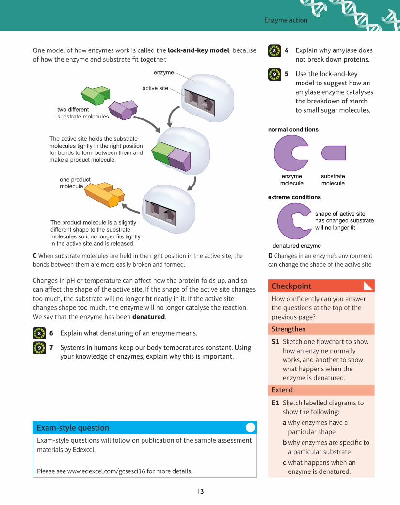
C When substrate molecules are held in the right position in the active site, the bonds between them are more easily broken and formed.
normal conditions
extreme conditions
enzymemolecule
denatured enzyme
shape of active sitehas changed substrate will no longer fit
substratemolecule
D Changes in an enzyme's environment can change the shape of the active site.
6 Explain what denaturing of an enzyme means.
7 Systems in humans keep our body temperatures constant. Using your knowledge of enzymes, explain why this is important.
8
9
4 Explain why amylase does not break down proteins.
5 Use the lock-and-key model to suggest how an amylase enzyme catalyses the breakdown of starch to small sugar molecules.
8
9
Changes in pH or temperature can affect how the protein folds up, and so can affect the shape of the active site. If the shape of the active site changes too much, the substrate will no longer fit neatly in it. If the active site changes shape too much, the enzyme will no longer catalyse the reaction. We say that the enzyme has been denatured.
One model of how enzymes work is called the lock-and-key model, because of how the enzyme and substrate fit together.
two di�erentsubstrate molecules
The active site holds the substrate molecules tightly in the right position for bonds to form between them andmake a product molecule.
one productmolecule
The product molecule is a slightlydi�erent shape to the substratemolecules so it no longer fits tightlyin the active site and is released.
active site
enzyme

14
Enzymes are affected by the conditions in their surroundings. The results of a series of experiments that measure the time taken for an enzyme to complete the breakdown of a substrate at different temperatures can be combined to produce a graph.
• How is enzyme activity affected by temperature, pH and substrate concentration?
• How do you calculate the rate of enzyme activity?
• Why is enzyme activity affected by temperature, pH and substrate concentration?
Progression questions
Specification reference: B1.9; B1.10; B1.11
CB1g Enzyme activity
Many human enzymes have an optimum temperature of around 37 °C.
Did you know?
A results from experiments at different temperatures combined on to one graph
1 Use graph A to identify how long it took for the complete breakdown of starch at the following temperatures:
a 10 °C b 40 °C c 50 °C.
2 Suggest an explanation for the difference in reaction rates at 40 °C and 50 °C.
6
7
Graph A can be converted to a graph showing the rate of reaction by calculating the amount of substrate broken down or product formed in a given time. For example, graph A shows that, at 30 °C, 100 g of starch was broken down in 5 min. The mean rate of reaction was:
1005
= 20 g/min
Converting the values in graph A in this way gives graph B.
B the data in graph A shown as a rate of reaction graph
Why does graph B have this shape?
• As the temperature increases, molecules move faster. Higher speeds increase the chance of substrate molecules bumping into enzyme molecules and slotting into the active site.
• However, when the temperature gets too high, the shape of the enzyme molecule starts to change. The amount of change increases as the temperature increases. So it becomes more and more difficult for a substrate molecule to fit into the active site.
The temperature at which an enzyme works fastest is called its optimum temperature.
3 a Identify the optimum temperature for the enzyme shown in graph B.
b Explain your answer.
4 Explain why enzymes work more slowly when the temperature is:
a below the optimum
b above the optimum.
6
7
9
9
Tim
e ta
ken
for c
ompl
ete
dige
stio
n of
100
g o
f sta
rch
(min
utes
)
30
20
10
00 10 20 30 40 50 60
Temperature (°C)
Time taken for amylase to digest starchdepends on temperature
30
20
10
5
15
25
35
00 10 20 30 40 50 60
Temperature (°C)
Rat
e of
reac
tion
(am
ount
of s
tarc
h di
gest
ed) (
g/m
in)
How the rate of starch breakdown usingamylase depends on temperature

Enzyme activity
15
Exam-style questions will follow on publication of the sample assessment materials by Edexcel.
Please see www.edexcel.com/gcsesci16 for more details.
Exam-style question
How confidently can you answer the questions at the top of the previous page?
Strengthen
S1 Close the book, then sketch and annotate a graph to show how temperature affects the rate of an enzyme-controlled reaction.
Extend
E1 A manufacturer is testing several high-temperature cellulase enzymes to break down plant cell walls in plant waste used for making biofuels. Suggest how the manufacturer might carry out the test and how they would decide which is the best enzyme for this process.
Checkpoint
Enzymes are now used in many processes in industry. Some industrial processes take place at high temperatures, so the search for new enzymes that have a high optimum temperature is important.
Some other factors that affect the rate of an enzyme-controlled reaction are pH and the concentration of the substrate, as shown in graphs D and E.
D the effect of pH on the rate of an enzyme-controlled reaction
6 Explain the effect of substrate concentration on the rate of an enzyme-controlled reaction.
7 The pH in the stomach is about 2, but in the small intestine it is about 6. Explain why different protease enzymes are found in the two digestive organs.
9
9
E the effect of substrate concentration on the rate of an enzyme-controlled reaction
C The bacterium Thermus aquaticus was discovered growing in this hotspring pool, which is at about 70 °C.
5 a Sketch a graph to show the effect of pH on the enzyme pepsin, which has an optimum pH of 2.
b Annotate your sketch to explain the shape.
8
9
pH
Rat
e of
reac
tion
How the rate of an enzyme-controlledreaction depends on pH
optimum pH
At pHs below and abovethe optimum, the shape
of the active site isa�ected and so the
enzyme does not workso well.
Substrate concentration
Rat
e of
reac
tion
How the rate of an enzyme-controlledreaction depends on substrate concentration
At high concentrations, most enzymeactive sites contain substrate molecules,
and the rate of reaction is as fast as it can be.
At low concentrations, many enzymemolecules have empty active sites
so the rate of reaction is slow.

16
Bacteria living on your body cause body odour. The smelly substances they produce are released into the air and reach our noses.
• What is the difference between diffusion and osmosis?
• How do cells move substances against a concentration gradient?
• How do you calculate a percentage change in mass?
Progression questions
Specification reference: B1.15; B1.17
CB1h Transporting substances
Smells spread by diffusion. Particles in gases and liquids are constantly moving past each other in random directions. This causes an overall movement of particles from where there are more of them (a higher concentration) to where there are fewer (a lower concentration).
A an experiment to assess body odour
higher concentration lower concentration
di�usion
The number of particles decreases as you go down a concentration gradient.
B diffusion occurs down a concentration gradient
Diffusion allows small molecules (such as oxygen and carbon dioxide) to move into and out of cells.
2 a A dish of perfume is put at the front of a lab. Describe the perfume’s concentration gradient after 5 minutes.
b Describe the overall movement of the perfume molecules.
3 Muscle cells in the leg use up oxygen but are surrounded by a fluid containing a lot of oxygen. Explain why oxygen moves into the cells.
7
7
7
OsmosisA membrane that allows some molecules through and not others is semi-permeable.
Cell membranes are semi-permeable and trap large soluble molecules inside cells, but water molecules can diffuse through the membrane. If there are more water molecules in a certain volume on one side of the membrane than the other, there will be an overall movement of water molecules from the side where there are more water molecules (a more dilute solute concentration) to the side where there are fewer water molecules (a more concentrated solution of solute). This diffusion of small molecules of a solvent, such as water, through a semi-permeable membrane is called osmosis. The overall movement of solvent molecules will stop when the concentration of solutes is the same on both sides of a membrane.
1 Explain why smells spread.6
A difference between two concentrations forms a concentration gradient. Particles diffuse down a concentration gradient. The bigger the difference between concentrations, the steeper the concentration gradient and the faster diffusion occurs.

Transporting substances
17
Exam-style questions will follow on publication of the sample assessment materials by Edexcel.
Please see www.edexcel.com/gcsesci16 for more details.
Exam-style question
How confidently can you answer the questions at the top of the previous page?
Strengthen
S1 A small number of sugar molecules are in your small intestine. Describe how they will be absorbed into cells in the small intestine and why they need to be absorbed in this way.
Extend
E1 Sorbitol is a sweet-tasting substance that is not broken down or absorbed by the body. It is used in some sugar-free sweets. Explain why eating too many of these sweets can cause diarrhoea.
Checkpoint
4 a In diagram C, in which direction will water flow, X to Y or Y to X?
b Explain why this flow occurs.
5 Red blood cells contain many solute molecules. Explain why red blood cells burst if put in pure water.
Osmosis can cause tissues to gain or lose mass. To calculate the mass change:
• work out the difference between the mass of tissue at the start and at the end (start mass – final mass)
• divide this difference by the start mass
• multiply by 100.
So, percentage change in mass = (start mass – final mass)start mass
× 100
A negative answer is a percentage loss in mass.
7
9
9
Active transportCells may need to transport molecules against a concentration gradient or transport molecules that are too big to diffuse through the cell membrane. They can do this using active transport.
This process is carried out by transport proteins in cell membranes. The transport proteins capture certain molecules and carry them across the cell membrane. This is an active process and so requires energy. Osmosis and diffusion are passive processes, so do not require an input of energy.
soluble moleculethat is too large topass through themembrane(e.g. sucrose)
moreconcentrated
solution
more dilutesolution
partially permeablemembrane allowsmolecules to passthrough if they aresmall enough
watermolecule
YX
C In osmosis, a solvent flows from a dilute solution of a solute to a more concentrated one.
conc
entra
tion
grad
ient
energy
transporter proteincell membrane
A moleculesticks to thetransporter
protein.
The transporter protein changesshape and carries the molecule
across the cell membrane.
D active transport
8 Explain how cells that carry out a lot of active transport would be adapted to their function.
7
6 An 8 g piece of potato is left in water for an hour. Its mass becomes 8.5 g. Calculate the percentage change in mass.
9
7 Look at diagram D. Explain why active transport is needed to move the molecules.
7