c.c. : chest pain for 5 years anterior, mid-chest area · c.c. : chest pain for 5 years anterior,...
TRANSCRIPT

C.C. : Chest pain for 5 years
Anterior, mid-chest area
Aggravated by exercise,
Improved by resting
Dizziness (+), Palpitation (+)
C.C. : Chest pain for 5 years
Anterior, mid-chest area
Aggravated by exercise,
Improved by resting
Dizziness (+), Palpitation (+)


T2-axial
STIR-axial

T1-axialT1-axial
T1-Gd(+)T1-Gd(+)
T1-Gd(+)-10minT1-Gd(+)-10min

T2-SAT2-SA

What do you have in your mind?
1.Lymphoma2.Hypertrophic Cardiomyopathy3.ARVD4.Vascular Tumor


DIAGNOSIS:Heart, transplantation:
Capillary hemangioma, 7cm in greatest dimension, right ventricle,with 1) lipomatous fatty infiltration.
2) myocardial disarray.
DIAGNOSIS:Heart, transplantation:
Capillary hemangioma, 7cm in greatest dimension, right ventricle,with 1) lipomatous fatty infiltration.
2) myocardial disarray.

5-10% of benign tumorAny location of the heartAt any ageShape• In myocardium: ill-defined, sponge-like• In endocardium: well-defined, myxoidChance of spontaneous regressionCT: Calcification in tumorMRI: T2 high, heterogeneous strong enhancement
5-10% of benign tumorAny location of the heartAt any ageShape• In myocardium: ill-defined, sponge-like• In endocardium: well-defined, myxoidChance of spontaneous regressionCT: Calcification in tumorMRI: T2 high, heterogeneous strong enhancement
Grebenc et al. Radiographics 2000;20:1073-1103

C.C : palpable neck massPast medical history:
3 months ago intraarterial thrombolytic therapy for acute infarction of left MCA territory
C.C : palpable neck massPast medical history:
3 months ago intraarterial thrombolytic therapy for acute infarction of left MCA territory
Presented by Joon Won Kang, Tae-Hwan Lim.Ulsan University Asan Medical Center (AMC), Seoul, Korea


4 chamber view Mitral inflow DT = 140msE/E’ = 21

T2WI
T1WI
Delayed-enhancement

AmyloidosisAmyloidosis
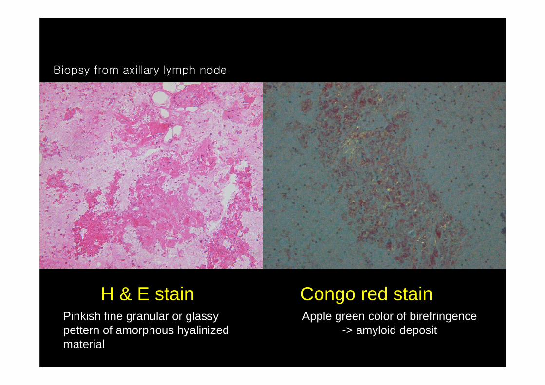
H & E stainPinkish fine granular or glassy pettern of amorphous hyalinized material
H & E stainPinkish fine granular or glassy pettern of amorphous hyalinized material
Congo red stainApple green color of birefringence
-> amyloid deposit
Congo red stainApple green color of birefringence
-> amyloid deposit
Biopsy from axillary lymph node

Abnormal extracellular deposition and accumulation of protein and protein derivativesDiagnosis
Congo red staining‘Apple green birefringence’ under polarized light
ClassificationPrimary (underlying plasma cell disorder or no associated disease)
vs. secondary (underlying chronic abnormality)
Systemic vs. localizedType of deposited amyloid (Ig light chains, AL)

Lymph node involvementUp to 37% of systemic amyloidosis
Punctuate calcification in 50% of amyloid LNs
TuberculosisSarcoidosisCastleman's diseaseAmyloidosis
Lymph node involvementUp to 37% of systemic amyloidosis
Punctuate calcification in 50% of amyloid LNs
TuberculosisSarcoidosisCastleman's diseaseAmyloidosis
– Scleroderma– Metastasis– Hodgkin’s disease after
irradiation