ccoolllleeggee ooff oonnccoollooggyy cancer v2.2012 (ex... · ccoolllleeggee ooff oonnccoollooggyy...
TRANSCRIPT

CCOOLLLLEEGGEE OOFF OONNCCOOLLOOGGYY
NNaattiioonnaall CClliinniiccaall PPrraaccttiiccee GGuuiiddeelliinneess
GGaassttrriicc CCaanncceerr VVeerrssiioonn 22..22001122

NATIONAL GUIDELINES GASTRIC CANCER _________________________________________________________________________________________________
V2.2012 © 2012 College of Oncology
Gastric Cancer Guidelines Expert Panel Prof. dr. Marc Peeters
Coordinator, University Hospital Antwerp Field of expertise: gastroenterology
Prof. dr. Tom Boterberg
University Hospital Ghent Field of expertise: radiotherapy
Prof. dr. Gert De Hertogh
University Hospital Leuven Field of expertise: pathology
Prof. Dr. Johan De Mey Universitair Ziekenhuis Brussel Field of expertise: radiology
Prof. dr. Pierre Deprez Clinques Universitaires Saint-Luc Field of expertise: gastroenterology
Prof. dr. Patrick Flamen Jules Bordet Institute Brussels Field of expertise: nuclear medicine
Prof. dr. Antoon Lerut University Hospital Leuven Field of expertise: surgery
Prof. dr. Piet Pattyn University Hospital Ghent Field of expertise: surgery
Prof. dr. Jean-Luc Van Laethem ULB Hôpital Erasme Bruxelles Field of expertise: gastroenterology
Dr. Sabine Stordeur
Belgian Health Care Knowledge Centre
Dr. Leen Verleye
Belgian Health Care Knowledge Centre
Dr. Joan Vlayen
Belgian Health Care Knowledge Centre
This report was supported by the Belgian Healthcare Knowledge Centre. The full scientific report can be consulted at the KCE website (www.kce.fgov.be).
Reference: Lerut T, Stordeur S, Verleye L, Vlayen J, Boterberg T, De Hertogh G, De Mey J, Deprez P, Flamen P, Pattyn P, Van Laethem J-L, Peeters M. Update van de praktijkrichtlijn voor slokdarm- en maagkanker. Good Clinical Practice (GCP). Brussel: Federaal Kenniscentrum voor de Gezonheidszorg (KCE); 2012. KCE reports 179A. D/2012/10.273/32).
or
Reference: Lerut T, Stordeur S, Verleye L, Vlayen J, Boterberg T, De Hertogh G, De Mey J, Deprez P, Flamen P, Pattyn P, Van Laethem J-L, Peeters M. Actualisation des recommandations cliniques pour le cancer de l’oesophage et de l’estomac. Good Clinical Practice (GCP). Bruxelles: Centre Fédéral d’Expertise des Soins de Santé (KCE). 2012. KCE Report 179B. D/2012/10.273/33.
NATIONAL GUIDELINES GASTRIC CANCER _________________________________________________________________________________________________
V1.2012 © 2012 College of Oncology
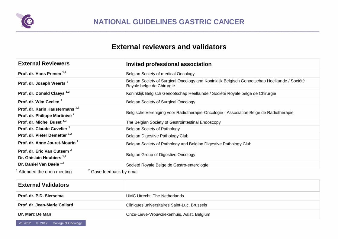
External reviewers and validators
External Reviewers Invited professional association
Prof. dr. Hans Prenen 1,2
Belgian Society of medical Oncology
Prof. dr. Joseph Weerts 2
Belgian Society of Surgical Oncology and Koninklijk Belgisch Genootschap Heelkunde / Société Royale belge de Chirurgie
Prof. dr. Donald Claeys 1,2
Koninklijk Belgisch Genootschap Heelkunde / Société Royale belge de Chirurgie
Prof. dr. Wim Ceelen 2 Belgian Society of Surgical Oncology
Prof. dr. Karin Haustermans 1,2
Prof. dr. Philippe Martinive 2
Belgische Vereniging voor Radiotherapie-Oncologie - Association Belge de Radiothérapie
Prof. dr. Michel Buset 1,2
The Belgian Society of Gastrointestinal Endoscopy
Prof. dr. Claude Cuvelier 1 Belgian Society of Pathology
Prof. dr. Pieter Demetter 1,2
Belgian Digestive Pathology Club
Prof. dr. Anne Jouret-Mourin 1 Belgian Society of Pathology and Belgian Digestive Pathology Club
Prof. dr. Eric Van Cutsem 2
Dr. Ghislain Houbiers 1,2
Belgian Group of Digestive Oncology
Dr. Daniel Van Daele 1,2
Societé Royale Belge de Gastro-enterologie 1 Attended the open meeting 2 Gave feedback by email
External Validators
Prof. dr. P.D. Siersema UMC Utrecht, The Netherlands
Prof. dr. Jean-Marie Collard Cliniques universitaires Saint-Luc, Brussels
Dr. Marc De Man Onze-Lieve-Vrouwziekenhuis, Aalst, Belgium
NATIONAL GUIDELINES GASTRIC CANCER _________________________________________________________________________________________________
V1.2012 © 2012 College of Oncology
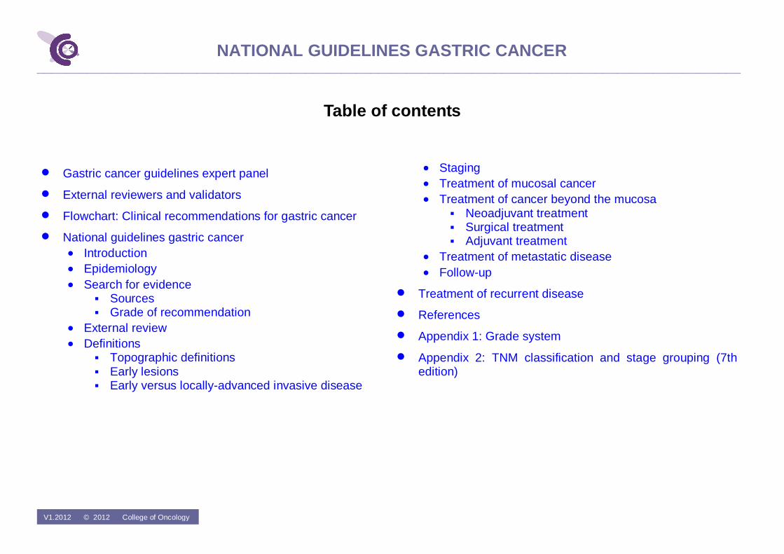
Table of contents
Gastric cancer guidelines expert panel
External reviewers and validators
Flowchart: Clinical recommendations for gastric cancer
National guidelines gastric cancer
Introduction
Epidemiology
Search for evidence Sources Grade of recommendation
External review
Definitions Topographic definitions Early lesions Early versus locally-advanced invasive disease
Staging
Treatment of mucosal cancer
Treatment of cancer beyond the mucosa Neoadjuvant treatment Surgical treatment Adjuvant treatment
Treatment of metastatic disease
Follow-up
Treatment of recurrent disease
References
Appendix 1: Grade system
Appendix 2: TNM classification and stage grouping (7th edition)

NATIONAL GUIDELINES GASTRIC CANCER _________________________________________________________________________________________________
V1.2012 © 2012 College of Oncology
Flowchart: Clinical recommendations for gastric cancer
Staging CT neck/thorax/abdomen
EUS +/- FNAC* FDG-PET/CT*
Extramucosal cancer
Mucosal cancer
F
O
L
L
O
W
U
P
Multidisciplinary team meeting
Invasive cancer
Endoscopic mucosal resection/endoscopic submucosal
dissection +/- ablation* +/- surgery*
* Option to be discussed at the MDT meeting
Inoperable disease/M1
Gastrectomy + D2 lymphadenectomy
Neoadjuvant chemotherapy*
Palliative treatment
Adjuvant chemo*

NATIONAL GUIDELINES GASTRIC CANCER _________________________________________________________________________________________________
1 V1.2012 © 2012 College of Oncology
National Guidelines Gastric Cancer
INTRODUCTION [1]
This document presents the updated clinical practice guidelines on gastric cancer which was first published in 2008 [1]. It covers a broad range of topics: staging, treatment and follow-up of patients with confirmed invasive gastric cancer.
Importantly, the following topics that were part of the previous version were not included in the update:
work-up of pre-invasive lesions, i.e. dysplastic lesions, including high-grade dysplasia
treatment of gastric lymphoma
treatment of gastrointestinal stromal tumors (GIST)
For more in-depth information and the scientific background, we would like to ask the readers to consult the full scientific report at www.kce.fgov.be.
The guidelines are developed by a panel of experts (see 'expert panel') comprising clinicians of different specialties and were reviewed by relevant professional associations (see 'external reviewers and validators').
The aim of these guidelines is to assist all care providers involved in the care of patients with gastric cancer.
EPIDEMIOLOGY[2-12] With an estimated 988 000 new cases in 2008 worldwide (7.8% of all new cancer cases), gastric cancer is in fourth place behind cancers of the lung, breast, and colon and rectum, with more than 70% of the cases occurring in developing countries. It is the second most common cause of death from cancer.
In Belgium, the crude incidence rate of gastric cancer declined from 17.4 per 100.000 males in 2004 to 15.4 per 100 000 males in 2009. In females, the crude incidence rate remained quite stable between 2004 and 2009 (9.4/100 000 females in 2009). Similar trends are reported for the age standardised incidence.
While the incidence rates of gastro-oesophageal junction (GOJ) tumours recently increased, the incidence rates of ‘real’ gastric tumours declined.
SEARCH FOR EVIDENCE [13-16]
Sources
Systematic reviews and meta-analyses were searched in the following databases: OVID Medline and PreMedline, EMBASE, Cochrane Database of Systematic Reviews, Database of Abstracts of Reviews of Effects (DARE), Health Technology Assessment (HTA) database. RCTs were searched in OVID Medline, PreMedline, EMBASE and CENTRAL, while diagnostic accuracy studies were searched in OVID Medline, PreMedline and EMBASE.

NATIONAL GUIDELINES GASTRIC CANCER _________________________________________________________________________________________________
2 V2.2012 © 2012 College of Oncology
A date limit was set from August 2007 (i.e. the search date of the previous version) until 2011.
Grade of recommendation
A grade of recommendation was assigned to each recommendation using the GRADE system (Appendix 1).
EXTERNAL REVIEW The guidelines prepared by the expert panel were circulated to the
relevant professional associations (see 'external reviewers'). Each
association was asked to assign two key persons to discuss the recommendations during an open meeting. As a preparation of the meeting all invited experts were asked to score each recommendation on a 5-point Likert-scale to indicate their agreement with the recommendation, with a score of ‘1’ indicating ‘completely disagree’, ‘2’ indicating ‘somewhat disagree’, ‘3’ indicating ‘unsure’, ‘4’ indicating ‘somewhat agree’, and ‘5’ indicating ‘completely agree’ (the experts were also able to answer ‘not applicable’ in case they were not familiar with the underlying evidence). In case an expert disagreed with the recommendation (score ‘1’ or ‘2’), (s)he was asked to provide appropriate evidence. All scores were then anonymized and summarized into a median score, minimum score, maximum score and % of ‘agree’-scores (score ‘4’ and ‘5’) to allow a targeted discussion. The recommendations were then discussed during a face-to-face meeting on March 30th 2012. Based on this discussion a final draft of the recommendations was prepared.
As part of the standard KCE procedures, an external scientific validation
of the report was conducted by three independent experts. Following this validation procedure, some recommendations were finally adapted if strong arguments supported a change in the formulation.
DEFINITIONS
Topographic definitions [17-20]
A tumour of which the epicentre is within 5 cm of the GOJ and extending into the oesophagus is to be classified as an oesophageal tumour.
Tumours with an epicentre in the stomach greater than 5 cm from the GOJ or those within 5 cm of the GOJ without extension in the oesophagus are to be classified as a gastric tumour.
Early lesions [21-50]
Several classifications are available for dysplasia. For the physician, the used classification should be clinically relevant.
Early versus locally-advanced invasive disease
Definitions of early and locally-advanced cancer are not uniform and controversial. Therefore, to avoid discussion, an attempt will be made to define the eligible population for our recommendations as accurate as possible.
NATIONAL GUIDELINES GASTRIC CANCER _________________________________________________________________________________________________
3 V2.2012 © 2012 College of Oncology
STAGING [51,52-56,61-70] Conclusions of the literature update
For T-staging, CT seems to have a low sensitivity for the diagnosis of T1-2 tumours and a moderate sensitivity for higher T-stages (low level of evidence; Anzidei 2009, Cidon 2009, Hwang 2010, Hye 2009, Kim 2011, Kim 2009, Lee 2009, Lee 2009, Makino 2011, Moschetta 2010, Pan 2010). EUS seems to have a better diagnostic accuracy for the detection of T1 tumours, and has a moderate sensitivity and specificity for the distinction between T1a and T1b tumours (low level of evidence; Mocellin 2011, Kwee 2008).
For N-staging, CT has a moderate sensitivity and specificity (low level of evidence; Seevaratnam 2011, Kwee 2009). EUS and MRI do not seem to have a better diagnostic accuracy (low level of evidence; Mocellin 2011, Seevaratnam 2011, Kwee 2009). PET and PET/CT have a lower sensitivity but a higher specificity (low level of evidence; Seevaratnam 2011, Kwee 2009). Sentinel lymph node biopsy in patients with T1-2 tumours has a sensitivity similar to that of CT, but a higher specificity (low level of evidence, Wang 2011).
For the detection of liver metastases, CT and PET(/CT) have a similar low sensitivity and high specificity (low level of evidence, Wang 2011). The diagnostic accuracy of MRI seems to be better (low level of evidence, Wang 2011).
For the detection of peritoneal metastases, CT, EUS and PET(/CT) have a low sensitivity and a high specificity (low level of evidence, Wang 2011). Laparoscopy seems to have a better diagnostic accuracy (low level of evidence, Leake 2011).
Final recommendations
All patients diagnosed with gastric cancer should be discussed at a multidisciplinary team meeting (strong recommendation, low level of evidence).
In patients with newly diagnosed gastric cancer, CT scan of the chest and abdomen should always be performed (strong recommendation, low level of evidence).
Endoscopic ultrasonography (EUS) can be considered in patients planned for curative treatment on the basis of clinical presentation and/or CT (weak recommendation, low level of evidence). Fine-needle aspiration cytology of suspicious lymph nodes or metastases can be considered if technically feasible.
The following examinations can be considered for specific indications: PET scan, magnetic resonance imaging, laparoscopy (weak recommendation, low level of evidence).
Good clinical practice
Multi-detector, multi-planar reformatted CT scan should be performed with IV contrast and gastric distension with oral contrast or water. The liver should at least be imaged in the arterial and portal venous phase.
TREATMENT OF MUCOSAL CANCER [51,71-79] Conclusions of the literature update
Based on observational studies, excellent cure rates and survival can be achieved with endoscopic treatment of early gastric cancer (T1) if

NATIONAL GUIDELINES GASTRIC CANCER _________________________________________________________________________________________________
4 V2.2012 © 2012 College of Oncology
standard indications are applied (low level of evidence; Bennett 2009).
Based on observational studies, endoscopic submucosal dissection seems to be associated with a higher curative resection rate, a lower local recurrence rate, but a higher perforation rate than endoscopic mucosal resection. Both interventions are equally effective in terms of survival (low level of evidence; Park 2011).
Insufficient evidence is available to draw conclusions on the effectiveness of photodynamic therapy, laser or argon plasma coagulation for the treatment of early gastric cancer.
Final recommendations
Endoscopic mucosal resection (EMR) or endoscopic submucosal dissection (ESD) should be performed whenever possible for a T1a gastric cancer aiming at staging and curative resection. If the staging and R0 resection is pathologically confirmed, the procedure can be considered therapeutic, taking into account other well-defined criteria relating to size, histological type, lymphovascular invasion and differentiation grade (weak recommendation, low level of evidence).
(Destructive) mucosal ablative techniques cannot be recommended as a curative option for patients with T1a gastric cancer and should be limited to centres with appropriate expertise (weak recommendation, very low level of evidence).
Good clinical practice
Resection specimens of EMR and ESD should be reviewed by an experienced pathologist in this area and discussed at a multidisciplinary meeting with access to the clinical information.
TREATMENT OF CANCER BEYOND THE MUCOSA
Neoadjuvant treatment [57,80-87]
Conclusions of the literature update
Neoadjuvant chemotherapy is associated with a survival benefit compared with surgery alone in patients with locally advanced gastric cancer eligible for potentially curative surgery (moderate level of evidence; Li 2010). The benefit seems to be larger in T3-4 tumours (low level of evidence; Li 2010).
Perioperative chemotherapy seems to be associated with a survival benefit in patients with gastric cancer (low level of evidence; Cunningham 2006).
Preoperative radiotherapy seems to improve local control and 5-year survival in patients with resectable gastric cancer compared to no adjuvant therapy (low level of evidence; Valentini 2009).
Final recommendations
If after multidisciplinary discussion neoadjuvant treatment is considered for a locally-advanced gastric tumour, neoadjuvant chemotherapy is recommended (strong recommendation, moderate level of evidence).

NATIONAL GUIDELINES GASTRIC CANCER _________________________________________________________________________________________________
5 V2.2012 © 2012 College of Oncology
Surgical treatment [51,58,88-98]
Conclusions of the literature update
Randomized studies to support the use of surgery as standard treatment for patients with resectable gastric cancer is lacking.
In patients with resectable gastric cancer, D2 lymphadenectomy is associated with similar overall survival and recurrence rates as D1 lymphadenectomy (high level of evidence; Memon 2011), but with a significantly increased morbidity (low level of evidence; Memon 2011). This morbidity seems to be related to the splenectomy and/or pancreatectomy performed as part of the D2 lymphadenectomy (low level of evidence; Roberts 2011).
In patients with resectable gastric cancer, para-aortic lymph node dissection is not associated with a survival benefit (moderate level of evidence; Zheng 2011, Lustosa 2008).
Pouch reconstruction seems to be associated with better food intake and improved quality of life (high level of evidence; Gertler 2009).
In patients with resectable gastric cancer, laparoscopic gastrectomy is associated with fewer postoperative complications compared with open gastrectomy, but a longer operation time and a reduced number of lymph nodes harvested (low level of evidence; Zorcolo 2011, Chen 2009).
Postoperative mortality after gastrectomy for gastric cancer seems to be associated with the surgeon and hospital volume (low level of evidence; Halm 2002, Killeen 2005).
Final recommendations
Surgical resection should be considered standard treatment for patients with resectable gastric cancer (strong recommendation, low level of evidence).
Surgery for gastric cancer should aim at achieving an R0 resection (strong recommendation, low level of evidence).
D2 lymphadenectomy should be standard during gastrectomy and performed in high-volume, specialized centres with experience and/or specialist training (weak recommendation, low level of evidence).
Splenectomy and pancreatectomy should not be considered standard practice during gastrectomy if no disease infiltration in the spleen or pancreas is present (weak recommendation, low level of evidence).
Laparoscopic surgery should be restricted to clinical studies (weak recommendation, low level of evidence).
Adjuvant treatment [51,57,59,82,85-86,99-118]
Conclusions of the literature update
Postoperative adjuvant chemotherapy significantly improves overall survival in patients with resectable gastric cancer compared to no adjuvant therapy (moderate level of evidence; GASTRIC 2010, Sun 2009, Liu 2008).
There are indications that perioperative chemotherapy improves the overall survival of patients with gastric cancer (low level of evidence; Cunningham 2006).
Adjuvant oral fluorinated pyrimidines are associated with a survival benefit compared to surgery alone (high level of evidence; Sun 2009).

NATIONAL GUIDELINES GASTRIC CANCER _________________________________________________________________________________________________
6 V2.2012 © 2012 College of Oncology
Postoperative radiotherapy is not associated with a survival benefit in patients with gastric cancer (low level of evidence; Valentine 2009).
No definite conclusions can be drawn on the effectiveness of postoperative chemoradiation (low level of evidence; CCO 2003).
Postoperative hyperthermic intraperitoneal chemotherapy can improve overall survival in patients with resectable gastric cancer compared to no adjuvant therapy (moderate level of evidence; Yan 2007).
The benefits of immunochemotherapy are currently unclear (low level of evidence; CCO 2003).
Final recommendations
Patients with gastric cancer who received neoadjuvant chemotherapy can be considered for postoperative chemotherapy (weak recommendation; low level of evidence).
Postoperative chemotherapy and chemoradiotherapy can be considered for patients with gastric cancer who did not receive neoadjuvant chemotherapy (weak recommendation; low level of evidence).
Postoperative radiotherapy alone is not recommended for patients with gastric cancer (weak recommendation; low level of evidence).
TREATMENT OF METASTATIC DISEASE [51,60-61,119-139] Conclusions of the literature update
Palliative gastrectomy seems to be associated with a lower mortality than gastric bypass for patients with unresectable gastric cancer (low level of evidence; SIGN 2006).
Laparoscopic gastrojejunostomy seems to be associated with a shorter time to oral intake and a shorter hospital stay than open gastric bypass for patients with unresectable gastric cancer (low level of evidence; Mahar 2011).
There is weak evidence that adding HIPEC to cytoreductive surgery in patients with peritoneal metastases is associated with a survival benefit (low level of evidence; Yang 2011).
In patients with malignant gastric outlet obstruction, endoscopic stenting and surgical gastroenterostomy have similar survival rates (low level of evidence; Zheng 2011, Ly 2010). Inconsistent evidence is available on the associated risks.
In patients with advanced or metastatic gastric cancer, combination chemotherapy has a survival benefit compared to single agent chemotherapy (high level of evidence; Wagner 2010).
Oral 5-FU analogues seem to be associated with a survival benefit compared with intravenous 5-FU (moderate level of evidence; Huang 2010, Ma 2011).
In HER2-positive gastric cancer, adding trastuzumab to standard chemotherapy seems to be associated with a survival benefit (low level of evidence; Bang 2010).
Final recommendations
Palliative gastric surgery is limited to symptomatic stenoses, bleeding tumours and perforation (weak recommendation, low level of evidence).

NATIONAL GUIDELINES GASTRIC CANCER _________________________________________________________________________________________________
7 V2.2012 © 2012 College of Oncology
For patients with malignant gastric outlet obstruction, treatment options include endoscopic stenting or surgical gastroenterostomy (weak recommendation, low level of evidence).
In patients with locally advanced or metastatic cancer of the stomach with good performance status combination chemotherapy is recommended (strong recommendation, high level of evidence).
Patients with advanced gastric cancer should have access to a specialist palliative care team, in particular in relation to comfort and symptom control, nutrition and quality of life (strong recommendation, low level of evidence).
FOLLOW-UP [51,75,139-140] Conclusions of the literature update
In routine follow-up, CT seems to have a low sensitivity and moderate specificity to detect recurrence after curative gastrectomy for gastric cancer (low level of evidence; Kim 2009). The diagnostic accuracy of PET/CT is not clearly better (low level of evidence; Kim 2009, Graziosi 2011, Sim 2009).
Conflicting evidence is available on the diagnostic accuracy of PET/CT in patients with a suspected recurrence of gastric cancer (low level of evidence; Bilici 2011, Park 2009).
Final recommendations
It is recommended that the follow-up of patients treated for gastric cancer includes a physical examination and blood analysis every three months, with targeted imaging if needed. A CT scan can be considered
every six months in the first year and then annually until the fifth year (weak recommendation; very low level of evidence).
Patients treated with endoscopic mucosal resection (EMR) or endoscopic submucosal dissection (ESD) should have a follow-up endoscopy after three months, then every six months in the first two years, and then annually (weak recommendation; very low level of evidence).
Treatment of recurrent disease [141-147] Conclusions of the literature update
No good evidence is available about the optimal treatment for patients with recurrent gastric cancer.
Final recommendations
In patients with recurrent gastric cancer, treatment options should be discussed in the multidisciplinary team (strong recommendation; very low level of evidence).
NATIONAL GUIDELINES GASTRIC CANCER _________________________________________________________________________________________________
8 V2.2012 © 2012 College of Oncology
1. Peeters M, Lerut T, Vlayen J, Mambourg F, Ectors N, Deprez P, et al. Guideline pour la prise en charge du cancer oesophagien et gastrique: éléments scientifiques à destination du Collège d’Oncologie. Bruxelles: Centre fédéral d'expertise des soins de santé (KCE); 2008. KCE Reports 75B (D/2008/10.273/17)
2. International Agency for Research on Cancer (IARC) GLOBOCAN 2008. Cancer incidence, mortality and prevalence worldwide in 2008 [2008 [cited March 1st 2012]. Available from: http://globocan.iarc.fr/
3. Crane LM, Schaapveld M, Visser O, Louwman MW, Plukker JT, van Dam GM. Oesophageal cancer in The Netherlands: increasing incidence and mortality but improving survival. Eur J Cancer. 2007;43(9):1445-51.
4. Qiu D, Kaneko S. Comparison of esophageal cancer mortality in five countries: France, Italy, Japan, UK and USA from the WHO mortality database (1960-2000). Jpn J Clin Oncol. 2005;35(9):564-7.
5. Lagergren J, Mattsson F. No further increase in the incidence of esophageal adenocarcinoma in Sweden. Int J Cancer. 2011;129(2):513-6.
6. Steevens J, Botterweck AA, Dirx MJ, van den Brandt PA, Schouten LJ. Trends in incidence of oesophageal and stomach cancer subtypes in Europe. Eur J Gastroenterol Hepatol. 2010;22(6):669-78.
7. Vizcaino AP, Moreno V, Lambert R, Parkin DM. Time trends incidence of both major histologic types of esophageal carcinomas in selected countries, 1973-1995. Int J Cancer. 2002;99(6):860-8.
8. Belgian Cancer Registry (BCR) [Tables on an anual basis] [Brussels;2012 [cited March 1st 2012]. Available from: http://www.kankerregister.org/Statistieken_tabellen_jaarbasis
9. Parkin DM, Bray F, Ferlay J, Pisani P. Global cancer statistics, 2002. CA Cancer J Clin. 2005;55(2):74-108.
10. Dassen AE, Lemmens VE, van de Poll-Franse LV, Creemers GJ, Brenninkmeijer SJ, Lips DJ, et al. Trends in incidence, treatment and
survival of gastric adenocarcinoma between 1990 and 2007: a population-based study in the Netherlands. Eur J Cancer. 2010;46(6):1101-10.
11. Wijnhoven BP, Louwman MW, Tilanus HW, Coebergh JW. Increased incidence of adenocarcinomas at the gastro-oesophageal junction in Dutch males since the 1990s. Eur J Gastroenterol Hepatol. 2002;14(2):115-22.
12. Crane SJ, Richard Locke G, 3rd, Harmsen WS, Diehl NN, Zinsmeister AR, Joseph Melton L, 3rd, et al. The changing incidence of oesophageal and gastric adenocarcinoma by anatomic sub-site. Aliment Pharmacol Ther. 2007;25(4):447-53.
13. Whiting PF, Rutjes AW, Westwood ME, Mallett S, Deeks JJ, Reitsma JB, et al. QUADAS-2: a revised tool for the quality assessment of diagnostic accuracy studies. Ann Intern Med. 2011;155(8):529-36.
14. Balshem H, Helfand M, Schunemann HJ, Oxman AD, Kunz R, Brozek J, et al. GRADE guidelines: 3. Rating the quality of evidence. J Clin Epidemiol. 2011;64(4):401-6.
15. Guyatt G, Oxman AD, Akl EA, Kunz R, Vist G, Brozek J, et al. GRADE guidelines: 1. Introduction-GRADE evidence profiles and summary of findings tables. J Clin Epidemiol. 2011;64(4):383-94.
16. Guyatt GH, Oxman AD, Kunz R, Falck-Ytter Y, Vist GE, Liberati A, et al. Going from evidence to recommendations. BMJ. 2008;336(7652):1049-51.
17. Siewert JR, Stein HJ. Classification of adenocarcinoma of the oesophagogastric junction. Br J Surg. 1998;85(11):1457-9.
18. Spechler SJ, Dixon MF, Genta R, Hainaut P, Lambert R, Siewert R. Adenocarcinoma of the esophago-gastric junction. In: Hamilton SR, Aaltonen LA, editors. Pathology and Genetics of Tumours of the Digestive System. Lyon, France: IARC Press; 2000. p. 31-6.
19. (UICC) IUAC. TNM classification of malignant tumours. 6th ed. ed. Berlin: Springer-Verlag; 2002.
References
NATIONAL GUIDELINES GASTRIC CANCER _________________________________________________________________________________________________
9 V2.2012 © 2012 College of Oncology
20. (UICC) IUAC. TNM classification of malignant tumours. 7th ed. ed. Sobin LH, Gospodarowicz MK, Wittekind C, editor.: Wiley-Blackwell; 2009.
21. Skinner DB, Walther BC, Riddell RH, Schmidt H, Iascone C, DeMeester TR. Barrett's esophagus. Comparison of benign and malignant cases. Ann Surg. 1983;198(4):554-65.
22. Paull A, Trier JS, Dalton MD, Camp RC, Loeb P, Goyal RK. The histologic spectrum of Barrett's esophagus. N Engl J Med. 1976;295(9):476-80.
23. Naef AP, Savary M, Ozzello L. Columnar-lined lower esophagus: an acquired lesion with malignant predisposition. Report on 140 cases of Barrett's esophagus with 12 adenocarcinomas. J Thorac Cardiovasc Surg. 1975;70(5):826-35.
24. Haggitt RC, Tryzelaar J, Ellis FH, Colcher H. Adenocarcinoma complicating columnar epithelium-lined (Barrett's) esophagus. Am J Clin Pathol. 1978;70(1):1-5.
25. Reid BJ, Haggitt RC, Rubin CE, Rabinovitch PS. Barrett's esophagus. Correlation between flow cytometry and histology in detection of patients at risk for adenocarcinoma. Gastroenterology. 1987;93(1):1-11.
26. Spechler SJ, Goyal RK. The columnar-lined esophagus, intestinal metaplasia, and Norman Barrett. Gastroenterology. 1996;110(2):614-21.
27. Sampliner RE. Practice guidelines on the diagnosis, surveillance, and therapy of Barrett's esophagus. The Practice Parameters Committee of the American College of Gastroenterology. Am J Gastroenterol. 1998;93(7):1028-32.
28. Sharma P, McQuaid K, Dent J, Fennerty MB, Sampliner R, Spechler S, et al. A critical review of the diagnosis and management of Barrett's esophagus: the AGA Chicago Workshop. Gastroenterology. 2004;127(1):310-30.
29. British Society of Gastroenterology. Guidelines for the diagnosis and management of Barrett’s columnar-lined oesophagus. London: British Society of Gastroenterology; 2005 August 2005.
30. Flejou JF, Svrcek M. Barrett's oesophagus--a pathologist's view. Histopathology. 2007;50(1):3-14.
31. Takubo K, Sasajima K, Yamashita K, Tanaka Y, Fujita K. Double muscularis mucosae in Barrett's esophagus. Hum Pathol. 1991;22(11):1158-61.
32. Nigro JJ, Hagen JA, DeMeester TR, DeMeester SR, Peters JH, Oberg S, et al. Prevalence and location of nodal metastases in distal esophageal adenocarcinoma confined to the wall: implications for therapy. J Thorac Cardiovasc Surg. 1999;117(1):16-23; discussion -5.
33. Westerterp M, Koppert LB, Buskens CJ, Tilanus HW, ten Kate FJ, Bergman JJ, et al. Outcome of surgical treatment for early adenocarcinoma of the esophagus or gastro-esophageal junction. Virchows Arch. 2005;446(5):497-504.
34. Watanabe H, Jass JR, Sobin LH. Histological typing of oesophageal and gastric tumours. 2nd ed. ed. Berlin: Springer-Verlag; 1990.
35. Rubio CA, Liu FS, Zhao HZ. Histological classification of intraepithelial neoplasias and microinvasive squamous carcinoma of the esophagus. Am J Surg Pathol. 1989;13(8):685-90.
36. Riddell RH, Goldman H, Ransohoff DF, Appelman HD, Fenoglio CM, Haggitt RC, et al. Dysplasia in inflammatory bowel disease: standardized classification with provisional clinical applications. Hum Pathol. 1983;14(11):931-68.
37. Montgomery E. Is there a way for pathologists to decrease interobserver variability in the diagnosis of dysplasia? Arch Pathol Lab Med. 2005;129(2):174-6.
38. Montgomery E, Bronner MP, Goldblum JR, Greenson JK, Haber MM, Hart J, et al. Reproducibility of the diagnosis of dysplasia in Barrett esophagus: a reaffirmation. Hum Pathol. 2001;32(4):368-78.
39. Reid BJ, Haggitt RC, Rubin CE, Roth G, Surawicz CM, Van Belle G, et al. Observer variation in the diagnosis of dysplasia in Barrett's esophagus. Hum Pathol. 1988;19(2):166-78.
40. Schlemper RJ, Borchard F, Dixon MF, Koike M, Mueller J, Stolte M, et al. International comparability of the pathological diagnosis for early cancer of the digestive tract: Munich meeting. J Gastroenterol. 2000;35 Suppl 12:102-10.

NATIONAL GUIDELINES GASTRIC CANCER _________________________________________________________________________________________________
10 V2.2012 © 2012 College of Oncology
41. Schlemper RJ, Dawsey SM, Itabashi M, Iwashita A, Kato Y, Koike M, et al. Differences in diagnostic criteria for esophageal squamous cell carcinoma between Japanese and Western pathologists. Cancer. 2000;88(5):996-1006.
42. Schlemper RJ, Itabashi M, Kato Y, Lewin KJ, Riddell RH, Shimoda T, et al. Differences in diagnostic criteria for gastric carcinoma between Japanese and western pathologists. Lancet. 1997;349(9067):1725-9.
43. Antonioli DA, Wang HH. Morphology of Barrett's esophagus and Barrett's-associated dysplasia and adenocarcinoma. Gastroenterol Clin North Am. 1997;26(3):495-506.
44. Haggitt RC. Barrett's esophagus, dysplasia, and adenocarcinoma. Hum Pathol. 1994;25(10):982-93.
45. Rugge M, Correa P, Dixon MF, Hattori T, Leandro G, Lewin K, et al. Gastric dysplasia: the Padova international classification. Am J Surg Pathol. 2000;24(2):167-76.
46. Schlemper RJ, Riddell RH, Kato Y, Borchard F, Cooper HS, Dawsey SM, et al. The Vienna classification of gastrointestinal epithelial neoplasia. Gut. 2000;47(2):251-5.
47. Schlemper RJ, Kato Y, Stolte M. Diagnostic criteria for gastrointestinal carcinomas in Japan and Western countries: proposal for a new classification system of gastrointestinal epithelial neoplasia. J Gastroenterol Hepatol. 2000;15 Suppl:G49-57.
48. Schlemper RJ, Kato Y, Stolte M. Review of histological classifications of gastrointestinal epithelial neoplasia: differences in diagnosis of early carcinomas between Japanese and Western pathologists. J Gastroenterol. 2001;36(7):445-56.
49. Hamilton SR, Aaltonen LA. Pathology and genetics of tumours of the digestive system. Hamilton SR, Aaltonen LA, editor. Lyon, France: IARC Press; 2000.
50. Odze RD. Diagnosis and grading of dysplasia in Barrett's oesophagus. J Clin Pathol. 2006;59(10):1029-38.
51. Scottish Intercollegiate Guidelines Network. Management of oesophageal and gastric cancer. A national clinical guideline. Edinburgh: SIGN; 2006 June 2006.
52. Mortensen MB, Fristrup CW, Ainsworth AP, Pless T, Nielsen HO, Hovendal C. Combined preoperative endoscopic and laparoscopic ultrasonography for prediction of R0 resection in upper gastrointestinal tract cancer. British Journal of Surgery. 2006;93(6):720-5.
53. Patkar V, Acosta D, Davidson T, Jones A, Fox J, Keshtgar M. Cancer multidisciplinary team meetings: evidence, challenges, and the role of clinical decision support technology. Int J Breast Cancer. 2011;2011:831605.
54. Davies AR, Deans DAC, Penman I, Plevris JN, Fletcher J, Wall L, et al. The multidisciplinary team meeting improves staging accuracy and treatment selection for gastro-esophageal cancer.[see comment]. Diseases of the Esophagus. 2006;19(6):496-503.
55. Stephens MR, Lewis WG, Brewster AE, Lord I, Blackshaw GR, Hodzovic I, et al. Multidisciplinary team management is associated with improved outcomes after surgery for esophageal cancer. Dis Esophagus. 2006;19(3):164-71.
56. Jobe BA, Enestvedt CK, Thomas CR, Jr. Disease-specific multidisciplinary care: a natural progression in the management of esophageal cancer. Dis Esophagus. 2006;19(6):417-8.
57. Cunningham D, Allum WH, Stenning SP, Thompson JN, Van de Velde CJH, Nicolson M, et al. Perioperative chemotherapy versus surgery alone for resectable gastroesophageal cancer.[see comment]. New England Journal of Medicine. 2006;355(1):11-20.
58. Halm EA, Lee C, Chassin MR. Is volume related to outcome in health care? A systematic review and methodologic critique of the literature. Ann Intern Med. 2002;137(6):511-20.
59. Macdonald JS, Smalley SR, Benedetti J, Hundahl SA, Estes NC, Stemmermann GN, et al. Chemoradiotherapy after surgery compared with surgery alone for adenocarcinoma of the stomach or gastroesophageal junction. N Engl J Med. 2001;345(10):725-30.

NATIONAL GUIDELINES GASTRIC CANCER _________________________________________________________________________________________________
11 V2.2012 © 2012 College of Oncology
60. Kuchler T, Bestmann B, Rappat S, Henne-Bruns D, Wood-Dauphinee S. Impact of psychotherapeutic support for patients with gastrointestinal cancer undergoing surgery: 10-year survival results of a randomized trial.[see comment]. Journal of Clinical Oncology. 2007;25(19):2702-8.
61. Fédération Nationale des Centres de Lutte Contre le Cancer (FNCLCC). Recommandations pour la pratique clinique: Standards, Options et Recommandations 2003 pour la prise en charge des patients atteints d’adénocarcinomes de l’estomac (cancers du cardia, autres types histologiques exclus) (rapport intégral). Paris: FNCLCC; 2004 January 2004. Standards, Options et Recommandations
62. Kwee RM, Kwee TC. Imaging in local staging of gastric cancer: a systematic review. Journal of Clinical Oncology. 2007;25(15):2107-16.
63. Chen C-Y, Hsu J-S, Wu D-C, Kang W-Y, Hsieh J-S, Jaw T-S, et al. Gastric cancer: preoperative local staging with 3D multi-detector row CT--correlation with surgical and histopathologic results. Radiology. 2007;242(2):472-82.
64. Seevaratnam R, Cardoso R, McGregor C, Lourenco L, Mahar A, Sutradhar R, et al. How useful is preoperative imaging for tumor, node, metastasis (TNM) staging of gastric cancer? A meta-analysis. 2011:1-16.
65. Anzidei M, Napoli A, Zaccagna F, Di Paolo P, Zini C, Cavallo Marincola B, et al. Diagnostic performance of 64-MDCT and 1.5-T MRI with high-resolution sequences in the T staging of gastric cancer: a comparative analysis with histopathology. Radiol Med (Torino). 2009;114(7):1065-79.
66. Cidon EU, Cuenca IJ. Gastric Adenocarcinoma: Is Computed Tomography (CT) Useful in Preoperative Staging? Clinical Medicine Oncology. 2009;3:91-7.
67. Hye SA, Lee HJ, Yoo MW, Sang GK, Jong PI, Se HK, et al. Diagnostic accuracy of T and N stages with endoscopy, stomach protocol CT, and endoscopic ultrasonography in early gastric cancer. J. Surg. Oncol. 2009;99(1):20-7.
68. Lee SM, Kim SH, Lee JM, Im SA, Bang YJ, Kim WH, et al. Usefulness of CT volumetry for primary gastric lesions in predicting pathologic response to neoadjuvant chemotherapy in advanced gastric cancer. Abdom. Imaging. 2009;34(4):430-40.
69. Pan Z, Zhang H, Yan C, Du L, Ding B, Song Q, et al. Determining gastric cancer resectability by dynamic MDCT. Eur. Radiol. 2010;20(3):613-20.
70. Goseki N, Takizawa T, Koike M. Differences in the mode of the extension of gastric cancer classified by histological type: new histological classification of gastric carcinoma. Gut. 1992;33(5):606-12.
71. Ono H, Kondo H, Gotoda T, Shirao K, Yamaguchi H, Saito D, et al. Endoscopic mucosal resection for treatment of early gastric cancer. Gut. 2001;48(2):225-9.
72. Tajima Y, Nakanishi Y, Ochiai A, Tachimori Y, Kato H, Watanabe H, et al. Histopathologic findings predicting lymph node metastasis and prognosis of patients with superficial esophageal carcinoma: analysis of 240 surgically resected tumors. Cancer. 2000;88(6):1285-93.
73. Gotoda T, Yanagisawa A, Sasako M, Ono H, Nakanishi Y, Shimoda T, et al. Incidence of lymph node metastasis from early gastric cancer: estimation with a large number of cases at two large centers. Gastric Cancer. 2000;3(4):219-25.
74. Kitajima K, Fujimori T, Fujii S, Takeda J, Ohkura Y, Kawamata H, et al. Correlations between lymph node metastasis and depth of submucosal invasion in submucosal invasive colorectal carcinoma: a Japanese collaborative study. J Gastroenterol. 2004;39(6):534-43.
75. Wang YP, Bennett C, Pan T. Endoscopic mucosal resection for early gastric cancer. Cochrane Database of Systematic Reviews. 2006(1):CD004276.
76. Oka S, Tanaka S, Kaneko I, Mouri R, Hirata M, Kawamura T, et al. Advantage of endoscopic submucosal dissection compared with EMR for early gastric cancer. Gastrointest Endosc. 2006;64(6):877-83.
77. Bennett C, Wang Y, Pan T. Endoscopic mucosal resection for early gastric cancer. Cochrane Database Syst Rev. 2009(4):CD004276.
78. Japanese Gastric Cancer A. Japanese Classification of Gastric Carcinoma - 2nd English Edition. Gastric Cancer. 1998;1(1):10-24.
79. Park YM, Cho E, Kang HY, Kim JM. The effectiveness and safety of endoscopic submucosal dissection compared with endoscopic mucosal

NATIONAL GUIDELINES GASTRIC CANCER _________________________________________________________________________________________________
12 V2.2012 © 2012 College of Oncology
resection for early gastric cancer: a systematic review and metaanalysis. Surg Endosc. 2011;25(8):2666-77.
80. Wu AW, Xu GW, Wang HY, Ji JF, Tang JL. Neoadjuvant chemotherapy versus none for resectable gastric cancer. Cochrane Database of Systematic Reviews. 2007(2):CD005047.
81. Boige V, Pignon J, Saint-Aubert B, Lasser P, Conroy T, Bouche O, et al. Final results of a randomized trial comparing preoperative 5-fluorouracil (F)/cisplatin (P) to surgery alone in adenocarcinoma of stomach and lower esophagus (ASLE): FNLCC ACCORD07-FFCD 9703 trial. J Clin Oncol (Meeting Abstracts). 2007;25(18_suppl):4510-.
82. Earle CC, Maroun J, Zuraw L, and members of the Gastrointestinal Cancer Disease Site Group. Neoadjuvant or Adjuvant Therapy for Resectable Gastric Cancer. Practice Guideline Report #2-14. Ottawa: CCO; 2003.
83. Romano F, Piacentini MG, Franciosi C, Caprotti R, De Fina S, Cesana G, et al. Phase-II randomized study of preoperative IL-2 administration in radically operable gastric cancer patients. Hepato-Gastroenterology. 2004;51(60):1872-6.
84. Li W, Qin J, Sun YH, Liu TS. Neoadjuvant chemotherapy for advanced gastric cancer: a meta-analysis. World J Gastroenterol. 2010;16(44):5621-8.
85. Fiorica F, Cartei F, Enea M, Licata A, Cabibbo G, Carau B, et al. The impact of radiotherapy on survival in resectable gastric carcinoma: a meta-analysis of literature data. Cancer Treat Rev. 2007;33(8):729-40.
86. Valentini V, Cellini F, Minsky BD, Mattiucci GC, Balducci M, D'Agostino G, et al. Survival after radiotherapy in gastric cancer: systematic review and meta-analysis. Radiother Oncol. 2009;92(2):176-83.
87. Biffi R, Fazio N, Luca F, Chiappa A, Andreoni B, Zampino MG, et al. Surgical outcome after docetaxel-based neoadjuvant chemotherapy in locally-advanced gastric cancer. World J Gastroenterol. 2010;16(7):868-74.
88. McCulloch P, Nita ME, Kazi H, Gama-Rodrigues J. Extended versus limited lymph nodes dissection technique for adenocarcinoma of the stomach.[update of Cochrane Database Syst Rev. 2003;(4):CD001964;
PMID: 14583942]. Cochrane Database of Systematic Reviews. 2004(4):CD001964.
89. Roukos DH, Kappas AM. Perspectives in the treatment of gastric cancer. Nat Clin Pract Oncol. 2005;2(2):98-107.
90. Hayashi H, Ochiai T, Shimada H, Gunji Y. Prospective randomized study of open versus laparoscopy-assisted distal gastrectomy with extraperigastric lymph node dissection for early gastric cancer. Surgical Endoscopy. 2005;19(9):1172-6.
91. Hosono S, Arimoto Y, Ohtani H, Kanamiya Y. Meta-analysis of short-term outcomes after laparoscopy-assisted distal gastrectomy. World Journal of Gastroenterology. 2006;12(47):7676-83.
92. Huscher CGS, Mingoli A, Sgarzini G, Sansonetti A, Di Paola M, Recher A, et al. Laparoscopic versus open subtotal gastrectomy for distal gastric cancer: five-year results of a randomized prospective trial. Annals of Surgery. 2005;241(2):232-7.
93. Lee JH, Han HS, Lee JH. A prospective randomized study comparing open vs laparoscopy-assisted distal gastrectomy in early gastric cancer: early results. Surgical Endoscopy. 2005;19(2):168-73.
94. Zorcolo L, Rosman AS, Pisano M, Marcon F, Restivo A, Nigri GR, et al. A meta-analysis of prospective randomized trials comparing minimally invasive and open distal gastrectomy for cancer. J Surg Oncol. 2011;104(5):544-51.
95. Yakoub D, Athanasiou T, Tekkis P, Hanna GB. Laparoscopic assisted distal gastrectomy for early gastric cancer: is it an alternative to the open approach? Surg Oncol. 2009;18(4):322-33.
96. Chen XZ, Hu JK, Yang K, Wang L, Lu QC. Short-term evaluation of laparoscopy-assisted distal gastrectomy for predictive early gastric cancer: a meta-analysis of randomized controlled trials. Surg Laparosc Endosc Percutan Tech. 2009;19(4):277-84.
97. Martinez-Ramos D, Miralles-Tena JM, Cuesta MA, Escrig-Sos J, Van der Peet D, Hoashi JS, et al. Laparoscopy versus open surgery for advanced and resectable gastric cancer: a meta-analysis. Rev Esp Enferm Dig. 2011;103(3):133-41.

NATIONAL GUIDELINES GASTRIC CANCER _________________________________________________________________________________________________
13 V2.2012 © 2012 College of Oncology
98. Callahan MA, Christos PJ, Gold HT, Mushlin AI, Daly JM. Influence of surgical subspecialty training on in-hospital mortality for gastrectomy and colectomy patients. Ann Surg. 2003;238(4):629-36; discussion 36-9.
99. Oba K, Morita S, Tsuburaya A, Kodera Y, Kobayashi M, Sakamoto J. Efficacy of adjuvant chemotherapy using oral fluorinated pyrimidines for curatively resected gastric cancer: a meta-analysis of centrally randomized controlled clinical trials in Japan. Journal of Chemotherapy. 2006;18(3):311-7.
100. Bouche O, Ychou M, Burtin P, Bedenne L, Ducreux M, Lebreton G, et al. Adjuvant chemotherapy with 5-fluorouracil and cisplatin compared with surgery alone for gastric cancer: 7-year results of the FFCD randomized phase III trial (8801). Annals of Oncology. 2005;16(9):1488-97.
101. Chipponi J, Huguier M, Pezet D, Basso N, Hay J-M, Quandalle P, et al. Randomized trial of adjuvant chemotherapy after curative resection for gastric cancer. American Journal of Surgery. 2004;187(3):440-5.
102. Nashimoto A, Nakajima T, Furukawa H, Kitamura M, Kinoshita T, Yamamura Y, et al. Randomized trial of adjuvant chemotherapy with mitomycin, Fluorouracil, and Cytosine arabinoside followed by oral Fluorouracil in serosa-negative gastric cancer: Japan Clinical Oncology Group 9206-1.[see comment]. Journal of Clinical Oncology. 2003;21(12):2282-7.
103. Nitti D, Wils J, Dos Santos JG, Fountzilas G, Conte PF, Sava C, et al. Randomized phase III trials of adjuvant FAMTX or FEMTX compared with surgery alone in resected gastric cancer. A combined analysis of the EORTC GI Group and the ICCG. Annals of Oncology. 2006;17(2):262-9.
104. Group G, Paoletti X, Oba K, Burzykowski T, Michiels S, Ohashi Y, et al. Benefit of adjuvant chemotherapy for resectable gastric cancer: a meta-analysis. JAMA. 2010;303(17):1729-37.
105. Liu TS, Wang Y, Chen SY, Sun YH. An updated meta-analysis of adjuvant chemotherapy after curative resection for gastric cancer. Eur J Surg Oncol. 2008;34(11):1208-16.
106. Sun P, Xiang JB, Chen ZY. Meta-analysis of adjuvant chemotherapy after radical surgery for advanced gastric cancer. Br J Surg. 2009;96(1):26-33.
107. Di Costanzo F, Gasperoni S, Manzione L, Bisagni G, Labianca R, Bravi S, et al. Adjuvant chemotherapy in completely resected gastric cancer: a randomized phase III trial conducted by GOIRC. J Natl Cancer Inst. 2008;100(6):388-98.
108. Kulig J, Kolodziejczyk P, Sierzega M, Bobrzynski L, Jedrys J, Popiela T, et al. Adjuvant chemotherapy with etoposide, adriamycin and cisplatin compared with surgery alone in the treatment of gastric cancer: a phase III randomized, multicenter, clinical trial. Oncology. 2010;78(1):54-61.
109. Zhang XL, Shi HJ, Cui SZ, Tang YQ, Ba MC. Prospective, randomized trial comparing 5-FU/LV with or without oxaliplatin as adjuvant treatment following curative resection of gastric adenocarcinoma. Eur J Surg Oncol. 2011;37(6):466-72.
110. Sui H, Bai Y, Han Y, Wang K. Arsenic trioxide enhances the therapeutic efficacy of adjuvant post-operative chemotherapy of gastric carcinoma while protecting bone marrow. Chin. Ger. J. Clin. Oncol. 2009;8(7):406-10.
111. Bamias A, Karina M, Papakostas P, Kostopoulos I, Bobos M, Vourli G, et al. A randomized phase III study of adjuvant platinum/docetaxel chemotherapy with or without radiation therapy in patients with gastric cancer. Cancer Chemother Pharmacol. 2010;65(6):1009-21.
112. Kwon HC, Kim MC, Kim KH, Jang JS, Oh SY, Kim SH, et al. Adjuvant chemoradiation versus chemotherapy in completely resected advanced gastric cancer with D2 nodal dissection. Asia Pac J Clin Oncol. 2010;6(4):278-85.
113. Xu D-Z, Zhan Y-Q, Sun X-W, Cao S-M, Geng Q-R. Meta-analysis of intraperitoneal chemotherapy for gastric cancer. World Journal of Gastroenterology. 2004;10(18):2727-30.
114. Yan TD, Black D, Sugarbaker PH, Zhu J, Yonemura Y, Petrou G, et al. A systematic review and meta-analysis of the randomized controlled trials on adjuvant intraperitoneal chemotherapy for resectable gastric cancer. Ann Surg Oncol. 2007;14(10):2702-13.
115. Miyashiro I, Furukawa H, Sasako M, Yamamoto S, Nashimoto A, Nakajima T, et al. Randomized clinical trial of adjuvant chemotherapy with intraperitoneal and intravenous cisplatin followed by oral fluorouracil (UFT) in serosa-positive gastric cancer versus curative resection alone: final

NATIONAL GUIDELINES GASTRIC CANCER _________________________________________________________________________________________________
14 V2.2012 © 2012 College of Oncology
results of the Japan Clinical Oncology Group trial JCOG9206-2. Gastric Cancer. 2011;14(3):212-8.
116. Oba K, Teramukai S, Kobayashi M, Matsui T, Kodera Y, Sakamoto J. Efficacy of adjuvant immunochemotherapy with polysaccharide K for patients with curative resections of gastric cancer. Cancer Immunology, Immunotherapy. 2007;56(6):905-11.
117. Popiela T, Kulig J, Czupryna A, Szczepanik AM, Zembala M. Efficiency of adjuvant immunochemotherapy following curative resection in patients with locally advanced gastric cancer. Gastric Cancer. 2004;7(4):240-5.
118. Jeung HC, Moon YW, Rha SY, Yoo NC, Roh JK, Noh SH, et al. Phase III trial of adjuvant 5-fluorouracil and adriamycin versus 5-fluorouracil, adriamycin, and polyadenylic-polyuridylic acid (poly A:U) for locally advanced gastric cancer after curative surgery: final results of 15-year follow-up. Ann Oncol. 2008;19(3):520-6.
119. Mahar AL, Coburn NG, Karanicolas PJ, Viola R, Helyer LK. Effective palliation and quality of life outcomes in studies of surgery for advanced, non-curative gastric cancer: a systematic review. 2011:1-8.
120. Gill RS, Al-Adra DP, Nagendran J, Campbell S, Shi X, Haase E, et al. Treatment of gastric cancer with peritoneal carcinomatosis by cytoreductive surgery and HIPEC: a systematic review of survival, mortality, and morbidity. J Surg Oncol. 2011;104(6):692-8.
121. Yang XJ, Huang CQ, Suo T, Mei LJ, Yang GL, Cheng FL, et al. Cytoreductive surgery and hyperthermic intraperitoneal chemotherapy improves survival of patients with peritoneal carcinomatosis from gastric cancer: final results of a phase III randomized clinical trial. Ann Surg Oncol. 2011;18(6):1575-81.
122. Hosono S, Ohtani H, Arimoto Y, Kanamiya Y. Endoscopic stenting versus surgical gastroenterostomy for palliation of malignant gastroduodenal obstruction: a meta-analysis. Journal of Gastroenterology. 2007;42(4):283-90.
123. Jeurnink SM, van Eijck CHJ, Steyerberg EW, Kuipers EJ, Siersema PD. Stent versus gastrojejunostomy for the palliation of gastric outlet obstruction: a systematic review. BMC Gastroenterology. 2007;7:18.
124. Zheng B, Wang X, Ma B, Tian J, Jiang L, Yang K. Endoscopic stenting versus gastrojejunostomy for palliation of malignant gastric outlet obstruction. Dig. Endosc. 2011.
125. Ly J, O'Grady G, Mittal A, Plank L, Windsor JA. A systematic review of methods to palliate malignant gastric outlet obstruction. Surg Endosc. 2010;24(2):290-7.
126. Jeurnink SM, Steyerberg EW, van Hooft JE, van Eijck CH, Schwartz MP, Vleggaar FP, et al. Surgical gastrojejunostomy or endoscopic stent placement for the palliation of malignant gastric outlet obstruction (SUSTENT study): a multicenter randomized trial. Gastrointest Endosc. 2010;71(3):490-9.
127. Casaretto L, Sousa PLR, Mari JJ. Chemotherapy versus support cancer treatment in advanced gastric cancer: a meta-analysis. Brazilian Journal of Medical & Biological Research. 2006;39(4):431-40.
128. Wagner AD, Grothe W, Haerting J, Kleber G, Grothey A, Fleig WE. Chemotherapy in advanced gastric cancer: a systematic review and meta-analysis based on aggregate data. Journal of Clinical Oncology. 2006;24(18):2903-9.
129. Wagner AD, Unverzagt S, Grothe W, Kleber G, Grothey A, Haerting J, et al. Chemotherapy for advanced gastric cancer. Cochrane Database Syst Rev. 2010(3):CD004064.
130. Montagnani F, Turrisi G, Marinozzi C, Aliberti C, Fiorentini G. Effectiveness and safety of oxaliplatin compared to cisplatin for advanced, unresectable gastric cancer: a systematic review and meta-analysis. Gastric Cancer. 2011;14(1):50-5.
131. Narahara H, Iishi H, Imamura H, Tsuburaya A, Chin K, Imamoto H, et al. Randomized phase III study comparing the efficacy and safety of irinotecan plus S-1 with S-1 alone as first-line treatment for advanced gastric cancer (study GC0301/TOP-002). Gastric Cancer. 2011;14(1):72-80.
132. Lee KH, Hyun MS, Kim HK, Jin HM, Yang J, Song HS, et al. Randomized, multicenter, phase III trial of heptaplatin 1-hour infusion and 5-fluorouracil combination chemotherapy comparing with cisplatin and 5-fluorouracil

NATIONAL GUIDELINES GASTRIC CANCER _________________________________________________________________________________________________
15 V2.2012 © 2012 College of Oncology
combination chemotherapy in patients with advanced gastric cancer. Cancer Res. 2009;41(1):12-8.
133. Huang J, Cao Y, Wu L, Liao C, He Y, Gao F. S-1-based therapy versus 5-FU-based therapy in advanced gastric cancer: a meta-analysis. Med. Oncol. 2010:1-8.
134. Ma Y, Tang L, Wang HX, Xu YC, Zhang FC. Capecitabine for the treatment for advanced gastric cancer: Efficacy, safety and ethnicity. J. Clin. Pharm. Ther. 2011.
135. Zagouri F, Papadimitriou CA, Dimopoulos MA, Pectasides D. Molecularly targeted therapies in unresectable-metastatic gastric cancer. A systematic review. Cancer Treat. Rev. 2011;37(8):599-610.
136. Ohtsu A, Shah MA, Van Cutsem E, Rha SY, Sawaki A, Park SR, et al. Bevacizumab in Combination With Chemotherapy As First-Line Therapy in Advanced Gastric Cancer: A Randomized, Double-Blind, Placebo-Controlled Phase III Study. J Clin Oncol. 2011;29(30):3968-76.
137. Bang YJ, Van Cutsem E, Feyereislova A, Chung HC, Shen L, Sawaki A, et al. Trastuzumab in combination with chemotherapy versus chemotherapy alone for treatment of HER2-positive advanced gastric or gastro-oesophageal junction cancer (ToGA): a phase 3, open-label, randomised controlled trial.[Erratum appears in Lancet. 2010 Oct 16;376(9749):1302]. Lancet. 2010;376(9742):687-97.
138. Kuramoto M, Shimada S, Ikeshima S, Matsuo A, Yagi Y, Matsuda M, et al. Extensive intraoperative peritoneal lavage as a standard prophylactic strategy for peritoneal recurrence in patients with gastric carcinoma. Ann Surg. 2009;250(2):242-6.
139. Whiting J, Sano T, Saka M, Fukagawa T, Katai H, Sasako M. Follow-up of gastric cancer: a review. Gastric Cancer. 2006;9(2):74-81.
140. Park MJ, Lee WJ, Lim HK, Park KW, Choi JY, Kim BT. Detecting recurrence of gastric cancer: The value of FDG PET/CT. Abdom. Imaging. 2009;34(4):441-7.
141. Ohashi M, Katai H, Fukagawa T, Gotoda T, Sano T, Sasako M. Cancer of the gastric stump following distal gastrectomy for cancer. Br J Surg. 2007;94(1):92-5.
142. Chen L, Tian H, Chen J, He ZG, Tao SF, Lokesh G, et al. Surgical management of gastric stump cancer: a report of 37 cases. J Zhejiang Univ Sci B. 2005;6(1):38-42.
143. Hirai I, Kimura W, Fuse A, Isobe H, Hachiya O, Moriya T, et al. Surgical management for metastatic liver tumors. Hepatogastroenterology. 2006;53(71):757-63.
144. Lehnert T, Rudek B, Buhl K, Golling M. Surgical therapy for loco-regional recurrence and distant metastasis of gastric cancer. Eur J Surg Oncol. 2002;28(4):455-61.
145. Oka S, Tanaka S, Kaneko I, Mouri R, Hirata M, Kanao H, et al. Endoscopic submucosal dissection for residual/local recurrence of early gastric cancer after endoscopic mucosal resection. Endoscopy. 2006;38(10):996-1000.
146. Yokoi C, Gotoda T, Hamanaka H, Oda I. Endoscopic submucosal dissection allows curative resection of locally recurrent early gastric cancer after prior endoscopic mucosal resection. Gastrointest Endosc. 2006;64(2):212-8.
147. Thuss-Patience PC, Kretzschmar A, Bichev D, Deist T, Hinke A, Breithaupt K, et al. A randomised phase III study of the Arbeitsgemeinschaft Internistische Onkologie (AIO). Eur. J. Cancer. 2011;47(15):2306-14.

NATIONAL GUIDELINES GASTRIC CANCER _________________________________________________________________________________________________
16 V2.2012 © 2012 College of Oncology
Appendix 1: GRADE system Levels of evidence according to the GRADE system
Quality level Definition Methodological Quality of Supporting Evidence
High We are very confident that the true effect lies close to that of the estimate of the effect
RCTs without important limitations or overwhelming evidence from observational studies
Moderate We are moderately confident in the effect estimate: the true effect is likely to be close to the estimate of the effect, but there is a possibility that it is substantially different
RCTs with important limitations (inconsistent results, methodological flaws, indirect, or imprecise) or exceptionally strong evidence from observational studies
Low Our confidence in the effect estimate is limited: the true effect may be substantially different from the estimate of the effect
RCTs with very important limitations or observational studies or case series
Very low We have very little confidence in the effect estimate: the
true effect is likely to be substantially different from the estimate of the effect
Strength of recommendations according to the GRADE system
Grade Definition
Strong The desirable effects of an intervention clearly outweigh the undesirable effects (the intervention is to be put into practice), or the undesirable effects of an intervention clearly outweigh the desirable effects (the intervention is not to be put into practice)
Weak The desirable effects of an intervention probably outweigh the undesirable effects (the intervention probably is to be put into practice), or the undesirable effects of an intervention probably outweigh the desirable effects (the intervention probably is not to be put into practice)
NATIONAL GUIDELINES GASTRIC CANCER _________________________________________________________________________________________________
17 V2.2012 © 2012 College of Oncology
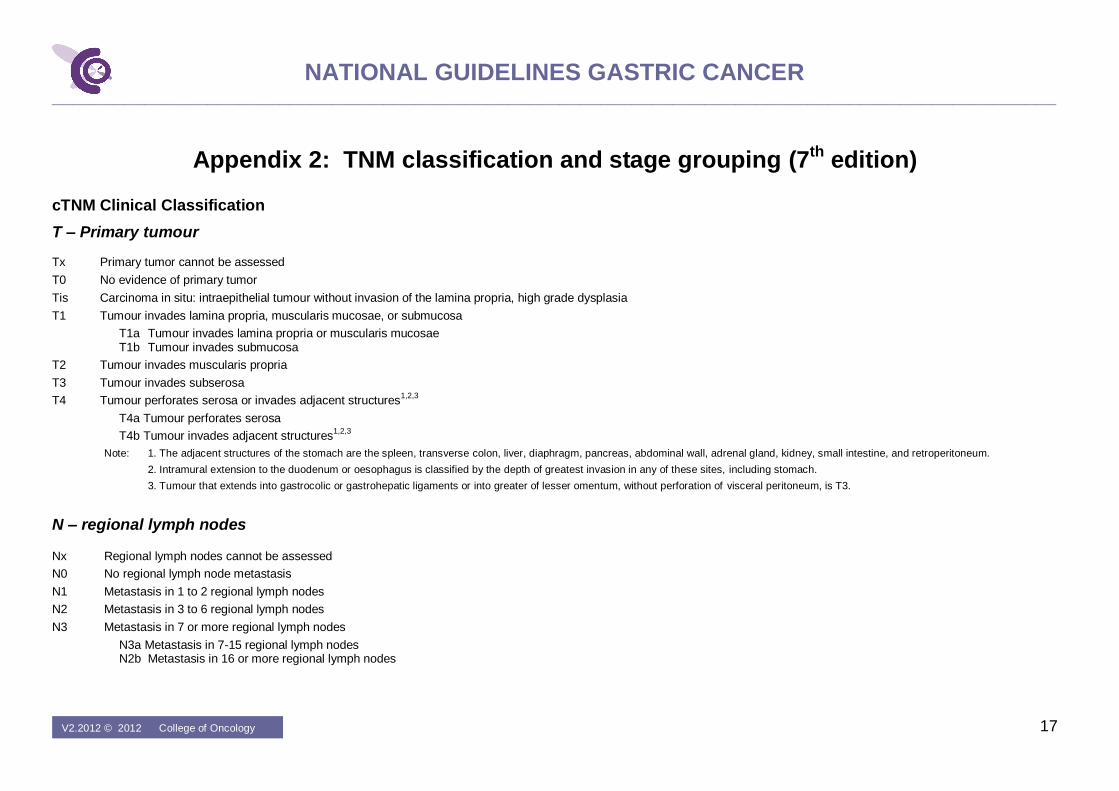
Appendix 2: TNM classification and stage grouping (7th edition) cTNM Clinical Classification
T – Primary tumour
Tx Primary tumor cannot be assessed
T0 No evidence of primary tumor
Tis Carcinoma in situ: intraepithelial tumour without invasion of the lamina propria, high grade dysplasia
T1 Tumour invades lamina propria, muscularis mucosae, or submucosa
T1a Tumour invades lamina propria or muscularis mucosae T1b Tumour invades submucosa
T2 Tumour invades muscularis propria
T3 Tumour invades subserosa
T4 Tumour perforates serosa or invades adjacent structures1,2,3
T4a Tumour perforates serosa
T4b Tumour invades adjacent structures1,2,3
Note: 1. The adjacent structures of the stomach are the spleen, transverse colon, liver, diaphragm, pancreas, abdominal wall, adrenal gland, kidney, small intestine, and retroperitoneum.
2. Intramural extension to the duodenum or oesophagus is classified by the depth of greatest invasion in any of these sites, including stomach.
3. Tumour that extends into gastrocolic or gastrohepatic ligaments or into greater of lesser omentum, without perforation of visceral peritoneum, is T3.
N – regional lymph nodes
Nx Regional lymph nodes cannot be assessed
N0 No regional lymph node metastasis
N1 Metastasis in 1 to 2 regional lymph nodes
N2 Metastasis in 3 to 6 regional lymph nodes
N3 Metastasis in 7 or more regional lymph nodes
N3a Metastasis in 7-15 regional lymph nodes N2b Metastasis in 16 or more regional lymph nodes

NATIONAL GUIDELINES GASTRIC CANCER _________________________________________________________________________________________________
18 V2.2012 © 2012 College of Oncology
M – Distant metastasis
M0 No distant metastasis
M1 Distant metastasis
Note: Distant metastasis includes peritoneal seeding, positive peritoneal cytology, and omental tumour not part of continuous extension.
pTNM Pathological Classification
The pT and pN categories correspond to the T and N categories.
pN0 Histological examination of a regional lymphadenectomy specimen will ordinarily include 16 or more lymph nodes.
If the lymph nodes are negative, but the number ordinarily examined is not met, classify as pN0.
pM – Distant Metastasis
pM1 Distant metastasis microscopically confirmed
Note: pM0 and pMx are not valid categories

NATIONAL GUIDELINES GASTRIC CANCER _________________________________________________________________________________________________
19 V2.2012 © 2012 College of Oncology
Stage grouping
Stage 0 Tis N0 M0
Stage IA T1 N0 M0
Stage IB T2
T1
N0
N1
M0
M0
Stage IIA T3
T2
T1
N0
N1
N2
M0
M0
M0
Stage IIB T4a
T3
T2
T1
N0
N1
N2
N3
M0
M0
M0
M0
Stage IIIA T4a
T3
T2
N1
N2
N3
M0
M0
M0
Stage IIIB T4b
T4a
T3
N0, N1
N2
N3
M0
M0
M0
Stage IIIC T4a
T4b
N3
N2, N3
M0
M0
Stage IV Any T Any N M1