ce-symm jlbr talk
TRANSCRIPT

Spencer Bliven September 30, 2014
jLBR Seminar

1
From Google Earth. Data SIO, NOAA, U.S. Navy, NGA, GEBCO, Image Landsat, Image IBCAO

2
PDP:2AEFAa PDP:3IPRAa
PDP:2FN9Ab
d1iiba_
d2r4qa1
PDP:3L9WAa
d1pdoa_
PDP:3A1DAa
d1djla_d1shux_
d1d4oa_
d1nrjb_
PDP:2IYEAa
PDP:2FN9Aa
PDP:2ZXEAa
PDP:2VOYIa
d1wa5a_
PDP:3QELBa
PDP:3QELBb
PDP:2O1EAb
d1nrza_
PDP:2OSVAa
PDP:2OSVAb
PDP:3HH8Aa
PDP:2O1EAa
PDP:3HH8Ab
PDP:3IPCAb
PDP:2Q8PAa
d1id1a_
d2hmva1
PDP:2WI8Aa
PDP:3QJGAa
PDP:3P2YAb
d1ls1a2
PDP:2FEWBa
PDP:3RBZAb
d1vkra_PDP:2VQ3Aa
PDP:3IPCAa
SCOP:d1su3a2 d1pt2a_ d1c5ka1 d1k3ia3 d1h9ya2
! What’s the nature of protein fold space? ! How do new folds evolve? ! How do gene rearrangements relate to protein
structure? ! What can internal symmetry tell us about protein
function and evolution? ! What algorithms are best for detecting remote
homology?

Hemoglobin [PDB:4HHB]
C2
GTP Cyclohydrolase I
[1A8R] D5
Rhinovirus 2 [3DPR]
Icosahedral
AmtB Ammonia Channel [1U7G]
C3
3

Ferredoxin-like [SCOP:d2j5aa1]
C2
Beta-Propeller [SCOP:d1u6dx_]
C6
Beta-trefoil [3JUT]
C3
TIM barrel [1TIM]
C8
Key: Crystallographic/NCS axis Pseudosymmetry axis
4

! Function ! Allosteric regulation/cooperativity ! Bind ligands symmetrically (e.g.
metals, palindromic DNA, channels)
Monod, J., Wyman, J., & Changeux, J.-P. (1965). J Mol Biol, 12, 88–118.
5
TATA Binding Protein [1TGH]
Hemoglobin [4HHB]

! Function ! Allosteric regulation/cooperativity ! Bind ligands symmetrically (e.g.
metals, palindromic DNA, channels)
! Folding ! Prevent infinite assembly ! Subunits fold quasi-
independently TATA Binding Protein
[1TGH] Monod, J., Wyman, J., & Changeux, J.-P. (1965). J Mol Biol, 12, 88–118. Wolynes, P. G. (1996). PNAS, 93(25), 14249–14255.
6
Hemoglobin [4HHB]

! Evolution ! Identify duplications & fusions ! Many examples of homologous quaternary symmetric/
internally symmetric proteins ! Tradeoff between monomer & oligomer
Lee and Blaber. PNAS (2011) vol. 108 (1) pp. 126-30
7
3OL0 3O49
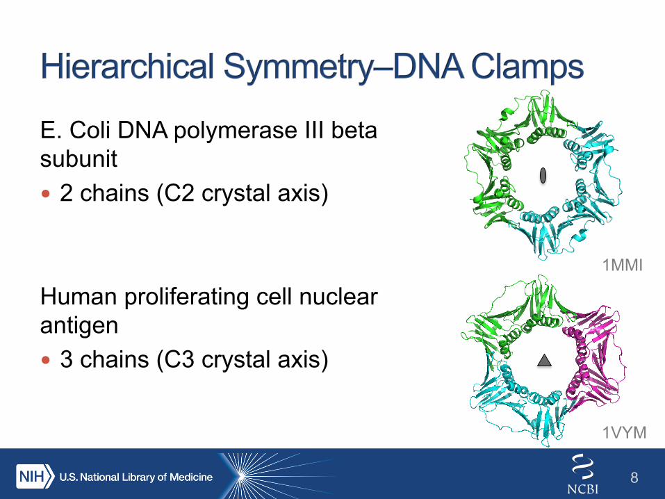
E. Coli DNA polymerase III beta subunit ! 2 chains (C2 crystal axis)
Human proliferating cell nuclear antigen ! 3 chains (C3 crystal axis)
8
1MMI
1VYM

E. Coli DNA polymerase III beta subunit ! 2 chains ! 6 domains (pseudo C6)
Human proliferating cell nuclear antigen ! 3 chains ! 6 domains (pseudo C6)
9
1MMI
1VYM

! 2-3 chains ! 6 domains ! 12 structural repeats (pseudo D6)
Ancient 12-mer?
Ancient 6-mer
Bacterial Dimer Eukaryotic/Archaeal/Viral Trimer
Kelman, Z., & O'Donnell, M. (1995). Nucleic Acids Research, 23(18), 3613–3620. Neuwald, A. F., & Poleksic, A. (2000). Nucleic Acids Research, 28(18), 3570–3580.
10

Glyoxalase I from Clostridium acetobutylicum [3HDP] (Nickel; Dimer)
Glyoxalase I from E. coli [1F9Z] (Nickel; Dimer)
1,2-dihydroxy-naphthalene dioxygenase from Pseudomonas sp. strain C18 [2EHZ] (Iron; Octamer)
11

12
Glyoxalase I from Clostridium acetobutylicum [3HDP] (Nickel; Dimer)
Glyoxalase I from E. coli [1F9Z] (Nickel; Dimer)
1,2-dihydroxy-naphthalene dioxygenase from Pseudomonas sp. strain C18 [2EHZ] (Iron; Octamer)
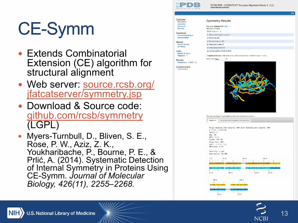
! Extends Combinatorial Extension (CE) algorithm for structural alignment
! Web server: source.rcsb.org/jfatcatserver/symmetry.jsp
! Download & Source code: github.com/rcsb/symmetry (LGPL)
! Myers-Turnbull, D., Bliven, S. E., Rose, P. W., Aziz, Z. K., Youkharibache, P., Bourne, P. E., & Prlić, A. (2014). Systematic Detection of Internal Symmetry in Proteins Using CE-Symm. Journal of Molecular Biology, 426(11), 2255–2268.
13

14
PDB:1MER Chain A

! 1007 structures from SCOP superfamilies
! Manually curated ! Excludes small proteins
(<4 SSEs) ! 24% of superfamilies
have internal symmetry or large structural repeats
Order Superfamilies % Asymmetric 766 76.10%
Rotational
2 166 16.5%
3 10 1.0%
4 2 0.2%
5 3 0.3%
6 9 0.9%
7 9 0.9%
8 21 2.1%
Dihedral
2 2 0.2%
4 1 0.1%
Helical
2 9 0.9%
3 2 0.2%
Non-integral 2 0.2%
Superhelical 2 0.2%
Translational 3 0.3%
15

! AUC = .95 ! 86% True Positive
Rate ! 3.3% False
Positive Rate
SymD: Kim, C., Basner, J., & Lee, B. (2010). BMC Bioinformatics, 11, 303.
16

! All domains from SCOPe 2.03 ! Interactive results:
source.rcsb.org/jfatcatserver/scopResults.jsp ! Underestimate based on conservative thresholds
SCOP Class Superfamilies % Symmetric α 507 18.5% β 354 24.6% α/β 244 16.8% α+β 551 14.3% Membrane 109 23.8% Overall 1831 18.0%
17

18
BtuF
BtuC
BtuD
Vitamin B12 transporter BtuCD–F from E. coli [4FI3]
Periplasmic-binding protein
Transmembrane domain
Nucleotide-binding domain

19
BtuF
BtuC
BtuD
Vitamin B12 transporter BtuCD–F from E. coli [4FI3]

20
BtuF [1N4A]
BtuF
BtuC
BtuD
Vitamin B12 transporter BtuCD–F from E. coli [4FI3]

! PTS sorbitol transporter subunit IIA ! Novel fold ! Solved by the Protein Structure Initiative ! Structural alignment reveals a conserved sequence
motif between halves
2F9H
21

22
! Systematically compare symmetry units across the PDB
! CE-Symm improvements ! Improve order detection
(C8, not C4 or C2)
! Visualization & usability
! Find mixed quaternary & internal symmetry

! Paul Scherrer Institute ! Guido Capitani ! Kumaran Baskaran ! Jose Duarte ! Joseph Somody ! LBR members
! UC San Diego/RCSB ! Douglas Myers-Turnbull ! Andreas Prlić ! Peter Rose ! Zaid Aziz ! RCSB & Bourne Lab members
! NIH ! Philip Bourne ! Philippe Youkharibache ! David Landsman
Resources: ! source.rcsb.org/jfatcatserver/
symmetry.jsp ! github.com/rcsb/symmetry ! www.slideshare.net/sbliven
Funding: NSF, NIH, DOE, Open Science Grid
23


! Racemases and epimerases are enriched
25
0
1
2
3
4
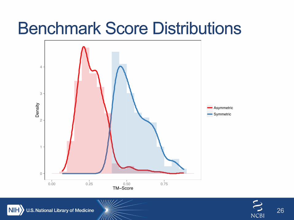
0.00 0.25 0.50 0.75TM−Score
Density
AsymmetricSymmetric
26

Fibroblast Growth Factor [3JUT]
120° 120°
Myers-Turnbull, D., Bliven, S. E., Rose, P. W., Aziz, Z. K., Youkharibache, P., Bourne, P. E., & Prlić, A. (2014). Journal of Molecular Biology, 426(11), 2255–2268.
27

Fibroblast Growth Factor [3JUT]
120° 120°
Myers-Turnbull, D., Bliven, S. E., Rose, P. W., Aziz, Z. K., Youkharibache, P., Bourne, P. E., & Prlić, A. (2014). Journal of Molecular Biology, 426(11), 2255–2268.
28
This work is licensed under a Creative Commons Attribution-ShareAlike 3.0 Unported License.
29