cell-free translation reconstituted with purified …arep.med.harvard.edu/pdf/shimuzu01.pdfcell-free...
TRANSCRIPT
http://biotech.nature.com • AUGUST 2001 • VOLUME 19 • nature biotechnology
Cell-free translation reconstituted with purifiedcomponents
Yoshihiro Shimizu1, Akio Inoue2, Yukihide Tomari1, Tsutomu Suzuki2, Takashi Yokogawa3, Kazuya Nishikawa3, and Takuya Ueda2*
We have developed a protein-synthesizing system reconstituted from recombinant tagged protein factors puri-fied to homogeneity. The system was able to produce protein at a rate of about 160 µg/ml/h in a batch modewithout the need for any supplementary apparatus. The protein products were easily purified within 1 h usingaffinity chromatography to remove the tagged protein factors. Moreover, omission of a release factor allowedefficient incorporation of an unnatural amino acid using suppressor transfer RNA (tRNA).
RESEARCH ARTICLE
The development of cell-free strategies based on purified enzymes isan expanding field in biotechnology. DNA polymerases have made itpossible to amplify DNA rapidly and efficiently by PCR without theneed for cloning. RNA polymerases are used to synthesize novelRNA molecules through in vitro evolution methods. Reverse tran-scriptases generate complementary DNA (cDNA), which does notexist in natural living systems. The availability of these purifiedenzymes essential to the “central-dogma” pathway of molecular biol-ogy has removed many of the constraints in conventional gene-manipulation techniques that rely on living cells. So far, however, thetranslation process has eluded efficient reconstitution in vitro, inlarge part because of its complexity. The achievement of this goalwould offer advantages in many areas of biotechnology.
Two approaches have guided efforts to achieve cell-free transla-tion. One approach, developed over the past decade, is based oncrude cell extract, often derived from Escherichia coli, rabbit reticulo-cytes, or wheat germ1,2. But the use of such extracts inevitablyencounters two problems: a rapid depletion of energy charge, inde-pendent of peptide bond formation3,4, and degradation of proteinproducts or template nucleic acids by proteases or nucleases. Theseproblems have been partly overcome by a continuous-flow cell-freeprotein-synthesis system5,6.
The second approach attempts to reconstitute protein synthesisfrom purified components of the translation machinery. More than100 molecules participate in prokaryotic and eukaryotic translation,many of which have been individually purified for biochemical stud-ies of their functions and structures. In 1977, Weissbach’s groupendeavored to reconstitute a translation system using purified solu-ble components7 but did not achieve a satisfactory production ofproteins, perhaps because of a complex purification protocol and theabsence of release factor 3 (RF3) and ribosome-recycling factor(RRF). A few years later, Ganoza and co-workers reconstructed acell-free translation system from precharged aminoacyl-tRNAs andpurified translation factors8. Although this system did not requireaminoacyl-tRNA synthetases, the tRNAs were not recycled, resultingin low productivity. More recently, Pavlov and co-workers producedan in vitro translation system using a partially purified aminoacyl-tRNA synthetase mixture with purified translation factors9. Theyalso constructed a completely purified in vitro translation system
using short artificial messenger RNA (mRNA)10. However, this sys-tem lacked a full set of aminoacyl-tRNA synthetases and its produc-tivity was relatively low.
Here we describe a cell-free translation system reconstructed frompurified histidine (His)-tagged translation factors and “pro-grammed” by natural mRNA. The system—termed the “protein syn-thesis using recombinant elements” (PURE) system—contains allnecessary translation factors, purified with high specific activity, andallows efficient protein production. We succeeded in producingactive dihydrofolic acid reductase (DHFR) with a productivity onthe order of submilligrams in a 1 ml reaction volume. The PURE sys-tem is also advantageous in terms of product purification. Proteinproducts in the native form (with no His tag) were purified easilyand rapidly through ultrafiltration followed by affinity chromatog-raphy to remove the His-tagged translation factors. Omission ofrelease factor 1 (RF1) allowed the suppression of an amber codonusing chemically synthesized UAG-specific suppressor tRNA with achemically attached valine (Val–tRNAsup), demonstrating that thePURE system can be used for the efficient synthesis of proteins con-taining unnatural amino acids.
ResultsThe PURE system. Protein biosynthesis proceeds in three steps: ini-tiation, elongation, and termination. In E. coli, the translation factorsresponsible for completing these steps are three initiation factors(IF1, IF2, and IF3), three elongation factors (EF-G, EF-Tu, and EF-Ts), and three release factors (RF1, RF2, and RF3), as well as RRF fortermination. However, RF2 is not required for the translation ofgenes terminating with the codons UAG or UAA. The PURE systemincludes 32 components that we purified individually: IF1, IF2, IF3,EF-G, EF-Tu, EF-Ts, RF1, RF3, RRF, 20 aminoacyl-tRNA synthetases(ARSs), methionyl-tRNA transformylase (MTF), T7 RNA poly-merase, and ribosomes. In addition, the system contains 46 tRNAs,NTPs, creatine phosphate, 10-formyl-5,6,7,8-tetrahydrofolic acid, 20amino acids, creatine kinase, myokinase, nucleoside-diphosphatekinase, and pyrophosphatase.
Purification of translation factors. We overexpressed in E. coli andpurified IF1, IF2, IF3, EF-G, EF-Tu, EF-Ts, RF1, RF3, RRF, 20 ARSs,MTF, and T7 RNA polymerase. We reconstructed these genes in the
1Department of Chemistry and Biotechnology, Graduate School of Engineering, and 2Department of Integrated Biosciences, Graduate School of Frontier Sciences, TheUniversity of Tokyo, Bldg FSB401, 5-1-5, Kasiwanoha, Kashiwa, Chiba prefecture 277-8562, Japan. 3Department of Biomolecular Science, Faculty of Engineering,
Gifu University, Yanagido, Gifu 501-1193, Japan. *Corresponding author ([email protected]).
751
©20
01 N
atu
re P
ub
lish
ing
Gro
up
h
ttp
://b
iote
ch.n
atu
re.c
om
© 2001 Nature Publishing Group http://biotech.nature.com
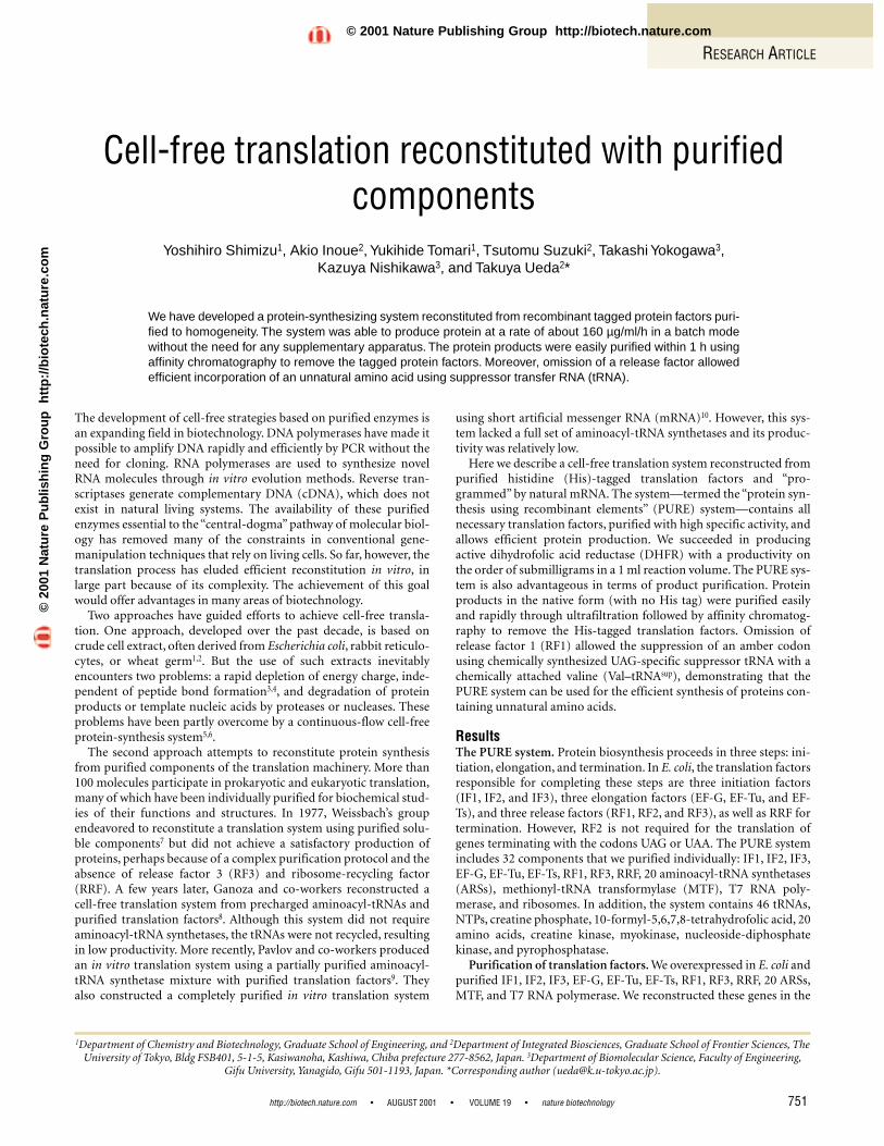
His-tagged fusion form for easy and high-grade protein purificationby a Ni2+ column. An imidazole gradient strategy was successful inachieving effective purification. Figure 1 shows the sodium dodecylsulfate–polyacrylamide gel electrophoresis (SDS–PAGE) gel patternsof the purified translation factors (Fig. 1A) and enzymes (Fig. 1B).The electrophoretic pattern indicated that the proteins were purifiedto homogeneity.
Activities of the purified translation components. We evaluatedthe activities of the purified translation factors and enzymes exceptfor RF1, RF3, and RRF from the reduction in protein synthesiscaused by excluding each component in turn from the PURE system(Fig. 1C). The results indicated that most of the components wereindispensable for translation and simultaneously ruled out the pos-sibility of sample contamination by other E. coli proteins. The resultsalso suggested that IF1 and MTF were least essential for protein syn-thesis. These observations are consistent with previous reportsshowing that deletion of the gene for MTF results in an extremereduction of the viability but not the lethality of E. coli11, and thatformation of the initiation complex can be achieved in the absenceof IF1 (ref. 12). However, it is likely that IF1 and MTF contribute tothe efficiency and accuracy of protein synthesis.
We evaluated the activities of RF1,RF3, and RRF by determining peptiderelease from ribosomes according tothe method of Pavlov et al.10, using ashort synthetic mRNA (mMFL) withthe sequence AUGUUCUUGUAA,corresponding to fMet–Phe–Leu–Stop(fMFL). Complete peptide release wasdependent on the presence of RF1(Table 1). The addition of RRF signifi-cantly increased the amount of peptidereleased and gave multiple-turnoverprotein synthesis. The observation thatthe turnover rate mediated by RF1 andRRF was enhanced by supplementationwith RF3 indicated that RF3 was puri-fied as the active form.
From the results of the two experi-ments, we concluded that all the puri-fied translation components wereactive despite the His tags.
Additonal information about thecloning vectors used for expression ofthe translation factors and enzymes, theyields following purification, the con-centrations and specific activities of the
purified proteins, and the units used per 50 µl reaction is available onthe Web Extras page of Nature Biotechnology Online.
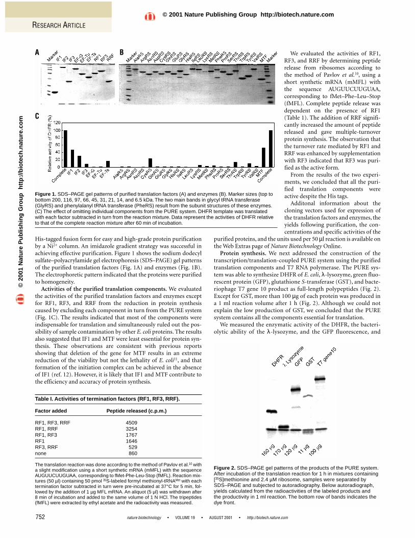
Protein synthesis. We next addressed the construction of thetranscription/translation-coupled PURE system using the purifiedtranslation components and T7 RNA polymerase. The PURE sys-tem was able to synthesize DHFR of E. coli, λ-lysozyme, green fluo-rescent protein (GFP), glutathione S-transferase (GST), and bacte-riophage T7 gene 10 product as full-length polypeptides (Fig. 2).Except for GST, more than 100 µg of each protein was produced ina 1 ml reaction volume after 1 h (Fig. 2). Although we could notexplain the low production of GST, we concluded that the PUREsystem contains all the components essential for translation.
We measured the enzymatic activity of the DHFR, the bacteri-olytic ability of the λ-lysozyme, and the GFP fluorescence, and
RESEARCH ARTICLE
nature biotechnology • VOLUME 19 • AUGUST 2001 • http://biotech.nature.com752
Table I. Activities of termination factors (RF1, RF3, RRF).
Factor added Peptide released (c.p.m.)
RF1, RF3, RRF 4509RF1, RRF 3254RF1, RF3 1767RF1 1646RF3, RRF 529none 860
The translation reaction was done according to the method of Pavlov et al.10 witha slight modification using a short synthetic mRNA (mMFL) with the sequenceAUGUUCUUGUAA, corresponding to fMet-Phe-Leu-Stop (fMFL). Reaction mix-tures (50 µl) containing 50 pmol 35S-labeled formyl methionyl-tRNAMet with eachtermination factor subtracted in turn were pre-incubated at 37°C for 5 min, fol-lowed by the addition of 1 µg MFL mRNA. An aliquot (5 µl) was withdrawn after8 min of incubation and added to the same volume of 1 N HCl. The tripeptides(fMFL) were extracted by ethyl acetate and the radioactivity was measured.
Figure 1. SDS–PAGE gel patterns of purified translation factors (A) and enzymes (B). Marker sizes (top tobottom 200, 116, 97, 66, 45, 31, 21, 14, and 6.5 kDa. The two main bands in glycyl tRNA transferase(GlyRS) and phenylalanyl tRNA transferase (PheRS) result from the subunit structures of these enzymes.(C) The effect of omitting individual components from the PURE system. DHFR template was translatedwith each factor subtracted in turn from the reaction mixture. Data represent the activities of DHFR relativeto that of the complete reaction mixture after 60 min of incubation.
Figure 2. SDS–PAGE gel patterns of the products of the PURE system.After incubation of the translation reaction for 1 h in mixtures containing[35S]methionine and 2.4 µM ribosome, samples were separated bySDS–PAGE and subjected to autoradiography. Below autoradiograph,yields calculated from the radioactivities of the labeled products and the productivity in 1 ml reaction. The bottom row of bands indicates the dye front.
A B
C
©20
01 N
atu
re P
ub
lish
ing
Gro
up
h
ttp
://b
iote
ch.n
atu
re.c
om
© 2001 Nature Publishing Group http://biotech.nature.com
RESEARCH ARTICLE
found that these proteins were synthesized in their active forms.The specific activity of the synthesized DHFR was 16.7 ± 1.1U/mg, which is similar to the specific activity value of DHFR thatwe overexpressed in cells and purified (16.0 ± 0.80 U/mg). Fromthese data we concluded that the components of the PURE systemare sufficient to produce active proteins.
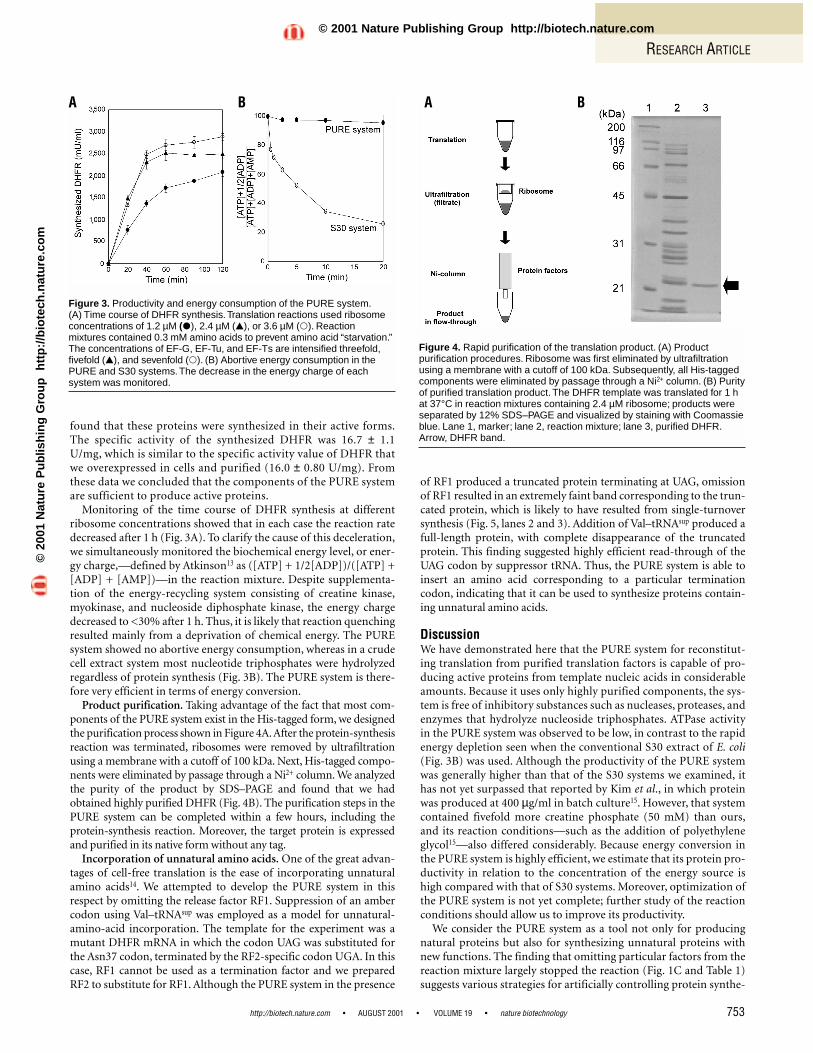
Monitoring of the time course of DHFR synthesis at differentribosome concentrations showed that in each case the reaction ratedecreased after 1 h (Fig. 3A). To clarify the cause of this deceleration,we simultaneously monitored the biochemical energy level, or ener-gy charge,—defined by Atkinson13 as ([ATP] + 1/2[ADP])/([ATP] +[ADP] + [AMP])—in the reaction mixture. Despite supplementa-tion of the energy-recycling system consisting of creatine kinase,myokinase, and nucleoside diphosphate kinase, the energy chargedecreased to <30% after 1 h. Thus, it is likely that reaction quenchingresulted mainly from a deprivation of chemical energy. The PUREsystem showed no abortive energy consumption, whereas in a crudecell extract system most nucleotide triphosphates were hydrolyzedregardless of protein synthesis (Fig. 3B). The PURE system is there-fore very efficient in terms of energy conversion.
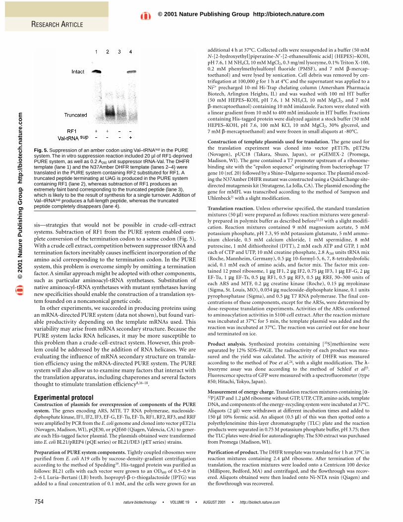
Product purification. Taking advantage of the fact that most com-ponents of the PURE system exist in the His-tagged form, we designedthe purification process shown in Figure 4A.After the protein-synthesisreaction was terminated, ribosomes were removed by ultrafiltrationusing a membrane with a cutoff of 100 kDa. Next, His-tagged compo-nents were eliminated by passage through a Ni2+ column. We analyzedthe purity of the product by SDS–PAGE and found that we hadobtained highly purified DHFR (Fig. 4B). The purification steps in thePURE system can be completed within a few hours, including the protein-synthesis reaction. Moreover, the target protein is expressedand purified in its native form without any tag.
Incorporation of unnatural amino acids. One of the great advan-tages of cell-free translation is the ease of incorporating unnaturalamino acids14. We attempted to develop the PURE system in thisrespect by omitting the release factor RF1. Suppression of an ambercodon using Val–tRNAsup was employed as a model for unnatural-amino-acid incorporation. The template for the experiment was amutant DHFR mRNA in which the codon UAG was substituted forthe Asn37 codon, terminated by the RF2-specific codon UGA. In thiscase, RF1 cannot be used as a termination factor and we preparedRF2 to substitute for RF1. Although the PURE system in the presence
of RF1 produced a truncated protein terminating at UAG, omissionof RF1 resulted in an extremely faint band corresponding to the trun-cated protein, which is likely to have resulted from single-turnoversynthesis (Fig. 5, lanes 2 and 3). Addition of Val–tRNAsup produced afull-length protein, with complete disappearance of the truncatedprotein. This finding suggested highly efficient read-through of theUAG codon by suppressor tRNA. Thus, the PURE system is able toinsert an amino acid corresponding to a particular terminationcodon, indicating that it can be used to synthesize proteins contain-ing unnatural amino acids.
DiscussionWe have demonstrated here that the PURE system for reconstitut-ing translation from purified translation factors is capable of pro-ducing active proteins from template nucleic acids in considerableamounts. Because it uses only highly purified components, the sys-tem is free of inhibitory substances such as nucleases, proteases, andenzymes that hydrolyze nucleoside triphosphates. ATPase activityin the PURE system was observed to be low, in contrast to the rapidenergy depletion seen when the conventional S30 extract of E. coli(Fig. 3B) was used. Although the productivity of the PURE systemwas generally higher than that of the S30 systems we examined, ithas not yet surpassed that reported by Kim et al., in which proteinwas produced at 400 µg/ml in batch culture15. However, that systemcontained fivefold more creatine phosphate (50 mM) than ours,and its reaction conditions—such as the addition of polyethyleneglycol15—also differed considerably. Because energy conversion inthe PURE system is highly efficient, we estimate that its protein pro-ductivity in relation to the concentration of the energy source ishigh compared with that of S30 systems. Moreover, optimization ofthe PURE system is not yet complete; further study of the reactionconditions should allow us to improve its productivity.
We consider the PURE system as a tool not only for producingnatural proteins but also for synthesizing unnatural proteins withnew functions. The finding that omitting particular factors from thereaction mixture largely stopped the reaction (Fig. 1C and Table 1)suggests various strategies for artificially controlling protein synthe-
http://biotech.nature.com • AUGUST 2001 • VOLUME 19 • nature biotechnology 753
Figure 4. Rapid purification of the translation product. (A) Productpurification procedures. Ribosome was first eliminated by ultrafiltrationusing a membrane with a cutoff of 100 kDa. Subsequently, all His-taggedcomponents were eliminated by passage through a Ni2+ column. (B) Purityof purified translation product. The DHFR template was translated for 1 hat 37°C in reaction mixtures containing 2.4 µM ribosome; products wereseparated by 12% SDS–PAGE and visualized by staining with Coomassieblue. Lane 1, marker; lane 2, reaction mixture; lane 3, purified DHFR.Arrow, DHFR band.
Figure 3. Productivity and energy consumption of the PURE system.(A) Time course of DHFR synthesis.Translation reactions used ribosomeconcentrations of 1.2 µM (�), 2.4 µM (�), or 3.6 µM (�). Reactionmixtures contained 0.3 mM amino acids to prevent amino acid “starvation.”The concentrations of EF-G, EF-Tu, and EF-Ts are intensified threefold,fivefold (�), and sevenfold (�). (B) Abortive energy consumption in thePURE and S30 systems.The decrease in the energy charge of eachsystem was monitored.
A B A B
©20
01 N
atu
re P
ub
lish
ing
Gro
up
h
ttp
://b
iote
ch.n
atu
re.c
om
© 2001 Nature Publishing Group http://biotech.nature.com
sis—strategies that would not be possible in crude-cell-extract systems. Subtraction of RF1 from the PURE system enabled com-plete conversion of the termination codon to a sense codon (Fig. 5).With a crude cell extract, competition between suppressor tRNA andtermination factors inevitably causes inefficient incorporation of theamino acid corresponding to the termination codon. In the PUREsystem, this problem is overcome simply by omitting a terminationfactor. A similar approach might be adopted with other components,such as particular aminoacyl-tRNA synthetases. Substitution ofnative aminoacyl-tRNA synthetases with mutant synthetases havingnew specificities should enable the construction of a translation sys-tem founded on a noncanonical genetic code.
In other experiments, we succeeded in producing proteins usingan mRNA-directed PURE system (data not shown), but found vari-able productivity depending on the template mRNAs used. Thisvariability may arise from mRNA secondary structure. Because thePURE system lacks RNA helicases, it may be more susceptible tothis problem than a crude-cell-extract system. However, this prob-lem could be addressed by the addition of RNA helicases. We areevaluating the influence of mRNA secondary structure on transla-tion efficiency using the mRNA-directed PURE system. The PUREsystem will also allow us to examine many factors that interact withthe translation apparatus, including chaperones and several factorsthought to stimulate translation efficiency8,16–18.
Experimental protocolConstruction of plasmids for overexpression of components of the PUREsystem. The genes encoding ARS, MTF, T7 RNA polymerase, nucleoside-diphosphate kinase, IF1, IF2, IF3, EF-G, EF-Tu, EF-Ts, RF1, RF2, RF3, and RRFwere amplified by PCR from the E. coli genome and cloned into vector pET21a(Novagen, Madison, WI), pQE30, or pQE60 (Qiagen, Valencia, CA) to gener-ate each His-tagged factor plasmid. The plasmids obtained were transformedinto E. coli BL21/pREP4 (pQE series) or BL21/DE3 (pET series) strains.
Preparation of PURE system components. Tightly coupled ribosomes werepurified from E. coli A19 cells by sucrose-density-gradient centrifugationaccording to the method of Spedding19. His-tagged protein was purified asfollows: BL21 cells with each vector were grown to an OD600 of 0.5–0.9 in2–6 L Luria–Bertani (LB) broth. Isopropyl-β-D-thiogalactoside (IPTG) wasadded to a final concentration of 0.1 mM, and the cells were grown for an
additional 4 h at 37°C. Collected cells were resuspended in a buffer (50 mMN-[2-hydroxyethyl]piperazine-N’-[2-ethanesulfonic acid] (HEPES)–KOH,pH 7.6, 1 M NH4Cl, 10 mM MgCl2, 0.3 mg/ml lysozyme, 0.1% Triton X-100,0.2 mM phenylmethylsulfonyl fluoride (PMSF), and 7 mM β-mercap-toethanol) and were lysed by sonication. Cell debris was removed by cen-trifugation at 100,000 g for 1 h at 4°C and the supernatant was applied to aNi2+ precharged 10-ml Hi-Trap chelating column (Amersham PharmaciaBiotech, Arlington Heights, IL) and was washed with 100 ml HT buffer (50 mM HEPES–KOH, pH 7.6, 1 M NH4Cl, 10 mM MgCl2, and 7 mM β-mercaptoethanol) containing 10 mM imidazole. Factors were eluted witha linear gradient from 10 mM to 400 mM imidazole in HT buffer. Fractionscontaining His-tagged protein were dialyzed against a stock buffer (50 mMHEPES–KOH, pH 7.6, 100 mM KCl, 10 mM MgCl2, 30% glycerol, and 7 mM β-mercaptoethanol) and were frozen in small aliquots at -80°C.
Construction of template plasmids used for translation. The gene used forthe translation experiment was cloned into vector pET17b, pET29a(Novagen), pUC18 (Takara, Shuzo, Japan), or pGEMEX-2 (Promega,Madison, WI). The gene contained a T7 promoter upstream of a ribosome-binding site with the “epsilon sequence” originating from bacteriophage T7gene 10 (ref. 20) followed by a Shine–Dalgarno sequence. The plasmid encod-ing the N37Amber DHFR mutant was constructed using a QuickChange site-directed mutagenesis kit (Stratagene, La Jolla, CA). The plasmid encoding thegene for mMFL was transcribed according to the method of Sampson andUhlenbeck21 with a slight modification.
Translation reaction. Unless otherwise specified, the standard translationmixtures (50 µl) were prepared as follows: reaction mixtures were general-ly prepared in polymix buffer as described before22,23 with a slight modifi-cation. Reaction mixtures contained 9 mM magnesium acetate, 5 mMpotassium phosphate, pH 7.3, 95 mM potassium glutamate, 5 mM ammo-nium chloride, 0.5 mM calcium chloride, 1 mM spermidine, 8 mMputrescine, 1 mM dithiothreitol (DTT), 2 mM each ATP and GTP, 1 mMeach of CTP and UTP, 10 mM creatine phosphate, 2.8 A260 units tRNA mix(Roche, Mannheim, Germany), 0.5 µg 10-formyl-5, 6, 7, 8-tetrahydrofolicacid, 0.1 mM each of amino acids, and factor mix. The factor mix con-tained 12 pmol ribosome, 1 µg IF1, 2 µg IF2, 0.75 µg IF3, 1 µg EF-G, 2 µgEF-Tu, 1 µg EF-Ts, 0.5 µg RF1, 0.5 µg RF3, 0.5 µg RRF, 30–300 units ofeach ARS and MTF, 0.2 µg creatine kinase (Roche), 0.15 µg myokinase(Sigma, St. Louis, MO), 0.054 µg nucleoside-diphosphate kinase, 0.1 unitspyrophosphatase (Sigma), and 0.5 µg T7 RNA polymerase. The final con-centrations of these components, except for the ARSs, were determined bydose-response translation experiments. Activities of the ARSs conformedto aminoacylation activities in S100 cell extract. After the reaction mixturewas incubated at 37°C for 5 min, the template plasmid was added and thereaction was incubated at 37°C. The reaction was carried out for one hourand terminated on ice.
Product analysis. Synthesized proteins containing [35S]methionine wereseparated by 12% SDS–PAGE. The radioactivity of each product was mea-sured and the yield was calculated. The activity of DHFR was measuredaccording to the method of Poe et al.24, with a slight modification. The λ-lysozyme assay was done according to the method of Schleif et al25.Fluorescence spectra of GFP were measured with a spectrofluorometer (type850; Hitachi, Tokyo, Japan).
Measurement of energy charge. Translation reaction mixtures containing [α-32P]ATP and 1.2 µM ribosome without GTP, UTP, CTP, amino acids, templateDNA, and components of the energy-recycling system were incubated at 37°C.Aliquots (2 µl) were withdrawn at different incubation times and added to 150 µl 10% formic acid. An aliquot (0.5 µl) of this was then spotted onto apolyethyleneimine thin-layer chromatography (TLC) plate and the reactionproducts were separated in 0.75 M potassium phosphate buffer, pH 3.75; thenthe TLC plates were dried for autoradiography. The S30 extract was purchasedfrom Promega (Madison, WI).
Purification of product. The DHFR template was translated for 1 h at 37°C inreaction mixtures containing 2.4 µM ribosome. After termination of thetranslation, the reaction mixtures were loaded onto a Centricon 100 device(Millipore, Bedford, MA) and centrifuged, and the flowthrough was recov-ered. Aliquots obtained were then loaded onto Ni-NTA resin (Qiagen) andthe flowthrough was recovered.
RESEARCH ARTICLE
nature biotechnology • VOLUME 19 • AUGUST 2001 • http://biotech.nature.com754
Fig. 5. Suppression of an amber codon using Val–tRNAsup in the PUREsystem. The in vitro suppression reaction included 20 µl of RF1-deprivedPURE system, as well as 0.2 A260 unit suppressor tRNA-Val. The DHFRtemplate (lane 1) and the N37Amber DHFR template (lanes 2–4) weretranslated in the PURE system containing RF2 substituted for RF1. Atruncated peptide terminating at UAG is produced in the PURE systemcontaining RF1 (lane 2), whereas subtraction of RF1 produces anextremely faint band corresponding to the truncated peptide (lane 3),which is likely to be the result of synthesis for a single turnover. Addition ofVal–tRNAsup produces a full-length peptide, whereas the truncatedpeptide completely disappears (lane 4).
©20
01 N
atu
re P
ub
lish
ing
Gro
up
h
ttp
://b
iote
ch.n
atu
re.c
om
© 2001 Nature Publishing Group http://biotech.nature.com
RESEARCH ARTICLE
http://biotech.nature.com • AUGUST 2001 • VOLUME 19 • nature biotechnology 755
1. Stiege, W. & Erdmann, V.A. The potentials of the in vitro protein biosynthesis sys-tem. J. Biotechnol. 41, 81–90 (1995).
2. Jermutus, L., Ryabova, L.A. & Pluckthun, A. Recent advances in producing andselecting functional proteins by using cell-free translation. Curr. Opin. Biotechnol.9, 534–548 (1998).
3. Kitaoka, Y., Nishimura, N. & Niwano, M. Cooperativity of stabilized mRNA andenhanced translation activity in the cell-free system. J. Biotechnol. 48, 1–8 (1996).
4. Matveev, S.V. et al. Effect of the ATP level on the overall protein biosynthesis ratein a wheat germ cell-free system. Biochim. Biophys. Acta 1293, 207–212 (1996).
5. Spirin, A.S., Baranov, V.I., Ryabova, L.A., Ovodov, S.Y. & Alakhov, Y.B. A continu-ous cell-free translation system capable of producing polypeptides in high yield.Science 242, 1162–1164 (1988).
6. Kim, D.M. & Choi, C.Y. A semicontinuous prokaryotic coupled transcription/trans-lation system using a dialysis membrane. Biotechnol. Prog. 12, 645–649 (1996).
7. Kung, H.F. et al. DNA-directed in vitro synthesis of beta-galactosidase. Studieswith purified factors. J. Biol. Chem. 252, 6889–6894 (1977).
8. Ganoza, M.C., Cunningham, C. & Green, R.M. Isolation and point of action of afactor from Escherichia coli required to reconstruct translation. Proc. Natl. Acad.Sci. USA 82, 1648–1652 (1985).
9. Pavlov, M.Y. & Ehrenberg, M. Rate of translation of natural mRNAs in an optimizedin vitro system. Arch. Biochem. Biophys. 328, 9–16 (1996).
10. Pavlov, M.Y., Freistroffer, D.V., Heurgue-Hamard, V., Buckingham, R.H. &Ehrenberg, M. Release factor RF3 abolishes competition between release factorRF1 and ribosome recycling factor (RRF) for a ribosome binding site. J. Mol. Biol.273, 389–401 (1997).
11. Guillon, J.M., Mechulam, Y., Schmitter, J.M., Blanquet, S. & Fayat, G. Disruption ofthe gene for Met-tRNA(fMet) formyltransferase severely impairs growth ofEscherichia coli. J. Bacteriol. 174, 4294–4301 (1992).
12. Benne, R., Arentzen, R. & Voorma, H.O.The mechanism of action of inititation fac-tor F1 from Escherichia coli. Biochim. Biophys. Acta 269, 304–310 (1972).
13. Atkinson, D.E. The energy charge of the adenylate pool as a regulatory parame-ter. Interaction with feedback modifiers. Biochemistry 7, 4030–4034 (1968).
14. Noren, C.J., Anthony-Cahill, S.J., Griffith, M.C. & Schultz, P.G. A general methodfor site-specific incorporation of unnatural amino acids into proteins. Science 244,
182–188 (1989).15. Kim, D.M., Kigawa, T., Choi, C.Y. & Yokoyama, S. A highly efficient cell-free protein
synthesis system from Escherichia coli. Eur. J. Biochem. 239, 881–886 (1996).16. Glick, B.R. & Ganoza, M.C. Identification of a soluble protein that stimulates pep-
tide bond synthesis. Proc. Natl. Acad. Sci. USA 72, 4257–4260 (1975).17. Kossel, H. Purification and properties of peptidyl-tRNA hydrolase from
Escherichia coli. Biochim. Biophys. Acta 204, 191–202 (1970).18. Lu, J., Aoki, H. & Ganoza, M.C. Molecular characterization of a prokaryotic trans-
lation factor homologous to the eukaryotic initiation factor eIF4A. Int. J. Biochem.Cell. Biol. 31, 215–229 (1999).
19. Spedding, G. Isolation and analysis of ribosomes from prokaryotes, eukaryotes,and organelles. In Ribosomes and protein synthesis: a practical approach. (ed.Spedding, G.) 1–29 (IRL Press at Oxford University Press, New York, NY;1990).
20. Olins, P.O. & Rangwala, S.H. A novel sequence element derived from bacterio-phage T7 mRNA acts as an enhancer of translation of the lacZ gene inEscherichia coli. J. Biol. Chem. 264, 16973–16976 (1989).
21. Sampson, J.R. & Uhlenbeck, O.C. Biochemical and physical characterization of anunmodified yeast phenylalanine transfer RNA transcribed in vitro. Proc. Natl.Acad. Sci. USA 85, 1033–1037 (1988).
22. Jelenc, P.C. & Kurland, C.G. Nucleoside triphosphate regeneration decreases thefrequency of translation errors. Proc. Natl. Acad. Sci. USA 76, 3174–3178 (1979).
23. Wagner, E.G., Jelenc, P.C., Ehrenberg, M. & Kurland, C.G. Rate of elongation ofpolyphenylalanine in vitro. Eur. J. Biochem. 122, 193–197 (1982).
24. Poe, M., Greenfield, N.J., Hirshfield, J.M., Williams, M.N. & Hoogsteen, K.Dihydrofolate reductase. Purification and characterization of the enzyme from anamethopterin-resistant mutant of Escherichia coli. Biochemistry 11, 1023–1030(1972).
25. Schleif, R.F. L-arabinose operon messenger of Escherichia coli. Its inducibility andtranslation efficiency relative to lactose operon messenger. J. Mol. Biol. 61,275–279 (1971).
26. Short, G.F., Golovine, S.Y. & Hecht, S.M. Effect of release factor 1 on in vitro pro-tein translation and the elaboration of protein containing unnatural amino acids.Biochemistry 38, 8808–8819 (1999).
Preparation of valyl-tRNAsup. Suppressor tRNA lacking the 3′-terminal –CAsequence was transcribed using T7 RNA polymerase21 from the plasmidencoding the yeast suppressor tRNAPheCUA. The products were purified bydenaturing 12% PAGE, chemically misacylated with N-(4-pentenoyl)valyl-pdCpA, and “de-protected” according to the method of Short et al26.
Note: Supplementary information can be found on the Nature Biotechnologywebsite in Web Extras (http://biotech.nature.com/web_extras)
AcknowledgmentsWe thank S. Yokoyama, The University of Tokyo, for providing the expressionplasmid encoding the ArgRS gene. This work was supported by a grant (JSPS-RFTF 96100306) from The Japan Society for the Promotion of Science (toT.U.).
Received 29 January 2001; accepted 23 May 2001
©20
01 N
atu
re P
ub
lish
ing
Gro
up
h
ttp
://b
iote
ch.n
atu
re.c
om
© 2001 Nature Publishing Group http://biotech.nature.com