cell-induced response by tetracyclines on human bone ... · cell-induced response by tetracyclines...
TRANSCRIPT

Available online at www.sciencedirect.com
Acta Biomaterialia 4 (2008) 630–637
www.elsevier.com/locate/actabiomat
Cell-induced response by tetracyclines on human bonemarrow colonized hydroxyapatite and Bonelike�
P.S. Gomes a, J.D. Santos b,c, M.H. Fernandes a,*
a Laboratorio de Farmacologia e Biocompatibilidade Celular, Faculdade de Medicina Dentaria, Universidade do Porto (FMDUP),
Rua Dr. Manuel Pereira da Silva, s/n 4200-392 Porto, Portugalb Faculdade de Engenharia, Universidade do Porto (FEUP), Secc�ao de Materiais, DEMM, Rua Dr. Roberto Frias, 4200-465 Porto, Portugal
c Instituto de Engenharia Biomedica (INEB), Laboratorio de Biomateriais, Rua do Campo Alegre, 823, 4150-180 Porto, Portugal
Received 10 August 2007; received in revised form 21 November 2007; accepted 18 December 2007Available online 8 January 2008
Abstract
Semi-synthetic tetracyclines are commonly used antibiotics that also seem to play an important role in the modulation of theimmuno-inflammatory imbalance, verified in several bone diseases. The association of a therapeutic agent (that prevents bacterialinfection and induces tissue formation) to a biomaterial aiming to repair/regenerate bone defects could contribute to a more predict-able clinical outcome. The present study intends to evaluate the proliferation and functional activity of osteoblast-induced humanbone marrow cells, cultured on the surface of hydroxyapatite (HA) and Bonelike�, in the presence of therapeutic concentrationsof doxycycline and minocycline. First passage bone marrow cells were cultured for 35 days on the surface of HA and Bonelike� discs,in the absence or presence of 1 lg ml�1 doxycycline and minocycline. Cultures performed in standard tissue culture plates were used ascontrol.
Doxycycline or minocycline induced cell proliferation and increased the extent of matrix mineralization in osteoblastic cell culturesestablished in the three substrates. Also, an improved biological behavior was verified in seeded Bonelike� compared with HA. Theresults suggest that the local delivery of tetracyclines might associate the antimicrobial activity in implant-related bone infection withan eventual induction of osteoblastic proliferation and maintenance of the characteristic biological activity of these cells.� 2008 Acta Materialia Inc. Published by Elsevier Ltd. All rights reserved.
Keywords: Tetracyclines; Osteoblasts; Hydroxyapatite; Bonelike
1. Introduction
Tetracyclines are commonly used bacteriostatic antibi-otics active against a wide range of both aerobic and anaer-obic Gram-positive bacteria. Their antimicrobial activity isdue to the inhibition of bacterial protein synthesis, by bind-ing to the 30S ribosome subunit, blocking the bond to thetRNA, on the mRNA–ribosome complex [1].
In the last years, several observations have testified thetherapeutic effectiveness of tetracycline (as well as itssemi-synthetic derivatives, minocycline and doxycycline)in the modulation of the immuno-inflammatory imbalance
1742-7061/$ - see front matter � 2008 Acta Materialia Inc. Published by Else
doi:10.1016/j.actbio.2007.12.006
* Corresponding author. Tel.: +351 226074900.E-mail address: [email protected] (M.H. Fernandes).
verified in several animal and human bone diseases [2–4].Different mechanisms, distinct from the antimicrobialaction, have been proposed to justify the pro-anabolicactivity of these pharmacological agents regarding bonemetabolism. These include enhancing bone formation,decreasing connective tissue breakdown and diminishingbone resorption [5–10]. Clinical application of these agentstargeting bone might also be helped by their cation quela-tion activity and consequent avidity for mineralized tissue[11].
Ceramic-based biomaterials are currently used in bonetissue repair strategies because of their adequate mechani-cal properties and chemical composition similar to thoseof the bone tissue. One of the biomaterials, hydroxyapatite(HA), has generated a great deal of interest in the last years
vier Ltd. All rights reserved.
P.S. Gomes et al. / Acta Biomaterialia 4 (2008) 630–637 631
[12]. This synthetic bone graft substitute, although lackingosteogenic properties that can only be found in autologousgrafts, still offers advantages that include a reduced localtissue morbidity, the absence of complications at the donorsite and unlimited material availability [13]. This biomate-rial, being similar to the mineral component of naturalbone, revealed good osteoconductivity and bone bondingability [14]. However, HA presents low load-bearing capac-ity [15], and the introduction of phosphate-based glasses asa sintering aid is known to reinforce HA mechanically [16].Glass-reinforced HA with bioactive properties has beenapplied with success in medical and dental clinical applica-tions aiming to regenerate the bone tissue [17,18]. Bone-like� is a registered and patented glass-reinforced HAwith improved mechanical properties and enhanced bioac-tivity that result from the addition of CaO–P2O5-basedglasses during the liquid phase sintering process of HA,leading to several ionic substitutions in the lattice thatare responsible for the reduction of porosity and grain size[19,20]. Recently, it has been successfully applied in regen-erative procedures aiming to restore bone structure andfunction in oral, maxillofacial and orthopedic procedures[21,22].
Despite the wide application of synthetic biomaterialsin the repair/regeneration of bone tissue, several clinicalcomplications have been established and have proved dif-ficult to remedy. Among them are osteomyelitis, septicarthritis and prosthetic joint infection, which are causedspecially by Gram-positive organisms and are known tocontribute to a heavy clinical and economic burden[23]. Treatment is often complicated at sites of reducedvascularization, requiring prolonged antimicrobial use,usually associated with surgical drainage or debridement[24]. In this way, the selection of the most effective ther-apeutic approach, based on several parameters thatinclude the specificity of the pathogenic agents, their sen-sitivity profile, pharmacokinetics of the drug, local vascu-lar supply, and the presence or absence of a biomaterialand individual factors is essential in order to minimizetissue and function loss, as well as to reduce discomfortand need of further medical/surgical intervention [24].Also, it is known that local and systemic measures tocontrol the colonization of the surgical wound at theearly healing phase, associated with reduction of theinfection’s spreading, may increase the predictability ofthe results [25]. Tetracyclines have been proved to beeffective in several bone- and joint-related infections[26–28].
Regarding bone regeneration strategies, the associationof a biomaterial and a therapeutic agent that might inducebone formation while preventing bacterial infection could,undoubtedly, contribute to a more predictable clinicaloutcome. In this way, the objective of this research wasto evaluate the proliferation and functional activity ofosteoblast-induced human bone marrow cells, cultured onthe surface of HA and Bonelike�, in the presence oftherapeutic concentrations of doxycycline and minocycline.
2. Materials and methods
2.1. Preparation of samples
Bonelike� was prepared with the chemical composi-tion of 65P2O5–15CaO–10CaF2–10Na2O in mol.% fromreagent grade chemicals using conventional glass-makingtechniques. The composite was obtained by adding themilled glass to HA powder to 2.5 wt.%, using isopropa-nol as a solvent. The powders were then dried and sievedto less then 75 lm under a nitrogen atmosphere. Discsamples were therefore prepared for in vitro testing byuniaxial pressing at 200 MPa using steel dies to obtain12 mm diameter discs. The discs were then sintered at1300 �C (using a ramp rate of 4 �C min�1), with the tem-perature maintained for 1 h, followed by natural coolinginside the furnace. Phase identification and quantificationwas assessed by X-ray diffraction and Rietveld analysis.X-ray diffraction (XRD) was performed on Bonelike�
samples using a Siemens D5000 diffractometer with CuKa radiation (k = 1.5418 A). The scans were made inthe range of 25–35� (2), with a step size of 0.02� and acount time of 2 s step�1.
Detailed description of Bonelike� preparation has beenpreviously reported [20].
For in vitro testing, discs were mechanically polished tothe same final topology of 1 lm using silicon carbidepaper, ultrasonically degreased, cleaned with ethanol fol-lowed by deionised water and finally sterilized, prior tocell culture.
HA samples were also prepared as 12 mm diameter discsin order to compare their biological behavior with that ofBonelike�.
2.2. Cell culture
Human bone marrow was obtained from orthopedicsurgical procedures conducted in adult patients (agedbetween 25 and 45 years). Informed consent was obtainedfor the use of this biological material, which would other-wise have been discarded. Bone marrow was cultured ina-MEM culture medium containing 10% fetal bovineserum, 50 lg ml�1 gentamicin, 2.5 lg ml�1 fungizone and50 lg ml�1 ascorbic acid. Primary cultures were main-tained in a humidified atmosphere (5% CO2 in air at37 �C) for 10–15 days until sub-confluence was reached.At this stage, cells were released enzymatically (0.05%trypsin and 0.025% collagenase) and the resultant suspen-sion was cultured at a density of 104 cell cm�2, in the cul-ture medium described previously, further supplementedwith 10 mM b-glycerophosphate and 10 nM dexametha-sone. Cell cultures were established for 35 days and main-tained on the surface of the culture plate (controlcultures), HA or Bonelike� in the absence or presenceof doxycycline or minocycline (1 lg ml�1). Tetracyclineswere both renewed at every medium change, whichoccurred twice a week.

Fig. 1. XRD pattern of Bonelike�. Bonelike� is composed of HA and a-and b-TCP phases.
632 P.S. Gomes et al. / Acta Biomaterialia 4 (2008) 630–637
2.3. Culture characterization
2.3.1. Cell viability/proliferation and total protein content
MTT assay was used to estimate cell viability/prolifera-tion. This assay is based on the reduction of 3-(4,5-dimeth-ylthiazol-2-yl)-2,5-diphenyltetrazolium bromide to a purpleformazan product by viable cells. Cultured cells were incu-bated with 0.5 mg ml�1 of MTT during the last 4 h of eachtest. The medium was then decanted, the stained productdissolved with dimethylsulfoxide and absorbance determi-nation was conducted at 600 nm in an ELISA reader.Results were expressed as absorbance per square centime-ter (A cm�2).
Total protein content was determined by Lowry methodafter treatment of the cell layer with 0.1 M NaOH for 1 h.Bovine serum albumin was used as a standard and absor-bance determination was conducted at 750 nm.
2.3.2. Alkaline phosphatase (ALP) activity
Activity of ALP was determined in cell lysates (obtainedafter treatment of cultured cells with 0.1% triton) by thehydrolysis of p-nitrophenyl phosphate (30 min at 37 �C) inan alkaline buffer solution (pH 10.3). Colorimetric determi-nation of p-nitrophenol was established at 405 nm. Enzymeactivity was normalized by total protein content and resultswere expressed as nanomoles of p-nitrophenol produced perminute per milligram of protein (nmol min�1 lg�1 protein).
2.3.3. Ionized calcium (Cai) loss from the culture medium
Culture medium from cultures in control and experi-mental conditions was collected every 2–3 days (and cul-tures reefed with fresh medium) between days 10 and 35of the culture. Analysis of Cai content was conducted usingSigma Diagnostics Kit procedure number 587. Resultswere expressed as millimoles per litre of ionized calciumloss from medium (Cai mmol l�1).
2.3.4. Scanning electron microscopy (SEM)Glutaraldehyde (3%) fixed cultures were dehydrated in
graded alcohols (70, 80, 2 � 90 and 99.8%), critical-pointdried, sputter-coated with gold and analyzed in a JeoLJSM 6301F scanning electron microscope equipped withan X-ray energy dispersive spectroscopy (EDX) microanal-ysis capability (Voyager XRMA System, NoranInstruments).
2.4. Statistical analyses
Results presented in this study are from three separateexperiments using cell cultures from different patients.There were three replicates for each experimental situation.Groups of data were evaluated using a two-way analysis ofvariance (ANOVA) and no significant differences in thepattern of the cell behaviour were found. Statistical differ-ences found between control and experimental conditionswere determined by Bonferroni’s method. Values ofp 6 0.05 were considered significant.
3. Results
Human bone marrow stromal cells were characterizedfor proliferation and differentiation events on the surfaceof the culture plate, HA and Bonelike�, in the presenceof an osteogenic inducing medium, further supplementedwith 1 lg ml�1 doxycycline or minocycline.
XRD analysis revealed that, due to the reaction betweenthe HA matrix and P2O5-based glass during the sinteringprocess, the Bonelike� microstructure had a main crystal-line phase of HA with b- and a-tricalcium phosphate as sec-ondary phases (Fig. 1).
3.1. Cell viability/proliferation
The results regarding the evaluation of cell proliferationby the MTT assay are presented in Fig. 2A.
Cultures grown on the surface of the culture plate pro-liferated gradually until day 21, followed by a decrease inthe remaining time of culture. Seeded Bonelike� presenteda similar behavior to the control, whereas cultures estab-lished on the surface of HA showed an initial lag phaseof approximately 2 weeks, with maximal MTT reductionvalues only being achieved by day 28. The presence ofdoxycycline or minocycline (1 lg ml–1) increased cell prolif-eration in control cultures and on seeded sample materials.Results were statistically significant (p 6 0.05) at days 21,28 and 35 for cultures grown on the culture plate, and atmaximal MTT reduction values for cultures establishedon the surface of the materials – day 21 for Bonelike�
and day 28 for HA. The stimulatory effect was evident afteran initial lag stage (during approximately the first week),and maximal MTT values were around 30 and 40% higherin the presence of minocycline and doxycycline, respec-tively, compared with those obtained on non-treatedcultures.
3.2. Alkaline phosphatase activity
Results regarding the activity of alkaline phosphataseare presented in Fig. 2B.
ALP activity was low during the first week of culturebefore increasing significantly, achieving maximal values

Fig. 2. Cell viability/proliferation (A) and alkaline phosphatase activity (B) of human bone marrow osteoblastic cells cultured for 35 days on the surfaceof culture plate, Bonelike� and HA in the absence (�) and presence of 1 lg ml–1 doxycycline (j) or minocycline (N). *Significantly different from controlcultures (p 6 0.05).
P.S. Gomes et al. / Acta Biomaterialia 4 (2008) 630–637 633
by day 14 in control and cultures established on the surfaceof Bonelike�, and later, by day 21, on seeded HA. Subse-quently, ALP activity decreased throughout the remainingculture time. Compared with control cultures, reducedenzymatic activity was verified in seeded Bonelike� andHA.
No significant differences were found in ALP activity inthe presence of doxycycline, although, in minocycline-trea-ted cultures, a tendency for a reduction in the enzyme activ-ity was verified, which was statistically significant (p6 0.05) in the cultures grown on the surface of the cultureplate.
3.3. Mineralized deposits in the extracellular matrix
The presence of mineralized deposits (calcium phos-phate) in the cell layer was evaluated by the analysis ofCai loss from the culture medium throughout days 10–35and by SEM observation.
Fig. 3. Levels of ionized calcium (Cai) loss from the culture medium regardingthe culture plate, Bonelike� and HA. Cai loss was not cumulative, as the medmeasured reflect changes occurring in intervals of 2–3 days throughout the1 lg ml�1 doxycycline (j) or minocycline (N). *Significantly different from co
3.3.1. Cai loss from the culture medium
The Cai (in association with Pi) is consumed in the for-mation of calcium phosphate deposits in the extracellularmatrix, reflecting the mineralization process. In the presentwork, the Cai consumption reflects the changes occurringbetween every medium change (intervals of 2–3 days), sincethe medium was totally replaced at every change, so thatthe values were not cumulative. The results are presentedon Fig. 3.
Levels of Cai were determined in the absence of culturedcells and a relatively steady value of 1.7 mmol l�1 was ver-ified in the absence of materials. A higher variation of Cailevels (between 1.6 and 1.8 mmol ml�1) occurred in incu-bated Bonelike� and HA samples.
Cultures grown on the surface of the culture platerevealed almost no Cai loss from the culture medium,between days 10 and 17. From this day onwards, a signif-icant and progressive increase of the Cai loss was verifiedand, at day 30, values around 1.35 mmol l�1 were attained.
human bone marrow osteoblastic cell cultures performed on the surface ofium was totally replaced at each medium change (twice a week), so levelsculture period. Cultures were grown in the absence (�) and presence ofntrol cultures (p 6 0.05).
634 P.S. Gomes et al. / Acta Biomaterialia 4 (2008) 630–637
Medium collected from cultures established on the surfaceof Bonelike� revealed a similar behavior. Cai loss regard-ing cultures grown on the surface of HA was significantfrom day 20 onwards, achieving approximately1.15 mmol l�1 around day 30.
In the presence of doxycycline and minocycline, levels ofCai followed a similar pattern. However, in cultures estab-lished on the surface of culture plate and Bonelike�, theinitial rate of calcium deposition (between days 17 and23) was higher compared with non-treated cultures, espe-cially in the presence of doxycycline (results statistically sig-nificant, p 6 0.05).
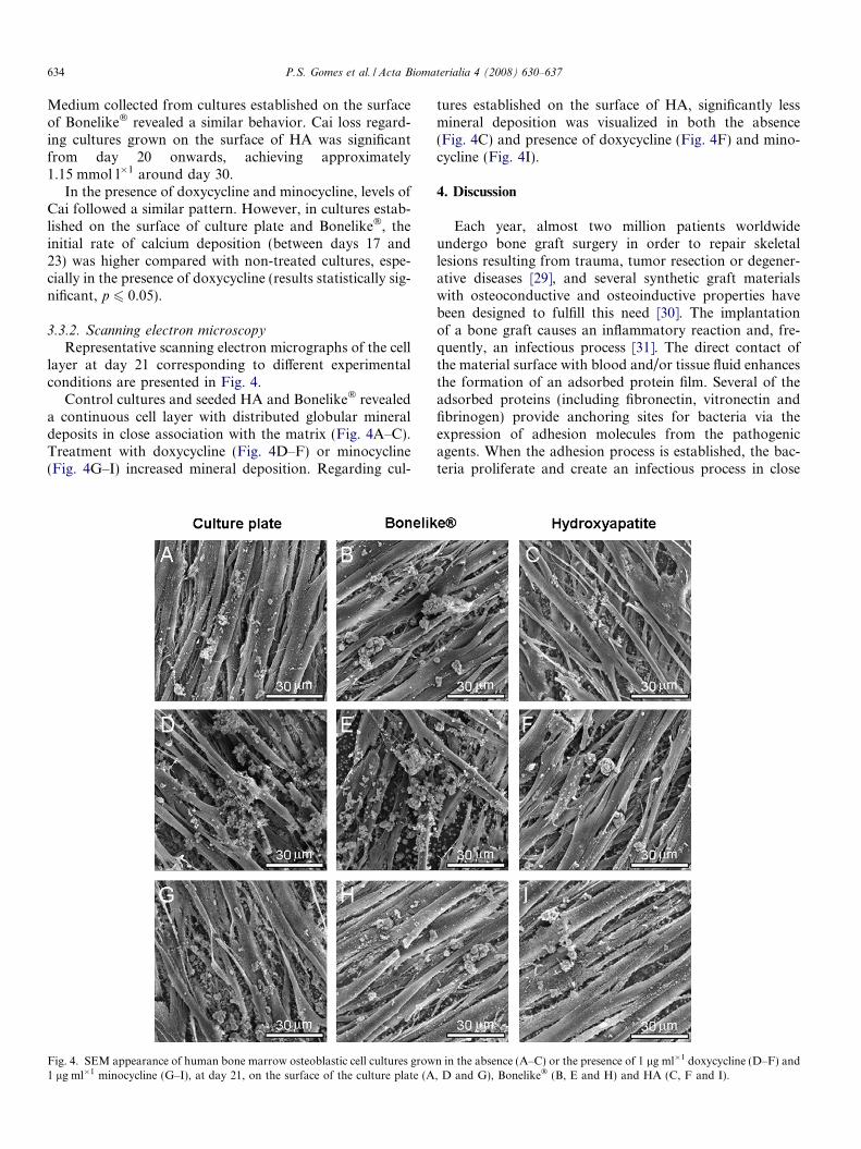
3.3.2. Scanning electron microscopyRepresentative scanning electron micrographs of the cell
layer at day 21 corresponding to different experimentalconditions are presented in Fig. 4.
Control cultures and seeded HA and Bonelike� revealeda continuous cell layer with distributed globular mineraldeposits in close association with the matrix (Fig. 4A–C).Treatment with doxycycline (Fig. 4D–F) or minocycline(Fig. 4G–I) increased mineral deposition. Regarding cul-
Fig. 4. SEM appearance of human bone marrow osteoblastic cell cultures grow1 lg ml�1 minocycline (G–I), at day 21, on the surface of the culture plate (A
tures established on the surface of HA, significantly lessmineral deposition was visualized in both the absence(Fig. 4C) and presence of doxycycline (Fig. 4F) and mino-cycline (Fig. 4I).
4. Discussion
Each year, almost two million patients worldwideundergo bone graft surgery in order to repair skeletallesions resulting from trauma, tumor resection or degener-ative diseases [29], and several synthetic graft materialswith osteoconductive and osteoinductive properties havebeen designed to fulfill this need [30]. The implantationof a bone graft causes an inflammatory reaction and, fre-quently, an infectious process [31]. The direct contact ofthe material surface with blood and/or tissue fluid enhancesthe formation of an adsorbed protein film. Several of theadsorbed proteins (including fibronectin, vitronectin andfibrinogen) provide anchoring sites for bacteria via theexpression of adhesion molecules from the pathogenicagents. When the adhesion process is established, the bac-teria proliferate and create an infectious process in close
n in the absence (A–C) or the presence of 1 lg ml�1 doxycycline (D–F) and, D and G), Bonelike� (B, E and H) and HA (C, F and I).

P.S. Gomes et al. / Acta Biomaterialia 4 (2008) 630–637 635
association with the implanted material [32]. In order tocontrol the infection, several methodologies have been pro-posed. Local control offers many advantages over systemicantibiotic chemotherapy [25,32] and, in addition, the asso-ciation of antimicrobial activity with a positive modulationof the host response contributes to a more predictable andrapid clinical outcome in bone regeneration therapies [33].
Tetracyclines are broad spectrum antibiotics importantin the treatment of bone and joint infections [26–28], andhave been associated with a positive effect in bone metabo-lism [5–10]. In previous studies, performed in standard tis-sue culture plates, we reported that therapeutic levels ofdoxycycline and minocycline were able to induce prolifera-tion of osteoblastic bone marrow cells while maintainingtheir specific phenotype [34]. In the present study, HAand Bonelike�, a glass-reinforced HA, were cultured withhuman bone marrow cells in a medium known to induceosteogenic differentiation [35,36], and cell proliferationand function were assessed in the presence of 1 lg ml�1
doxycycline or minocycline, representative of the plasmaticlevels attained with standard therapeutic systemic dosages[37].
Bone marrow cells grown on standard polystyrene cul-ture plates, in the absence of tetracyclines, presentedactive proliferation during the first three weeks associatedwith expression of high levels of ALP. The maximumlevels of ALP, achieved on day 14, indicate the subjacentdifferentiation process being established at this stage.ALP is associated with the availability of high levels ofphosphate ions, essential to the onset of the mineraliza-tion process, being subsequently down-regulated [38].Accordingly, the pattern of Cai loss from the culturemedium and SEM observation showed that mineraldeposition closely associated with the cell layer occurredfrom day 17 onwards. In the last 2 weeks of culture, cellproliferation was greatly reduced by the embedding ofthe cells in the mineralized extracellular matrix, in accor-dance with the established model for the in vitro devel-opment of the osteoblastic phenotype [35,36,38]. Thecell response on the surface of Bonelike� was similar.HA showed a poor performance during the initial stagesof the culture, reflected by a long lag phase, whichresulted in delayed maximal values in the MTT assayand ALP activity compared with Bonelike�. Upon cellseeding, surface chemical and topographic changes result-ing from the interactions between the material surfaceand the culture medium are of particular importance tothe subsequent cell growth and differentiation events.Appropriate surface features for normal cell behaviorseem to take place earlier on Bonelike� than on HA.Bonelike� is composed of an HA matrix with solubleb- and a-tricalcium phosphate phases (Fig. 1), and theongoing release/deposition events of Ca and P ionic spe-cies leads to the rapid formation of an apatite layer,according to previous studies [39]. This behavior appearsto contribute to the improved biological performance ofBonelike�, both in vitro [40,41] and in vivo [22,42,43]. In
comparison, formation of an apatite layer on the surfaceof HA occurs slowly [39], with favorable surface condi-tions for cell growth being observed later. However,the interfacial reactions tend to an equilibrium renderingthe surface of the two materials with similar features.Accordingly, after 2 weeks, cell behavior on the HA sur-face showed a pattern similar to that observed on Bone-like� samples. Also, the presence of F in the compositionof Bonelike� and/or its release may also contribute tothe improved initial response, as this ion is reported tostimulate osteoblastic proliferation [44].
Doxycycline or minocycline affected the proliferation ofosteoblastic cells in a time-dependent manner. After a smalllag phase during the first week, induced cell proliferationwas observed in all experimental situations, i.e. control cul-tures, seeded HA and Bonelike�. ALP activity was similar,although somewhat reduced in minocycline-treated cul-tures grown on standard culture plates. Rate of mineraldeposition, as assessed by the pattern of Cai loss fromthe culture medium, was slightly higher in tetracycline-trea-ted cultures, which is probably related with the presence ofincreased cell number of functional active cells in theseexperimental conditions.
Osteoblastic cell response to tetracyclines has beenaddressed in only a few previous studies, performed onstandard tissue culture plates. Minocycline (up to3 lg ml�1) was reported to increase the efficiency of ratbone marrow stromal cells regarding colony-formingcapacity [45]. Also, induction of early differentiation eventsand cell proliferation improvement were observed in osteo-blast-like MG63 cells cultured in tetracycline pre-coateddentin [46]. In contrast, doxycycline failed to cause anyanabolic effect in murine calvarial osteoblasts and MG63osteoblast-like cells [47]. A recent study assessed the effectof a variety of antibiotics on human trabecullar bonederived cells and on the cell line MG63 exposed for 48 h,and found a mean IC20 of 60 lg ml�1 for tetracycline[48]. As mentioned above, we reported previously thatlong-term exposure of osteogenic-induced human bonemarrow cells to 1 lg ml�1 doxycycline and minocyclineresulted in increased cell growth, while higher levels causeddose-dependent deleterious effects [34]. Positive effects oftetracyclines over osteoblastic cells were also reported inan in vivo model of normal bone metabolism [10]. Tetracy-cline administered to squirrel monkeys over a period of 17days increased osteoblastic activity and osteoid formationin alveolar bone [10], consistent with similar effects of othertetracycline compounds in animal models of bone-defi-ciency diseases [5–8].
The mechanisms underlying the effects of tetracyclinesover osteoblastic cells remain unclear. However, changesin the collagenous extracellular matrix, expected from thewell-established inhibitory effect of tetracyclines overmatrix metalloproteinases [4,49,50], might play a role. Tet-racyclines can inhibit collagenase and/or the breakdown ofcollagen under a variety of conditions [51–54]. Also,additional effects on collagen metabolism may occur,

636 P.S. Gomes et al. / Acta Biomaterialia 4 (2008) 630–637
particularly increased collagen synthesis, after tetracyclineadministration, as verified in pathological models ofbone-associated diseases and cell populations from soft tis-sues [53,54]. In addition, it is interesting to note that pro-gressive consolidation of collagenous connective tissue atthe apical portion of periodontal pockets is reported tooccur in patients receiving subantimicrobial doses of doxy-cycline [55,56]. These events might result in improvedosteoblastic behavior, since a stable collagenous matrix isknow to play a significant role in the osteoblastic prolifer-ation/differentiation sequence [38,57,58].
In the present study, treatment with 1 lg ml�1 doxycy-cline or minocycline induced cell proliferation andincreased the extent of matrix mineralization in humanosteoblastic cell cultures growing on HA or Bonelike�.Results suggest that the local delivery of tetracyclinesmight associate the antimicrobial activity in implant-related bone infection with an eventual induction of osteo-blastic proliferation and maintenance of the characteristicbiological activity of these cells. In this way, tetracyclinesmay become suitable candidates for drug confinementand delivery applications regarding the use of ceramicmatrices for hard tissue regeneration.
Acknowledgements
Finantial support was from Faculdade de MedicinaDentaria (FMDUP), Universidade do Porto, Portugaland FCT – POCI/CTM/59091/2004.
References
[1] Chambers H. Protein synthesis inhibitors and miscellaneous antibac-terial agents. In: Brunton L, editor. Goodman and Gilman’s – ThePharmacological Basis of Therapeutics. Germany: McGraw-Hill;2006. p. 1173–202.
[2] Alkan A, Erdem E, Gunhan O, Karasu C. Histomorphometricevaluation of the effect of doxycycline on the healing of bone defectsin experimental diabetes mellitus: a pilot study. J Oral MaxillofacSurg 2002;60:898.
[3] Buchter A, Kleinheinz J, Meyer U, Joos U. Treatment of severe peri-implant bone loss using autogenous bone and a bioabsorbablepolymer that delivered doxycycline (AtridoxTM). Brit J Oral Maxil-lofacial Surg 2004;42:452–6.
[4] Ramamurthy N, Rifkin B, Greenwald R, Xu J-W, Liu Y, Turner G,et al. Inhibition of matrix metalloproteinase-mediated periodontalbone loss in rats: a comparison of 6 chemically modified tetracyclines.J Periodontol 2005;73:726–34.
[5] Golub L, Ramamurthy N, Kaneko H, Sasaki T, Rifkin B, McNa-mara T. Tetracycline administration prevents diabetes-induced oste-openia in the rat: initial observations. Res Commun Chem PatholPharmacol 1990;68:27.
[6] Williams S, Wakisaka A, Zeng Q, Barnes J, Martin G, Wechter W.Minocycline prevents the decrease in bone mineral density andtrabecular bone in ovariectomized aged rats. Bone 1996;19:637–44.
[7] Williams S, Wakisaka A, Zeng Q, Barnes J, Seyedin S, Martin G,et al. Effect of minocycline on osteoporosis. Adv Dental Res1998;12:71–5.
[8] Pytlik M, Folwarczna J, Janiec W. Effects of doxycycline onmechanical properties of bones in rats with ovariectomy-inducedosteopenia. Calcified Tissue Int 2004;75:225–30.
[9] Holmes S, Still K, Buttle D, Bishop N, Grabowski P. Chemicallymodified tetracyclines act through multiple mechanisms directly onosteoclast precursors. Bone 2004;35:471–8.
[10] Polson A, Bouwsma O, McNamara T, Golub L. Enhancement ofalveolar bone formation after tetracycline administration in squirrelmonkeys. J Appl Res Clin Dent 2005:32–42.
[11] Yamaguchi A, Udagawa T, Sawai T. Transport of divalent cationswith tetracycline as mediated by the transposon Tn10-encodedtetracycline resistance protein. J Biol Chem 1990;265:4809–13.
[12] Li S, de Wijn J, Layrolle P, de Groot K. Synthesis of macroporoushydroxyapatite scaffolds for bone tissue engineering. J Biomed MaterRes 2002;61:109–20.
[13] Giannoudis P, Dinopoulos H, Tsiridis E. Bone substitutes: an update.Int J Care Injured 2005;365:220.
[14] LeGeros R. Properties of osteoconductive biomaterials: calciumphosphates. Clin Orthop 2002;395:81–98.
[15] Suchanek W, Yoshimura M. Processing and properties of hydroxy-apatite-based biomaterials for use as hard tissue replacementimplants. J Mater Res 1998;13:94–117.
[16] Lopes M, Santos J, Monteiro F, Knowles J. Glass reinforcedhydroxyapatite: a comprehensive study of the effect of glass compo-sition on the crystallography of the composite. J Biomed Mater Res1998;39:244–50.
[17] Hench L. Bioactive materials: the potential for tissue regeneration. JBiomed Mater Res 1998;41:511–8.
[18] Kellomaki M, Niiranen H, Puumanen KAN, Waris T, Tormala P.Bioabsorbable scaffolds for guided bone, regeneration, and genera-tion. Biomaterials 2000;21:2495–505.
[19] Santos J, Reis R, Monteiro F, Knowles J, Hastings G. Liquid phasesintering of hydroxyapatite by phosphate and silicate glass additionsstructure and properties of the composites. J Mater Sci Mat Med1995;6:348.
[20] Santos J, Silva P, Knowles J, Talal S, Monteiro F. Reinforcement ofhydroxyapatite by adding P2O5–CaO glasses with Na2O, K2O andMgO. J Mater Sci Mat Med. 1996;7:187.
[21] Duarte F, Santos J, Afonso A. Medical applications of Bonelike inmaxillofacial surgery. Mater Sci Forum 2004;370:455–6.
[22] Gutierres M, Hussain N, Afonso A, Almeida L, Cabral A, Lopes M,et al. Biological behaviour of bonelike� graft implanted in the tibia ofhumans. Key Eng Mater 2005;1041:284–6.
[23] Brause B. Infections with prostheses in bones and joints. In: MandellG, Bennet J, Dolin R, editors. Mandell, Douglas and Bennett’sPrinciples and Practice of Infectious Diseases. 5th ed. Philadel-phia: Churchill Livingstone; 2000. p. 1196–200.
[24] Darley E, MacGowan A. Antibiotic treatment of Gram-positive boneand joint infections. J Antimicrobial Chemotherapy 2004;53:928–35.
[25] Zucchelli G, Sforza N, Clauser C, Cesari C, De Sanctis M. Topicaland systemic antimicrobial therapy in guided tissue regeneration. JPeriodontol 1999;70:239–47.
[26] Yukna R, Sepe W. Clinical evaluation of localized periodontosisdefects treated with freeze-dried bone allografts combined with localand systemic tetracyclines. Int J Periodontics Restorative Dent1982;2:8–21.
[27] Shirtliff M, Mader J. Acute septic arthritis. Clin Microbiol Rev2002;15:527–44.
[28] Sunder M, Babu N, Victor S, Kumar K, Kumar T. Biphasic calciumphosphates for antibiotic release. Trends Biomater Artif Organs2005;18:213–8.
[29] Lieberman J, Daluiski A, Einhorn T. The role of growth factors in therepair of bone: biology and clinical applications. J Bone Joint Surg2002;84:1032–44.
[30] Carson J, Bostrom M. Synthetic bone scaffolds and fracture repair.Injury 2007;38:S33–70.
[31] Regı-Vallet M, Balas F, Colilla M, Manzano M. Drug confinementand delivery in ceramic implants. Drug Metabol Lett 2007;1:37–40.
[32] Lin T-L, Lu F-M, Conroy S, Sheu M-S, Su S-H, Tang L.Antimicrobial coatings: a remedy for medical device-related infec-tions. Med Device Technol. 2001;12:26–30.

P.S. Gomes et al. / Acta Biomaterialia 4 (2008) 630–637 637
[33] Beardmore A, Brooks D, Wenke J, Thomas D. Effectiveness of localantibiotic delivery with an osteoinductive and osteoconductive bone-graft substitute. J Bone Joint Surg 2005;87:107–12.
[34] Gomes P, Fernandes M. Effect of therapeutic levels of doxycyclineand minocycline in the proliferation and differentiation of humanbone marrow osteoblastic cells. Arch Oral Biol 2007;52:251–9.
[35] Coelho M, Fernandes M. Human bone cell cultures in biocompat-ibility testing Part II. Effect of ascorbic acid, b-glycerophosphate anddexamethasone on osteoblastic differentiation. Biomaterials2000;21:1095–102.
[36] Jørgensen N, Henriksena Z, Sørensena O, Civitelli R. Dexametha-sone, BMP-2, and 1,25-dihydroxyvitamin D enhance a more differ-entiated osteoblast phenotype: validation of an in vitro model forhuman bone marrow-derived primary osteoblasts. Steroids2004;69:219–26.
[37] Sakellari D, Goodson J, Socransky S, Kolokotronis A, Konstantin-idis A. Concentration of 3 tetracyclines in plasma, gingival crevicefluid and saliva. J Clin Periodontol 2000;27:53–60.
[38] Stein G, Lian J. Molecular mechanisms mediating proliferation/differentiation interrelationships during progressive development ofthe osteoblast phenotype. Endocr Rev 1993;14:424–42.
[39] Zhang Y, Lopes M, Santos J. CaO-P2O5 Glass ceramics containingbioactive phases: crystallization and in vitro bioactivity studies. KeyEng Mater 2001;192:643–6.
[40] Lopes M, Knowles J, Santos J, Monteiro F, Olsen I. Direct andindirect effects of P2O5-glass reinforced hydroxyapatite on thegrowth and function of osteoblast-like cells. Biomaterials2000;21:1165–72.
[41] Costa M, Gutierres M, Almeida L, Lopes M, Santos J, Fernandes M.In vitro mineralisation of human bone marrow cells cultured onBonelike. Key Eng Mater 2004:254–6 (821–4).
[42] Lopes M, Santos J, Monteiro F, Ohtsuki C, Osaka A, Kaneko S,et al. Push-out testing and histological evaluation of glass reinforcedhydroxyapatite composites implanted in the tibia of rabbits. J BiomedMater Res 2001;54:463–9.
[43] Gutierres M, Hussain N, Lopes M, Afonso A, Cabral A, Almeida L,et al. Histological and scanning electron microscopy analyses of bone/implant interface using the novel Bonelike� synthetic bone graft. JOrtho Res 2006;24:953.
[44] Kima H-W, Leea E-J, Kima H-E, Salihb V, Knowles J. Effect offluoridation of hydroxyapatite in hydroxyapatite–polycaprolactonecomposites on osteoblast activity. Biomaterials 2005;26:4395–404.
[45] Williams S, Barnes J, Wakisaka A, Ogasa H, Liang C. Treatment ofosteoporosis with MMP inhibitors. Ann NY Acad Sci1999;878:191–200.
[46] Schwartz Z, Lohmann C, Wieland M, Cochran D, Dean D, TextorM, et al. Osteoblast proliferation and differentiation on dentin slices
are modulated by pretreatment of the surface with tetracycline orosteoclasts. J Periodontol 2000;71:586–97.
[47] Bettany J, Wolowacz R. Tetracycline derivatives induce apoptosisselectively in cultured monocytes and macrophages but not inmesenchymal cells. Adv Dent Res 1998;12:136–43.
[48] Duewelhenke N, Krut O, Eysel P. Influence on mythochondria andcytotoxicity of different antibiotics administered in high concentra-tions on primary human osteoblasts and cell lines. AntimicrobialAgents and Chemotherapy 2007;51:54–63.
[49] Golub L, Wolff M, Roberts S, Lee H-M, Leung M, Payonk G.Treating periodontal diseases by blocking tissue-destructive enzymes.JADA 1994;125:163–9.
[50] Woessner J, Nagase H. Inhibition of MMPs. In: Woessner J, NagaseH, editors. MMPs and Timps. Oxford: Oxford University Press; 2000.p. 109–25.
[51] Golub L, Ramamurthy N, McNamara T, Gomes B, Wolff M, CasinoA, et al. Tetracyclines inhibit tissue collagenase activity. A newmechanism in the treatment of periodontal disease. J Periodontal Res1984;19:651–5.
[52] Yu Z, Ramamurthy N, Leung M, Chang K, McNamara T, Golub L.Chemically-modified tetracycline normalizes collagen metabolism indiabetic rats: a dose–response study. J Periodontal Res1993;28:420–8.
[53] Karimbux N, Ramamurthy N, Golub L, Nishimura I. The expressionof collagen I and XII on RNAs in porphyromonas gingivalis-inducedperiodontitis in rats: The effect of doxycycline and chemically-modified tetracycline (CMT-1). J Periodontol 1998;69:39–40.
[54] Ramamurthy N, Pirila E, Lee H-M. Delayed wound healing, alteredcollagen level and keratinocyte growth factor expression in skin ofovariectomized rat is normalized by estrogen or chemically modifieddoxycycline. In: Schneider H, editor. Menopause – The State of theArt in Research and Management. The Proceedings of the TenthWorld Congress. New York: Parthenon Publishing Group; 2003. p.236–43.
[55] Gurkan A, Cunarcuk S, Huseyinov A. Adjunctive subantimicrobialdose doxycycline: effect on clinical parameters and gingival crevicularfluid transforming growth factor-b1 levels in severe, generalizedchronic periodontitis. J Clin Periodontol 2005;32:244–53.
[56] Caton J. Evaluation of Periostat for patient management. CompendContin Edu Dent 1999;20:451–6.
[57] Salasznyk R, Williams W, Boskey A, Batorsky A, Plopper G.Adhesion to vitronectin and collagen I promotes osteogenic differen-tiation of human mesenchymal stem cells. J Biomed Biotechnol2004;1:24–34.
[58] Gerstenfeld L, Chipman S, Kelly C, Hodgens K, Lee D, Landis W.Collagen expression, ultrastructural assembly, and mineralization incultures of chicken embryo osteoblasts. J Cell Biol 1988;106:979–89.