cell interaction with cellulose-based caffolds for tissue engineering … · 2019. 1. 31. · cell...
TRANSCRIPT

In: Cellulose and Cellulose Composites ISBN: 978-1-63483-553-4
Editor: Md. Ibrahim H. Mondal © 2015 Nova Science Publishers, Inc.
Chapter 13
CELL INTERACTION WITH CELLULOSE-BASED
SCAFFOLDS FOR TISSUE ENGINEERING: A REVIEW
Lucie Bacakova1,*
, Katarina Novotna1,
Tomas Sopuch2 and Pavel Havelka
3
1Department of Biomaterials and Tissue Engineering,
Institute of Physiology of the Czech Academy of Sciences, Prague, Czech Republic 2Holzbecher s.r.o., Ceska Skalice - Zlic, Czech Republic
3Lonza Biotec s.r.o., Kourim, Czech Republic
ABSTRACT
Cellulose is a structural polysaccharide consisting of a linear chain of several hundred to
over ten thousand β(1→4) linked D-glucose units. This natural polymer is synthesized by
herbs, wooden plants, many forms of algae, fungi and some species of bacteria. Cellulose
has been widely used in biomedical applications, including clinical applications as wound
dressings, carriers for drug delivery, preparations for treatment of ophthalmological
disorders, membranes for prevention of postoperative adhesions, meshes for hernia
repair, materials for hemostasis, membranes for hemodialysis, and also as materials for
plastic, reconstructive and aesthetic surgery. In addition, cellulose-based materials have
been experimentally tested as cell carriers for tissue engineering, and some of these
results have been introduced into clinical practice. This review summarizes the status of
the use of cellulose-based materials over the past 30 years for engineering, reconstruction
and regeneration of various tissues, namely blood vessels, cardiac muscle, heart valves,
skeletal muscle, skin, liver, pancreatic islets, the peripheral nervous system, the central
nervous system, the urinary system, bone, cartilage, tendon and ligament. The experience
of our group in vascular and bone tissue engineering using cellulose-based materials,
such as viscose, dialdehyde cellulose, cotton and 6-carboxycellulose, is also included.
Keywords: polysaccharides, cellulose, biomaterial, tissue engineering, regenerative
medicine, cell therapy
E-mail: [email protected].
No part of this digital document may be reproduced, stored in a retrieval system or transmitted commercially in any form or by any means. The publisher has taken reasonable care in the preparation of this digital document, but makes no expressed or implied warranty of any kind and assumes no responsibility for any errors or omissions. No liability is assumed for incidental or consequential damages in connection with or arising out of information contained herein. This digital document is sold with the clear understanding that the publisher is not engaged in rendering legal, medical or any other professional services.
Lucie Bacakova, Katarina Novotna, Tomas Sopuch et al. 342
INTRODUCTION
Cellulose is a structural polysaccharide consisting of a linear chain of several hundred to
over ten thousand β(1→4) linked D-glucose units. It was discovered and isolated from green
plants by Payen [1] (for a review, see [2]). It is the most abundant biopolymer on Earth,
synthesized by herbs, wooden plants, many forms of algae, fungi and some species of
bacteria, namely by Gluconacetobacter xylinus (formerly referred to as Acetobacter xylinum).
Bacterial cellulose is identical to plant cellulose in chemical structure, but it can be produced
without contaminant molecules, such as lignin and hemicelluloses, and does not require
intensive purification processes. In addition, it is remarkable for its mechanical strength and
biocompatibility, so it is suitable for biomedical applications, particularly for tissue
engineering, where the cell carriers should not only to be well-tolerated by the organism, but
they should also match the mechanical properties of the replaced tissue (for a review, see [3]).
Cellulose-based materials have achieved a remarkably wide range of applications in
clinical practice. These materials serve as wound dressings, carriers for drug delivery,
preparations for treatment of ophthalmological disorders, membranes for prevention of
postoperative adhesions, meshes for hernia repair, materials for hemostasis, membranes for
hemodialysis, and also as materials for plastic, reconstructive and aesthetic surgery.
Cellulose-based wound dressings have been applied for treating acute and chronic skin
wounds, such as burns [4], leg venous ulcers [5], and immune (allergic) disorders [6, 7]. For
treatment of burns, a viscose cellulose sponge Cellonex™ was used. Although this material
evoked some inflammatory reaction, it induced earlier vascularization than Integra®, a
commonly used material in skin wound treatment [4]. In the case of leg venous ulcers, there
was a more improved periulcer skin condition using a biosynthetic cellulose dressing than
when standard care with a foam dressing was applied [5]. An advanced wound dressing made
of crystalline cellulose (Veloderm) accelerated healing of skin wounds caused by burns or by
reconstructive plastic surgery, and also required a dressing change less frequently than
Vaseline gauze, which had traditionally been applied [8]. Oxidized regenerated cellulose
accelerated re-epithelialization of oral mucosal lesions in comparison with conventional
medication [9]. Cellulose materials have been combined with antimicrobial agents, such as
antibiotics [10] or silver [11], in order to prevent secondary infection of wounds, and thus to
accelerate healing. Clothing made of cellulose fibers enriched with silver ions [6], and also
citric acid-coated cellulose textiles improved healing and the barrier function of skin affected
by atopic eczema [7].
Well-known cellulosic materials used in the moist wound care are hydrocolloids (usually
a mixture of sodium salt of carboxymethylcellulose with pectin and gelatin) [12],
hydrocolloidal fibers from sodium salt of carboxymethylcellulose [13] known as Aquacel®
(Convatec), or there is also a similar product Durafiber® (Smith and Nephew). Durafiber
® is a
non-woven fabric made of the mixture of cellulose and cellulose ethylsulfonate [14]. Both
Durafiber® and Aquacel
®, creating soft gel on the wound, are also available in the form of
silver-containing antimicrobial dressing. The acidic carboxymethylated cellulose wound
dressing available as Hcel® HT (Holzbecher) [15].
In addition to their skin care applications, cellulose-based materials have also been used
as carriers for delivering drugs into other tissues, including tumors. For example,
hydroxypropyl methylcellulose capsules containing Dabrafenib were administered orally to
Cell Interaction with Cellulose-Based Scaffolds for Tissue Engineering 343
patients to treat BRAF V600 mutation-positive melanoma, and these capsules were shown to
dissolve a higher percentage of dabrafenib than gelatin capsules [16]. Vaccination with
glioma-associated antigen peptides stabilized in carboxymethylcellulose was used for
treatment of gliomas in child patients [17]. Orally disintegrated films containing
hydroxypropyl methylcellulose facilitated oral administration of drugs (donepezil) in
Alzheimer disease patients [18]. Hydroxypropyl methyl cellulose, Carbopol 934, served for
delivering an antimicrobial agent metronidazole for treatment of periodontal diseases [19].
Ophthalmology is another important field for the clinical application of cellulose-based
materials. Hydroxypropyl cellulose ophthalmic inserts (LACRISERT®) have been used
successfully for treating the dry eye syndrome occurring e.g., in eye lens wearers and during
autoimmune diseases [20, 21]. In addition, lubricant eye drops (Can-C) designed as 1% N-
acetylcarnosine prodrug of L-carnosine containing a mucoadhesive cellulose-based
compound combined with corneal absorption promoters improved the vision in patients with
age-related cataracts, and thus they have potential for non-surgical treatment of this disorder
[22].
Cellulose-based materials efficiently prevented postoperative adhesions after
gynaecological, abdominal and lumbar surgery. Oxidized regenerated cellulose (Interceed)
adhesion reduced the incidence of pelvic adhesion formation in both laparoscopy and
laparotomy. Chemically modified sodium hyaluronate/carboxymethylcellulose (Seprafilm)
was effective in preventing adhesion formation, especially following myomectomies [23].
Bioresorbable hyaluronic acid/carboxymethylcellulose membrane prevented abdominal and
perihepatic adhesions in metastatic colorectal cancer patients requiring 2-stage hepatectomy
[24]. Hyaluronate-carboxymethylcellulose also reduced the incidence of reoperations for
adhesive small bowel obstruction [25], and showed beneficial anti-adhesive and anti-
inflammatory effects after tympanomastoid surgery, resulting in postoperative hearing
improvement [26]. Carboxymethylcellulose/polyethylene oxide gel reduced posterior dural
adhesions in the spine, lower-back pain and leg pain in patients with lumbar discectomy for
herniated lumbar discs [27]. Sheets of regenerated oxidized cellulose (Tabotamp) with
bupivacaine significantly reduced pain after video-laparoscopic cholecystectomy and
significantly reduced the use of postoperative pain killers [28]. A composite mesh with
incorporated oxidized regenerated cellulose (Proceed) was used for minimally invasive
laparoscopic repair of ventral hernia [29].
For hemostasis, cellulose-based materials have been used in the form of a powder or
various tissue-like substrates, such a patches, cuffs, bolsters and tampons. A powder made of
cellulose proved to be a good haemostatic agent following sinus surgery [30]. The Veriset™
haemostatic patch,i.e., a topical haemostat made of oxidized cellulose and self-adhesive
hydrogel components, proved to be effective for hemostasis in patients undergoing hepatic
resection. This material achieved hemostasis significantly faster than another clinically
applied material, i.e a TachoSil®
fibrin sealant patch [31]. Oxidized regenerated cellulose has
also been applied in laparoscopy to achieve hemostasis during surgery [32]. Tampons
prepared from oxidized regenerated cellulose (EpiCell) were clinically tested in patients with
inherited bleeding disorders, such as Glanzmann disease or von-Willebrand disease [33].
Cellulose bolsters were used for hemostasis during laparoscopic partial nephrectomy in
patients with renal tumors [34]. Cellulose porous beads were efficient for the preoperative
embolization of the vascular bed of meningiomas [35].
Lucie Bacakova, Katarina Novotna, Tomas Sopuch et al. 344
Cellulose has been widely used for constructing membranes for hemodialysis, which
have been applied in clinical practice. However, these membranes have often been considered
to be less biocompatible than membranes made of synthetic polymers, e.g., polysulfone [36-
38]. For example, the development of amyloid disease and plasma triglyceride values were
higher in patients treated with cellulose membranes than in patients treated with synthetic
membranes [36]. However, the biocompatibility of cellulose membranes can be enhanced by
various cellulose modifications, e.g., by coating with vitamin E, which reduced circulating
biomarkers of lipid peroxidation [39], or by chemical substitution of hydroxyl group of
cellulose for the carboxyl group, which converts the cellulose into cellulose triacetate. These
membranes have a homogeneous structure and can be produced with a wide range of
permeability (i.e., from low-flux performance to super high-flux performance), with high
diffusive efficiency and with a uniform pore size distribution [40].
In plastic and reconstructive surgery, oxidized regenerated cellulose has been applied for
breast conserving surgery [41], particularly for preventing cosmetic defects in patients
undergoing oncoplastic procedures for breast cancer (Tabotamp fibrillar, Johnson, Johnson;
Ethicon, USA). In addition to improved cosmetic results, these implants reduced the rate of
post-operative bleeding and infection at the surgical site [42]. Carboxymethylcellulose
hydrogel implants have several advantages over conventionally used silicone gel, such as
higher radiotranslucency and easier insertion through a small incision, because of the highly
elastic shell [43]. Carboxymethylcellulose also proved to be an excellent material for
correcting facial defects, such as nasolabial folds, perioral wrinkles, and low lip volume,
through intradermal injections [44].
Similarly, cellulose has been used for engineering practically all tissues and organs in the
mammalian organism. These applications include soft and hard tissue engineering,i.e., blood
vessels, cardiac muscle, heart valves, skeletal muscle, skin, liver, pancreatic islets, the
peripheral nervous system and the central nervous system, the urinary system (kidneys and
bladder), bone, cartilage, tendon and ligament. Although most of these applications were
carried out in experimental in vitro and in vivo systems, some of them have been introduced
into clinical practice, e.g., the use of cellulose-based materials for cell therapy of pancreatic
cancer in human patients [45], for replacing the dura mater covering the brain [46, 47], and
for filling bone defects in patients after tooth extraction [48], as will be mentioned in more
detail below. Our contribution to tissue engineering of blood vessels and bone on the basis of
cellulose will also be mentioned in greater detail [49, 50].
USE OF CELLULOSE IN SPECIFIC FIELDS OF TISSUE ENGINEERING
Cellulose in Vascular Tissue Engineering
One of the first attempts at vascular tissue engineering was carried out with microfibers
made of regenerated cellulose, which is purified plant cellulose chemically converted from
short fibers into long fibers for use in textiles and nonwovens. These fibers were modified
with fibronectin in order to improve cell adhesion, and were successfully applied for
constructing three-dimensional vascularized tissue in vitro [51]. In vascular tissue
engineering, the most frequently used type of cellulose is bacterial cellulose. The research
Cell Interaction with Cellulose-Based Scaffolds for Tissue Engineering 345
group of Backdahl et al. investigated three-dimensional nanofibrous bacterial cellulose, which
allowed adhesion and proliferation of human saphenous vein smooth muscle cells (SMC) on
the surface of and inside the scaffolds [52, 53].
The cellulose-based scaffolds were attractive not only for vascular SMC, but also for
vascular endothelial cells. The adhesion, spreading, formation of an actin cytoskeleton and
maturation of human saphenous vein cells was supported by nanofibrous bacterial cellulose
scaffolds, particularly those functionalized with xyloglucan-bearing RGD-containing
oligopeptides,i.e., ligands for integrin adhesion receptors on cells [54, 55]. Similarly, the
proliferation of endothelial cells and the spontaneous formation of capillary tube-like
structures in vitro were improved on nano- and micro-fibrous cellulose acetate scaffolds after
they were combined with chitosan [56]. The angiogenic response to cellulose was also
observed under in vivo conditions,i.e., after implantation of bacterial cellulose scaffolds in the
form of a dorsal skinfold chamber into Syrian golden hamsters [57].
Bacterial cellulose has also been used for creating tubular scaffolds to replace small-
caliber vessels. The construction of functional small-caliber vascular replacements is
relatively complicated, because these grafts are associated with the highest risk of restenosis
and failure. Optimal prevention of restenosis is achieved by covering the inner surface of
these grafts with endothelial cells. In vivo experiments showed that replacement of the carotid
arteries with small-diameter bacterial cellulose grafts resulted in the development of a
confluent inner endothelial cell layer [58] as well as a layer consisting of SMC [59, 60]. The
mechanical properties of tubular structures formed from bacterial cellulose seemed to be
advantageous for vascular tissue engineering [61]. In addition, blood compatibility results
obtained on vascular grafts made of bacterial cellulose were favourable. These results showed
that bacterial cellulose demonstrates no significant difference in platelet consumption, as
compared to clinically used poly(ethylene terephthalate) (PET) or expanded
polytetrafluoroethylene (ePTFE) [62].
Some novel cellulose nanocomposites have been developed for potential use in vascular
tissue engineering. For example, nanocrystalline cellulose and fibrin nanocomposites have
been synthesized. The degree of oxidation of nanocrystalline cellulose and the nanocrystalline
cellulose-to-fibrin ratio resulted in variable strength and elongation of the nanocomposites
[63]. Another example is a scaffold consisting of cellulose nanowhiskers embedded in a
matrix of cellulose acetate. This biomaterial delivered excellent mechanical stability [64].
Although these findings are encouraging, cellulose is not an ideal scaffold for tissue
engineering in terms of its degradation ability. As mentioned above, ideal scaffolds should be
constructed from resorbable materials that degrade in proportion to the regeneration of the
tissue [65]. However, cellulose in the organism behaves as a non-degradable or very slowly
degradable material. For example, the degradation time of viscose cellulose sponges
implanted subcutaneously into rats was longer than 60 weeks [66]. Oxidation is an efficient
method for inducing degradability of cellulose [67, 68]; for a review see [50]. Oxidized
cellulose is degradable by hydrolysis, mediated by hydrolytic enzymes present in the serum
supplement of cell culture media in vitro and in macrophages in vivo [69, 70]. Cellulose
oxidation induces conversion of the glucose residues to glucuronic acid residues containing -
COOH groups. The concentration of these groups modulates not just the degradation time of
cellulose, but also its pH, its swelling in a water environment, its mechanical stability, drug
loading efficiency and other behavior of the material [71]. In addition, the -COOH groups,
Lucie Bacakova, Katarina Novotna, Tomas Sopuch et al. 346
which are polar and negatively charged, can be used for functionalizing the oxidized cellulose
with various biomolecules [72, 73].
In our experiments, we have focused on preparing and testing cellulose-based materials
modified with oxidation and/or functionalization with biomolecules. Namely, 6-
carboxycellulose with 2.1 or 6.6 wt.% of –COOH groups was further functionalized with
arginine or with chitosan in order to balance the relatively acid character of the oxidized
cellulose molecules. Materials were seeded with vascular SMC, and the adhesion,
proliferation and phenotypic maturation of the cells was evaluated. We found that oxidized
cellulose with 2.1 wt.% of –COOH groups functionalized with chitosan was the most
appropriate of all the tested materials for colonization with vascular SMC. This conclusion
was based on the highest numbers of cells found on these samples after 7 days of cultivation,
either on the material itself, or on the bottoms of polystyrene culture dishes in the presence of
this material. Accordingly, the adhered cells were elongated in shape on cellulose with 2.1
wt.% of –COOH groups, while they tended to be spherical in shape on the other materials. In
addition, the concentration of contractile proteins alpha-actin and SM1 and SM2 myosins
were significantly higher on oxidized cellulose with 2.1 wt.% of –COOH groups.
Functionalization of the material with arginine and chitosan further slightly increased the
concentrations of these proteins in cells grown on these samples. However, it should be
mentioned that the overall proliferation of the vascular SMC on the cellulose materials was
low, when compared to the control polystyrene culture dish. However, uncontrolled and
massive proliferation of vascular SMC is not desired in vascular tissue engineering [50]. In
our earlier study, besides carboxycellulose with 2.1 or 6.6 wt.% of –COOH groups, we also
tested viscose and dialdehyde cellulose. The stability of dialdehyde cellulose proved to be
very low due to its loose network, which resulted in poor attachment of the cells on this
material. Carboxycellulose with 6.6 wt.% of –COOH groups, due to its relatively high acidity
was also very unstable in the cell culture system. Viscose, on the other hand, was the most
stable of all the tested materials, with almost no tendency to degrade [49]; for a review, see
[3].
Another experiment was performed on polyethylene (PE) foils doped with various
concentrations (0-20 wt.%) of calcium salt of oxidized cellulose (OKCEL Ca-L, Synthesia,
Pardubice, Czech Republic). On all samples doped with oxidized cellulose, the vascular SMC
cells proliferated better than on non-doped PE, but the highest cell numbers were found on
samples with lower concentrations of oxidized cellulose,i.e., 1-5 wt.% [74].
Cellulose in Cardiac Muscle Regeneration and Engineering
Cellulose based-polymers have been used particularly for stimulating the regeneration
and function of myocardial tissue after infarction, which is the main cause of heart failure.
For this purpose, cellulose-based materials have been used in the form of injectable
hydrogels, which are cell-free [75], containing cells, particularly stem cells [76-78] or
containing the extracellular matrix (ECM) from bone marrow, which is considered to
stimulate angiogenesis and tissue repair following ischemia-reperfusion injury [79].
Injection of thermo-reversible methylcellulose modified with ECM-derived RGD and
HepIII peptides, which bind cell adhesion receptors of the integrin superfamily, into the
aneurysmal infarct region of the left ventricle of rats improved left ventricular function,
Cell Interaction with Cellulose-Based Scaffolds for Tissue Engineering 347
increased angiogenesis, decreased infarct size, and increased the cardiomyocyte number
within the infarct region [75].
A thermo-responsive methylcellulose hydrogel system was also used for fabricating cell
sheet fragments containing human amniotic fluid stem cells (hAFSC) with their own ECM.
These cell sheet fragments were then injected into the peri-ischemic area of an immune-
suppressed rat model after experimentally-induced myocardial infarction. The hAFSC cell
sheet fragments had better ability to retain cells, to support cell proliferation, tissue
vascularization and to reduce the infarct size than the control dissociated hAFSCs
transplanted to the myocardium. In addition, histological and qPCR analyses suggested that
the hAFSCs transplanted in the form of cell sheet fragments can be differentiated into
cardiomyocyte-like cells and cells of endothelial lineages, and modulated expression of
multiple angiogenic cytokines and cardiac protective factor, which improved the ventricular
function [76].
Similar results were also obtained when the thermo-responsive methylcellulose hydrogel
system was used for constructing spherically symmetric cell bodies containing hAFSCs for
cellular cardiomyoplasty [77], or for constructing cell sheet fragments, containing autologous
bone marrow-derived mesenchymal stem cells (MSCs) in a porcine model [78]. MSC sheet
fragments in infarcted hearts attenuated the adverse ventricular dilation, preserved the cardiac
function, prevented scar expansion and left ventricle remodeling. Immunohistochemical
analysis demonstrated that the engrafted MSCs can differentiate into endothelial cells and
smooth muscle cells, implying that angiogenesis and subsequent regional perfusion
improvement is a promising mechanism for ameliorating post-infarcted cardiac function.
However, the transplanted MSCs may provoke arrhythmia [78].
Interestingly, ECM alone,i.e., incorporated in cellulose-based gels without cells, proved
to be sufficient to induce significant repair of the infarcted myocardial tissue. Injecting bone
marrow ECM in a methylcellulose carrier gel in a rat model of myocardial infarction reduced
the infarct area, decreased cell apoptosis, improved fractional shortening, enhanced
angiogenesis, and led to significantly lower macrophage counts in the infarct border [79].
In experimental cardiac tissue engineering, cellulose has also been used for constructing
three-dimensional scaffolds. Cellulose acetate and regenerated cellulose scaffolds were
prepared in the form of wavy microscale fibers or three-dimensional grooved topographies by
casting the materials onto micromachined surfaces. These scaffolds provided good support for
the adhesion and growth of cardiac cells isolated from neonatal rats; the support was
comparable with the control polystyrene dishes. In addition, the molding capabilities of the
materials down to the nanoscale were comparable with the current favorite in soft
lithography,i.e., polydimethylsiloxane, and the scaffold biodegradability can be controlled by
hydrolysis, de-acetylization of cellulose acetate and cytocompatible enzyme (i.e., cellulase)
action [80].
Cellulose in Heart Valve Reconstruction
Cellulose-based materials have been used relatively rarely for reconstructing heart valves,
although these materials are biocompatible and can be elaborated with good mechanical
properties. The main problem with polymeric heart valves is that they usually fail in long-
term use owing to tearing and calcification of the leaflets under high dynamic tensile bending
Lucie Bacakova, Katarina Novotna, Tomas Sopuch et al. 348
stress and due to oxidative reactions with blood. Cellulose-based materials offer the
possibility to create artificial valve leaflets which mimic the structure and the mechanical
properties of the native valve leaflet. A composite containing polyvinyl alcohol (PVA) matrix
reinforced with bacterial cellulose (BC) fibers is used for this purpose. A combination of 15
wt.% PVA and 0.5 wt.% BC seemed to be optimal, because the mechanical properties of this
material, evaluated by tensile testing and stress relaxation testing, were similar to those of the
porcine heart valve in both the circumferential direction and the axial tissue direction [81-83].
Cellulose in Skeletal Muscle Tissue Engineering
Skeletal muscle is an oriented tissue, so its proper engineering requires oriented
substrates. On glass coverslips coated with radially-oriented cellulose nanowhiskers, C2C12
cells, derived from the thigh muscle of C3H mice after a crush injury, adopted oriented
morphologies and fused into myotubes, which increased with increasing orientation of the
nanowhiskers and was less apparent on the control flat surfaces [84, 85]. Multilayer coatings
of polycationic chitosan paired with polyanionic semi-synthetic cellulose sulfates or heparin
supported the adhesion and growth of C2C12 cells, which was more apparent on the material
with an intermediate degree of sulfation than on highly sulfated materials [86].
Cellulose in Skin Tissue Engineering
Skin treatment products can be divided into two main groups: wound dressings and
scaffolds for skin tissue engineering. While wound dressings should be easily removable
without causing skin tissue damage, tissue engineering scaffolds should be able to adhere to
the wound and support cell proliferation during skin regeneration. These distinct adherence
features can be adjusted in composite cellulose acetate/gelatin scaffolds by changing the ratio
of cellulose acetate and gelatin. High proliferation of human dermal fibroblasts on
electrospun cellulose acetate/gelatin 25:75 confirmed the capability of cellulose
acetate/gelatin 25:75 nanofibers as a tissue-engineered scaffold, while the electrospun
cellulose acetate/gelatin 75:25 can be a potential low-adherent wound dressing [87].
Other cellulose-based materials supporting the adhesion and growth of dermal fibroblasts
are transfer membranes made of enzyme-digestible cellulose [88], 2,3 dialdehyde cellulose
hydrogel membranes [89] or composite electrospun nanofibrous scaffolds containing
polyurethane, cellulose acetate and zein [90]. Surprisingly, bacterial cellulose supported
colonization with fibroblasts to a lower extent than the adhesion, spreading and growth of
keratinocytes [91], which was further enhanced by combining cellulose with chitosan [92].
However, in another study performed on degradable transfer membranes, keratinocyte
proliferation was lower on membranes based on cellulose and chitosan than on collagen
membranes [88].
An ordered cellulose film scaffold, termed a nematic ordered cellulose template, is an
interesting material. This material allowed 3D proliferation of human epidermal keratinocyte
layers in the perpendicular direction, which was in accordance with the basic concept of skin
formation [93]. Other advanced growth supports for keratinocytes are porous three-
Cell Interaction with Cellulose-Based Scaffolds for Tissue Engineering 349
dimensional hydrogel matrices composed of carboxymethylcellulose and a silk cocoon
protein sericin [94], or nanofibrous composites of cellulose, silk fibroin and lysozyme [95].
Cellulose and Liver Engineering
In the first phase of the application of cellulose-based materials in liver replacement,
these materials were used for encapsulating hepatocytes for constructing extracorporeal
bioartificial liver support systems in order to improve the condition of patients with hepatic
failure, and to survive the period until a suitable donor liver is available. In addition, this
technology enables the use of hepatocytes of allogenic or xenogenic origin, or even in the
form of tumor-derived cell lines. For this purpose, a material composed of polysaccharides
(carboxymethylcellulose, chondroitin sulfate A, chitosan, and polygalacturonate) was
developed, and was found to be superior to widely used alginate-polylysine capsules [96, 97].
Murine hepatocytes encapsulated in permeable multicomponent capsules, formed by
polyelectrolyte complexation between sodium alginate, cellulose sulfate and poly(methylene-
co-guanidine) hydrochloride, retained their specific functions, namely transaminase activity,
urea synthesis, and protein secretion during the first four days of culture in a minimum
medium. The technology for encapsulating hepatocytes is also useful for fundamental
research, e.g., in analyses of drug metabolism, intercellular regulations, and metabolic
pathways, and also for establishing a tissue bank for storing and supplying hepatocytes [98].
Biodegradable microcapsules containing cellulose were used for engineering vascularized
liver tissue in vitro. In these experiments, hepatocytes were suspended in glycosaminoglycan
(GAG) solutions (4%/1.5% chondroitin sulfate/carboxymethylcellulose, or 1.5 wt.%
hyaluronan) and encapsulated by forming chitosan-GAG polyelectrolyte complex membranes
around droplets of the cell suspension [99].
Cellulose-based microcarriers in the form of porous micro-sized membranes [100] and
microspheres [101] have also been applied for constructing a bioartificial liver support.
Multiporous membrane-like cellulose microcarriers were used for immobilizing hepatocytes
and for studies on their metabolic activity in a floating culture, in a newly developed
bioreactor, and under perfusion of a hollow-fiber-based hybrid artificial liver support system
[100]. Cellulose microspheres containing cell-adhesive Gly-Arg-Gly-Asp-Ser (GRGDS)
peptides,i.e., ligands for cell adhesion receptors, excellently immobilized hepatocytes on the
surface of microspheres in a high number and quality [101]. Cellulose beads were used for
reconstructing liver organoids in a radial-flow bioreactor using a functional human
hepatocellular carcinoma cell line FLC-5 as hepatocytes, mouse immortalized sinusoidal
endothelial cell line M1 and mouse immortalized hepatic stellate cell (line A7) as non-
parenchymal cells [102]. Cellulose acetate modified with 2-methacryloyloxyethyl
phosphorylcholine copolymers was used for constructing a hollow fiber membrane bioreactor
for use in an extracorporeal therapy. Modification with these copolymers increased the
functionality of hepatocytes in terms of urea synthesis and albumin synthesis [103].
Another three-dimensional system which promoted phenotypic maturation and function
of hepatocytes consisted of wood-derived nanofibrillar cellulose combined with hyaluronan-
gelatin hydrogels. This material induced the formation of 3D multicellular spheroids of
HepaRG liver progenitor cells, characterized by apicobasal polarity and functional bile
canaliculi-like structures, i.e., structural hallmarks of liver tissue. In addition, the cells
Lucie Bacakova, Katarina Novotna, Tomas Sopuch et al. 350
expressed the mRNA for hepatocyte markers albumin and cytochrome P450 3A4 (CYP3A4),
and showed metabolic activity of this enzyme [104, 105]. A thin 3D-microstructured fibrous
substrate consisting of a microfibrillated cellulose sheet coating a highly O2-permeable
polydimethylsiloxane (PDMS) membrane proved to be suitable for obtaining stably-attached
and functional rat hepatocyte 3D cultures in the form of hemispheroids, and appeared
interesting for drug/chemical screenings in a microplate format, and also for microfluidic
applications [106].
Planar cellulose membranes and sheets also provided good supports for cultures of
hepatocytes. Some of the first materials used for this purpose were Cuprophan cellulose
membranes. When coated with collagen or fibronectin, these membranes induced spherical or
polygonal flattening of hepatocytes, as confirmed by computer-aided time-lapse video
analysis [107]. Flat sheet membranes made of cellulose acetate or aminated cellulose acetate
supported the adhesion, viability and function of rat hepatocytes (manifested by urea
synthesis and ammonia utilization), though to a lower extent than polysulfone-based
membranes [108]. The adhesion and growth of hepatocytes on cellulose acetate membranes
was also positively influenced by the free surface energy, wettability and surface tension of
these membranes. This is a typical feature of the cell behavior in cultures on various
biomaterials [109]. Other cellulose-based substrates suitable for hepatocyte cultivation were
carboxymethylcellulose membranes, particularly due to their mechanical and cell-interaction
properties [110], and lactose-modified cellulose films, which promoted hepatocyte adhesion
through a direct interaction between galactose residues on the cellulose films and
asialoglycoprotein receptors on the cell surface [111].
Cellulose and Pancreatic Islet Delivery
Cellulose-based or cellulose-containing materials have been used massively for
encapsulation, immunoisolation, cryoprotection and long-term storage (banking) of
Langerhans islets for transplantation. The first experiments in this field started as long as 30
years ago (in 1985), when cellulose sulfate and regenerated cellulose were used for
cultivating pancreatic islets, and for encapsulating them and implanting them into rats.
Pancreatic islets cultured in the presence of 2% cellulose sulfate for up to 3 weeks are
characterized by unchanged insulin content, secretion and biosynthesis when compared to
appropriate controls [112]. The application of cellulose sulfate in the combination of sodium
alginate, poly(methylene-co-guanidine) hydrochloride, calcium chloride, and sodium chloride
proved to be particularly promising for pancreatic islet encapsulation from the point of view
of islet viability, immunoisolation, insulin secretion and the mechanical strength of the
capsules, which was markedly superior to the widely-used alginate/poly(L-lysine) capsules
[113, 114]. Recently, sodium cellulose sulfate has been tested for islet cryopreservation and
banking [115], and particularly for cell therapy of pancreatic cancer in human patients. In a
clinical trial performed on patients with non-resectable pancreatic cancer, genetically
modified 293-derived cells overexpressing a cytochrome P450 enzyme were encapsulated in
cellulose sulfate and were angiographically placed into the tumour vasculature of the patients.
Cytochrome P450 enzymes produced by these cells then activated a chemotherapeutic agent
ifosfamide, and thus facilitated the pancreatic tumor therapy [45].
Cell Interaction with Cellulose-Based Scaffolds for Tissue Engineering 351
Regenerated cellulose proved to be permeable for insulin secreted by the islet cells [116].
Implantation of chambers with membranes of regenerated cellulose, containing Langerhans
islets, into the retroperitoneal space of rats with experimental diabetes significantly lowered
or even normalized the glycemia in these rats [117]. Even cellophane,i.e., a thin sheet made of
regenerated cellulose, has been tested for potential islet wrapping [118].
Other materials experimentally used for pancreatic islet encapsulation and long-term
cultivation were methylcellulose, applied in porcine islets [119] and carboxymethylcellulose,
used for xenotransplantation of rat islets into mice [120, 121] and mouse islets into rats [122].
In these cases, carboxymethylcellulose was used as a component of multilayered capsules,
which also contained agarose, polystyrene sulfonic acid and polybrene [120-122]. A cellulose
molecular dialysis Spectra/Por 2 membrane was also succesfully tested as a potential
candidate for pancreatic islet encapsulation [123].
Cellulose acetate and cellulose acetate phthalate were interesting materials. Cellulose
acetate was used for constructing a microsensor for measuring the concentration of oxygen
around and within pancreatic islets, which is needed for adequate insulin secretion [124].
Cellulose acetate phthalate proved to be an efficient antimicrobial agent, capable of blocking
the infection of human immature Langerhans cells with HIV-1 virus [125].
Cellulose and Nervous System Regeneration
Cellulose-based materials have been widely used for repairing and regenerating
peripheral nerves and also for the central nervous system, namely spinal cord and brain.
These applications began relatively early,i.e., at the end of the 1980s.
For the peripheral nerves, the pioneering experiments were performed in vitro on
nitrocellulose in the form of paper soaked with basic fibroblast growth factor, known as
mitogen for endothelial cells, fibroblasts and Schwann cells,i.e., cell types present in the
peripheral nerve [126], or on nitrocellulose in the form of blots containing neuronotrophic
factors [127]. The nitrocellulose paper was applied in experiments in vivo for anastomosis and
regeneration of transected sciatic nerves in mice [128]. Other constructs applied for
experimental sciatic nerve regeneration included methylcellulose gels loaded with platelet-
derived growth factor and insulin-like growth factor-I [129, 130], a carboxymethylcellulose
vehicle with a pyrimidine analog Xymedon [131], and particularly woven oxidized
regenerated cellulose gauze with amniotic fluid mesenchymal stem cells [132]. Also lignin, a
macromolecule crosslinking various plant polysaccharides, including cellulose, induced
differentiation of embryonic stem cells into neuroectodermal cells, namely ocular cells and
neural cells [133].
Electroactive substances or dynamic cultivation enhanced the supportive effects of
cellulose-based materials on nerve regeneration. Loading porous cellulose aerogels with
electrically conductive polypyrrole nanoparticles enhanced the adhesion, proliferation and
neurite extension in rat pheochromocytoma PC12 cells, which have neuroblastic potential
[134]. Aligned synthetic microfiber scaffolds of viscose rayon and electrospun polystyrene
have been used for establishing an advanced dynamic bioreactor for tissue engineering of
peripheral nerve conduits in vitro. Schwann cells seeded on these scaffolds and exposed to
continuous medium flow increased in number markedly compared to a static culture [135].
Lucie Bacakova, Katarina Novotna, Tomas Sopuch et al. 352
Cellulose-based materials also promoted peripheral nerve regeneration by preventing
perineural scar formation. In experiments in vivo performed on rats and rabbits, composites of
hyaluronic acid and carboxymethylcellulose in the form of an injectable solution [136] or
membranes [137], and also bacterial cellulose in the form of tubes [138], supported nerve
regeneration by preventing infiltration of the perineural tissue with inflammatory cells, by
preventing excessive proliferation of connective tissue cells, and also by accumulating
neurotrophic factors in the site of injury.
In spine surgery, nitrocellulose was the first cellulose-based material to be used for
regenerating injured dorsal roots. Intraspinal implantation of nitrocellulose carrying the nerve
growth factor promoted axonal regrowth from the damaged roots in rats [139]. Another
material that was applied at an early stage in spine surgery was carboxymethylcellulose,
which prevented epidural scar formation following laminectomy in rabbits [140]. Another
cellulose-based material used for preventing epidural fibrosis and adhesion was
carboxymethylcellulose with polyethylene oxide (Oxiplex/SP Gel, FzioMed, Inc., San Luis
Obispo, CA), which was clinically applied in patients who underwent surgery for unilateral
herniation of the lumbar discs and suffered from severe leg pain and lower-extremity
weakness [141]. A hydrogel material composed of poly(2-hydroxyethyl methacrylate-co-
methyl methacrylate (pHEMA-MMA) with guidance channels containing methylcellulose
improved axonal regeneration after complete spinal cord transection in rats [142]. A hydrogel
blend of hyaluronan and methylcellulose, covalently modified with recombinant rat platelet-
derived growth factor-A was applied as an injectable carrier for delivering adult brain-derived
neural stem/progenitor cells (NSPCs) into rats with an experimental spinal cord injury. These
rats then showed improved behavioral recovery, a significant reduction in cavitation,
improved graft survival, increased oligodendrocytic differentiation, and sparing of
perilesional oligodendrocytes and neurons [143]. Other injectable hydrogels suitable for
delivering cells and neurotrophing into the injured spinal cord are based on poly(N-
isopropylacrylamide) (PNIPAAm), lightly cross-linked with polyethylene glycol or
methylcellulose [144].
Attempts to regenerate damaged brain tissue also started with nitrocellulose.
Nitrocellulose soaked with a conditioned medium originating from regenerating fish optic
nerves was implanted into rabbits in order to regenerate an experimental injury of the optical
nerve, which is anatomically considered as an extension of the brain tissue [145]. Other
promising materials include methylcellulose-based materials. Injectable methylcellulose
hydrogels proved to be suitable substrates for the repair of an experimental brain injury in rats
[146]. Hyaluronan-methylcellulose composite hydrogels were used as carriers for local
delivery of erythropoietin, epidermal growth factor and Cyclosporin to mouse brain in order
to repair the tissue damage after a stroke by means of activating endogenous neural stem and
progenitor cells, maintaining their viability and reducing the inflammatory response to the
stroke [147-149]. Oxidized cellulose also holds potential for treating brain damage. Oxidized
regenerated cellulose in fibrillar form significantly reduced bleeding in hemorrhagic cerebral
contusions in rats, and its effect was comparable to that of fibrillar collagen [150].
Cellulose-based materials were also effective in repairing damaged dura mater on the
brain. Biosynthetic cellulose,i.e., a Brazilian-manufactured membrane used in plastic surgery
as a temporary skin substitute for second degree burns, has also been applied for treating
experimental defects of the dura mater covering the brain or spine in dogs and sheep [151,
152]. Biosynthetic cellulose grafts have been applied clinically in human patients as dural
Cell Interaction with Cellulose-Based Scaffolds for Tissue Engineering 353
replacements [46]. Other clinically-applied cellulose materials include oxidized cellulose
reinforced by fibrin glue, which has been used for sutureless closure of minor dural defects
[47]. Bacterial cellulose is another material that is promising as a dural substitute [153].
Cellulose and Urinary System Reconstruction
Similarly as in neural tissue regeneration, the use of cellulose in surgery, repair and
regeneration of the urinary system started as early as the 1960s and 1970s, when oxidized
cellulose was used as hemostatic agent after experimental nephrectomy [154, 155]. Oxidized
cellulose was also tested for prevention of a urinary fistula after laparoscopic partial
nephrectomy [156], and for closing a bladder neck fistula complicated by urethral and vaginal
stenosis [157]. Microporous scaffolds made of bacterial cellulose and seeded with human
urine-derived stem cells supported the formation of a multilayered urothelium, which
expressed urothelial differentiation markers uroplakin Ia and cytokeratins AE1/AE3. These
constructs thus hold promise for forming tissue-engineered urinary conduits for urinary
reconstruction and diversion [158].
Cellulose acetate in the form of porous membranes has been applied for constructing a
bioartificial renal tubule system. This system promoted the proliferation and functional
differentiation of Lewis-lung cancer porcine kidney 1 (LLC-PK1) cells, manifested by the
expression of glucose transporters, which was not observed on conventional nonporous
polystyrene plates [159]. Cellulose acetate in the form of electrospun porous microfibrous
three-dimensional scaffolds has been tested for potential reconstruction of the urinary bladder
[160].
Cellulose and Bone Tissue Engineering
Cellulose shows great potential in bone tissue engineering, mainly due to its
biocompatibility and its ability to promote osteoblast proliferation and osteogenic cell
differentiation [161, 162]. However, it has usually been tested as a part of more complex
composites, since cellulose itself does not have the necessary mechanical strength for load-
bearing applications [163-166]. One of the most widely-used materials in bone tissue
engineering is hydroxyapatite (HAp), which has been applied in combination with cellulose
to improve its mechanical properties [164-168].
Liuyun et al. [164] developed composite membranes of chitosan-carboxymethylcellulose
polyelectrolyte filled with nano-HAp. The addition of nano-HAp to the cellulose-based
membranes improved their microstructure compatibility, mechanical properties, swelling
behavior, their degradation and bioactivity in vitro, when compared to membranes without
HAp. The most appropriate mechanical properties were achieved when 40 wt.% of nano-HAp
was used. Accordingly, the research group of Jiang et al. [166] investigated membranes
composed of chitosan, sodium carboxymethylcellulose and nano-HAp. Osteoblasts cultivated
on the membrane with 60 wt.% of nano-HAp exhibited the highest cell viability and
osteocalcin expression. Moreover, an important role in this study was played by the spiral-
cylindrical arrangement of the scaffold, which promoted complete infiltration with bone
tissue in vivo. Another study investigated bacterial cellulose (BC) supplemented with HAp,
Lucie Bacakova, Katarina Novotna, Tomas Sopuch et al. 354
and concluded that there was significantly increased osteoblast adhesion and growth on
BC/HAp membranes as compared to BC alone, and also greater bone nodule formation and
mineralization [167].
Cellulose-based hydrogels form another group that has been widely tested in bone tissue
engineering. The use of hydrogels for bone regeneration is problematic due to their low
modulus to support cell adhesion and proliferation. Carboxymethylcellulose-HAp hybrid
hydrogel was evaluated by using human osteoblast-like MG-63 cell line. Addition of HAp to
the hydrogel enhanced the cell proliferation and promoted the production of mineralized
extracellular matrix [168]. Another study developed multifunctional polysaccharide hydrogels
composed of methylcellulose, chitosan and agarose. Their stiffness was increased by
crosslinking the chitosan with increasing amounts of genipin. A positive correlation was
found between increasing gel stiffness and increasing osteoblast and fibroblast proliferation
[169]. In addition, studies suggested that cellulose-based hydrogels promote differentiation of
human mesenchymal stem cells into osteoblastic phenotype [170], or promote mineralization
of regenerated cellulose hydrogel surfaces by human bone marrow stromal cells [171].
Successful osteogenic differentiation of human adipose-derived stem cells was also achieved
on bacterial cellulose [162].
Another way to approach mineralization of the cellulose surface is indirectly via
immersion of cellulose in simulated body fluid (SBF). Experiments resulted in effectual
coverage of cellulose with hydroxyapatite crystals after 14 days of immersion. Subsequently,
the adhesion and growth of osteoblast cells was improved on mineralized cellulose scaffolds
[172]. Another recent study used electrospun hydroxyethyl cellulose/polyvinyl alcohol
nanofibers and immersed them in concentrated simulated body fluid, resulting in nanofibers
coated in bone-like apatite [173].
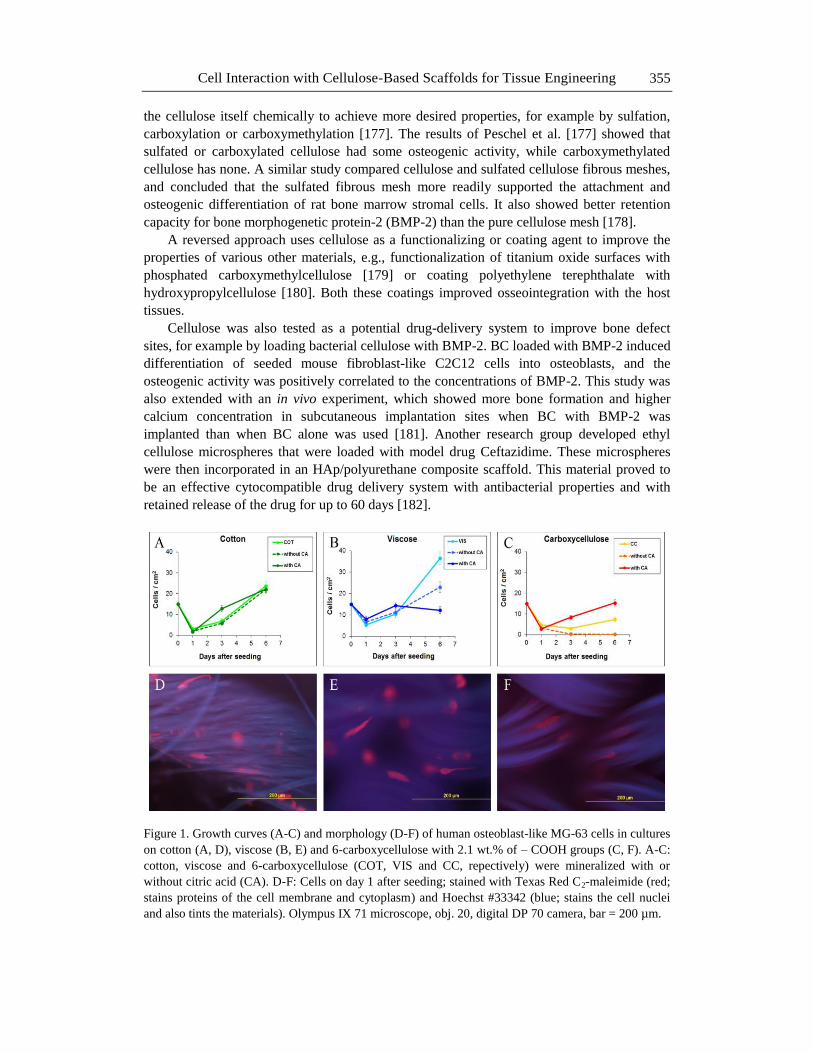
Our studies have also contributed to knowledge on the mineralization of cellulose
scaffolds in SBF and their potential further use for bone tissue engineering. Plant-derived
cellulose in the form of cotton fabrics, viscose fabrics, and also 6-carboxycellulose with 2.1
wt.% of -COOH groups proved to be suitable for vascular tissue engineering [49, 50]. These
samples were mineralized in SBF with or without citric acid, which acts a modulator of the
mineralization of various materials in SBF. For example, in the concentration range from 0.3
to 2 mM in SBF, citric acid promoted the mineralization of collagen membranes [174]. In our
experiments, all three cellulose-based materials promoted cell adhesion and subsequent
growth, though the cells adhered in relatively low initial densities on day 1 after seeding
(Figure 1 A-C). Nevertheless, the cells were able to spread and to gain a spindle-shaped
morphology, oriented along the fibers (Figure 1 D-F) and to proliferate within a period of 6
days. The final cell number on day 6 was highest on viscose and lowest on 6-carboxycellulose
with 2.1 wt.% of -COOH groups. The cell number was usually higher on the materials
mineralized in SBF with citric acid than on the non-mineralized materials and on the material
mineralized without citric acid, which was most apparent on 6-carboxycellulose with 2.1
wt.% of -COOH groups (Figure 1 A-C). This could be explained by higher mineralization of
the materials due to hydrogen bonding of citric acid to the cellulose cloth and its ability to
form chelates with calcium ions [175].
The properties of cellulose materials can also be improved and made suitable for bone
tissue engineering applications by functionalizing them with various bioactive agents, e.g.,
functionalization of bacterial cellulose with osteogenic growth peptide [176], or by modifying
Cell Interaction with Cellulose-Based Scaffolds for Tissue Engineering 355
the cellulose itself chemically to achieve more desired properties, for example by sulfation,
carboxylation or carboxymethylation [177]. The results of Peschel et al. [177] showed that
sulfated or carboxylated cellulose had some osteogenic activity, while carboxymethylated
cellulose has none. A similar study compared cellulose and sulfated cellulose fibrous meshes,
and concluded that the sulfated fibrous mesh more readily supported the attachment and
osteogenic differentiation of rat bone marrow stromal cells. It also showed better retention
capacity for bone morphogenetic protein-2 (BMP-2) than the pure cellulose mesh [178].
A reversed approach uses cellulose as a functionalizing or coating agent to improve the
properties of various other materials, e.g., functionalization of titanium oxide surfaces with
phosphated carboxymethylcellulose [179] or coating polyethylene terephthalate with
hydroxypropylcellulose [180]. Both these coatings improved osseointegration with the host
tissues.
Cellulose was also tested as a potential drug-delivery system to improve bone defect
sites, for example by loading bacterial cellulose with BMP-2. BC loaded with BMP-2 induced
differentiation of seeded mouse fibroblast-like C2C12 cells into osteoblasts, and the
osteogenic activity was positively correlated to the concentrations of BMP-2. This study was
also extended with an in vivo experiment, which showed more bone formation and higher
calcium concentration in subcutaneous implantation sites when BC with BMP-2 was
implanted than when BC alone was used [181]. Another research group developed ethyl
cellulose microspheres that were loaded with model drug Ceftazidime. These microspheres
were then incorporated in an HAp/polyurethane composite scaffold. This material proved to
be an effective cytocompatible drug delivery system with antibacterial properties and with
retained release of the drug for up to 60 days [182].
Figure 1. Growth curves (A-C) and morphology (D-F) of human osteoblast-like MG-63 cells in cultures
on cotton (A, D), viscose (B, E) and 6-carboxycellulose with 2.1 wt.% of – COOH groups (C, F). A-C:
cotton, viscose and 6-carboxycellulose (COT, VIS and CC, repectively) were mineralized with or
without citric acid (CA). D-F: Cells on day 1 after seeding; stained with Texas Red C2-maleimide (red;
stains proteins of the cell membrane and cytoplasm) and Hoechst #33342 (blue; stains the cell nuclei
and also tints the materials). Olympus IX 71 microscope, obj. 20, digital DP 70 camera, bar = 200 µm.
Lucie Bacakova, Katarina Novotna, Tomas Sopuch et al. 356
Scientists agree that a certain degree of microporosity within the scaffold is beneficial to
osteoblast ingrowth and subsequent mineralization of the scaffold [183, 184]. Zaborowska et
al. [183] compared nanoporous and microporous bacterial cellulose scaffolds, which were
seeded with MC3T3-E1 osteoprogenitor cells. The cells formed denser mineral deposits on
microporous scaffolds than on nanoporous scaffolds. The micropores in this study ranged
between 300-500 µm. Another research group developed nanofibrous cellulose scaffolds with
laser induced micropores ranging between 50-300 μm that enhanced osteoblast attachment at
the edge of these pores [184]. However, bigger pores ranging up to 750 µm formed in
cellulose matrix were also shown to be effectual for osteoblast adhesion and growth [172].
Although most studies with cellulose scaffolds are held as in vitro tests, there have also
been several in vivo experiments. Besides the studies already mentioned above, which were
performed partially in vivo [164, 181], there was a study which tested the biological
properties of bacterial cellulose-HAp membranes. These membranes were implanted in non-
critical bone defects in rat tibiae. After 4 weeks, the defects were filled with new bone [165].
Using a subcutaneous implantation rat model, an analysis was made of a new injectable
material composed of beta-tricalcium phosphate, methylcellulose and hyaluronic acid. Tissue
reaction with the implant resulted in increased vascularization and a longer in vivo lifetime in
comparison with implants consisting of beta-tricalcium phosphate alone [185]. A long-term
experiment lasting for 36 months evaluated calcium phosphate bone cement with
carboxymethylcellulose implanted in vertebral bone defects of the sheep. On average, after 36
months the defect section consisted of approximately 14% bone, 82% cement, and 4% bone
marrow, with no fibrous tissue [186]. A later study used the sheep model in a similar manner
[187]. This study evaluated the reaction of tissue to oxidized cellulose scaffolds and
compared it with the reaction of tissue to collagen scaffolds. No significant difference was
found between the two materials in the rate of repair of the bone defects, which were
completely repaired by lamellar bone at 6-8 weeks.
An injectable bone substitute was also applied clinically for filling bone defects in
patients after tooth extraction. This substitute was prepared by suspending biphasic calcium
phosphate microparticles in a water-soluble cellulose polymer carrier phase. Three years after
surgery, small biopsies of the implanted areas revealed gradual substitution of the filler by
bone tissue and preservation of the height of the alveolar bone crest [48].
Cellulose and Cartilage Tissue Engineering
Cellulose has been used for engineering various types of cartilage, e.g., articular
cartilage, meniscus, intervertebral discs and auricular cartilage. Cartilage tissue engineering is
considered difficult due to the fact that cartilage is an avascular tissue with very low
spontaneous regeneration potential. In addition, chondrocytes are prone to dedifferentiation
towards fibroblast-like cell phenotype. Last but not least, the cartilage replacement should
meet relatively high requirements for mechanical resistance, particularly in load-bearing
applications.
For engineering articular cartilage, cellulose and its derivatives and composites have
often been used in the form of injectable hydrogels with encapsulated cells in order to fill the
cartilage tissue defects with minimal invasion and pain. The cellulose-based hydrogels
included, e.g., silanized hydroxypropyl methylcellulose [188, 189], and particularly
Cell Interaction with Cellulose-Based Scaffolds for Tissue Engineering 357
thermoresponsive polymers, such as chitosan-beta glycerophosphate-hydroxyethyl cellulose
[190, 191] or poly(N-isopropylacrylamide)-g-methylcellulose [192]. These polymers are
characterized by a sol-gel transition at 37°C, which enables them to be injected in their liquid
state and then to gel at body temperature. Amidic derivatives of carboxymethylcellulose have
also been developed in order to mimic the advantageous physicochemical properties of
hyaluronan while overcoming its excessively fast degradation time [193]. For better
mechanical strength, hydrogels have been reinforced with multi-wall carbon nanotubes [194]
or by induction of a fibrous component, e.g., by critical point drying in a bacterial cellulose
hydrogel [195]. In some hydrogels, pores were created in order to facilitate the penetration of
cells inside the scaffolds, e.g., bacterial nanocellulose hydrogels perforated using a laser
[196].
Cellulose-based materials for articular cartilage engineering have also been constructed,
primarily in the form of fibrous, porous or combined scaffolds, such as nanofibrous bacterial
cellulose [197], composites of nanofibrous bacterial cellulose and poly(vinyl alcohol) matrix
[198], bacterial cellulose scaffolds with pores created by extrusion of wax particles [199],
cellulose and cellulose/recombinant type II collagen sponges [200], and composites
containing cellulose nanofibers, derived from rice straw and coated with a starch film, in
which the pores were created by a salt leaching technique [201]. Cellulose-based scaffolds
were also used for engineering the osteochondral interface, e.g., asymmetric porous
composites consisting of cellulose acetate matrix and bioactive glass particles, prepared by
phase separation techniques [202], or non-woven cellulose fabrics activated in a saturated
Ca(OH)2 solution and subsequently coated with a calcium phosphate layer precipitated from a
supersaturated physiological solution [203].
Injectable cellulose-based hydrogels, particularly photocrosslinked carboxy-
methylcellulose hydrogels with encapsulated cells, are also applicable for engineering nucleus
pulposus, an important component of intervertebral discs [204-206]. Bacterial cellulose has
been used for replacing auricular cartilage and meniscus. Bacterial cellulose is composed of
highly hydrated nanofibrils (99% water) with high mechanical strength. Bacterial cellulose
with an increased cellulose content of 17% exhibited mechanical strength
similar to that of auricular cartilage, a minimal cytotoxic response in vitro, tested on murine
fibrosarcoma L929 cells, and a minimal foreign body response in vivo, evaluated after
intradermal implantation into rabbits [207]. Bacterial cellulose can also be fabricated into
patient-specific auricular shapes [208]. In the case of meniscus replacement, the Young's
moduli of bacterial cellulose gel and pig meniscus were similar in magnitude under a
compression load of 2 kPa and had five times better mechanical properties than a reference
collagen material [209].
For encapsulation into hydrogels or for seeding on various scaffolds, chondrogenic cells
were used in the form of cell lines, such as murine ATDC5 cells [192] or differentiated
chondrocytes derived from articular cartilage [190, 193]. This approach is appropriate for
testing the materials in vitro, but for in vivo testing or for potential clinical application, a more
advantageous approach is to use autologous nasal chondrocytes [188] or autologous
mesenchymal stem cells derived from bone marrow [191, 195, 206] or from adipose tissue
[189]. Cells derived from nucleus pulposus are often applied for engineering intervertebral
discs [204, 205]. Chondrogenic cell differentiation is supported by transforming growth factor
β3 added into the cell culture medium or released by the scaffolds [191, 206]. Some specific
scaffold components, such as addition of silk to cellulose, have also been reported to support
Lucie Bacakova, Katarina Novotna, Tomas Sopuch et al. 358
chondrogenic cell differentiation [210]. This differentiation is manifested by the expression of
COL2A1, ACAN, SOX9, and COMP genes and by synthesis of cartilage-specific
components of extracellular matrix, namely aggrecan and type II collagen [188, 189, 206,
211]. In order to make cellulose-based scaffolds degradable, novel bacterial cellulose was
formed in metabolically engineered Gluconacetobacter xylinus. This cellulose is lysozyme-
susceptible, and can gradually be replaced by newly formed regenerated cartilage tissue
[211].
Cellulose for Tendon and Ligament Repair
The first application of cellulose-based materials in tendon surgery was to prevent the
formation of adhesions between the healing tendon and the surrounding structures, such as
bone, muscle, skin, tendon sheath, or other tendons, via scar tissue. For these purposes, the
following formulation was tested in rabbits and rats: Interceed TC7 (Johnson, Johnson, USA),
which is a fabric comprised of oxidized regenerated cellulose [212, 213], and Seprafilm
Bioresorbable Membrane (Genzyme Corporation, Cambridge, MA), containing sodium
hyaluronate and carboxymethylcellulose [214]. It was concluded that these materials
significantly reduced peritendinous adhesions in experimental animals and are promising for
clinical application in human patients.
Cellulose-based materials have also been used for osseointegration of ligament
replacements and for ligament and tendon tissue engineering. For example, hydroxypropyl
cellulose coating of polyethylene terephthalate artificial ligaments enhances graft
osseointegration in the bone tunnel in the proximal tibia of rabbits [215]. Fibrous networks of
cellulose nanofibers and collagen crosslinked using a bio-based crosslinker genipin promoted
the adhesion, growth and differentiation of human ligament cells and human endothelial cells.
In addition, the material showed mechanical performance similar to that of the natural
ligament and tendon [216]. Similar results were obtained with electrospun cellulose
nanofibers, which improved their tensile strength, elastic modulus and thermal stability after
being reinforced with cellulose nanocrystals, and supported the adhesion and oriented growth
of human fibroblasts [217].
Cellulose-based materials are also promising for periodontal ligament repair. Suspensions
of nano-hydroxyapatite/sodium carboxymethylcellulose in cell culture medium accelerated
the proliferation of human periodontal ligament cells (HPLCs) by shortening the G1 phase of
the cell cycle [218]. When HPLCs were cultured on a polyelectrolyte complex consisting of
chitosan and sulfated cellulose, treatment with 10-7
-10-9
M dexamethason promoted HPLC
growth and inhibited the production of cell aggregates [219].
CONCLUSION AND FURTHER PERSPECTIVES
Cellulose is the most abundant biopolymer on Earth, and it accordingly also has abundant
biomedical applications, including clinical applications. Cellulose has been used for repairing,
regenerating and reconstructing practically all of the tissues in the mammalian organism. In
its clinical applications, however, cellulose supports the healing and the function of tissues
Cell Interaction with Cellulose-Based Scaffolds for Tissue Engineering 359
and organs rather indirectly, e.g., by covering wounds and releasing drugs into them, by
preventing postoperative adhesions, by hemostasis, hemodialysis or by covering and filling
various tissue defects. Direct clinical applications of cellulose-based materials as scaffolds for
tissue engineering and cell delivery are still relatively rare, although extensive research has
been carried out in this field under in vitro and in vivo experimental conditions. The practical
use of cellulose-based materials in tissue engineering and in cell therapies now needs to be
promoted. For this purpose, cellulose-based scaffolds can be tailored by a wide range of
modifications, including combinations of cellulose with other natural or synthetic polymers,
ceramics, metals or carbon materials, loading with bioactive molecules (drugs, growth
factors), functionalization with various chemical groups, oxidation, preparation of scaffolds
with various morphologies, etc. All these modifications can control the physical and chemical
properties of cellulose-based scaffolds, which are essential for cell-material interaction. They
can also enhance the degradability of the material, which is an important property for
advanced tissue engineering, in which material scaffolds act as a temporary support for the
formation of new tissue.
ACKNOWLEDGMENT
Supported by the Grant Agency of the Czech Republic (grants No. P108/12/1168 and
P108/11/1857), the Ministry of Health of the Czech Republic (grant No. NT13297-4/2012),
and the Technological Agency of the Czech Republic (grant No. TA04010065). Mr. Robin
Healey (Czech Technical University, Prague, Czech Republic) is gratefully acknowledged for
his language revision of the manuscript.
REFERENCES
[1] Payen, A. (1838). Mémoire sur la composition du tissu propre des plantes et du ligneux
(Memoir on the composition of the tissue of plants and of woody material). Comptes
Rendus, 7, 1052-1056.
[2] O'Sullivan, A. C. (1997). Cellulose: the structure slowly unravels. Cellulose 4, 173-207.
[3] Bacakova, L., Novotna, K., Parizek, M. (2014). Polysaccharides as cell carriers for
tissue engineering: the use of cellulose in vascular wall reconstruction. Physiol. Res.,
63(Suppl. 1), S29-S47.
[4] Lagus, H., Sarlomo-Rikala, M., Böhling, T., Vuola, J. (2013). Prospective study on
burns treated with Integra®, a cellulose sponge and split thickness skin graft:
comparative clinical and histological study--randomized controlled trial. Burns, 39(8),
1577-1587.
[5] Dini, V., Romanelli, M., Andriessen, A., Barbanera, S., Bertone, M. S., Brilli, C., Abel,
M. (2013). Improvement of periulcer skin condition in venous leg ulcer patients:
prospective, randomized, controlled, single-blinded clinical trial comparing a
biosynthetic cellulose dressing with a foam dressing. Adv. Skin Wound Care, 26(8),
352-359.
Lucie Bacakova, Katarina Novotna, Tomas Sopuch et al. 360
[6] Araújo, C. P., Gomes, J., Vieira, A. P., Ventura, F., Fernandes, J. C., Brito, C. (2013). A
proposal for the use of new silver-seaweed-cotton fibers in the treatment of atopic
dermatitis. Cutan. Ocul. Toxicol., 32(4), 268-274.
[7] Jaeger, T., Rothmaier, M., Zander, H., Ring, J., Gutermuth, J., Anliker, M. D. (2014).
Acid-coated textiles (pH 5.5-6.5) - a new therapeutic strategy for atopic eczema? Acta
Derm. Venereol., in press, doi: 10.2340/00015555-1916
[8] Liu, J., Li, Y., Rong, X., Lin, W., Zhang, T., Wang, B., Li, X. B., Jiang, S., Zhang, Q.
(2013). Application of crystalline cellulose membrane (Veloderm) on split-thickness
skin graft donor sites in burn or reconstructive plastic surgery patients. J. Burn Care
Res., 34(3), e176-82.
[9] Catalfamo, L., Belli, E., Nava, C., Mici, E., Calvo, A., D'Alessandro, B., De Ponte, F.
S. (2013). Bioengineering in the oral cavity: our experience. Int. J. Nanomedicine, 8,
3883-3886.
[10] Albaugh, K. W., Biely, S. A., Cavorsi, J. P. (2013). The effect of a cellulose dressing
and topical vancomycin on methicillin-resistant Staphylococcus aureus (MRSA) and
Gram-positive organisms in chronic wounds: a case series. Ostomy Wound Manage,
59(5), 34-43.
[11] Duteille, F., Jeffery, S. L. (2012). A phase II prospective, non-comparative assessment
of a new silver sodium carboxymethylcellulose (AQUACEL(®) Ag BURN) glove in
the management of partial thickness hand burns. Burns, 38(7), 1041-1050.
[12] http://www.woundsinternational.com/product-reviews/technology-update-under
standing-hydrocolloids
[13] http://www.woundsinternational.com/product-reviews/technology-update-under
standing- hydrofiber-technology
[14] http://www.smith-nephew.com/global/assets/pdf/products/wound/durafiber.pdf
[15] Sopuch, T., Drahovzalova, R., Rydl, J., Bures, I., Milichovsky, M., Vytrasova, J.,
Motková, P., Svorcik, V., Podlaha, J., Horakova, M., Masteikova, R., Vinklarkova, L.,
Suchy P. (2013). Celulózové materiály v ošetřování ran (in Czech). Hojení ran, 7(2),
ISSN 1802-6400 (GEUM).
[16] Ouellet, D., Grossmann, K. F., Limentani G., Nebot N., Lan K., Knowles L., Gordon
M. S., Sharma S., Infante J. R., Lorusso P. M., Pande G., Krachey E. C., Blackman S.
C., Carson S. W. (2013). Effects of particle size, food, and capsule shell composition on
the oral bioavailability of dabrafenib, a BRAF inhibitor, in patients with BRAF
mutation-positive tumors. J. Pharm. Sci., 102(9), 3100-3019.
[17] Pollack, I. F., Jakacki, R. I., Butterfield, L. H., Hamilton, R. L., Panigrahy, A., Potter,
D. M., Connelly, A. K., Dibridge, S. A., Whiteside T. L., Okada H. (2014). Antigen-
specific immune responses and clinical outcome after vaccination with glioma-
associated antigen peptides and polyinosinic-polycytidylic acid stabilized by lysine and
carboxymethylcellulose in children with newly diagnosed malignant brainstem and
nonbrainstem gliomas. J. Clin. Oncol. 32(19), 2050-2058.
[18] Liew, K. B., Tan, Y. T., Peh, K. K. (2014). Effect of polymer, plasticizer and filler on
orally disintegrating film. Drug Dev. Ind. Pharm., 40(1), 110-119.
[19] Labib, G. S., Aldawsari, H. M., Badr-Eldin, S. M. (2014). Metronidazole and
Pentoxifylline films for the local treatment of chronic periodontal pockets: preparation,
in vitro evaluation and clinical assessment. Expert Opin. Drug. Deliv., 11(6), 855-865.
Cell Interaction with Cellulose-Based Scaffolds for Tissue Engineering 361
[20] McDonald, M., D'Aversa, G., Perry, H. D., Wittpenn, J. R., Nelinson, D. S. (2010).
Correlating patient-reported response to hydroxypropyl cellulose ophthalmic insert
(LACRISERT®) therapy with clinical outcomes: tools for predicting response. Curr.
Eye Res., 35(10), 880-887.
[21] Wander, A. H. (2011). Long-term use of hydroxypropyl cellulose ophthalmic insert to
relieve symptoms of dry eye in a contact lens wearer: case-based experience. Eye
Contact Lens, 37(1), 39-44.
[22] Babizhayev, M. A., Burke L., Micans P., Richer, S. P. (2009). N-Acetylcarnosine
sustained drug delivery eye drops to control the signs of ageless vision: glare
sensitivity, cataract amelioration and quality of vision currently available treatment for
the challenging 50,000-patient population. Clin. Interv. Aging, 4, 31-50.
[23] Robertson, D., Lefebvre, G., Leyland, N., Wolfman, W., Allaire, C., Awadalla, A.,
Best, C., Contestabile, E., Dunn, S., Heywood, M., Perouc, N., Potestio, F., Rittenberg,
D., Senikas, V., Soucy, R., Singh, S. (2010). Adhesion prevention in gynaecological
surgery. J. Obstet. Gynaecol. Can., 32(6), 598-608.
[24] Dupré, A., Legrand, A., Buc, E., Delpero, J. R., Quenet, F., Passot, G., Evrard, S.,
Divoure, M. (2013). Use of bioresorbable membranes to reduce abdominal and
perihepatic adhesions in 2-stage hepatectomy of liver metastases from colorectal
cancer: results of a prospective, randomized controlled phase II trial. Ann. Surg.,
258(1), 30-36.
[25] ten Broek, R. P., Stommel, M. W., Strik, C., van Laarhoven, C. J., Keus, F., van Goor,
H. (2014). Benefits and harms of adhesion barriers for abdominal surgery: a systematic
review and meta-analysis. Lancet 383(9911), 48-59.
[26] Ahn, J. H., Lim, H. W., Hong, H. R. (2012). The clinical application and efficacy of
sodium hyaluronate-carboxymethylcellulose during tympanomastoid surgery.
Laryngoscope 122(4), 912-915.
[27] Liu, Z. C., Li, Y., Zang, Y., Cui, G., Sang, H. X., Ma, Z. S., Kong, L., Lei, W., Wu, Z.
X. (2013). Clinical assessment of a CMC/PEO gel to inhibit postoperative epidural
adhesion formation after lumbar discectomy: a randomized, control study. Arch.
Orthop. Trauma Surg. 133(3), 295-301.
[28] Derici, F., Kröning, K. C., Scatizzi, M. (2009). Effectiveness for pain after laparoscopic
cholecystectomy of 0.5% bupivacaine-soaked Tabotamp placed in the gallbladder bed:
a prospective, randomized, clinical trial. Surg Endosc., 23(10), 2214-2220.
[29] Berrevoet, F., Fierens, K., De Gols, J., Navez, B., Van Bastelaere, W., Metr, E.,
Ceulemans, R. (2009). Multicentric observational cohort study evaluating a composite
mesh with incorporated oxidized regenerated cellulose in laparoscopic ventral hernia
repair. Hernia, 13(1), 23-27.
[30] Al-Shaikh, S., Muddaiah, A., Lee, R. J., Bhutta, M. F. (2014). Oxidised cellulose
powder for haemostasis following sinus surgery: a pilot randomised trial. J. Laryngol.
Otol., 128(8), 709-713.
[31] Öllinger, R., Mihaljevic, A. L., Schuhmacher, C., Bektas, H., Vondran, F., Kleine, M.,
Sainz-Barriga, M., Weiss, S., Knebel, P., Pratschke, J., Troisi, R. I. (2013). A
multicentre, randomized clinical trial comparing the Veriset™ haemostatic patch with
fibrin sealant for the management of bleeding during hepatic surgery. HPB (Oxford),
15(7), 548-558.
Lucie Bacakova, Katarina Novotna, Tomas Sopuch et al. 362
[32] Tam, T., Harkins, G., Dykes, T., Gockley, A., Davies, M. (2014). Oxidized regenerated
cellulose resembling vaginal cuff abscess. JSLS, 18(2), 353-356.
[33] Eshghi, P., Jenabzade, A., Habibpanah, B. (2014). A self-controlled comparative
clinical trial to explore the effectiveness of three topical hemostatic agents for stopping
severe epistaxis in pediatrics with inherited coagulopathies. Hematology, 19(6), 361-
364.
[34] Gidaro, S. Cindolo, L., Lipsky, K., Zigeuner, R., Schips, L. (2009). Efficacy and safety
of the haemostasis achieved by Vivostat system during laparoscopic partial
nephrectomy. Arch. Ital. Urol. Androl., 81(4), 223-227.
[35] Kai, Y., Hamada, J., Morioka, M., Yano, S., Nakamura, H., Makino, K., Mizuno, T.,
Takeshima, H., Kuratsu, J. (2006). Clinical evaluation of cellulose porous beads for the
therapeutic embolization of meningiomas. AJNR Am. J. Neuroradiol., 27(5), 1146-
1150.
[36] MacLeod, A., Daly, C., Khan, I., Vale, L., Campbell, M., Wallace, S., Cody, J.,
Donaldson, C., Grant, A. (2001). Comparison of cellulose, modified cellulose and
synthetic membranes in the haemodialysis of patients with end-stage renal disease.
Cochrane Database Syst. Rev., 3, CD003234.
[37] Alonso, A., Lau, J., Jaber, B. L. (2008). Biocompatible hemodialysis membranes for
acute renal failure. Cochrane Database Syst. Rev., 1, CD005283.
[38] Bowry, S. K., Gatti, E., Vienken, J. (2011). Contribution of polysulfone membranes to
the success of convective dialysis therapies. Contrib. Nephrol. 173, 110-118.
[39] Sosa, M. A., Balk, E. M., Lau, J., Liangos, O., Balakrishnan, V. S., Madias, N. E.,
Pereira, B. J., Jaber, B. L. (2006). A systematic review of the effect of the Excebrane
dialyser on biomarkers of lipid peroxidation. Nephrol. Dial. Transplant., 21(10), 2825-
2833.
[40] Sunohara, T., Masuda, T. (2011). Cellulose triacetate as a high-performance membrane.
Contrib. Nephrol. 173, 156-163.
[41] Tanaka, S., Sato, N., Fujioka, H., Takahashi, Y., Kimura, K., Iwamoto, M., Uchiyama,
K. (2014). Breast conserving surgery using volume replacement with oxidized
regenerated cellulose: a cosmetic outcome analysis. Breast J., 20(2), 154-158.
[42] Franceschini, G., Visconti, G., Terribile, D., Fabbri, C., Magno, S., Di Leone, A.,
Salgarello, M., Masetti, R. (2012). The role of oxidized regenerate cellulose to prevent
cosmetic defects in oncoplastic breast surgery. Eur. Rev. Med. Pharmacol. Sci. 16(7),
966-971.
[43] Brunner, C. A., Gröner, R. W. (2006). Carboxy-methyl-cellulose hydrogel-filled breast
implants - an ideal alternative? A report of five years' experience with this device. Can.
J. Plast. Surg., 14(3), 151-154.
[44] Leonardis, M., Palange, A., Dornelles, R. F., Hund, F. (2010). Use of cross-linked
carboxymethyl cellulose for soft-tissue augmentation: preliminary clinical studies. Clin.
Interv. Aging, 5, 317-322.
[45] Günzburg, W. H., Salmons, B. (2005). Use of cell therapy as a means of targeting
chemotherapy to inoperable pancreatic cancer. Acta Biochim Pol, 52(3), 601-607.
[46] Rosen, C. L., Steinberg, G. K., DeMonte, F., Delashaw, J. B. Jr., Lewis, S. B., Shaffrey,
M. E., Aziz, K., Hantel, J., Marciano, F. F. (2011). Results of the prospective,
randomized, multicenter clinical trial evaluating a biosynthesized cellulose graft for
repair of dural defects. Neurosurgery, 69(5), 1093-1103.
Cell Interaction with Cellulose-Based Scaffolds for Tissue Engineering 363
[47] Gazzeri, R., Galarza, M., Alfieri, A., Neroni, M., Roperto, R. (2011). Simple
intraoperative technique for minor dural gap repair using fibrin glue and oxidized
cellulose. World Neurosurg., 76(1-2), 173-175.
[48] Weiss, P., Layrolle, P., Clergeau, L. P., Enckel, B., Pilet, P., Amouriq, Y., Daculsi, G.,
Giumelli, B. (2007). The safety and efficacy of an injectable bone substitute in dental
sockets demonstrated in a human clinical trial. Biomaterials, 28(22), 3295-3305.
[49] Novotna, K., Bacakova, L., Lisa, V., Havelka, P., Sopuch, T., Klepetar, J. (2009).
Vascular smooth muscle cells in cultures on biofunctionalized cellulose-based
scaffolds. Eng. Biomater., 12, 21-24.
[50] Novotna, K., Havelka, P., Sopuch, T., Kolarova, K., Vosmanska, V., Lisa, V., Svorcik,
V., Bacakova, L. (2013). Cellulose-based materials as scaffolds for tissue engineering.
Cellulose, 20, 2263-2278.
[51] Ko, I. K., Iwata, H. (2001). An approach to constructing three-dimensional tissue. Ann.
N. Y. Acad. Sci., 944, 443-455.
[52] Bäckdahl, H., Helenius, G., Bodin, A., Nannmark, U., Johansson, B. R., Risberg, B.,
Gatenholm, P. (2006). Mechanical properties of bacterial cellulose and interactions with
smooth muscle cells. Biomaterials., 27(9), 2141-2149.
[53] Bäckdahl, H., Esguerra, M., Delbro, D., Risberg, B., Gatenholm, P. (2008). Engineering
microporosity in bacterial cellulose scaffolds. J. Tissue Eng. Regen. Med., 2(6), 320-
330.
[54] Bodin, A., Ahrenstedt, L., Fink, H., Brumer, H., Risberg, B., Gatenholm, P. (2007).
Modification of nanocellulose with a xyloglucan-RGD conjugate enhances adhesion
and proliferation of endothelial cells: implications for tissue engineering.
Biomacromolecules, 8(12), 3697-3704.
[55] Fink, H., Ahrenstedt, L., Bodin, A., Brumer, H., Gatenholm, P., Krettek, A., Risberg, B.
(2011). Bacterial cellulose modified with xyloglucan bearing the adhesion peptide RGD
promotes endothelial cell adhesion and metabolism--a promising modification for
vascular grafts. J. Tissue Eng. Regen. Med., 5(6), 454-463.
[56] Rubenstein, D., Han, D., Goldgraben, S., El-Gendi, H., Gouma, P. I., Frame, M. D.
(2007). Bioassay chamber for angiogenesis with perfused explanted arteries and
electrospun scaffolding. Microcirculation, 14(7), 723-737.
[57] Esguerra, M., Fink, H., Laschke, M. W., Jeppsson, A., Delbro, D., Gatenholm, P.,
Menger, M. D., Risberg. B. (2010). Intravital fluorescent microscopic evaluation of
bacterial cellulose as scaffold for vascular grafts. J. Biomed. Mater. Res. A. 93(1), 140-
149.
[58] Malm, C. J., Risberg, B., Bodin, A., Bäckdahl. H., Johansson, B. R., Gatenholm, P.,
Jeppsson, A. (2012). Small calibre biosynthetic bacterial cellulose blood vessels: 13-
months patency in a sheep model. Scand. Cardiovasc., J. 46(1), 57-62.
[59] Wippermann, J., Schumann, D., Klemm, D., Kosmehl, H., Salehi-Gelani, S., Wahlers,
T. (2009). Preliminary results of small arterial substitute performed with a new
cylindrical biomaterial composed of bacterial cellulose. Eur. J. Vasc. Endovasc. Surg.,
37(5), 592-596.
[60] Scherner, M., Reutter, S., Klemm, D., Sterner-Kock, A., Guschlbauer, M., Richter, T.,
Langebartels, G., Madershahian, N., Wahlers, T., Wippermann, J. (2014). In vivo
application of tissue-engineered blood vessels of bacterial cellulose as small arterial
substitutes: proof of concept? J. Surg. Res., 189(2), 340-347.
Lucie Bacakova, Katarina Novotna, Tomas Sopuch et al. 364
[61] Zahedmanesh, H., Mackle, J. N., Sellborn, A., Drotz, K., Bodin, A., Gatenholm, P.,
Lally, C. (2011). Bacterial cellulose as a potential vascular graft: Mechanical
characterization and constitutive model development. J. Biomed. Mater. Res. B Appl.
Biomater., 97(1), 105-113.
[62] Fink, H., Hong, J., Drotz, K., Risberg, B., Sanchez, J., Sellborn, A. (2011). An in vitro
study of blood compatibility of vascular grafts made of bacterial cellulose in
comparison with conventionally-used graft materials. J. Biomed. Mater. Res A., 97A(1):
52–58.
[63] Brown, E. E., Hu, D., Abu Lail, N., Zhang, X. (2013). Potential of nanocrystalline
cellulose-fibrin nanocomposites for artificial vascular graft applications.
Biomacromolecules, 14(4), 1063-1071.
[64] Pooyan, P., Tannenbaum, R., Garmestani, H. (2012). Mechanical behavior of a
cellulose-reinforced scaffold in vascular tissue engineering. J. Mech. Behav. Biomed.
Mater., 7, 50-59.
[65] Collins, M. N., Birkinshaw, C. (2013). Hyaluronic acid based scaffolds for tissue
engineering-a review. Carbohydr. Polym., 92(2), 1262-1279.
[66] Märtson, M., Viljanto, J., Hurme, T., Laippala, P., Saukko, P. (1999). Is cellulose
sponge degradable or stable as implantation material? An in vivo subcutaneous study in
the rat. Biomaterials, 20(21), 1989-1995.
[67] Roychowdhury, P., Kumar, V. (2006). Fabrication and evaluation of porous 2,3-
dialdehydecellulose membrane as a potential biodegradable tissue-engineering scaffold.
J. Biomed. Mater. Res. A, 76(2), 300-309.
[68] Saito, T., Kimura, S., Nishiyama, Y., Isogai, A. (2007). Cellulose nanofibers prepared
by TEMPO-mediated oxidation of native cellulose. Biomacromolecules, 8(8), 2485-
2491.
[69] Dimitrijevich, S. D., Tatarko, M., Gracy, R. W., Linsky, C. B., Olsen, C. (1990a).
Biodegradation of oxidized regenerated cellulose. Carbohydr. Res., 195, 247–256.
[70] Dimitrijevich, S. D., Tatarko, M., Gracy, R. W., Wise, G. E., Oakford, L. X., Linsky, C.
B., Kamp, L. (1990b). In vivo degradation of oxidized, regenerated cellulose.
Carbohydr. Res. 198, 331–341.
[71] Zhu, L., Kumar, V., Banker, G. S. (2001). Examination of oxidized cellulose as a
macromolecular prodrug carrier: preparation and characterization of an oxidized
cellulose-phenylpropanolamine conjugate. Int. J. Pharm., 223(1-2), 35-47.
[72] Zimnitsky, D. S., Yurkshtovich, T. L., Bychkovsky, P. M. (2005). Multilayer
adsorption of amino acids on oxidized cellulose. J. Colloid Interf. Sci., 285(2), 502-508.
[73] Zimnitsky, D. S., Yurkshtovich, T. L., Bychkovsky, P. M. (2006). Adsorption of
zwitterionic drugs onto oxidized cellulose. J. Colloid Interf. Sci., 295(1), 33-40.
[74] Mikulikova, R., Kolarova, K., Bacakova, L., Svorcik, V. (2005). Biocompatibility of
polyethylene doped with oxycellulose. Mater. Sci. Forum, 482, 251-254.
[75] Mihardja, S. S., Gonzales, J. A., Gao, D., Sievers, R. E., Fang, Q., Stillson, C. A., Yu,
J., Peng, M., Lee, R. J. (2013). The effect of a peptide-modified thermo-reversible
methylcellulose on wound healing and LV function in a chronic myocardial infarction
rodent model. Biomaterials, 34(35), 8869-8877.
[76] Yeh, Y. C., Lee, W. Y., Yu, C. L., Hwang, S. M., Chung, M. F., Hsu, L. W., Chang, Y.,
Lin, W. W., Tsai, M. S., Wei, H. J., Sung, H. W. (2010). Cardiac repair with injectable
Cell Interaction with Cellulose-Based Scaffolds for Tissue Engineering 365
cell sheet fragments of human amniotic fluid stem cells in an immune-suppressed rat
model. Biomaterials, 31(25), 6444-6453.
[77] Lee, W. Y., Wei, H. J., Lin, W. W., Yeh, Y. C., Hwang, S. M., Wang, J. J., Tsai, M. S.,
Chang, Y., Sung, H. W. (2011). Enhancement of cell retention and functional benefits
in myocardial infarction using human amniotic-fluid stem-cell bodies enriched with
endogenous ECM. Biomaterials, 32(24), 5558-5567.
[78] Huang, C. C., Tsai, H. W., Lee, W. Y., Lin, W. W., Chen, D. Y., Hung, Y. W., Chen, J.
W., Hwang, S. M., Chang, Y., Sung, H. W. (2013). A translational approach in using
cell sheet fragments of autologous bone marrow-derived mesenchymal stem cells for
cellular cardiomyoplasty in a porcine model. Biomaterials, 34(19), 4582-4591.
[79] Ravi, S., Caves, J. M., Martinez, A. W., Xiao, J., Wen, J., Haller, C. A., Davis, M. E.,
Chaikof, E. L. (2012). Effect of bone marrow-derived extracellular matrix on cardiac
function after ischemic injury. Biomaterials, 33(31), 7736-7745.
[80] Entcheva, E., Bien, H., Yin, L., Chung, C. Y., Farrell, M., Kostov, Y. (2004).
Functional cardiac cell constructs on cellulose-based scaffolding. Biomaterials, 25(26),
5753-5762.
[81] Millon, L. E., Wan, W. K. (2006). The polyvinyl alcohol-bacterial cellulose system as a
new nanocomposite for biomedical applications. J. Biomed. Mater. Res. B Appl.
Biomater., 79(2), 245-253.
[82] Mohammadi, H., Boughner, D., Millon, L. E., Wan, W. K. (2009). Design and
simulation of a poly(vinyl alcohol)-bacterial cellulose nanocomposite mechanical aortic
heart valve prosthesis. Proc. Inst. Mech. Eng. H., 223(6), 697-711.
[83] Mohammadi, H. (2011). Nanocomposite biomaterial mimicking aortic heart valve
leaflet mechanical behaviour. Proc. Inst. Mech. Eng. H., 225(7), 718-722.
[84] Dugan, J. M., Gough, J. E., Eichhorn, S. J. (2010). Directing the morphology and
differentiation of skeletal muscle cells using oriented cellulose nanowhiskers.
Biomacromolecules, 11(9), 2498-2504.
[85] Dugan, J. M., Collins, R. F., Gough, J. E., Eichhorn, S. J. (2013). Oriented surfaces of
adsorbed cellulose nanowhiskers promote skeletal muscle myogenesis. Acta Biomater.,
9(1), 4707-4715.
[86] Aggarwal, N., Altgärde, N., Svedhem, S., Zhang, K., Fischer, S., Groth, T. (2014).
Study on multilayer structures prepared from heparin and semi-synthetic cellulose
sulfates as polyanions and their influence on cellular response. Colloids Surf. B
Biointerfaces, 116, 93-103.
[87] Vatankhah, E., Prabhakaran, M. P., Jin, G., Mobarakeh, L. G., Ramakrishna, S. (2014).
Development of nanofibrous cellulose acetate/gelatin skin substitutes for variety wound
treatment applications. J. Biomater. Appl., 28(6), 909-921.
[88] Johnen, C., Steffen, I., Beichelt, D., Bräutigam, K., Witascheck, T., Toman, N., Moser,
V., Ottomann, C., Hartmann, B., Gerlach, J. C. (2008). Culture of subconfluent human
fibroblasts and keratinocytes using biodegradable transfer membranes. Burns, 34(5),
655-663.
[89] Laçin, N. T. (2014). Development of biodegradable antibacterial cellulose based
hydrogel membranes for wound healing. Int. J. Biol. Macromol., 67, 22-27.
[90] Unnithan, A. R., Gnanasekaran, G., Sathishkumar, Y., Lee, Y. S., Kim, C. S. 4. (2014).
Electrospun antibacterial polyurethane-cellulose acetate-zein composite mats for wound
dressing. Carbohydr. Polym., 102, 884-892.
Lucie Bacakova, Katarina Novotna, Tomas Sopuch et al. 366
[91] Sanchavanakit, N., Sangrungraungroj, W., Kaomongkolgit, R., Banaprasert, T.,
Pavasant, P., Phisalaphong, M. (2006). Growth of human keratinocytes and fibroblasts
on bacterial cellulose film. Biotechnol. Prog., 22(4), 1194-1199.
[92] Kingkaew, J., Jatupaiboon, N., Sanchavanakit, N., Pavasant, P., Phisalaphong, M.
(2010). Biocompatibility and growth of human keratinocytes and fibroblasts on
biosynthesized cellulose-chitosan film. J. Biomater. Sci. Polym. Ed., 21(8-9), 1009-
1021.
[93] Seyama, T., Suh, E. Y., Kondo, T. (2013). Three-dimensional culture of epidermal cells
on ordered cellulose scaffolds. Biofabrication, 5(2), 025010.
[94] Batak, S., Kundu, S. C. (2014). Sericin-carboxymethyl cellulose porous matrices as
cellular wound dressing material. J. Biomed. Mater. Res. A., 102(6), 1928-1940.
[95] Xin, S., Li, X., Wang, Q., Juany, R., Xu, X., Lei, Z., Deng, H. (2014). Novel layer-by-
layer structured nanofibrous mats coated by protein films for dermal regeneration. J.
Biomed. Nanotechnol., 10(5), 803-810.
[96] Matthew, H. W., Salley, S. O., Peterson, W. D., Klein, M. D. (1993a). Complex
coacervate microcapsules for mammalian cell culture and artificial organ development.
Biotechnol. Prog., 9(5), 510-519.
[97] Matthew, H. W., Basu, S., Peterson, W. D., Salley, S. O., Klein, M. D. (1993b).
Performance of plasma-perfused, microencapsulated hepatocytes: prospects for
extracorporeal liver support. J. Pediatr. Surg. 28(11), 1423-1427.
[98] Canaple, L., Nurdin, N., Angelova, N., Hunkeler, D., Desvergne, B. (2001).
Development of a coculture model of encapsulated cells. Ann. N. Y. Acad. Sci., 944,
350-361.
[99] Tiruvannamalai-Annamalai, R., Armant, D. R., Matthew, H. W. (2014). A
glycosaminoglycan based, modular tissue scaffold system for rapid assembly of
perfusable, high cell density, engineered tissues. PLoS One, 9(1), e84287.
[100] Kino, Y., Sawa, M., Kasai, S., Mito, M. (1998). Multiporous cellulose microcarrier for
the development of a hybrid artificial liver using isolated hepatocytes. J. Surg. Res.,
79(1), 71-76.
[101] Kobayashi, N., Noguchi, H., Watanabe, T., Matsumura, T., Totsugawa, T., Fujiwara,
T., Taguchi, T., Urata, H., Kishimoto, N., Hayashi, N., Nakaji, S., Westerman, K. A.,
Leboulch, P., Murakami, T., Tanaka, N. (2000). A new approach to develop a biohybrid
artificial liver using a tightly regulated human hepatocyte cell line. Hum. Cell., 13(4),
229-235.
[102] Saito, M., Matsuura, T., Masaki, T., Maehashi, H., Shimizu, K., Hataba, Y., Iwahori,
T., Suzuki, T., Braet, F. (2006). Reconstruction of liver organoid using a bioreactor.
World J. Gastroenterol., 12(12), 1881-1888.
[103] Ho Ye, S., Watanabe, J., Takai, M., Iwasaki, Y., Ishihara, K. (2006). High functional
hollow fiber membrane modified with phospholipid polymers for a liver assist
bioreactor. Biomaterials, 27(9), 1955-1962.
[104] Bhattacharya, M., Malinen, M. M., Lauren, P., Lou, Y. R., Kuisma, S. W., Kanninen,
L., Lille, M., Corlu, A., GuGuen-Guillouzo, C., Ikkala, O., Laukkanen, A., Urtti, A.,
Yliperttula, M. (2012). Nanofibrillar cellulose hydrogel promotes three-dimensional
liver cell culture. J. Control Release, 164(3), 291-298.
[105] Malinen, M. M., Kanninen, L. K., Corlu, A., Isoniemi, H. M., Lou, Y. R., Yliperttula,
M. L., Urtti, A. O. (2014). Differentiation of liver progenitor cell line to functional
Cell Interaction with Cellulose-Based Scaffolds for Tissue Engineering 367
organotypic cultures in 3D nanofibrillar cellulose and hyaluronan-gelatin hydrogels.
Biomaterials, 35(19), 5110-5121.
[106] Evenou, F., Couderc, S., Kim, B., Fujii, T., Sakai, Y. (2011). Microfibrillated cellulose
sheets coating oxygen-permeable PDMS membranes induce rat hepatocytes 3D
aggregation into stably-attached 3D hemispheroids. J. Biomater. Sci. Polym. Ed.,
22(11), 1509-1522.
[107] Gerlach, J., Vienken, J., Walker, P., Affeld, K. (1990). Computer aided time-lapse
video analysis of hepatocyte morphology during adhesion to cellulose membranes. Int.
J. Artif. Organs, 13(6), 365-369.
[108] Jóźwiak, A., Karlik, W., Wiechetek, M., Weryński, A. (1998). Attachment and
metabolic activity of hepatocytes cultivated on selected polymeric membranes. Int. J.
Artif. Organs, 21(8), 460-466.
[109] De Bartolo, L., Morelli, S., Bader, A., Drioli, E. (2002). Evaluation of cell behaviour
related to physico-chemical properties of polymeric membranes to be used in
bioartificial organs. Biomaterials, 23(12), 2485-2497.
[110] Koç, A., Durkut, S., Elçin, A. E., Tan, E., Elçin, Y. M. (2007). Evaluation of modified
CMC and CMC-PVA as miscible polymer blend membranes for hepatocytes.
Macromol. Biosci., 7(5), 681-689.
[111] Esaki, K., Yokota, S., Egusa, S., Okutani, Y., Ogawa, Y., Kitaoka, T., Goto, M.,
Wariishi, H. (2009). Preparation of lactose-modified cellulose films by a nonaqueous
enzymatic reaction and their biofunctional characteristics as a scaffold for cell culture.
Biomacromolecules, 10(5), 1265-1269.
[112] Braun, K., Besch, W., Jahr, H., Loth, F., Dautzenberg, H., Hahn, H. J. (1985). The
encapsulation of pancreatic islets. Investigation of insulin secretion and content in vitro.
Biomed. Biochim. Acta, 44(1), 143-147.
[113] Wang, T., Lacík, I., Brissová, M., Anilkumar, A. V., Prokop, A., Hunkeler, D., Green,
R., Shahrokhi, K., Powers, A. C. (1997). An encapsulation system for the
immunoisolation of pancreatic islets. Nat. Biotechnol., 15(4), 358-362.
[114] Brissova, M., Lacik, I., Powers, A. C., Anilkumar, A. V., Wang, T. (1998). Control and
measurement of permeability for design of microcapsule cell delivery system. J.
Biomed. Mater. Res., 39(1), 61-70.
[115] Stiegler, P. B., Stadlbauer, V., Schaffellner, S., Halwachs, G., Lackner, C., Hauser, O.,
Iberer, F., Tscheliessnigg, K. (2006). Cryopreservation of insulin-producing cells
microencapsulated in sodium cellulose sulfate. Transplant. Proc., 38(9), 3026-3030.
[116] Bodziony, J., Gasior, U., Stanosek, J. (1985). Functional estimation of the isolated
islands of Langerhans in semipermeable membrane chambers. Z. Exp. Chir. Transplant.
Kunstliche Organe, 18(4), 204-214.
[117] Bodziony, J., Stanosek, J. (1985). Treatment of diabetes by implantation of isolated
islands of Langerhans in semipermeable chambers. Z. Exp. Chir. Transplant. Kunstliche
Organe, 18(4), 215-223.
[118] Rosenberg, L., Duguid, W. P., Vinik, A. I. (1989). The effect of cellophane wrapping of
the pancreas in the Syrian golden hamster: autoradiographic observations. Pancreas,
4(1), 31-37.
[119] Ye, H., Tai, J., Cheung, S. C., Tze, W. J. (1998). Improved function of porcine islets
after long-term culture in methylcellulose matrix. Transplant. Proc., 30(2), 386-387.
Lucie Bacakova, Katarina Novotna, Tomas Sopuch et al. 368
[120] Tun, T., Inoue, K., Hayashi, H., Aung, T., Gu, Y. J., Doi, R., Kaji, H., Echigo, Y.,
Wang, W. J., Setoyama, H., Imamura, M., Maetani, S., Morikawa, N., Iwata, H., Ikada,
Y. (1996). A newly developed three-layer agarose microcapsule for a promising
biohybrid artificial pancreas: rat to mouse xenotransplantation. Cell Transplant., 5(5
Suppl 1), S59-63.
[121] Kawakami, Y., Inoue, K., Tun, T., Hayashi, H., Setoyama, H., Gu, Y. J., Cui, W. X.,
Imamura, M., Iwata, H., Ikada, Y. (1997). Prolonged effect of troglitazone (CS-045) on
xenograft survival of hybrid artificial pancreas. Cell Transplant., 6(5), 547-550.
[122] Kawakami, Y., Inoue, K., Hayashi, H., Wang, W. J., Setoyama, H., Gu, Y. J., Imamura,
M., Iwata, H., Ikada, Y., Nozawa, M., Miyazaki, J. (1997). Subcutaneous
xenotransplantation of hybrid artificial pancreas encapsulating pancreatic B cell line
(MIN6): functional and histological study. Cell Transplant., 6(5), 541-545.
[123] Risbud, M. V., Bhonde, R. R. (2001). Suitability of cellulose molecular dialysis
membrane for bioartificial pancreas: in vitro biocompatibility studies. J. Biomed.
Mater. Res., 54(3), 436-444.
[124] Jung, S. K., Gorski, W., Aspinwall, C. A., Kauri, L. M., Kennedy, R. T. (1999).
Oxygen microsensor and its application to single cells and mouse pancreatic islets.
Anal. Chem., 71(17), 3642-3649.
[125] Kawamura, T., Cohen, S. S., Borris, D. L., Aquilino, E. A., Glushakova, S., Margolis,
L. B., Orenstein, J. M., Offord, R. E., Neurath, A. R., Blauvelt, A. (2000). Candidate
microbicides block HIV-1 infection of human immature Langerhans cells within
epithelial tissue explants. J. Exp. Med., 192(10), 1491-1500.
[126] Danielsen, N., Pettmann, B., Vahlsing, H. L., Manthorpe, M., Varon, S. (1988).
Fibroblast growth factor effects on peripheral nerve regeneration in a silicone chamber
model. J. Neurosci. Res., 20(3), 320-330.
[127] Bates, D. J., Ranford, J. A., Mangelsdorf, D. C. (1991). Blot and culture analysis of
neuronotrophic factors in nerve regeneration chamber fluids. Neurochem Res., 16(6),
621-628.
[128] Jones, N. S., Hall, S. M. (1991). Evaluation of a new method of neural anastomosis
using nitrocellulose paper. Clin. Otolaryngol. Allied Sci., 16(4), 383-387.
[129] Welch, J. A., Kraus, K. H., Wells, M. R., Blunt, D. G., Weremowitz, J. (1997). Effect
of combined administration of insulin-like growth factor and platelet-derived growth
factor on the regeneration of transected and anastomosed sciatic nerve in rats. Am. J.
Vet. Res., 58(9), 1033-1037.
[130] Wells, M. R., Kraus, K., Batter, D. K., Blunt, D. G., Weremowitz, J., Lynch, S. E.,
Antoniades, H. N., Hansson, H. A. (1997). Gel matrix vehicles for growth factor
application in nerve gap injuries repaired with tubes: a comparison of biomatrix,
collagen, and methylcellulose. Exp. Neurol., 146(2), 395-402.
[131] Masgutov, R., Raginov, I., Fomina, G., Kozlova, M., Chelyshev, Y. (2006). Stimulation
of the rat's sciatic nerve regeneration by local treatment with Xymedon. Cell. Mol.
Neurobiol., 26(7-8), 1413-1421.
[132] Pan, H. C., Yang, D. Y., Chiu, Y. T., Lai, S. Z., Wang, Y. C., Chang, M. H., Cheng, F.
C. (2006). Enhanced regeneration in injured sciatic nerve by human amniotic
mesenchymal stem cell. J. Clin. Neurosci., 13(5), 570-575.
Cell Interaction with Cellulose-Based Scaffolds for Tissue Engineering 369
[133] Inoue, Y., Hasegawa, S., Yamada, T., Date, Y., Mizutani, H., Nakata, S., Akamatsu, H.
(2013). Lignin Induces ES Cells to Differentiate into Neuroectodermal Cells through
Mediation of the Wnt Signaling Pathway. PLoS One, 8(6), e66376.
[134] Shi, Z., Gao, H., Feng, J., Ding, B., Cao, X., Kuga, S., Wang, Y., Zhang, L., Cai, J.
(2014). In situ synthesis of robust conductive cellulose/polypyrrole composite aerogels
and their potential application in nerve regeneration. Angew Chem. Int. Ed. Engl.,
53(21), 5380-5384.
[135] Sun, T., Norton, D., Vickers, N. L., McArthur, S., Neil, S. M., Ryan, A. J., Haycock, J.
W. (2008). Development of a bioreactor for evaluating novel nerve conduits.
Biotechnol. Bioeng., 99(5), 1250-1260.
[136] Park, J. S., Lee, J. H., Han, C. S., Chung, D. W., Kim, G. Y. (2011). Effect of
hyaluronic acid-carboxymethylcellulose solution on perineural scar formation after
sciatic nerve repair in rats. Clin. Orthop. Surg., 3(4), 315-324.
[137] Adanali, G., Verdi, M., Tuncel, A., Erdogan, B., Kargi, E. (2003). Effects of hyaluronic
acid-carboxymethylcellulose membrane on extraneural adhesion formation and
peripheral nerve regeneration. J. Reconstr. Microsurg., 19(1), 29-36.
[138] Kowalska-Ludwicka, K., Cala, J., Grobelski, B., Sygut, D., Jesionek-Kupnicka, D.,
Kolodziejczyk, M., Bielecki, S., Pasieka, Z. (2013). Modified bacterial cellulose tubes
for regeneration of damaged peripheral nerves. Arch. Med. Sci., 9(3), 527-534.
[139] Houle, J. D., Johnson, J. E. (1989). Nerve growth factor (NGF)-treated nitrocellulose
enhances and directs the regeneration of adult rat dorsal root axons through intraspinal
neural tissue transplants. Neurosci. Lett., 103(1), 17-23.
[140] Kitano, T., Zerwekh, J. E., Edwards, M. L., Usui, Y., Allen, M. D. (1991). Viscous
carboxymethylcellulose in the prevention of epidural scar formation. Spine (Phila Pa
1976), 16(7), 820-823.
[141] Kim, K. D., Wang, J. C., Robertson, D. P., Brodke, D. S., BenDebba, M., Block, K. M.,
diZerega, G. S. (2004). Reduction of leg pain and lower-extremity weakness for 1 year
with Oxiplex/SP gel following laminectomy, laminotomy, and discectomy. Neurosurg.
Focus., 17(1), ECP1.
[142] Tsai, E. C., Dalton, P. D., Shoichet, M. S., Tator, C. H. (2006). Matrix inclusion within
synthetic hydrogel guidance channels improves specific supraspinal and local axonal
regeneration after complete spinal cord transection. Biomaterials, 27(3), 519-533.
[143] Mothe, A. J., Tam, R. Y., Zahir, T., Tator, C. H., Shoichet, M. S. (2013). Repair of the
injured spinal cord by transplantation of neural stem cells in a hyaluronan-based
hydrogel. Biomaterials, 34(15), 3775-3783.
[144] Conova, L., Vernengo, J., Jin, Y., Himes, B. T., Neuhuber, B., Fischer, I., Lowman, A.,
Vernengo, J., Jin, Y., Himes, B. T., Neuhuber, B., Fischer, I., Lowman, A. (2011). A
pilot study of poly(N-isopropylacrylamide)-g-polyethylene glycol and poly(N-
isopropylacrylamide)-g-methylcellulose branched copolymers as injectable scaffolds
for local delivery of neurotrophins and cellular transplants into the injured spinal cord.
J. Neurosurg. Spine, 15(6), 594-604.
[145] Solomon, A., Lavie, V., Ben-Bassat, S., Belkin, M., Schwartz, M. (1991). New surgical
approach to overcome the inability of injured mammalian axons to grow within their
environment. J. Neural Transplant Plast., 2(3-4), 243-248.
[146] Tate, M. C., Shear, D. A., Hoffman, S. W., Stein, D. G., LaPlaca, M. C. (2001).
Biocompatibility of methylcellulose-based constructs designed for intracerebral
Lucie Bacakova, Katarina Novotna, Tomas Sopuch et al. 370
gelation following experimental traumatic brain injury. Biomaterials, 22(10), 1113-
1123.
[147] Wang, Y., Cooke, M. J., Morshead, C. M., Shoichet, M. S. (2012). Hydrogel delivery of
erythropoietin to the brain for endogenous stem cell stimulation after stroke injury.
Biomaterials, 33(9), 2681-2692.
[148] Wang, Y., Cooke, M. J., Sachewsky, N., Morshead, C. M., Shoichet, M. S. (2013).
Bioengineered sequential growth factor delivery stimulates brain tissue regeneration
after stroke. J. Control Release, 172(1), 1-11.
[149] Caicco, M. J., Cooke, M. J., Wang, Y., Tuladhar, A., Morshead, C. M., Shoichet, M. S.
(2013). A hydrogel composite system for sustained epi-cortical delivery of Cyclosporin
A to the brain for treatment of stroke. J. Control Release, 166(3), 197-202.
[150] Tessitore, E., Schatlo, B., Schaller, C., Schonauer, C. (2012). Fibrillary structure is key
for hemostasis: a similar effect of collagen fleece and oxidized cellulose on
experimental hemorrhagic brain injury. J. Neurol. Surg. A Cent. Eur. Neurosurg., 73(2),
89-92.
[151] Mello, L. R., Feltrin, L. T., Fontes Neto P. T., Ferraz, F. A. (1997). Duraplasty with
biosynthetic cellulose: an experimental study. J. Neurosurg., 86(1), 143-150.
[152] Sanchez e Oliveira, R. de C., Valente, P. R., Abou-Jamra, R. C., Araújo, A., Saldiva, P.
H., Pedreira, D. A. (2007). Biosynthetic cellulose induces the formation of a
neoduramater following pre-natal correction of meningomyelocele in fetal sheep. Acta
Cir. Bras., 22(3), 174-181.
[153] Xu, C., Ma, X., Chen, S., Tao, M., Yuan, L., Jing, Y. (2014). Bacterial cellulose
membranes used as artificial substitutes for dural defection in rabbits. Int. J. Mol. Sci.,
15(6), 10855-10867.
[154] Clarke, B. G., Sachs, B. F., Fayer, R., Gherardi, G. J. (1963). Surgical hemostasis and
repair of the kidney. II. Experimental evaluation of oxidized regenerated cellulose as a
hemostatic agent in experimental nephrotomy. Invest. Urol., 1, 31-44.
[155] Furka, I., Bornemissza, G., Mikó, I. (1976). Use of absorbable material for the closure
of experimental longitudinal nephrotomy. Int. Urol. Nephrol., 8(2), 107-112.
[156] Sabino, L., Andreoni, C., Faria, E. F., Ferreira, P. S., Paz, A. R., Kalil, W., De
Figueiredo, L. P., Ortiz, V. (2007). Evaluation of renal defect healing, hemostasis, and
urinary fistula after laparoscopic partial nephrectomy with oxidized cellulose. J.
Endourol., 21(5), 551-556.
[157] Mittal, S., Sharma, J. B., Gupta, N. (2006). Successful closure of a bladder neck fistula
complicated by urethral and vaginal stenosis, using oxidized cellulose (Surgicel) for
reinforcement. Int. Urogynecol. J. Pelvic. Floor Dysfunct., 17(4), 426-428.
[158] Bodin, A., Bharadwaj, S., Wu, S., Gatenholm, P., Atala, A., Zhang, Y. (2010). Tissue-
engineered conduit using urine-derived stem cells seeded bacterial cellulose polymer in
urinary reconstruction and diversion. Biomaterials, 31(34), 8889-8901.
[159] Sato, Y., Terashima, M., Kagiwada, N., Tun, T., Inagaki, M., Kakuta, T., Saito, A.
(2005). Evaluation of proliferation and functional differentiation of LLC-PK1 cells on
porous polymer membranes for the development of a bioartificial renal tubule device.
Tissue Eng., 11(9-10), 1506-1515.
[160] Han, D., Gouma, P. I. (2006) Electrospun bioscaffolds that mimic the topology of
extracellular matrix. Nanomedicine, 2(1), 37-41.
Cell Interaction with Cellulose-Based Scaffolds for Tissue Engineering 371
[161] Petreus, T., Stoica, B. A., Petreus, O., Goriuc, A., Cotrutz, C. E., Antoniac, I. V.,
Barbu-Tudoran, L. (2014). Preparation and cytocompatibility evaluation for
hydrosoluble phosphorous acid-derivatized cellulose as tissue engineering scaffold
material. J. Mater. Sci. Mater. Med., 25(4), 1115-1127.
[162] Zang, S., Zhuo, Q., Chang, X., Qiu, G., Wu, Z., Yang, G. (2014). Study of osteogenic
differentiation of human adipose-derived stem cells (HASCs) on bacterial cellulose.
Carbohydr. Polym., 104, 158-165.
[163] Kumbar, S. G., Toti, U. S., Deng, M., James, R., Laurencin, C. T., Aravamudhan, A.,
Harmon, M., Ramos, D. M. (2011). Novel mechanically competent polysaccharide
scaffolds for bone tissue engineering. Biomed Mater., 6(6), 065005.
[164] Liuyun, J., Yubao, L., Chengdong, X. (2009). A novel composite membrane of
chitosan-carboxymethyl cellulose polyelectrolyte complex membrane filled with nano-
hydroxyapatite I. Preparation and properties. J. Mater. Sci. Mater. Med., 20(8), 1645-
1652.
[165] Saska, S. I., Barud, H. S., Gaspar, A. M., Marchetto, R., Ribeiro, S. J., Messaddeq, Y.
(2011). Bacterial cellulose-hydroxyapatite nanocomposites for bone regeneration. Int. J.
Biomater., 2011, 175362.
[166] Jiang, H., Zuo, Y., Zou, Q., Wang, H., Du, J., Li, Y., Yang, X. (2013). Biomimetic
spiral-cylindrical scaffold based on hybrid chitosan/cellulose/nano-hydroxyapatite
membrane for bone regeneration. ACS Appl. Mater. Interf., 5(22), 12036-12044.
[167] Tazi, N., Zhang, Z., Messaddeq, Y., Almeida-Lopes, L., Zanardi, L. M., Levinson, D.,
Rouabhia, M. (2012). Hydroxyapatite bioactivated bacterial cellulose promotes
osteoblast growth and the formation of bone nodules. AMB Express, 2(1), 61.
[168] Pasqui, D., Torricelli, P., De Cagna, M., Fini, M., Barbucci, R. (2014). Carboxymethyl
cellulose-hydroxyapatite hybrid hydrogel as a composite material for bone tissue
engineering applications. J. Biomed. Mater. Res. A, 102(5), 1568-1579.
[169] Pandit, V., Zuidema, J. M., Venuto, K. N., Macione, J., Dai, G., Gilbert, R. J., Kotha, S.
P. (2013). Evaluation of multifunctional polysaccharide hydrogels with varying
stiffness for bone tissue engineering. Tissue Eng. Part A, 19(21-22), 2452-2463.
[170] Raucci, M. G., Alvarez-Perez, M. A., Demitri, C., Sannino, A., Ambrosio, L. (2012).
Proliferation and osteoblastic differentiation of hMSCs on cellulose-based hydrogels. J.
Appl. Biomater. Funct. Mater., 10(3), 302-307.
[171] Granja, P. L., De Jéso, B., Bareille, R., Rouais, F., Baquey, C., Barbosa, M. A. (2005).
Mineralization of regenerated cellulose hydrogels induced by human bone marrow
stromal cells. Eur. Cell Mater., 31-37.
[172] Petrauskaite, O., Gomes, P. de S., Fernandes, M. H., Juodzbalys, G., Stumbras, A.,
Maminskas, J., Liesiene, J., Cicciù, M. (2013). Biomimetic mineralization on a
macroporous cellulose-based matrix for bone regeneration. Biomed. Res. Int., 2013,
452750.
[173] Chahal, S., Fathima, S. J., Yusoff, M. B. (2014). Biomimetic growth of bone-like
apatite via simulated body fluid on hydroxyethyl cellulose/polyvinyl alcohol
electrospun nanofibers. Biomed. Mater. Eng., 24(1), 799-806.
[174] Rhee, S. H., Tanaka, J. Effect of citric acid on the nucleation of hydroxyapatite in a
simulated body fluid. Biomaterials 20(22), 2155-2160.
[175] Rhee, S. H., Tanaka. J. (2000). Hydroxyapatite formation on cellulose cloth induced by
citric acid. J. Mater. Sci. Mater. Med., 11(7), 449-452.
Lucie Bacakova, Katarina Novotna, Tomas Sopuch et al. 372
[176] Saska, S., Scarel-Caminaga, R. M., Teixeira, L. N., Franchi, L. P., Dos Santos, R. A.,
Gaspar, A. M., de Oliveira, P. T., Rosa, A. L., Takahashi, C. S., Messaddeq, Y.,
Ribeiro, S. J., Marchetto, R. (2012). Characterization and in vitro evaluation of bacterial
cellulose membranes functionalized with osteogenic growth peptide for bone tissue
engineering. J. Mater. Sci. Mater. Med., 23(9), 2253-2266.
[177] Peschel, D., Zhang, K., Fischer, S., Groth, T. (2012). Modulation of osteogenic activity
of BMP-2 by cellulose and chitosan derivatives. Acta Biomater., 8(1), 183-193.
[178] Filion, T. M., Kutikov, A., Song, J. (2011). Chemically modified cellulose fibrous
meshes for use as tissue engineering scaffolds. Bioorg. Med. Chem. Lett., 21(17), 5067-
5070.
[179] Pasqui, D., Rossi, A., Di Cintio, F., Barbucci, R. (2007). Functionalized titanium oxide
surfaces with phosphated carboxymethyl cellulose: characterization and bonelike cell
behavior. Biomacromolecules, 8(12), 3965-3972.
[180] Yang, J., Jiang, J., Li, Y., Li, H., Jing, Y., Wu, P., Yu, D., Chen, S. (2013). A new
strategy to enhance artificial ligament graft osseointegration in the bone tunnel using
hydroxypropylcellulose. Int. Orthop., 37(3), 515-521.
[181] Shi, Q., Li, Y., Sun, J., Zhang, H., Chen, L., Chen, B., Yang, H., Wang, Z. (2012). The
osteogenesis of bacterial cellulose scaffold loaded with bone morphogenetic protein-2.
Biomaterials, 33(28), 6644-6649.
[182] Liu, H., Zhang, L., Shi, P., Zou, Q., Zuo, Y., Li, Y. (2010).
Hydroxyapatite/polyurethane scaffold incorporated with drug-loaded ethyl cellulose
microspheres for bone regeneration. J. Biomed. Mater. Res. B Appl. Biomater., 95(1),
36-46.
[183] Zaborowska, M., Bodin, A., Bäckdahl, H., Popp, J., Goldstein, A., Gatenholm, P.
(2010). Microporous bacterial cellulose as a potential scaffold for bone regeneration.
Acta Biomater., 6(7), 2540-2547.
[184] Rodríguez, K., Sundberg, J., Gatenholm, P., Renneckar, S. (2014). Electrospun
nanofibrous cellulose scaffolds with controlled microarchitecture. Carbohydr. Polym.,
100, 143-149.
[185] Ghanaati, S., Barbeck, M., Hilbig, U., Hoffmann, C., Unger, R. E., Sader, R. A., Peters,
F., Kirkpatrick, C. J. (2011). An injectable bone substitute composed of beta-tricalcium
phosphate granules, methylcellulose and hyaluronic acid inhibits connective tissue
influx into its implantation bed in vivo. Acta Biomater., 7(11), 4018-4028.
[186] Kobayashi, H., Fujishiro, T., Belkoff, S. M., Kobayashi, N., Turner, A. S., Seim, H. B.
3rd, Zitelli, J., Hawkins, M., Bauer, T. W. (2009). Long-term evaluation of a calcium
phosphate bone cement with carboxymethyl cellulose in a vertebral defect model. J.
Biomed. Mater. Res. A 88(4), 880-888.
[187] Dias, G. J., Peplow. P. V., Teixeira, F. (2003). Osseous regeneration in the presence of
oxidized cellulose and collagen. J. Mater. Sci. Mater. Med., 14(9), 739-745.
[188] Vinatier, C., Gauthier, O., Fatimi, A., Merceron, C., Masson, M., Moreau, A., Moreau,
F., Fellah, B., Weiss, P., Guicheux, J. (2009). An injectable cellulose-based hydrogel
for the transfer of autologous nasal chondrocytes in articular cartilage defects.
Biotechnol. Bioeng. 102(4), 1259-1267.
[189] Merceron, C., Portron, S., Masson, M., Lesoeur, J., Fellah, B. H., Gauthier, O., Geffroy,
O., Weiss, P., Guicheux, J., Vinatier, C. (2011). The effect of two- and three-
dimensional cell culture on the chondrogenic potential of human adipose-derived
Cell Interaction with Cellulose-Based Scaffolds for Tissue Engineering 373
mesenchymal stem cells after subcutaneous transplantation with an injectable hydrogel.
Cell Transplant., 20(10), 1575-1588.
[190] Hao, T., Wen, N., Cao, J. K., Wang, H. B., Lü, S. H., Liu, T., Lin, Q. X., Duan, C. M.,
Wang, C. Y. (2010). The support of matrix accumulation and the promotion of sheep
articular cartilage defects repair in vivo by chitosan hydrogels. Osteoarthritis Cartilage,
18(2), 257-265.
[191] Naderi-Meshkin, H., Andreas, K., Matin, M. M., Sittinger, M., Bidkhori, H. R.,
Ahmadiankia, N., Bahrami, A. R., Ringe, J. (2014). Chitosan-based injectable hydrogel
as a promising in situ forming scaffold for cartilage tissue engineering. Cell Biol. Int.,
38(1), 72-84.
[192] Sá-Lima, H., Tuzlakoglu, K., Mano, J. F., Reis, R. L. (2011). Thermoresponsive
poly(N-isopropylacrylamide)-g-methylcellulose hydrogel as a three-dimensional
extracellular matrix for cartilage-engineered applications. J. Biomed. Mater. Res. A,
98(4), 596-603.
[193] Leone, G., Fini, M., Torricelli, P., Giardino, R., Barbucci, R. (2008). An amidated
carboxymethylcellulose hydrogel for cartilage regeneration. J. Mater. Sci. Mater. Med.,
19(8), 2873-2880.
[194] Xie, F., Weiss, P., Chauvet, O., Le Bideau, J., Tassin, J. F. (2010). Kinetic studies of a
composite carbon nanotube-hydrogel for tissue engineering by rheological methods. J.
Mater. Sci. Mater. Med., 21(4), 1163-1168.
[195] Favi, P. M., Benson, R. S., Neilsen, N. R., Hammonds, R. L., Bates, C. C., Stephens, C.
P., Dhar, M. S. (2013). Cell proliferation, viability, and in vitro differentiation of equine
mesenchymal stem cells seeded on bacterial cellulose hydrogel scaffolds. Mater. Sci.
Eng. C Mater. Biol. Appl., 33(4), 1935-1944.
[196] Ahrem, H., Pretzel, D., Endres, M., Conrad, D., Courseau, J., Müller, H., Jaeger, R.,
Kaps, C., Klemm, D. O., Kinne, R. W. (2014). Laser-structured bacterial nanocellulose
hydrogels support ingrowth and differentiation of chondrocytes and show potential as
cartilage implants. Acta Biomater., 10(3), 1341-1353.
[197] Svensson, A., Nicklasson, E., Harrah, T., Panilaitis, B., Kaplan, D. L., Brittberg, M.,
Gatenholm, P. (2005). Bacterial cellulose as a potential scaffold for tissue engineering
of cartilage. Biomaterials, 26(4), 419-431.
[198] Millon, L. E., Oates, C. J., Wan, W. (2009). Compression properties of polyvinyl
alcohol--bacterial cellulose nanocomposite. J. Biomed. Mater. Res. B Appl. Biomater.,
90(2), 922-929.
[199] Andersson, J., Stenhamre, H., Bäckdahl, H., Gatenholm, P. (2010). Behavior of human
chondrocytes in engineered porous bacterial cellulose scaffolds. J. Biomed. Mater. Res.
A, 94(4), 1124-1132.
[200] Pulkkinen, H., Tiitu, V., Lammentausta. E., Laasanen, M. S., Hämäläinen, E. R.,
Kiviranta, I., Lammi, M. J. (2006). Cellulose sponge as a scaffold for cartilage tissue
engineering. Biomed. Mater. Eng., 16(4 Suppl), S29-S35.
[201] Nasri-Nasrabadi, B., Mehrasa, M., Rafienia, M., Bonakdar, S., Behzad, T., Gavanji, S.
(2014). Porous starch/cellulose nanofibers composite prepared by salt leaching
technique for tissue engineering. Carbohydr. Polym., 108, 232-238.
[202] Zhang, K., Ma, Y., Francis, L. F. (2002). Porous polymer/bioactive glass composites
for soft-to-hard tissue interfaces. J. Biomed. Mater. Res., 61(4), 551-563.
Lucie Bacakova, Katarina Novotna, Tomas Sopuch et al. 374
[203] Müller, F. A., Müller, L., Hofmann, I., Greil, P., Wenzel, M. M., Staudenmaier, R.
(2006). Cellulose-based scaffold materials for cartilage tissue engineering.
Biomaterials, 27(21), 3955-3963.
[204] Reza, A. T., Nicoll, S. B. (2010). Characterization of novel photocrosslinked
carboxymethylcellulose hydrogels for encapsulation of nucleus pulposus cells. Acta
Biomater., 6(1), 179-186.
[205] Reza, A. T., Nicoll, S. B. (2010). Serum-free, chemically defined medium with TGF-
beta(3) enhances functional properties of nucleus pulposus cell-laden carboxymethyl-
cellulose hydrogel constructs. Biotechnol. Bioeng., 105(2), 384-395.
[206] Gupta, M. S., Cooper, E. S., Nicoll, S. B. (2011). Transforming growth factor-beta 3
stimulates cartilage matrix elaboration by human marrow-derived stromal cells
encapsulated in photocrosslinked carboxymethylcellulose hydrogels: potential for
nucleus pulposus replacement. Tissue Eng. Part A, 17(23-24), 2903-2910.
[207] Martínez Ávila, H., Schwarz, S., Feldmann, E. M., Mantas, A., von Bomhard, A.,
Gatenholm, P., Rotter, N. (2014). Biocompatibility evaluation of densified bacterial
nanocellulose hydrogel as an implant material for auricular cartilage regeneration. Appl.
Microbiol. Biotechnol., in press, DOI 10.1007/s00253-014-5819-z.
[208] Nimeskern, L., Martínez Ávila, H., Sundberg, J., Gatenholm, P., Müller, R., Stok, K. S.
(2013). Mechanical evaluation of bacterial nanocellulose as an implant material for ear
cartilage replacement. J. Mech. Behav. Biomed. Mater., 22, 12-21.
[209] Bodin, A., Concaro, S., Brittberg, M., Gatenholm, P. (2007). Bacterial cellulose as a
potential meniscus implant. J. Tissue Eng. Regen. Med., 1(5), 406-408.
[210] Singh, N., Rahatekar, S. S., Koziol, K. K., Ng, T. S., Patil, A. J., Mann, S., Hollander,
A. P., Kafienah, W. (2013). Directing chondrogenesis of stem cells with specific blends
of cellulose and silk. Biomacromolecules 14(5), 1287-1298.
[211] Yadav, V., Sun, L., Panilaitis, B., Kaplan, D. L. (2013). In vitro chondrogenesis with
lysozyme susceptible bacterial cellulose as a scaffold. J. Tissue Eng. Regen. Med., in
press, doi: 10.1002/term.1644.
[212] Meislin, R. J., Wiseman, D. M., Alexander, H., Cunningham, T., Linsky, C., Carlstedt,
C., Pitman, M., Casar, R. (1990). A biomechanical study of tendon adhesion reduction
using a biodegradable barrier in a rabbit model. J. Appl. Biomater., 1(1),
13-19.
[213] Temiz, A., Ozturk, C., Bakunov, A., Kara, K., Kaleli, T. (2008). A new material for
prevention of peritendinous fibrotic adhesions after tendon repair: oxidised regenerated
cellulose (Interceed), an absorbable adhesion barrier. Int. Orthop., 32(3), 389-394.
[214] Menderes, A., Mola, F., Tayfur, V., Vayvada, H., Barutçu, A. (2004). Prevention of
peritendinous adhesions following flexor tendon injury with seprafilm. Ann. Plast.
Surg., 53(6), 560-564.
[215] Yang, J., Jiang, J., Li, Y., Li, H., Jing, Y., Wu, P., Yu, D., Chen, S. (2013). A new
strategy to enhance artificial ligament graft osseointegration in the bone tunnel using
hydroxypropylcellulose. Int. Orthop., 37(3), 515-521.
[216] Mathew, A. P., Oksman, K., Pierron, D., Harmand, M. F. (2013). Biocompatible
fibrous networks of cellulose nanofibres and collagen crosslinked using genipin:
potential as artificial ligament/tendons. Macromol. Biosci., 13(3), 289-298.
[217] He, X., Xiao, Q., Lu, C., Wang, Y., Zhang, X., Zhao, J., Zhang, W., Zhang, X., Deng,
Y. (2014). Uniaxially aligned electrospun all-cellulose nanocomposite nanofibers
Cell Interaction with Cellulose-Based Scaffolds for Tissue Engineering 375
reinforced with cellulose nanocrystals: scaffold for tissue engineering.
Biomacromolecules, 15(2), 618-627.
[218] Li, F., Peng, J., Hu, R., Dong, X., Chen, W., Pan, Y., Tang, X., Xie, P. (2013). Effect of
nano-hydroxyapatite suspension on cell proliferation and cycle in human periodontal
ligament cells. J. Nanosci. Nanotechnol., 13(7), 4560-4564.
[219] Hamano, T., Chiba, D., Teramoto, A., Kondo, Y., Abe, K. (1998). Effect of
polyelectrolyte complex (PEC) on human periodontal ligament fibroblast (HPLF)
functions in the presence of glucocorticoids. J. Biomater. Sci. Polym. Ed., 9(9), 985-
1000.