cell of the month: x marks the spot
TRANSCRIPT
NATURE CELL BIOLOGY VOLUME 6 | NUMBER 7 | JULY 2004 593
C E L L O F T H E M O N T H
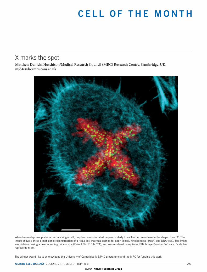
When two metaphase plates occur in a single cell, they become orientated perpendicularly to each other, seen here in the shape of an ‘X’. Theimage shows a three-dimensional reconstruction of a HeLa cell that was stained for actin (blue), kinetochores (green) and DNA (red). The imagewas obtained using a laser scanning microscope (Zeiss LSM 510 META), and was rendered using Zeiss LSM Image Browser Software. Scale barrepresents 5 µm.
The winner would like to acknowledge the University of Cambridge MB/PhD programme and the MRC for funding this work.
X marks the spotMatthew Daniels, Hutchison/Medical Research Council (MRC) Research Centre, Cambridge, UK,[email protected]
cell of the month July 14/6/04 4:20 PM Page 593
© 2004 Nature Publishing Group