cell reports report - universität bernboris.unibe.ch/53882/1/schoene2014_cellrep.pdf · cell...
TRANSCRIPT
Cell Reports
Report
Coreleased Orexin and GlutamateEvoke Nonredundant Spike Outputsand Computations in Histamine NeuronsCornelia Schone,1 John Apergis-Schoute,2 Takeshi Sakurai,3,4 Antoine Adamantidis,5,6 and Denis Burdakov1,7,*1Division of Neurophysiology, MRC National Institute for Medical Research, London NW7 1AA, UK2Department of Pharmacology, University of Cambridge, Cambridge CB2 1PD, UK3Department of Molecular Neuroscience and Integrative Physiology, Faculty of Medicine, Kanazawa University, Kanazawa, Ishikawa
920-8640, Japan4International Institute for Integrative Sleep Medicine, University of Tsukuba, Tsukuba, Ibaraki 305-8575, Japan5Neurology Department, Bern University Hospital, 3010 Bern, Switzerland6Department of Psychiatry, McGill University, Montreal, QC H3A 0G4, Canada7MRC Centre for Developmental Neurobiology, King’s College London, London WC2R 2LS, UK*Correspondence: [email protected]
http://dx.doi.org/10.1016/j.celrep.2014.03.055
This is an open access article under the CC BY license (http://creativecommons.org/licenses/by/3.0/).
SUMMARY
Stable wakefulness requires orexin/hypocretin neu-rons (OHNs) and OHR2 receptors. OHNs sensediverse environmental cues and control arousalaccordingly. For unknown reasons, OHNs containmultiple excitatory transmitters, including OH pep-tides and glutamate. To analyze their cotransmissionwithin computational frameworks for control, we op-togenetically stimulated OHNs and examined result-ing outputs (spike patterns) in a downstream arousalregulator, thehistamineneurons (HANs).OHR2swereessential for sustained HAN outputs. OHR2-depen-dent HAN output increased linearly during constantOHN input, suggesting that the OHN/HANOHR2
module may function as an integral controller. OHNstimulation evoked OHR2-dependent slow postsyn-aptic currents, similar to midnanomolar OH concen-trations. Conversely, glutamate-dependent outputtransiently communicated OHN input onset, peakingrapidly then decaying alongside OHN/HAN gluta-mate currents. Blocking glutamate-driven spikingdid not affect OH-driven spiking and vice versa,suggesting isolation (low cross-modulation) of out-puts. Therefore, in arousal regulators, cotransmittersmay translate distinct features of OHN activity intoparallel, nonredundant control signals for down-stream effectors.
INTRODUCTION
During chemical communication between neurons, transmitters
released by presynaptic activity evoke long-range postsynaptic
signals (action potentials, spikes). Small transmitters made in
presynaptic terminals (e.g., glutamate, GABA, ATP, acetylcho-
line) are recognized regulators of postsynaptic spiking (Bear
et al., 2001; Ren et al., 2011). They can be coreleased with larger
neuropeptides, which are encoded by the genome, highly
diverse, and widely present in central terminals (Bear et al.,
2001; Burnstock, 2004; Salio et al., 2006; Burbach, 2011). How-
ever, in the brain, knowledge of spike patterns arising from
activity-dependent neuropeptide release from defined neurons
remains imperfect.
To study input-output computations and spike patterns result-
ing from neural release of a behaviorally vital neuropeptide, we
probed relations between pre- and postsynaptic activity in a
brain microcircuit comprising orexin/hypocretin and histamine
neurons. Orexin/hypocretin neuropeptides (OH) are critical for
stable wakefulness, reward-seeking, and energy balance (de Le-
cea et al., 2006; Sakurai, 2007). OH-expressing neurons (OHNs)
are located in the hypothalamus, project widely throughout the
brain (Peyron et al., 1998), and are activated by diverse environ-
mental challenges such as sensory stimuli (e.g., sounds), fasting,
hypoglycemia, hypercapnia, and stress (Mileykovskiy et al.,
2005; Sakurai, 2007; Sakurai et al., 1998; Williams et al., 2007,
2008; Winsky-Sommerer et al., 2004). OHN firing promotes
awakening in a frequency-dependent manner (Adamantidis
et al., 2007), while OHN loss causes narcolepsy (Hara et al.,
2001; Thannickal et al., 2000; Mignot et al., 2002; Ripley et al.,
2001). Narcolepsy also results from lack of OH peptides or OH
type-2 G protein coupled receptors (OHR2), emphasizing the
importance of OH signaling (Chemelli et al., 1999; Lin et al.,
1999; Peyron et al., 2000; Willie et al., 2003). OH peptides are
stored in dense-core vesicles, in the same terminals as clear ves-
icles associated with small transmitters (de Lecea et al., 1998).
However, there is little direct evidence that OH peptides are
released by OHN firing to evoke spiking in downstream targets.
So far, the firing of OHNs has only been shown to release gluta-
mate (Schone et al., 2012). At the circuit level, the relative roles of
OH and glutamate remain unclear.
To address this, we used optogenetics (Petreanu et al., 2007;
Yizhar et al., 2011) to stimulate OHNs in situ. We measured
resulting responses in wake-promoting histamine neurons
Cell Reports 7, 697–704, May 8, 2014 ª2014 The Authors 697
D C
A B
E
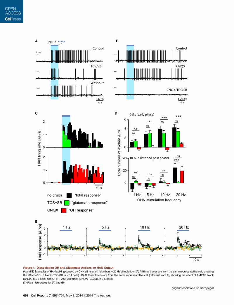
Figure 1. Dissociating OH and Glutamate Actions on HAN Output
(A and B) Examples of HAN spiking caused by OHN stimulation (blue bars = 20 Hz stimulation). (A) All three traces are from the same representative cell, showing
the effect of OHR block (TCS/SB, n = 11 cells). (B) All three traces are from the same representative cell (different from A), showing the effect of AMPAR block
(CNQX, n = 5 cells) and OHR + AMPAR block (CNQX/TCS/SB, n = 5 cells).
(C) Rate histograms for (A) and (B).
(legend continued on next page)
698 Cell Reports 7, 697–704, May 8, 2014 ª2014 The Authors
(HANs) of the tuberomammillary hypothalamus, one of the key
postsynaptic targets of OHNs expressing the ‘‘antinarcoleptic’’
OHR2s (Yamanaka et al., 2002; Willie et al., 2003; Haas et al.,
2008; Schone et al., 2012). Wakefulness instability produced by
global OHR2 deletion is reversed by local OHR2 rescue in tubero-
mammillary hypothalamus, consistent with the importance of this
circuit for brain state control (Haas et al., 2008; Mochizuki et al.,
2011;Sakurai, 2007).Simultaneouscontrol ofOHN input, pharma-
cological manipulation of transmission, and recording of HAN
output enabled us to compare circuit performance and computa-
tional operations enabled by OH versus glutamate transmission.
RESULTS
Pharmacological Dissociation of HistaminergicRepresentations of OHN ActivityTo study input-output computations in the OHN/HAN circuit,
we optically triggered spikes in OHNs, and measured responses
in HANs using intracellular patch-clamp recordings in mouse
brain slices (Figure S1; Supplemental Experimental Procedures).
Spontaneous OH or glutamate transmission was too low to
affect HAN firing in our preparation (Supplemental Experimental
Procedures, Section 3). To evoke transmission in the OHN/
HAN circuit, we first drove OHNs with brief optical stimuli (10 s
trains of flashes at 20 Hz), producing OHN spike bursts (Fig-
ure S1A) similar to those emitted by OHNs upon sensory stimu-
lation in vivo (Mileykovskiy et al., 2005). This produced rapid
postsynaptic excitation in �70% of HANs (Figure 1; n = 116/
173 cells). Continuing to analyze HAN firing pattern after high-
frequency stimulation revealed a late excitation (Figures 1A–
1D), blocked by a mixture of OH receptor antagonists (SB +
TCS; Figures 1A–1D). In the same cells, blocking glutamate
AMPA receptors (AMPARs) with CNQX abolished only rapid
excitation during the 10 s of stimulation (Figures 1B–1E).
Direct (versusmodulatory) control of neural firing has been dis-
cussed as a relatively minor action of naturally released neuro-
peptides (Salio et al., 2006; van den Pol, 2003; Schone and Bur-
dakov, 2012). However, for the same OHN stimulation (20 Hz for
10 s), we estimated that OH transmission generated �5-fold
more spikes than glutamate (during 0–60 s relative to the stimu-
lation; in CNQX: 25.2 ± 6.6 spikes, n = 16 versus in SB + TCS:
5.1 ± 0.9 spikes, n = 26; p < 0.001 by unpaired t test; see also Fig-
ure 1D). This suggests that OH and AMPARs may generate
distinct, temporarily and pharmacologically separable spike pat-
terns. The difference in speeds of the two spike responses pre-
sumably relates, in part, to the transmitters’ actions on the slow
metabotropic OHRs versus fast ionotropic AMPARs (Schone
et al., 2012; Sakurai, 2007).
Frequency Dependence of HistaminergicRepresentations of OHN ActivityStudies using nonselective stimulation (high potassium, electric
shocks) and unphysiological detection (e.g., radioimmuno-
(D) Evoked spikes (action potentials [APs]) versus stimulation frequency. Color-co
control: n = 11, 14, 13, and 13; SB/TCS: n = 9, 12, 12, and 11; CNQX: n = 3, 3, 3
(E) Temporal profiles of HAN firing responses across OHN stimulation frequencies
SEM; cells for 1, 5, 10, and 20 Hz, respectively: control: n = 9, 12, 11, and 10; SB
assay) suggested that neuropeptide release required more
stimulation than the release of smaller transmitters (Dutton
and Dyball, 1979; Haass et al., 1989; Lundberg et al., 1986; Ve-
rhage et al., 1991). However, physiological interpretation of this
could be confounded by (1) possible stimulation of off-target
neurons/axons, glial, endothelia, etc.; or (2) lower detection
sensitivity than may exist in intrinsic detectors, leading to
release underestimates. We re-examined stimulation require-
ments for neuropeptide action using the selective stimulation
and intrinsic detection in the OHN/HAN circuit across OHN
activity linked to behavior in vivo (1–20 Hz firing, Adamantidis
et al., 2007; Lee et al., 2005; Mileykovskiy et al., 2005). We
observed OH-dependent excitation only at upper frequencies
and glutamate-dependent excitation across all frequencies
(Figure 1D).
Synaptic inputs can be modulated by artificially (bath) applied
OH peptides (e.g., Lambe and Aghajanian, 2003; Ma et al., 2007;
van den Pol et al., 1998). We thus examined interactions be-
tween OH and glutamate-dependent firing across stimulation
frequencies. At every frequency, glutamate-dependent firing
was unaffected by blockers that abolished OH-dependent firing,
and vice versa (Figures 1D and 1E). OHNs also contain chemical
markers for other transmitters (Crocker et al., 2005; Furutani
et al., 2013). However, coapplication of OH and glutamate re-
ceptor blockers abolished the effects of OHN stimulation on
HAN firing (Figures 1B and 1E; evoked spikes at 20 Hz stimula-
tion = 0.7 ± 5, n = 5 cells, p > 0.2, one-sample t test).
This suggests that OH and glutamate are main drivers of
long-range output in the OHN/HAN circuit and that OH trans-
mission requires a higher presynaptic activity than glutamate
transmission.
Temporal Relations between OHN Input and itsHistaminergic RepresentationsCombining rapid reactions to stimulus trends with actions based
on longer stimulus histories is useful for the brain, and for control
systems in general (DiStefano et al., 2012). To explore howHANs
fire during prolonged input, we extended OHN stimulation to
30 s. This roughly mimics in vivo OHN firing during behavioral
transitions (e.g., from sleep to wakefulness) or during initiation
of food consumption (Lee et al., 2005; Mileykovskiy et al.,
2005). Prolonged OHN stimulation evoked two firing phases in
HANs: a fast transient firing peak (similar to short stimulation,
Figure 1E) followed by slow firing escalation overlapping in
time with OHN stimulation (Figure 2A). OHR2 blockade (with
10 mM TCS, expected to block OXR2, but not OXR1; Hirose
et al., 2003; Woldan-Tambor et al., 2011) abolished the slow
phase but did not affect the fast, CNQX-sensitive phase (Figures
2A and 2B). In turn, the OHR2-dependent phase was unaffected
by blockers of glutamate NMDA, AMPA, and GABAA/C receptors
(Figure 2A). This further suggests that OHN/HANOHR2 and
OHN/HANAMPAR signaling modules control HAN output inde-
pendently of each other and in complementary time domains.
ding as in (C). Data are means ± SEM; cells for 1, 5, 10, and 20 Hz, respectively:
, and 4.
. No drugs (black), SB/TCS (green), SB/TCS/CNQX (orange). Data are means ±
/TCS: n = 8, 11, 11, and 10; SB/TCS/CNQX: n = 5, 8, 8, and 7).
Cell Reports 7, 697–704, May 8, 2014 ª2014 The Authors 699
A B
C
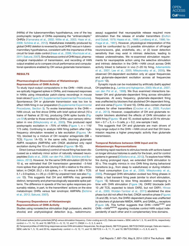
Figure 2. Time Courses of HAN Output in Relation to OHN Input
(A) Example (top trace, n = 17 cells) and group data (middle graph, means ± SEM) of HAN firing response to 30 s OHN stimulation (throughout the figure, blue
bars = 20 Hz stimulation). Bottom: same data compared at specific time points.
(B) Same as (A) but on longer timescale to illustrate recovery and individual variations (means in black, individual cells in gray). Statistical comparisons are relative
to baseline (arrow A).
(C) Relations between cumulative OHN input and HAN output during OH transmission (left graph, measured in CNQX/AP5/PiX) or glutamate transmission (right
graph, measured in TCS). OHN input was 20 Hz for 30 s. R and p are linear regression fit parameters. Data are means ± SEM.
We next asked what features of OHN input are conveyed by
the two HAN firing outputs. Amplitudes of glutamate-depen-
dent output (shown in Figure 1E, green traces, recorded in
OH blockers) correlated significantly with OHN stimulation fre-
quency (one-way ANOVA, F[3,36] = 3.45, p < 0.05). However,
decay time constants of the glutamate-dependent output did
not vary significantly with OHN stimulation frequency
(Figure 1E; extra sum-of-squares F test, F[3,778] = 0.648,
700 Cell Reports 7, 697–704, May 8, 2014 ª2014 The Authors
p > 0.05, based on monoexponential decay fits). The
OH-dependent output (which we observed and studied only
at high OHN frequency) continued to rise throughout the
30 s of OHN input (Figure 2A, middle). There was a strong
linear correlation between HAN output and cumulative (inte-
grated) OHN input during this time (Figure 2C). When OHR2s
were blocked, AMPARs could not maintain this input-output
relation (Figure 2C).

This suggests that OH and glutamate drive distinct temporal
patterns of HAN output and may thus be required for different
input-output computations (see Discussion).
HAN Membrane Currents Triggered by OHN ActivityThe main aim of our study was to reveal system-level relations
between OH input and HAN output. We assume that HAN firing
output is triggered by membrane currents evoked by coreleased
OHN transmitters (which generate firing patterns after a further
series of interactions with biophysical and geometric properties
of the HANmembrane; Haas and Reiner, 1988). To confirm exis-
tence of these currents, we performed whole-cell voltage-clamp
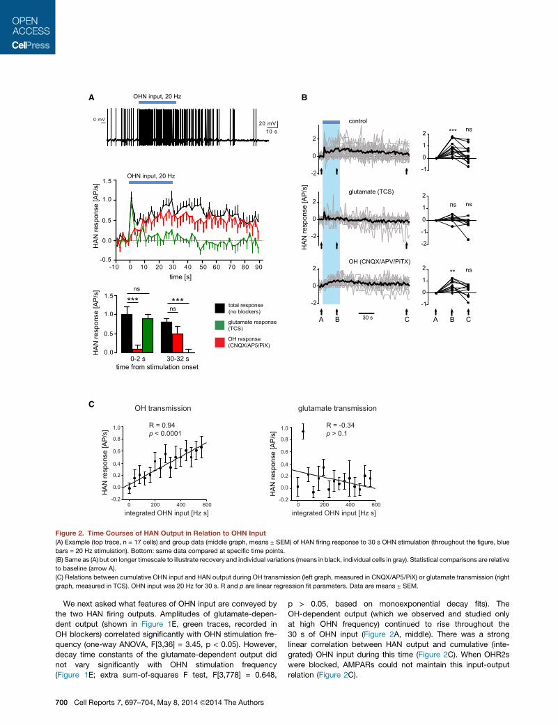
recordings from HANs. As expected from OHR2s present on
HANs (Eriksson et al., 2001; Willie et al., 2003), OHN stimulation
generated a significant progressive inward shift in the baseline
current (Figures 3A and 3B). The size of this current was the
same order of magnitude to currents evoked by a midnanomolar
concentration of bath-applied OH peptide (Figure 3B), possibly
providing a rough estimate of peptide levels arising from intrinsic
peptide release. The inward current shift was significant both in
control conditions (Figure 3B) and in CNQX (2.2 ± 0.6 pA, n = 8,
p < 0.01) and was confirmed to require OHR2 by block with TCS
(control + TCS: see Figure 3B; CNQX + TSC: 0.8 ± 0.7 pA, n = 7,
p > 0.2). The TCS-sensitive shift in inward current upon OHN
stimulation was not significantly different between control and
CNQX conditions (p > 0.05 in unpaired t test).
Mirroring the failure of glutamate to transmit a sustained firing
output (Figure 2A), there was a progressive falloff in glutamater-
gic currents (postsynaptic currents [PSCs]) during OHN stimula-
tion (note that CNQX-sensitive photostimulated PSCs were not
blocked by TCS, which was present throughout; Figures 3C–
3F). This falloff was seen in both the total excitatory PSC fre-
quency (Figure 3C) and in PSC success (i.e., increased failure
of flashes to evoke PSCs; Figure 3D). The amplitude of optically
evoked glutamatergic PSCs also tended to fall slightly with pro-
longed OHN stimulation (Figures 3E and 3F). Disappearance of
the OHN/HANAMPAR current response during prolonged
steady OHN stimulation was not due to irreversible vesicle
depletion, because in all cells tested, the response was seen
again after a 1–2 min ‘‘rest’’ without OHN stimulation (n = 40
cells).
DISCUSSION
Our results quantify the roles of OHN excitatory cotransmitters in
input-output operation of a key arousal-controlling module in the
brain. A central observation is that OH and glutamate convert the
same OHN input into strikingly different temporal patterns of
HAN spiking. These two spike responses could coexist in the
same postsynaptic cell (Figures 1A and 1B). OH cotransmission
was required for sustaining the postsynaptic firing responses
to OHN activity for physiologically relevant durations. This pro-
vides direct evidence that endogenous OH release mediates
spike transfer between brain circuits. In fact, under some condi-
tions, OH generated more spikes than coreleased glutamate
(Figure 1D).
We found that OH transmission required a higher presynaptic
activity than glutamate transmission, corroborating previous in-
ferences from less specific stimulation (Dutton and Dyball,
1979; Lundberg et al., 1986; Schone and Burdakov, 2012; van
den Pol, 2012; Verhage et al., 1991). At the level of spike output
of the OHN/HAN circuit, we found little evidence for interac-
tions betweenOH and glutamate, as implied by pharmacological
independence of the two outputs across stimulation intensities
and durations. This is surprising, because exogenously applied
OH peptides can modulate glutamate transmission in other cir-
cuits (Lambe and Aghajanian, 2003; van den Pol et al., 1998).
Perhaps OH-glutamate synergies depend on presynaptic
OHRs, which are differentially expressed and/or activated in
different circuits and at different levels of neural activity.
The glutamate firing response rose and fell rapidly, while OH
firing response escalated linearly during unchanging OHN stim-
ulation of physiological duration. If these differences reflected
the cutting of OHN axons in our preparation, we would expect
the reverse, i.e., OH transmission depleting rapidly, because
glutamate is made in the terminals but peptides are made in
the soma (Bear et al., 2001). The glutamate rundown was revers-
ible in the same cell after a stimulation break, suggesting func-
tional recycling. It is tempting to speculate that glutamate output
decay during unchanging input could help detection of subse-
quent input changes, similar to the role of adaptation in sensory
neurons tracking external stimuli (Carpenter, 2003).
Based on our data, the roles of glutamate and OH may be
viewed as extracting and encoding, in HAN output, two distinct
features of OHN input (Figure 4A). From this viewpoint, the tran-
sient AMPAR signaling may be seen as rapidly communicating
changes or trends in OHN activity (e.g., analogously to a deriva-
tive controller; DiStefano et al., 2012). Conversely, OHR2-depen-
dent operation (i.e., linear increase in output during constant
input) may function as an integral controller whose output is pro-
portional to integrated input (DiStefano et al., 2012). Interest-
ingly, when placed in a feedback loop, integral controllers pro-
vide the key operation required for stable output in diverse
systems (Astrom and Hagglund, 1995; Csete and Doyle, 2002;
DiStefano et al., 2012; Yi et al., 2000). Many existing lines of
experimental evidence argue that the OHN/HANmodule stabi-
lizes wakefulness and may be considered a part of a feedback
loop (e.g., via a hypothetical arrangement in Figure 4B). It may
therefore be important to investigate whether OH peptides
implement some form of integral control in the brain.
In summary, our data show that fast and slow cotransmitters
can convert OHN activity into parallel and nonredundant spike
streams in the same postsynaptic neuron. This clarifies func-
tional benefits of OH cotransmission for diversifying neural cir-
cuit performance and computation and offers a cybernetic
framework for reverse engineering pathophysiological OHR2
signaling.
EXPERIMENTAL PROCEDURES
Animal procedures followed United Kingdom Home Office regulations. ChR2
was introduced into OHNs using cre-dependent viruses in orexin-cre mice
(Schone et al., 2012; Matsuki et al., 2009). Effects of OH transmission were iso-
lated by blocking glutamate and GABA transmission and confirmed as
requiring OH receptors by blockade with 10 mM TCS-OX2-29 (‘‘TCS,’’ an
OHR2 receptor blocker; Huang et al., 2010; Smart et al., 2001; Xiao et al.,
2013) and/or 10 mMSB-334867 (‘‘SB,’’ an OHR1 blocker at this concentration;
Cell Reports 7, 697–704, May 8, 2014 ª2014 The Authors 701
A B
C D
E F
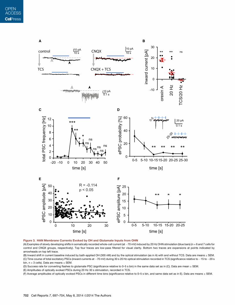
Figure 3. HAN Membrane Currents Evoked by OH and Glutamate Inputs from OHN
(A) Examples of slowly developing shifts in somatically recorded whole-cell current (at�70mV) induced by 20 Hz OHN stimulation (blue bars) (n = 6 and 7 cells for
control and CNQX groups, respectively). Top four traces are low-pass filtered for visual clarity. Bottom two traces are expansions at points indicated by
arrowheads on top left trace.
(B) Inward shift in current baseline induced by bath-applied OH (300 nM) and by the optical stimulation (as in A) with and without TCS. Data are means ± SEM.
(C) Time course of total excitatory PSCs (inward currents at �70 mV) during 30 s 20 Hz optical stimulation recorded in TCS (significance relative to�15 to�20 s
bin, n = 3 cells). Data are means ± SEM.
(D) Success rate for converting flashes to glutamate PSC (significance relative to 0–5 s bin) in the same data set as in (C). Data are mean ± SEM.
(E) Amplitudes of optically evoked PSCs during 20 Hz 30 s stimulation, recorded in TCS.
(F) Average amplitudes of optically evoked PSCs in different time bins (significance relative to 0–5 s bin, and same data set as in E). Data are means ± SEM.
702 Cell Reports 7, 697–704, May 8, 2014 ª2014 The Authors

+
-s
disturbance
e e w∫CX
w
A
B
OHR2
AMPAR
spikeoutput
input
OHN
HAN
Figure 4. Model for Functional Logic of OH-Glutamate Co-
transmission
(A) Cartoon of OHN/HAN circuit, overlayed with a theoretical engineering
scheme, viewing OHR2s and AMPARs as a control module generating
integral-derivative-like signals.
(B) A canonical integral feedback loop (simplified from Csete and Doyle, 2002;
Astrom and Hagglund, 1995). Integration in C compensates for disturbances
to output w, allowing w to follow s despite disturbance (Astrom and Hagglund,
1995; DiStefano et al., 2012). Hypothetically, to protect arousal signals (w) from
instability, C could correspond to OHR2-expressing cells (e.g., HANs) and e
could come from OHNs driven by positive inputs s (e.g., sounds; Mileykovskiy
et al., 2005) and negative-feedback inputs w (e.g., serotonin, Li et al., 2002).
The intermittent, disturbance-associated firing of OHN in vivo (Mileykovskiy
et al., 2005) is consistent with this position of OHNs in the feedback loop. Note
that integral (but not proportional or derivative) transformation of e by C is
necessary and sufficient for accurate and disturbance-resistant tracking of s
by w (Astrom and Hagglund, 1995). Indeed, when OH or OHR2 is knocked out,
OHNs cannot stabilize wakefulness (Chemelli et al., 1999; Lin et al., 1999;Willie
et al., 2003).
Smart et al., 2001). See Supplemental Experimental Procedures for further
detail.
SUPPLEMENTAL INFORMATION
Supplemental Information includes Supplemental Experimental Procedures
and one figure and can be found with this article online at http://dx.doi.org/
10.1016/j.celrep.2014.03.055.
ACKNOWLEDGMENTS
This work was supported by grants from the European Research Council (D.B.,
ref. 200500), UK Medical Research Council (D.B., ref. MC_UP_1202/2), and
Human Frontiers Science Foundation (D.B. and A.A., ref. RGY0076/2012).
We thank Lauren Mulholland for linguistic improvements to the text.
Received: June 29, 2013
Revised: February 5, 2014
Accepted: March 13, 2014
Published: April 24, 2014
REFERENCES
Adamantidis, A.R., Zhang, F., Aravanis, A.M., Deisseroth, K., and de Lecea, L.
(2007). Neural substrates of awakening probed with optogenetic control of hy-
pocretin neurons. Nature 450, 420–424.
Astrom, K., and Hagglund, T. (1995). PID Controllers: Theory, Design, and Tun-
ing (Research Triangle Park: Instrument Society of America).
Bear, M.F., Connors, B.W., and Paradiso, M.A. (2001). Neuroscience:
Exploring the Brain (Philadelphia: Lippincott Williams & Wilkins).
Burbach, J.P. (2011). What are neuropeptides? Methods Mol. Biol. 789, 1–36.
Burnstock, G. (2004). Cotransmission. Curr. Opin. Pharmacol. 4, 47–52.
Carpenter, R. (2003). Neurophysiology, Fourth Edition (London: Arnold).
Chemelli, R.M., Willie, J.T., Sinton, C.M., Elmquist, J.K., Scammell, T., Lee, C.,
Richardson, J.A., Williams, S.C., Xiong, Y., Kisanuki, Y., et al. (1999). Narco-
lepsy in orexin knockout mice: molecular genetics of sleep regulation. Cell
98, 437–451.
Crocker, A., Espana, R.A., Papadopoulou,M., Saper, C.B., Faraco, J., Sakurai,
T., Honda, M., Mignot, E., and Scammell, T.E. (2005). Concomitant loss of dy-
norphin, NARP, and orexin in narcolepsy. Neurology 65, 1184–1188.
Csete, M.E., and Doyle, J.C. (2002). Reverse engineering of biological
complexity. Science 295, 1664–1669.
de Lecea, L., Kilduff, T.S., Peyron, C., Gao, X., Foye, P.E., Danielson, P.E., Fu-
kuhara, C., Battenberg, E.L., Gautvik, V.T., Bartlett, F.S., 2nd., et al. (1998). The
hypocretins: hypothalamus-specific peptides with neuroexcitatory activity.
Proc. Natl. Acad. Sci. USA 95, 322–327.
de Lecea, L., Jones, B.E., Boutrel, B., Borgland, S.L., Nishino, S., Bubser, M.,
and DiLeone, R. (2006). Addiction and arousal: alternative roles of hypothalam-
ic peptides. J. Neurosci. 26, 10372–10375.
DiStefano, J., Stubberud, A., and Williams, I. (2012). Feedback and Control
Systems, Second Edition (New York: McGraw-Hill Education).
Dutton, A., and Dyball, R.E. (1979). Phasic firing enhances vasopressin release
from the rat neurohypophysis. J. Physiol. 290, 433–440.
Eriksson, K.S., Sergeeva, O., Brown, R.E., and Haas, H.L. (2001). Orexin/hypo-
cretin excites the histaminergic neurons of the tuberomammillary nucleus.
J. Neurosci. 21, 9273–9279.
Furutani, N., Hondo, M., Kageyama, H., Tsujino, N., Mieda, M., Yanagisawa,
M., Shioda, S., and Sakurai, T. (2013). Neurotensin co-expressed in orexin-
producing neurons in the lateral hypothalamus plays an important role in regu-
lation of sleep/wakefulness states. PLoS ONE 8, e62391.
Haas, H.L., and Reiner, P.B. (1988). Membrane properties of histaminergic
tuberomammillary neurones of the rat hypothalamus in vitro. J. Physiol. 399,
633–646.
Haas, H.L., Sergeeva, O.A., and Selbach, O. (2008). Histamine in the nervous
system. Physiol. Rev. 88, 1183–1241.
Haass, M., Cheng, B., Richardt, G., Lang, R.E., and Schomig, A. (1989). Char-
acterization and presynaptic modulation of stimulation-evoked exocytotic
co-release of noradrenaline and neuropeptide Y in guinea pig heart. Naunyn
Schmiedebergs Arch. Pharmacol. 339, 71–78.
Hara, J., Beuckmann, C.T., Nambu, T., Willie, J.T., Chemelli, R.M., Sinton,
C.M., Sugiyama, F., Yagami, K., Goto, K., Yanagisawa, M., and Sakurai, T.
(2001). Genetic ablation of orexin neurons in mice results in narcolepsy, hypo-
phagia, and obesity. Neuron 30, 345–354.
Hirose, M., Egashira, S., Goto, Y., Hashihayata, T., Ohtake, N., Iwaasa, H.,
Hata, M., Fukami, T., Kanatani, A., and Yamada, K. (2003). N-acyl 6,7-dime-
thoxy-1,2,3,4-tetrahydroisoquinoline: the first orexin-2 receptor selective
non-peptidic antagonist. Bioorg. Med. Chem. Lett. 13, 4497–4499.
Huang, S.C., Dai, Y.W., Lee, Y.H., Chiou, L.C., and Hwang, L.L. (2010). Orexins
depolarize rostral ventrolateral medulla neurons and increase arterial pressure
and heart rate in rats mainly via orexin 2 receptors. J. Pharmacol. Exp. Ther.
334, 522–529.
Lambe, E.K., and Aghajanian, G.K. (2003). Hypocretin (orexin) induces calcium
transients in single spines postsynaptic to identified thalamocortical boutons
in prefrontal slice. Neuron 40, 139–150.
Lee, M.G., Hassani, O.K., and Jones, B.E. (2005). Discharge of identified
orexin/hypocretin neurons across the sleep-waking cycle. J. Neurosci. 25,
6716–6720.
Li, Y., Gao, X.B., Sakurai, T., and van den Pol, A.N. (2002). Hypocretin/Orexin
excites hypocretin neurons via a local glutamate neuron-A potential
Cell Reports 7, 697–704, May 8, 2014 ª2014 The Authors 703

mechanism for orchestrating the hypothalamic arousal system. Neuron 36,
1169–1181.
Lin, L., Faraco, J., Li, R., Kadotani, H., Rogers, W., Lin, X., Qiu, X., de Jong,
P.J., Nishino, S., and Mignot, E. (1999). The sleep disorder canine narcolepsy
is caused by a mutation in the hypocretin (orexin) receptor 2 gene. Cell 98,
365–376.
Lundberg, J.M., Rudehill, A., Sollevi, A., Theodorsson-Norheim, E., and Ham-
berger, B. (1986). Frequency- and reserpine-dependent chemical coding of
sympathetic transmission: differential release of noradrenaline and neuropep-
tide Y from pig spleen. Neurosci. Lett. 63, 96–100.
Ma, X., Zubcevic, L., Bruning, J.C., Ashcroft, F.M., and Burdakov, D. (2007).
Electrical inhibition of identified anorexigenic POMC neurons by orexin/hypo-
cretin. J. Neurosci. 27, 1529–1533.
Matsuki, T., Nomiyama, M., Takahira, H., Hirashima, N., Kunita, S., Takahashi,
S., Yagami, K., Kilduff, T.S., Bettler, B., Yanagisawa,M., and Sakurai, T. (2009).
Selective loss of GABA(B) receptors in orexin-producing neurons results in dis-
rupted sleep/wakefulness architecture. Proc. Natl. Acad. Sci. USA 106, 4459–
4464.
Mignot, E., Taheri, S., and Nishino, S. (2002). Sleeping with the hypothalamus:
emerging therapeutic targets for sleep disorders. Nat. Neurosci. Suppl. 5,
1071–1075.
Mileykovskiy, B.Y., Kiyashchenko, L.I., and Siegel, J.M. (2005). Behavioral cor-
relates of activity in identified hypocretin/orexin neurons. Neuron 46, 787–798.
Mochizuki, T., Arrigoni, E., Marcus, J.N., Clark, E.L., Yamamoto,M., Honer, M.,
Borroni, E., Lowell, B.B., Elmquist, J.K., and Scammell, T.E. (2011). Orexin re-
ceptor 2 expression in the posterior hypothalamus rescues sleepiness in
narcoleptic mice. Proc. Natl. Acad. Sci. USA 108, 4471–4476.
Petreanu, L., Huber, D., Sobczyk, A., and Svoboda, K. (2007). Channelrhodop-
sin-2-assisted circuit mapping of long-range callosal projections. Nat. Neuro-
sci. 10, 663–668.
Peyron, C., Tighe, D.K., van den Pol, A.N., de Lecea, L., Heller, H.C., Sutcliffe,
J.G., and Kilduff, T.S. (1998). Neurons containing hypocretin (orexin) project to
multiple neuronal systems. J. Neurosci. 18, 9996–10015.
Peyron, C., Faraco, J., Rogers, W., Ripley, B., Overeem, S., Charnay, Y., Nev-
simalova, S., Aldrich, M., Reynolds, D., Albin, R., et al. (2000). A mutation in a
case of early onset narcolepsy and a generalized absence of hypocretin pep-
tides in human narcoleptic brains. Nat. Med. 6, 991–997.
Ren, J., Qin, C., Hu, F., Tan, J., Qiu, L., Zhao, S., Feng, G., and Luo, M. (2011).
Habenula ‘‘cholinergic’’ neurons co-release glutamate and acetylcholine and
activate postsynaptic neurons via distinct transmission modes. Neuron 69,
445–452.
Ripley, B., Fujiki, N., Okura, M., Mignot, E., and Nishino, S. (2001). Hypocretin
levels in sporadic and familial cases of canine narcolepsy. Neurobiol. Dis. 8,
525–534.
Sakurai, T. (2007). The neural circuit of orexin (hypocretin): maintaining sleep
and wakefulness. Nat. Rev. Neurosci. 8, 171–181.
Sakurai, T., Amemiya, A., Ishii, M., Matsuzaki, I., Chemelli, R.M., Tanaka, H.,
Williams, S.C., Richardson, J.A., Kozlowski, G.P., Wilson, S., et al. (1998).
Orexins and orexin receptors: a family of hypothalamic neuropeptides and G
protein-coupled receptors that regulate feeding behavior. Cell 92, 573–585.
Salio, C., Lossi, L., Ferrini, F., and Merighi, A. (2006). Neuropeptides as synap-
tic transmitters. Cell Tissue Res. 326, 583–598.
Schone, C., and Burdakov, D. (2012). Glutamate and GABA as rapid effectors
of hypothalamic ‘‘peptidergic’’ neurons. Front. Behav. Neurosci. 6, 81.
704 Cell Reports 7, 697–704, May 8, 2014 ª2014 The Authors
Schone, C., Cao, Z.F., Apergis-Schoute, J., Adamantidis, A., Sakurai, T., and
Burdakov, D. (2012). Optogenetic probing of fast glutamatergic transmission
from hypocretin/orexin to histamine neurons in situ. J. Neurosci. 32, 12437–
12443.
Smart, D., Sabido-David, C., Brough, S.J., Jewitt, F., Johns, A., Porter, R.A.,
and Jerman, J.C. (2001). SB-334867-A: the first selective orexin-1 receptor
antagonist. Br. J. Pharmacol. 132, 1179–1182.
Thannickal, T.C., Moore, R.Y., Nienhuis, R., Ramanathan, L., Gulyani, S., Al-
drich, M., Cornford, M., and Siegel, J.M. (2000). Reduced number of hypocre-
tin neurons in human narcolepsy. Neuron 27, 469–474.
van den Pol, A.N. (2003). Weighing the role of hypothalamic feeding neuro-
transmitters. Neuron 40, 1059–1061.
van den Pol, A.N. (2012). Neuropeptide transmission in brain circuits. Neuron
76, 98–115.
van den Pol, A.N., Gao, X.B., Obrietan, K., Kilduff, T.S., and Belousov, A.B.
(1998). Presynaptic and postsynaptic actions and modulation of neuroendo-
crine neurons by a new hypothalamic peptide, hypocretin/orexin.
J. Neurosci. 18, 7962–7971.
Verhage, M., McMahon, H.T., Ghijsen, W.E., Boomsma, F., Scholten, G., Wie-
gant, V.M., and Nicholls, D.G. (1991). Differential release of amino acids,
neuropeptides, and catecholamines from isolated nerve terminals. Neuron 6,
517–524.
Williams, R.H., Jensen, L.T., Verkhratsky, A., Fugger, L., and Burdakov, D.
(2007). Control of hypothalamic orexin neurons by acid and CO2. Proc. Natl.
Acad. Sci. USA 104, 10685–10690.
Williams, R.H., Alexopoulos, H., Jensen, L.T., Fugger, L., and Burdakov, D.
(2008). Adaptive sugar sensors in hypothalamic feeding circuits. Proc. Natl.
Acad. Sci. USA 105, 11975–11980.
Willie, J.T., Chemelli, R.M., Sinton, C.M., Tokita, S., Williams, S.C., Kisanuki,
Y.Y., Marcus, J.N., Lee, C., Elmquist, J.K., Kohlmeier, K.A., et al. (2003).
Distinct narcolepsy syndromes in Orexin receptor-2 and Orexin null mice: mo-
lecular genetic dissection of Non-REM and REM sleep regulatory processes.
Neuron 38, 715–730.
Winsky-Sommerer, R., Yamanaka, A., Diano, S., Borok, E., Roberts, A.J., Sa-
kurai, T., Kilduff, T.S., Horvath, T.L., and de Lecea, L. (2004). Interaction be-
tween the corticotropin-releasing factor system and hypocretins (orexins): a
novel circuit mediating stress response. J. Neurosci. 24, 11439–11448.
Woldan-Tambor, A., Biega�nska, K.,Wiktorowska-Owczarek, A., and Zawilska,
J.B. (2011). Activation of orexin/hypocretin type 1 receptors stimulates cAMP
synthesis in primary cultures of rat astrocytes. Pharmacol. Rep. 63, 717–723.
Xiao, F., Jiang, M., Du, D., Xia, C., Wang, J., Cao, Y., Shen, L., and Zhu, D.
(2013). Orexin A regulates cardiovascular responses in stress-induced hyper-
tensive rats. Neuropharmacology 67, 16–24.
Yamanaka, A., Tsujino, N., Funahashi, H., Honda, K., Guan, J.L., Wang, Q.P.,
Tominaga, M., Goto, K., Shioda, S., and Sakurai, T. (2002). Orexins activate
histaminergic neurons via the orexin 2 receptor. Biochem. Biophys. Res. Com-
mun. 290, 1237–1245.
Yi, T.M., Huang, Y., Simon, M.I., and Doyle, J. (2000). Robust perfect adapta-
tion in bacterial chemotaxis through integral feedback control. Proc. Natl.
Acad. Sci. USA 97, 4649–4653.
Yizhar, O., Fenno, L.E., Davidson, T.J., Mogri, M., and Deisseroth, K. (2011).
Optogenetics in neural systems. Neuron 71, 9–34.