cell structure and function - collin...
TRANSCRIPT

Prokaryotes
Cell Structure and Function

Characteristics of Living Things
MetabolismNutrient sourceChemical processes
GrowthResponsivenessReproduction
AsexualSexual

Size Comparison

ComparisonEukaryotesProkaryotes
DNANucleusOther OrganellesCell wallPlasma MembraneOther characteristics
SizeShapeLocation

Prokaryotic Structure & Function Topics
Cell Wall AppendagesCell WallPlasma MembraneCytoplasmDNA regionOrganellesReproductionAntimicrobial actions
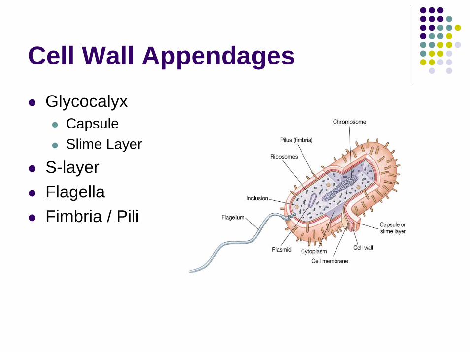
Cell Wall AppendagesGlycocalyx
CapsuleSlime Layer
S-layerFlagellaFimbria / Pili

GlycocalyxSecreted by all bacteria in some form
Produced insideExtruded to outside
Aid in survivabilityAid in pathogenicityNegatively chargedComposition
PolysaccharidesPolypetidesBoth

Glycocalyx: CapsuleOrganized repeating units ThickFirmly AttachedFunction
Protect from desiccation and other environmental hazardsEvade host defenses via phagocytosis

Glycocalyx: Slime LayerUnorganizedThinLoosely attachedViscousWater solubleFunction
AdherenceProtectionTrap nutrients
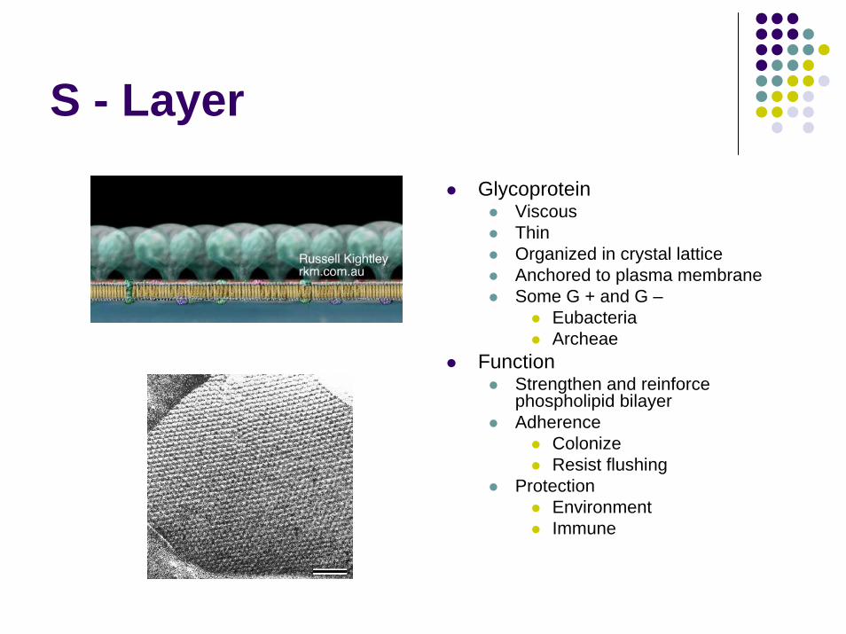
S - Layer
GlycoproteinViscousThinOrganized in crystal latticeAnchored to plasma membraneSome G + and G –
EubacteriaArcheae
FunctionStrengthen and reinforce phospholipid bilayerAdherence
ColonizeResist flushing
ProtectionEnvironmentImmune

BiofimsMicrobial community attached to surfaceResistant
ABImmune
TypesEnvironmentalInfectious
Dental PlaqueEndocarditisKidney StonesCF

FlagellaAppearance
10 -20 micrometersthin
ArrangementPartsFunction

Flagellar ArrangementMonotrichousAmphitrichousLophotrichous
TuftOne or both poles
PeritrichousVariations
Axial FilamentsAtrichous

Flagella ExamplesMonotrichous:
Pseudomonas aeruginosa
Amphitrichous:Spirullum volutans
Lophotrichous: E. coli
Peritrichous:Proteus vulgaris

Flagella
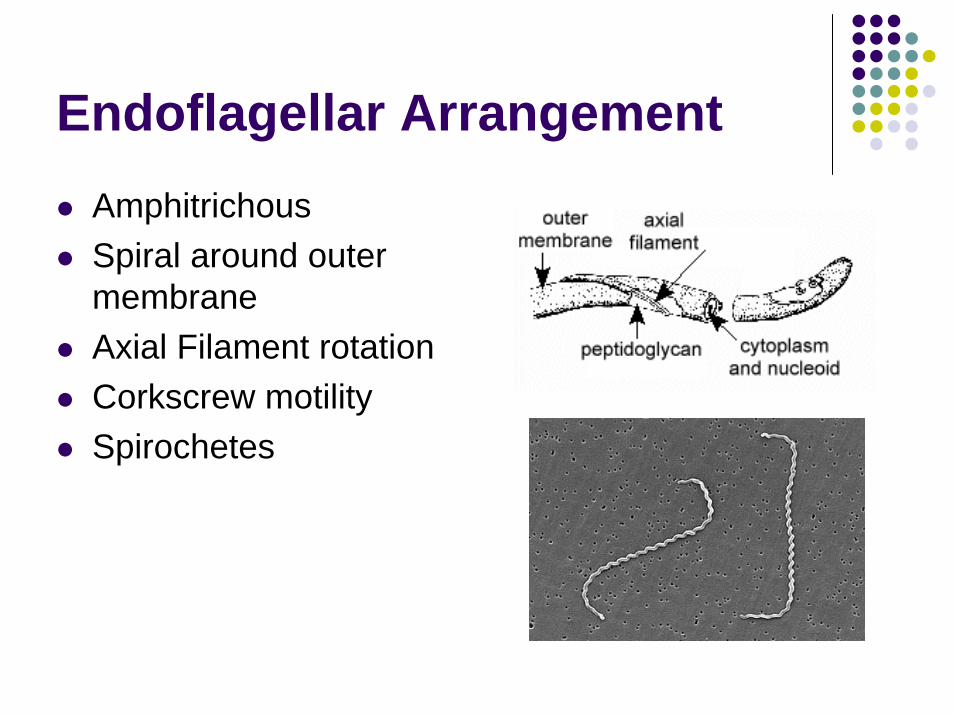
Endoflagellar ArrangementAmphitrichousSpiral around outer membraneAxial Filament rotationCorkscrew motilitySpirochetes

Flagella Anatomy: Exoflagella

Flagellar Parts: ExoflagellaFilament
Flagellin chains in helixHollow core for repairH protein antigen
HookProtein couplingL or curved shapeFunction: rotation
Basal BodyFunction: anchorCentral Protein RodRinged protein structures
4 rings for Gram Negative2 rings for Gram positive

Gram Negative Flagella

Flagellar StainSpecial StainDetermine
PresenceNumberLocation
Stains usedCarbolfuscinPararosanalineWith mordants

Flagellar MotilityRotationATP proton pumpFlagellar Motor
MotAMotBDriven by proton Gradient [outside to inside cell]
Moves toward food sourceClockwise= random movement [tumble]Counterclockwise=forward movement [run]

Motility Tests

Flagella SummaryFunction: Motility
PhototaxisChemotaxisPositive taxisNegative taxis
Proteins allow for classification into groups called serovarsTypes
Endoflagella [spirochetes]Exoflagella Arrangement
MonotrichousAmphitrichousLophotrichousPeritrichous
AnatomyFilamentHookBasal body
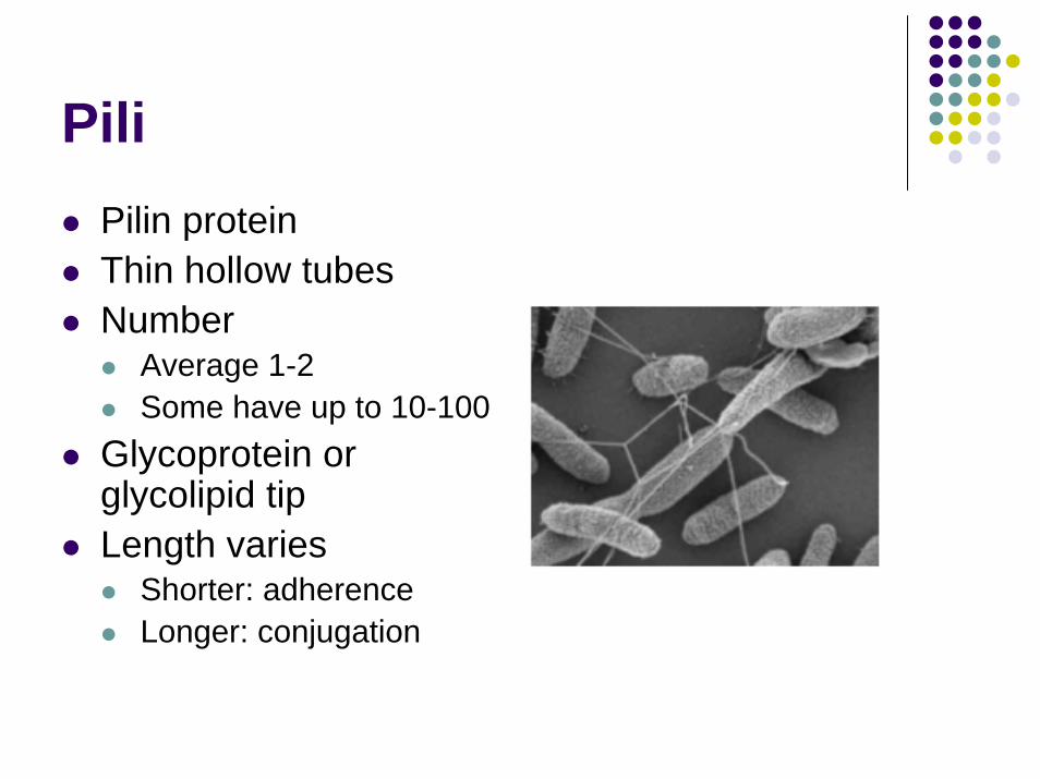
PiliPilin proteinThin hollow tubesNumber
Average 1-2Some have up to 10-100
Glycoprotein or glycolipid tipLength varies
Shorter: adherenceLonger: conjugation

Pili Structure

FimbriaGram Negative bacteriaShorter than flagella aka “Short pili”StickyFunction
AdherenceResist flushingBiofilm
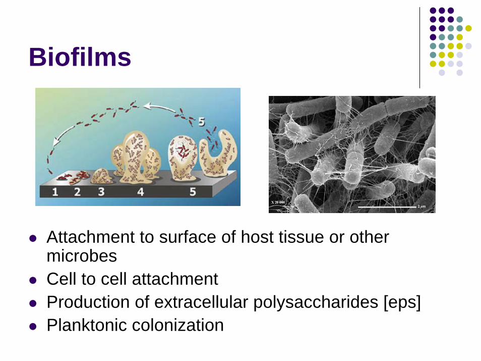
Biofilms
Attachment to surface of host tissue or other microbesCell to cell attachmentProduction of extracellular polysaccharides [eps]Planktonic colonization

Conjugation (Sex or F) Pili
Transfer DNA

Cell Wall StructurePeptidoglycan
Complex polysaccharideAlternating AminoSugars
LinkageTransglycolationBeta 1-4 Linkage
StructureNAM (muramic acid)NAG (glucosamine)
CrossbridgeLinkage: TranspeptidasesStructure
TetrapeptideFrom NAMBonded togetherShort chains
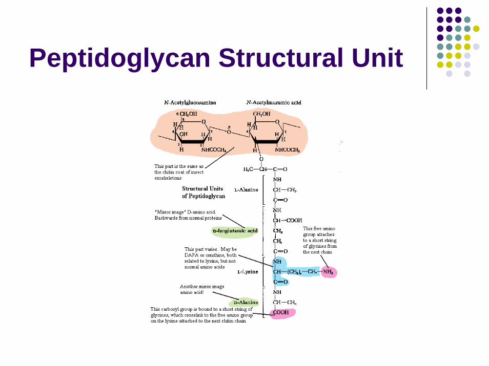
Peptidoglycan Structural Unit

Peptidoglycan Linkage
murein

Peptidoglycan Structure Compared

Cell Walls
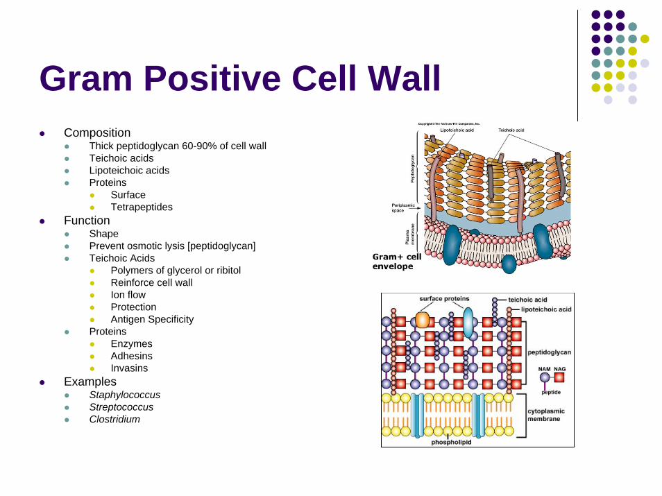
Gram Positive Cell WallComposition
Thick peptidoglycan 60-90% of cell wallTeichoic acidsLipoteichoic acidsProteins
SurfaceTetrapeptides
FunctionShapePrevent osmotic lysis [peptidoglycan]Teichoic Acids
Polymers of glycerol or ribitolReinforce cell wallIon flowProtectionAntigen Specificity
ProteinsEnzymesAdhesinsInvasins
ExamplesStaphylococcusStreptococcusClostridium

Gram Positive Bacteria

Gram Negative Cell WallComposition
Inner Cell Wall Peptidoglycan
1-2 layers make up 20%Intermediate cross linkageNO teichoic acid
Outer Cell MembraneLipid bilayer
PhospholipidsLipopolysaccharides [LPS]
Lipid AO AntigenCore Polysaccharide connects A-OLPS = Endotoxin
LipoproteinsProteins
Vary based on speciesPorins
FunctionPrevent osmotic lysisSemipermeable Reinforce membrane [LPS]Proteins
AdhesinsEnzymesInvasinsResist phagocytosisPassage of nutrients
ExamplesNeisseriaE coliPseudomonasProteus
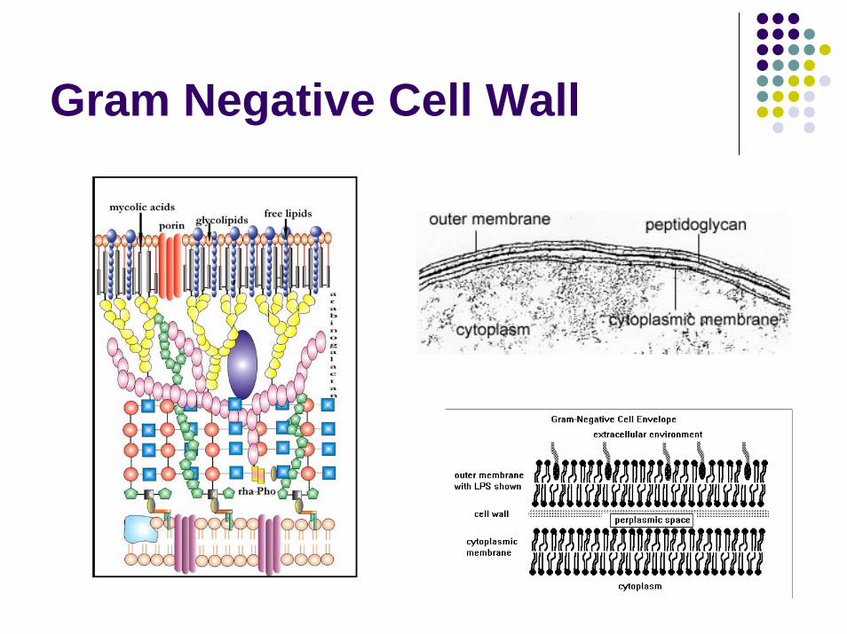
Gram Negative Cell Wall

Gram Negative Rods

LPSO Antigen
HexosesAntigenic specificitySmoothness to organism
Core PolysaccharideNAGPhosphorylated sugars [7,8 C]
Lipid ALipopolysaccharideGlucosamine moietiesToxic component
EndotoxinHyrophobicResistant: heat, acidReleased when cell wall disrupted

Cell Wall Comparison

Cell Wall Comparison #2

Gram Stain

Gram Stain Results

Acid Fast Cell WallComposition
PeptidoglycanArabinogalactan linkageGlycolipid: mycolic acidLipids
FreeGlycolipid
LipoarabinomannanPhosphatidyinositol mannosides
PeptidoglycolipidFunctions
Prevent osmotic lysisImpede entry of chemicals
Slower growing organismResistant to phagocytosis
ExamplesMycobacterium tuberculosisMycobacterium lepraeNocardia

Acid Fast Stain

Atypical Cell WallsArchaebacteria
No peptidoglycanPolysaccharidesS-layer [+/-]ProteinsGram +
ThickStain purple
Gram –Protein layerStain pink
Gram positive cell wall
Gram negative cell wall

Periplasm
CompositionGelatinous
LocationBetween peptidoglycan and cytoplasmic membrane
FunctionProteins
Enzymes for nutrient digestion [hyrolytic]Facilitate transfer of nutrientsStorage for toxins to be released into environment

Plasma MembraneComposition
PhospholipidPolarnonpolar
ProteinsIntegralPeripheral+/- polysaccharides
No sterols, hapanoidsFunction
Encloses cytoplasmSelectively permeableETCPeptidoglycan synthesis Aids in DNA replicationFlagella basal protein ringsWaste removalEndospore formation

Plasma Membrane TransportPassive
DiffusionOsmosisFacilitated Diffusion
UniporterChannel Proteins
WaterIons
ActiveAntiporterSymporterATP binding cassette
Gram negative bacteriaPeriplasm proteins
Group TranslocationChemical alterationMembrane impermeableExample: Sugars
GlucoseMannoseFructose

Cytoplasm: CytosolComposition
80% waterProteinsCH20LipidsIons
FunctionMetabolismEnzymes
ExoenzymesEndoenzymes

Organelles of Cytoplasm
Nucleoid regionRibosomesPlasmidsMesosomes

NucleoidComposition
ChromosomeSingle haploid moleculeDouble stranded DNAHelicalSupercoiled around protiens via topoisomerases
FunctionGenomeChemical reactions

Ribosomes: 70SComposition
rRNAProteinSubunits
50S30S
FunctionProtein synthesistRNAmRNA

PlasmidsComposition
DNASmall, helicalDouble strandedIndependent replication5-100 genes1-700 copies in cell
FunctionTypes
R plasmids: AB resistanceF plasmids: Fertility
Protein synthesisUnique proteins
ExotoxinsEndotoxins

Plasmid Replication

Plasmid Transformation

Transposons“Jumping Genes”Composition
DNANucleiodplasmid
FunctionCode for enzymes to transpose
Cut outRe-insert
AB resistance

Transposon cycle

Other OrganellesStorage Granules
SulfurGlycogenVolutin or Metachromic[Phosphate]Nitrogen
Gas vacuolesPhotosynthesisMagnetosomes

MesosomesInfoldings of cytoplasmic membraneConsidered artifact of slide preparation

EndosporesDormant alternate life formsSome Gram positive organismsIdentified
Size: large, smallShape: oval, rectangular, circular, club-shapedLocation : terminal, subterminal, central
ResistantHigh temperaturesDisinfectantsRadiationdrying
SurvivalGermination
Favorable ConditionsSpore coat rupturesVegetative cell forms

Spore StructureResistant coats
CortexSpore coat+/- exosporium
Nucleoid [DNA]RibosomesEnzymes

Spore FormationSporulation
DNA replicatesMembrane septums
One at endOne around DNA [forespore]Both synthesize peptidoglycan to form Cortex [inside layer]Calcium salts addedSpore Coat = keratin around cortexExosporium = outer lipid/protein

Spores: Gram Stain
Bacillus
Clostridium

Spores: Spore Stain

Atypical BacteriaMycoplasmas
Smallest free living cellLack cell wall
[no peptidoglycan]Sterols in cytoplasmic membraneMycolic acid [60%]Examples
Mycoplasma pneumoniaMycoplasma hominis

Atypical BacteriaRickettsia
PleomorphicObligate intracellular parasitesArthropod vectorsExamples
Rickettsia rickettsiitickRMSF
Rickettsia prowazekiiLouseEpidemic Typhus fever
Rickettsia typhiFleaEndemic Typhus fever

Atypical BacteriaChlamydia
CoccoidLack peptidoglycanObligate intracellular parasitesExamples
Chlamydia trachomatisSTD -> PID
Chlamydia pneumoniaEntire air way

Immune SystemCytokines
InterleukinsTNF
Complement ActivationInflammationPhagocytosis

Virulence Factors for Pathogenicity
Capsule [K antigen]Flagella [H antigen]Fimbria/Pili [adhesins]Outer membrane
LPS endotoxinProteins [porins]
Cell Wall PeptidoglycanProteins [A, M, T, R]
Periplasmic Space [enzymes]Plasma membraneEndosporesPlasmidsExotoxins
Enzyme action [hyaluronidase, DNAse, collagenase]Detergents [hemolysins, Staph a-toxinAlter cell metabolism [pertussis, cholera, diptheria toxins]Block Nerve function [tetanus, botulinum toxins]Others: dermonecrotic, erythrogenic

Physical and Chemical Actions on Bacterial Structures
Plasma MembranePolymyxinsDisinfectants: alcohol, chlorhexidine
Ribosomes30S: -cyclines
bind reversibly to distort tRNA/mRNA cannot align
50S: macrolides [-mycin]Bind reversiblyInhibit elongation

Chemical Actions on theBacterial Cell Wall

Summary

Questions?