cell transfer technology for tissue engineering
TRANSCRIPT

REVIEW Open Access
Cell transfer technology for tissueengineeringKeiko Akazawa1, Kengo Iwasaki2*, Mizuki Nagata1, Naoki Yokoyama3, Hirohito Ayame3, Kazumasa Yamaki3,Yuichi Tanaka3, Izumi Honda4, Chikako Morioka5, Tsuyoshi Kimura4, Motohiro Komaki2, Akio Kishida6,Yuichi Izumi1 and Ikuo Morita7
Abstract: We recently developed novel cell transplantation method “cell transfer technology” utilizingphotolithography. Using this method, we can transfer ex vivo expanded cells onto scaffold material in desiredpatterns, like printing of pictures and letters on a paper. We have investigated the possibility of this novel methodfor cell-based therapy using several disease models. We first transferred endothelial cells in capillary-like patterns onamnion. The transplantation of the endothelial cell-transferred amnion enhanced the reperfusion in mouse ischemiclimb model. The fusion of transplanted capillary with host vessel networks was also observed. The osteoblast- andperiodontal ligament stem cell-transferred amnion were next transplanted in bone and periodontal defects models.After healing period, both transplantations improved the regeneration of bone and periodontal tissues, respectively.This method was further applicable to transfer of multiple cell types and the transplantation of osteoblasts andperiodontal ligament stem cell-transferred amnion resulted in the improved bone regeneration compared withsingle cell type transplantation. These data suggested the therapeutic potential of the technology in cell-basedtherapies for reperfusion of ischemic limb and regeneration of bone and periodontal tissues. Cell transfertechnology is applicable to wide range of regenerative medicine in the future.
Keywords: Cell-based therapy, Cell transfer, Cell transplantation, Regeneration
BackgroundRecent progress in tissue engineering made it possible totreat various diseases using ex vivo expanded cells [1].The possibility of the cell-based therapy for many dis-eases has been widely studied. The selection of cell cul-ture methods, which facilitate therapeutic effect of thecells, and methods of transplantation, which include theideal carrier for the local transplantation, are essentialconsiderations in cell-based therapy [2]. We have devel-oped novel cell transplantation method “cell transfertechnology,” utilizing photolithography, which is oftenused for micropatterning formation in semiconductormanufacturing and printing [3]. This technology allowsus to transfer cultured cells onto scaffold material, likepictures and letters printed on a paper. We have investi-gated the possibility of this novel method for cell-basedtherapy using several disease models. In this review, we
outline the cell therapies that we have reported so farusing the cell transfer technique.
Cell transfer using photolithographyPhotolithography is a word with a prefix “photo” mean-ing light to “lithography,” which is originated from litho-graph. Literally, among various lithographic methods,photolithography uses the pattern made by light fordocument copy. Due to its precision, reproducibility,and mass productivity, photolithography is widely usedin the precision machinery industry and printing. Photo-lithography consists mainly of two steps, namely the de-piction of desired pattern on the substrate and “transfer”of the pattern to the product surface.We have developed “cell transfer technology” that en-
ables transfer of cultured cells onto the surface of trans-plantation scaffold. Figure 1 shows a schematic diagramof the cell transfer process by cell transfer technology.First, we made thin layer of tetraethyleneglycol (TEG) orpolyethyleneglycol (PEG) on glass substrate. Next, weapplied photomask on TEG/PEG layer and it was
* Correspondence: [email protected] of Nanomedicine (DNP), Graduate School of Medical andDental Sciences, Tokyo Medical and Dental University (TMDU), 1-5-45Yushima, Bunkyo-ku, Tokyo 113-8510, JapanFull list of author information is available at the end of the article
Inflammation and Regeneration
© The Author(s). 2017 Open Access This article is distributed under the terms of the Creative Commons Attribution 4.0International License (http://creativecommons.org/licenses/by/4.0/), which permits unrestricted use, distribution, andreproduction in any medium, provided you give appropriate credit to the original author(s) and the source, provide a link tothe Creative Commons license, and indicate if changes were made. The Creative Commons Public Domain Dedication waiver(http://creativecommons.org/publicdomain/zero/1.0/) applies to the data made available in this article, unless otherwise stated.
Akazawa et al. Inflammation and Regeneration (2017) 37:21 DOI 10.1186/s41232-017-0052-7

exposed to ultraviolet light. Ultraviolet irradiation par-tially collapses TEG/PEG chain and made the differencein the length of remaining TEG/PEG chain betweenphoto-masked and non-masked surface. The remaininglength of TEG/PEG appears as the difference between
hydrophilicity and hydrophobicity of the substrate sur-face. This difference is involved in the strength of celladhesion to the substrate surface (Fig. 2). Area with dis-rupted TEG/PEG is cell adhesive and area with pre-served TEG/PEG by photomasking is non-adhesive.
Fig. 1 Schema of cell transfer technology. a Procedure of cell transfer technology from construction of the transfer substrate to cell transfer. TEG/PEG layer (yellow) is formed on glass substrate. Following pattern drawing (photomask: red), UV light is radiated on the substrate. The surfaceexposed to UV light becomes cell adhesive area (green). Several hours of incubation after cell seeding on the substrate, the cells are transferredonto scaffold (pink) by making direct contact of the substrate to scaffold. After 18 to 20 h, cells are transferred onto scaffold. b Oseoblaststransferred using cell transfer substrate with grid patterning. Bar = 100 μm
Fig. 2 Non-adhesive and adhesive surface on cell transfer substrate. TEG/PEG chains are degraded by UV irradiation. Masked surface withpreserved TEG/PEG layer is hydrophobic and cell non- adhesive. Non-masked area, where TEG/PEG is degraded, is hydrophilic and cell adhesive
Akazawa et al. Inflammation and Regeneration (2017) 37:21 Page 2 of 6

Using this difference in hydrophilicity/hydrophilicity, itis possible to stick cells on substrate according to vari-ous patterns made by photomasking. Figure 1b demon-strates PKH26-labeled osteoblasts adhered to substratewith grid-like patterning. After adhesion of cells ontosubstrate, the substrate was placed onto scaffold materialmaking direct contact of the cell surface to scaffold.Eighteen to 24 h later, cells were transferred onto scaf-fold upon removal of the substrate. The transfer sub-strate was easily removed from scaffold without anydisturbance to the cells. In this step, the strength ofsubstrate-cell adhesion must be less than that betweencarrier and cells. This can be controlled by the strengthand duration of the UV irradiation on TEG/PEG surfaceafter masking. The degradation rate of PEG/TEG can beoptimized to maximize the cell transfer efficiency. Afterremoval of the transfer substrate from the scaffold, cellswere transferred onto the scaffold surface and were thenready for transplantation.For surface coating of the transfer substrate, we first
coated glass the plate with fluoroalkyl silane (FAS).However, because of the lower stability of patterning onthe substrate, FAS was replaced with PEG/TEG. We ob-served longer stability of patterns made with PEG/TEGon the substrate compared with those made with FAS.The time needed for cell transfer was also found to beshorter for PEG/TEG substrate than that with FAS.
Amnion as a scaffoldUsing cell transfer technology, ex vivo expanded cellswere transferred to scaffold materials including hydro-gels. Among these, we have used amnion, a part of am-niotic membrane, as a scaffold material for cell transferbecause of its elasticity, flexibility, and high success rateof cell transfer. Amniotic membrane is the biologicalmembrane forming amniotic sac, which keeps and pro-tects amniotic fluid and embryo inside [4]. Amnioticmembrane could be obtained at the time of delivery;however, it is discarded in general. The membrane iscomposed of amnion and chorion [5]. Amnion is theinner layer of the amniotic membrane. Amnion has beenused to treat dermal burns and ulceration, necrosis, andsevere inflammation of eyes as dressing material, takingadvantage of its anti-microbial and anti-fibrosis property[6, 7]. We isolated amnion from egg membrane by theremoval of chorion, and cell components were removedfrom amnion by high-hydrostatic pressure treatment [8,9]. The resulting decellurized amnion was used for celltransfer. Considering clinical applications, further evalu-ations are needed to determine the safety of the mem-brane in humans.Almost 100% of cells on transfer substrate were suc-
cessfully transferred onto amnion after several hours ofincubation. Amnion is great scaffold material for cell
transfer, as to facilitate high transfer efficiency. More-over, the most prominent characteristics of amnion uponcell transfer is the stability of transferred cells on themembrane [10, 11]. The transferred cells on amnion arefirmly adhered to amnion surface, and this makes it pos-sible to deform and trim the membrane with surgical in-struments [10, 11]. This unique characteristic allowseasy and reliable cell transplantation.
Patterned and layered cell transferOne of the notable features of photolithography is pre-cise transfer of substances according to the fine patteringdrawn on substrate. Taking advantage of this uniquecharacteristic of photolithography, we are able to trans-fer cells in any desired patterns onto scaffold. We madepatterns resembling to capillaries on glass substrate andtried to make capillaries by tissue engineering approach.We seeded bovine carotid artery endothelial cells(BCAECs) on transfer substrate with capillary-like pat-tering and transfer them onto amnion [12]. BCAECs,transferred onto amnion, showed capillary-like structure,and it was also revealed in electron microscopy that thecapillary was consisted of vessel wall by BCAECs andlumen inside. We transplanted the BCAEC-transferredamnion into mouse auricle and found that the capillarywas kept its structure until 5 days after transplantation.In vivo imaging demonstrated that the capillary func-tioned in host mouse ear. It is thus conceivable thatmimicking the anatomical structure of target tissue,prior to transplantation, may favor the results of celltransplantation. Microvasculature is one good exampleof this type of cell transplantation.On the other hand, we can also fabricate cell trans-
fer substrate with whole cell adhesive characteristics.With this substrate, cells are transferred onto amnionin layer structure. In case of cell transplantation re-quires cell numbers, not positioning, this sheet-likecell transplantation material is useful. We succeededin transfer of cells and fabricating cell sheet-like ma-terials using various cell types such as fibroblasts,mesenchymal stem cells (MSC), osteoblasts, andendothelial cells [10–13]. Approximately, 5 × 10⁵
cells/cm2 were transferred onto an amnion using thecell transfer technology. Furthermore, we transferredtwo different cell types in overlapping two layersusing cell transfer technology and named it “doublecell transfer” [11]. For this double cell transfer, wecultured two cell types on transfer substrate andtransfer them through single transfer process. Thismethod was applicable to three different cell typesand triple cell layers were successfully fabricated. Thismultiple cell transfer may enable unique cell trans-plantation considering three-dimensional cell structureand cell-cell communication.
Akazawa et al. Inflammation and Regeneration (2017) 37:21 Page 3 of 6
Various cell patterning and cell sheet formationmethods have been reported. For cell patterning, twomodalities have been mainly studied. One is to utilizethe specificity of cell adhesion to the extracellular matrixfor cell placement, and this can form sharp edge of pat-terns [14, 15]. However, it is difficult to form patternsusing more than two types of cells using this method.The other method involves the use of “force” to locatecells, including magnetic, electrokinetic, and fluidicforces [16–18]. These methods allow manipulation oflarge cell numbers, but some of them need labeling ofcells and may affect cell viability. The use of temperatureresponsive polymers has been studied and reported forcell sheet formation [19]. Cell sheets made through thismethod are too fragile to directly manipulate with surgi-cal instruments. Our method generates a cell sheet bytransferring a single cell layer onto a scaffold surfaceand endows physical strength that achieves stable celltransplantation.
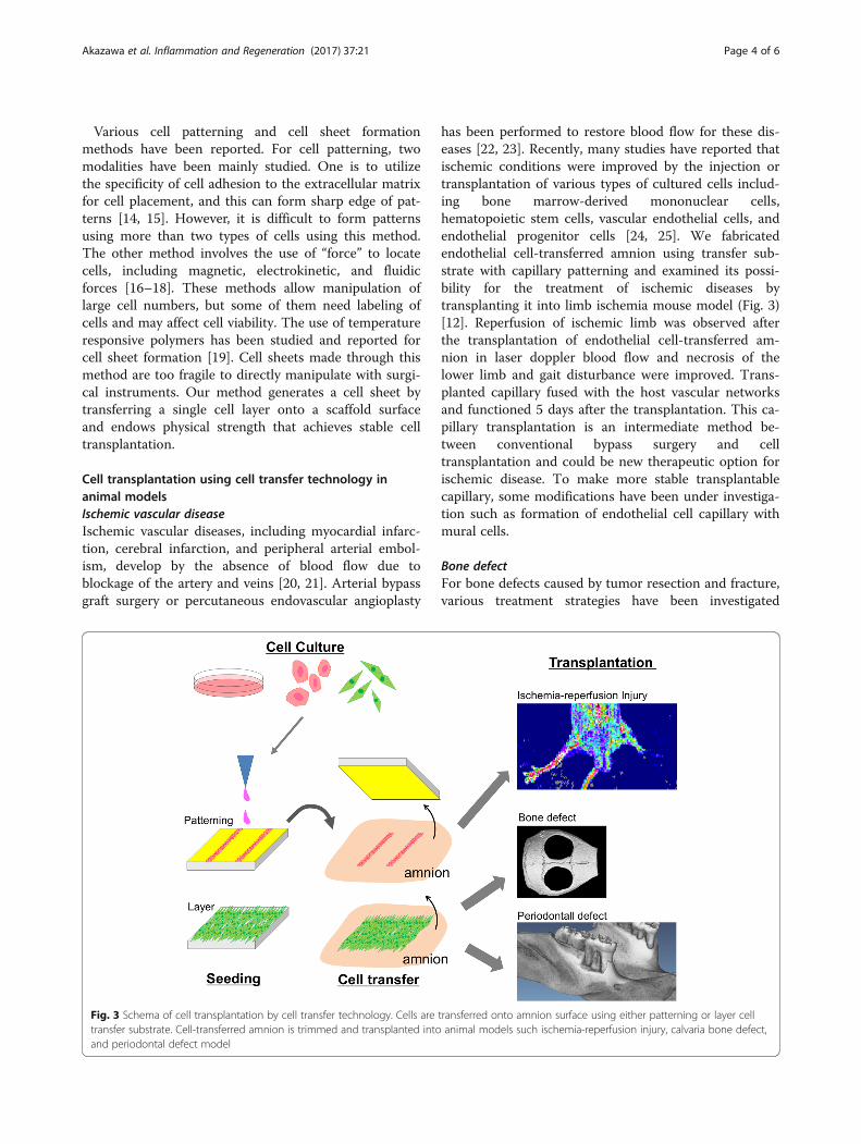
Cell transplantation using cell transfer technology inanimal modelsIschemic vascular diseaseIschemic vascular diseases, including myocardial infarc-tion, cerebral infarction, and peripheral arterial embol-ism, develop by the absence of blood flow due toblockage of the artery and veins [20, 21]. Arterial bypassgraft surgery or percutaneous endovascular angioplasty
has been performed to restore blood flow for these dis-eases [22, 23]. Recently, many studies have reported thatischemic conditions were improved by the injection ortransplantation of various types of cultured cells includ-ing bone marrow-derived mononuclear cells,hematopoietic stem cells, vascular endothelial cells, andendothelial progenitor cells [24, 25]. We fabricatedendothelial cell-transferred amnion using transfer sub-strate with capillary patterning and examined its possi-bility for the treatment of ischemic diseases bytransplanting it into limb ischemia mouse model (Fig. 3)[12]. Reperfusion of ischemic limb was observed afterthe transplantation of endothelial cell-transferred am-nion in laser doppler blood flow and necrosis of thelower limb and gait disturbance were improved. Trans-planted capillary fused with the host vascular networksand functioned 5 days after the transplantation. This ca-pillary transplantation is an intermediate method be-tween conventional bypass surgery and celltransplantation and could be new therapeutic option forischemic disease. To make more stable transplantablecapillary, some modifications have been under investiga-tion such as formation of endothelial cell capillary withmural cells.
Bone defectFor bone defects caused by tumor resection and fracture,various treatment strategies have been investigated
Fig. 3 Schema of cell transplantation by cell transfer technology. Cells are transferred onto amnion surface using either patterning or layer celltransfer substrate. Cell-transferred amnion is trimmed and transplanted into animal models such ischemia-reperfusion injury, calvaria bone defect,and periodontal defect model
Akazawa et al. Inflammation and Regeneration (2017) 37:21 Page 4 of 6

including local application of growth factor with bone-forming activity such as bone morphogenetic proteinsand transplantation of autologous or allogeanic bonegraft and artificial bone substitute materials such as hy-droxyapatite and beta-tricalcium phosphate [26, 27]. Re-cently, the therapeutic potential of cell transplantationwas proposed for bone defect and regeneration of bone de-fect have been reported by the transplantation of MSC andosteoblasts [28, 29]. We made transfer substrate with entirecell adhesive surface and transferred mouse osteoblasts(Kusa-A1) onto amnion (Fig. 3) [13]. Mouse calvaria bonydefect was created, and the osteoblast-transferred amnionwas placed to cover the defect. Approximately, 7.8 × 10⁴
cells were transplanted per defect. After 5 weeks, completeclosure of bone defect was observed in osteoblast-transplanted defect while control defect did not show thehealing. Compared with other test groups including injec-tion of osteoblast, only amnion transplantation, and notreatment, significant bone regeneration was found inosteoblast-transferred amnion defects. Transplanted osteo-blasts were found around newly formed bone 5 weeks postoperation, and it is suggested that those osteoblasts are dir-ectly involved in the regeneration of bone. The firm adhe-sion of osteoblasts to amnion may contribute to the longercell retention in bone defects and the prominent bone re-generation observed in osteoblast-amnion transplantationcompared with that in cell injection. These results suggestedthe therapeutic potential of osteoblast-transferred amnionfor the treatment of bone defect. We also observed theimprovement in bone healing by transplanting doublecell-transferred amnion, made using osteoblasts and MSCfrom periodontal ligament (periodontal ligament stemcells, PDLSC) [11]. We transferred human primary osteo-blasts and PDLSC onto amnion to form osteoblast-PDLSC layers on the membrane and transplanted theminto mouse calvarial bone defect model. Eight weeks afterthe transplantation, we observed the enhanced bone re-generation in osteoblast-PDLSC transplanted defectscompared with single cell transplanted (osteoblasts orPDLSC alone) defects. These results suggested the clinicalfeasibility of cell transfer technology in bone regeneration.
Periodontal defectPeriodontal disease is characterized by the chronic inflam-mation and destruction of tooth supporting tissues includ-ing bone, periodontal ligament, and cementum mainly dueto infection of gram-negative bacteria [30]. Conventionalperiodontal treatments consisted of mechanical removal ofbacterial factors, and it leads to reduction of inflammationand stability of disease status [31]. However, reconstructionof tooth supporting tissues, lost by the disease progression,was hardly observed. Although several regenerative ap-proaches have been applied clinically, sufficient regener-ation has not yet been achieved. Recently, it has been
demonstrated that cell transplantation is effective in regen-eration of periodontal tissues, including bone, periodontalligament, and cementum, using bone marrow-derivedMSC, adipose-derived MSC, PDLSC, and periosteum-derived cells [32]. We made periodontal defect model byremoving bone, cementum, and periodontal ligament in ratmaxillary molar and transplanted cells using cell transfertechnology (Fig. 3) [10]. As a cell type for transplantation,we selected PDLSC because they have been shown to pos-sess the differentiation capacity into various linages of cellssuch as osteoblast, adipocyte, chondrocyte, and cemento-blast [33]. We examined the regenerative potential ofPDLSC-transferred amnion (PDLSC-amnion) by trans-planting it into surgically created periodontal defect andcompared with the defect transplanted with amnion alone.After 4 weeks of healing period, enhanced periodontal tis-sue regeneration was observed in PDLSC-amnion trans-planted defects. Newly regenerated cementum, periodontalligament, and bone were observed in histological sections.These results suggested that transplantation of PDLSC-amnion could be a novel periodontal regenerative therapy.
ConclusionWe developed novel cell transplantation method “celltransfer technology” using photolithography technique.By transplanting cell transferred-amnion made by themethod, we have demonstrated the therapeutic potentialof the material in cell-based treatment including reperfu-sion of ischemic limb and regeneration of bone and peri-odontal tissues. Forming a fine patterning by culturedcell is the most unique characteristics of cell transfertechnique. In this regard, capillary patterning grafting istaking full advantage of this technology. However, thecell transplantation, which requires cell patterning,might be rather limited. Therefore, cell sheet-like am-nion, which we used for bone and periodontal defects,maybe widely applicable for tissue regeneration. Depend-ing on the target tissue to be regenerated, we can selectpatterned or non-patterned cell transfer substrate. Wealso found that the transferred cells are firmly andstably adhered on amnion and withstand against thedeformation and movement of the membrane by sur-gical manipulations. Additionally, the flexibility of am-nion enables us to transplant cultured cell in directcontact with defects or tissue surface, which is vitalimportance in certain regenerative cases. Taking theseadvantages and unique features, cell transfer technol-ogy is applicable to wide range of regenerative medi-cine in the future.
AbbreviationsBCAEC: Bovine carotid artery endothelial cell; MSC: Mesenchymal stem cells;PDLSC: Periodontal ligament stem cells; PEG: Polyethyleneglycol;TEG: Tetraethyleneglycol
Akazawa et al. Inflammation and Regeneration (2017) 37:21 Page 5 of 6

AcknowledgementsThe authors would like to thank all our laboratory members for their helpfuland constructive comments to this study.
FundingThis work is supported by the JSPS KAKENHI Grant Numbers 15K11381,15K11342, and 15K11380.
Availability of data and materialsNot applicable.
Authors’ contributionsKA, KI, MN, and MK performed the research. IH and CM contributed collectionof amniotic membrane. YT, NY, HA, and KY fabricated cell transfer base. TK, AK,and KA contributed decellularization of amnion. KA, KI, and IM wrote themanuscript. All authors read and approved the content of the manuscript.
Ethics approval and consent to participateNot applicable.
Competing interestsThe authors declare that they have no competing interests.
Publisher’s NoteSpringer Nature remains neutral with regard to jurisdictional claims inpublished maps and institutional affiliations.
Author details1Department of Periodontology, Graduate School of Medical and DentalSciences, Tokyo Medical and Dental University (TMDU), 1-5-45 Yushima,Bunkyo-ku, Tokyo 113-8510, Japan. 2Department of Nanomedicine (DNP),Graduate School of Medical and Dental Sciences, Tokyo Medical and DentalUniversity (TMDU), 1-5-45 Yushima, Bunkyo-ku, Tokyo 113-8510, Japan. 3LifeScience Laboratory, Research and Development Center, Dai Nippon PrintingCo., Ltd., 1-1-1 Kaga-cho, Shinjuku-ku, Tokyo 162-8001, Japan. 4Department ofComprehensive Reproductive Medicine, Graduate School of Medical andDental Science, Tokyo Medical and Dental University, 1-5-45 Yushima,Bunkyo-ku, Tokyo 113-8510, Japan. 5Department of Pediatrics andDevelopmental Biology, Graduate School of Medical and Dental Science,Tokyo Medical and Dental University (TMDU), 1-5-45 Yushima, Bunkyo-ku,Tokyo 113-8510, Japan. 6Department of Material-based Medical Engineering,Institute of Biomaterials and Bioengineering, Tokyo Medical and DentalUniversity (TMDU), 2-3-10, Kanda-Surugadai, Chiyoda-ku, Tokyo 101-0062,Japan. 7Department of Cellular Physiological Chemistry, Graduate School ofMedical and Dental Sciences, Tokyo Medical and Dental University (TMDU),1-5-45 Yushima, Bunkyo-ku, Tokyo 113-8510, Japan.
Received: 31 May 2017 Accepted: 18 September 2017
References1. Langer R, Vacanti JP. Tissue engineering. Science. 1993;260:920–6.2. Howard D, Buttery LD, Shakesheff KM, Roberts SJ. Tissue engineering:
strategies, stem cells and scaffolds. J Anat. 2008;213:66–72.3. Kobayashi A, Miyake H, Hattori H, Kuwana R, Hiruma Y, Nakahama K,
Ichinose S, Ota M, Nakamura M, Takeda S, Morita I. In vitro formation ofcapillary networks using optical lithographic techniques. Biochem BiophysRes Commun. 2007;358:692–7.
4. Niknejad H, Peirovi H, Jorjani M, Ahmadiani A, Ghanavi J, Seifalian AM.Properties of the amniotic membrane for potential use in tissueengineering. Eur Cell Mater. 2008;15:88–99.
5. Toda A, Okabe M, Yoshida T, Nikaido T. The potential of amnioticmembrane/amnion-derived cells for regeneration of various tissues. JPharmacol Sci. 2007;105:215–28.
6. Lo V, Pope E. Amniotic membrane use in dermatology. Int J Dermatol. 2009;48:935–40.
7. Altan-Yaycioglu R, Akova YA, Oto S. Amniotic membrane transplantation fortreatment of symblepharon in a patient with recessive dystrophicepidermolysis bullosa. Cornea. 2006;25:971–3.
8. Fujisato T, Minatoya K, Yamazaki S, Meng Y, Niwaya K, Kishida A, Nakatani T,Kitamura S. Preparation and recellularization of tissue engineered
bioscaffold for feat valve replacement. In: Mori H, Matsuda H, editors.Cardiovascular Regeneration Therapies Using Tissue EngineeringApproaches. Tokyo: Springer; 2005. p. 83–94.
9. Wilshaw SP, Kearney JN, Fisher J, Ingham E. Production of an acellular amnioticmembrane matrix for use in tissue engineering. Tissue Eng. 2006;12:2117–29.
10. Iwasaki K, Komaki M, Yokoyama N, Tanaka Y, Taki A, Honda I, Kimura Y, TakedaM, Akazawa K, Oda S, Izumi Y, Morita I. Tissue Eng Part A. 2014;20:693–704.
11. Akazawa K, Iwasaki K, Nagata M, Yokoyama N, Ayame H, Yamaki K, Tanaka Y,Honda I, Morioka C, Kimura T, Komaki M, Kishida A, Izumi Y, Morita I. SciRep. 2016;6:33286.
12. Akahori T, Kobayashi A, Komaki M, Hattori H, Nakahama K, Ichinose S, AbeM, Takeda S, Morita I. Implantation of capillary structure engineered byoptical lithography improves hind limb ischemia in mice. Tissue Eng Part A.2010;16:953–9.
13. Tsugawa J, Komaki M, Yoshida T, Nakahama K, Amagasa T, Morita I. Cell-printing and transfer technology applications for bone defects in mice. JTissue Eng Regen Med. 2011;5:695–703.
14. Fukuda J, Khademhosseini A, Yeh J, Eng G, Cheng J, Farokhzad OC, LangerR. Micropatterned cell co-cultures using layer-by-layer deposition ofextracellular matrix components. Biomaterials. 2006;27:1479–86.
15. Zhang S, Yan L, Altman M, Lässle M, Nugent H, Frankel F, Lauffenburger DA,Whitesides GM, Rich A. Biological surface engineering: a simple system forcell pattern formation. Biomaterials. 1999;20:1213–20.
16. Chiu DT, Jeon NL, Huang S, Kane RS, Wargo CJ, Choi IS, Ingber DE, WhitesidesGM. Patterned deposition of cells and proteins onto surfaces by using three-dimensional microfluidic systems. Proc Natl Acad Sci U S A. 2000;97:2408–13.
17. Ozkan M, Pisanic T, Scheel J, Barlow C, Esener S, Bhatia SN. Electro-opticalplatform for the manipulation of live cells. Langmuir. 2003;19:1532–8.
18. Chiou PY, Ohta AT, Wu MC. Massively parallel manipulation of single cellsand microparticles using optical images. Nature. 2005;436:370–2.
19. Elloumi-Hannachi I, Yamato M, Okano T. Cell sheet engineering: a uniquenanotechnology for scaffold-free tissue reconstruction with clinicalapplications in regenerative medicine. J Intern Med. 2010;267:54–70.
20. Schaller B, Graf R. Cerebral ischemia and reperfusion: the pathophysiologicconcept as a basis for clinical therapy. J Cereb Blood Flow Metab. 2004;24:351–71.
21. Shimokawa H, Yasuda S. Myocardial ischemia: current concepts and futureperspectives. J Cardiol. 2008;52:67–78.
22. Ferket BS, Spronk S, Colkesen EB, Hunink MG. Systematic review of guidelineson peripheral artery disease screening. Am J Med. 2012;125:198–208.
23. Allemang MT, Rajani RR, Nelson PR, Hingorani A, Kashyap VS. Prescribingpatterns of antiplatelet agents are highly variable after lower extremityendovascular procedures. Ann Vasc Surg. 2013;27:62–7.
24. Aranguren XL, Verfaillie CM, Luttun A. Emerging hurdles in stem cell therapyfor peripheral vascular disease. J Mol Med. 2009;87:3–16.
25. Sieveking DP, Ng MK. Cell therapies for therapeutic angiogenesis: back tothe bench. Vasc Med. 2009;14:153–66.
26. Dimitriou R, Jones E, McGonagle D, Giannoudis PV. Bone regeneration:current concepts and future directions. BMC Med. 2011;9:66.
27. McAllister BS, Haghighat K. Bone augmentation techniques. J Periodontol.2007;78:377–94.
28. Colnot C. Skeletal cell fate decisions within periosteum and bone marrowduring bone regeneration. J Bone Miner Res. 2009;24:274–82.
29. Akahane M, Nakamura A, Ohgushi H, Shigematsu H, Dohi Y, Takakura Y.Osteogenic matrix sheet-cell transplantation using osteoblastic cell sheetresulted in bone formation without scaffold at an ectopic site. J Tissue EngRegen Med. 2008;2:196–201.
30. Pihlstrom BL, Michalowicz BS, Johnson NW. Periodontal diseases. Lancet.2005;366:1809–20.
31. Graziani F, Karapetsa D, Alonso B, Herrera D. Nonsurgical and surgicaltreatment of periodontitis: how many options for one disease? Periodontol2000. 2017;75:152–88.
32. Ishikawa I, Iwata T, Washio K, Okano T, Nagasawa T, Iwasaki K, Ando T. Cellsheet engineering and other novel cell-based approaches to periodontalregeneration. Periodontol 2000. 2009;51:220–38.
33. Seo BM, Miura M, Gronthos S, Bartold PM, Batouli S, Brahim J, Young M,Robey PG, Wang CY, Shi S. Investigation of multipotent postnatal stem cellsfrom human periodontal ligament. Lancet. 2004;364:149–55.
Akazawa et al. Inflammation and Regeneration (2017) 37:21 Page 6 of 6