cell, volume 135 supplemental data the p. furiosus mre11 ... · carney). the p. furiosus hera ......
TRANSCRIPT

1
Cell, Volume 135
Supplemental Data The P. furiosus Mre11/Rad50 Complex
Promotes 5′ Strand Resection at a
DNA Double-Strand Break Ben B. Hopkins and Tanya T. Paull
Supplemental Experimental Procedures Expression constructs Pyrococcus furiosus Mre11/Rad50 was expressed from a bicistronic pET27b vector (gift from J. Carney). The P. furiosus HerA and NurA genes were amplified from P. furiosus genomic DNA (ATCC). The HerA and NurA genes were cloned separately into pETDuet-1 (Novagen), creating pTP1044 (HerA) and pTP1045 (NurA) expression constructs. The RadA gene was PCR amplified from P. furiosus genomic DNA and cloned into pETDuet-1 to create pTP1184. Point mutations were constructed using QuikChange Site-Directed Mutagenesis (Stratagene). Mutations were confirmed by DNA sequencing of the complete open reading frame. Details of construction available upon request. Protein expression and purification The PfMR complex was coexpressed in BL21 DE3 codonplus E. coli as previously described (Hopfner et al., 2000) with minor changes. Briefly, cells were grown to an OD600 0.8-1.0 and induced using 0.5 mM IPTG for 4-6 hrs at 37°C. Cells were harvested by centrifugation, flashed frozen in liquid N2, and lysed in high salt Nickel A buffer (50 mM KH2PO4 pH 7.0, 500 mM KCl, 2.5 mM imidazole, 10% glycerol, 20 mM β-mercaptoethanol [β-ME]) containing 1 mM phenylmethylsulphonyl fluoride (PMSF) and 0.5% Tween-20. Cells were lysed using a French Press (Thermo Fisher), sonicated, and insoluble material was pelleted by spinning at 100,000 × g for 1 hr at 4°C. The supernatant was heated at 70°C for 15 min and the precipitated protein was pelleted by spinning at 45,000 × g for 15 min at 4°C. The soluble fraction was loaded onto a Ni-NTA column (Qiagen) in low salt Nickel A buffer (50 mM KH2PO4 pH 7.0, 50 mM KCl, 2.5 mM imidazole, 10% glycerol, 20 mM β-ME) and washed with 10% Nickel B (Nickel A buffer containing 250 mM imidazole). Proteins were resolved with a linear gradient from 10-100% Nickel B buffer. Fractions containing MR were pooled, loaded onto a HiTrap Q HP column (GE) and washed in Buffer A (25 mM Tris pH 8.0, 100 mM NaCl, 10% glycerol, 1 mM dithiothreitol (DTT)). MR was eluted with 50% Buffer B (25 mM Tris pH 8.0, 1 M NaCl, 10% glycerol, 1 mM DTT). Peak fractions were pooled and mixed with 0.2% Tween-20 before running on a Superdex 200 10/300 GL column (GE) in Buffer A. Fractions containing the MR complex were aliquoted and flash frozen in liquid N2. Protein expression and recombinant protein purification for HerA and NurA was performed similar to the MR protocol with the exception that NurA was loaded onto a HiTrap SP HP instead of Q HP. RadA expression and purification was performed as previously described (Komori et al., 2000). Mre11, HerA, and NurA were expressed as N-

2
terminal 6×His fusion proteins, while Rad50 and RadA were expressed as untagged proteins. Protein concentrations were quantitated using BSA standards on SDS-PAGE gels. Gel filtration Wild-type purified HerA (290 nM hexamer), NurA (7 µM monomer), and Mre11/Rad50 (500 nM M2R2) were incubated as indicated in Buffer A (25 mM Tris pH 8.0, 100 mM NaCl, 10% glycerol) at 55°C for 15 min. Tween-20 was added to 0.1% before resolving on a Superdex 200 PC 3.2/30 column run in Buffer A. Fractions were run on 10% SDS-PAGE gels, transferred to PVDF membranes (Millipore), and probed with α-His antibodies conjugated to IRDYE-800 dye (Rockland). Western blots were analyzed on a Licor Odyssey system. Oligonucleotide DNA substrates The substrates in Fig. 5A were constructed as follows: Substrate 1 consisted of 5’ [32P]-labeled TP74 (5’-CTGCAGGGTTTTTGTTCCAGTCTGTAGCACTGTGTAAGACAGGCCA-3’) annealed to TP124 (5’-CATCTGGCCTGTCTTACACAGTGCTACAGACTGGAACAAAAACCCTGCAG-3’). Substrate 2 consisted of 5’ [32P]-labeled TP124 annealed to TP125 (5’-CTGCAGGGTTTTTGTTCCAGTCTGTAGCACTGTGTAAGACAGGCCAGATG-3’). Substrate 3 consisted of 5’ [32P]-labeled TP580 (5’-CTGCAGGGTTTTTGTTCCAGTCTGTAGCACTGTGTAAGACAGGCCsAsGsAsTsG-3’) annealed to TP124. Phosphorothioate bonds are indicated by “s” between the nucleotides. Substrate 4 consisted of 5’ [32P]-labeled TP694 (5’-AAGTAAATCATTAATCTAACAATGCGCTCATCGTCsAsTsCsCsT-3’) annealed to TP695 (5’-AGGATGACGATGAGCGCATTGTTAGATTAATGATTsTsAsCsTsT-3’). The oligonucleotide substrate in Fig. S4A consisted of 3’ [32P]-labeled TP124 annealed to TP580. The helicase assay substrate in Fig. S4C was constructed by annealing 5’ [32P]-labeled TP1760 (5’-CCACTACGTGAACCATCACCCAAATCAAGTTTTTTGGGGTCGAGGTGC-3’) to M13 single-stranded DNA. 3’ or 5’ [32P]-labeling was performed as described below. Preparation of oligonucleotide and DNA substrates Oligonucleotide DNA substrates (MWG) were either 3’ end-labeled with [α-32P]cordycepin (3’ deoxyadenosine) (NEN) and Terminal deoxytransferase (TdT) (Roche) or 5’ end-labeled with [γ-32P]ATP (NEN) and T4 Polynucleotide Kinase (PNK) (NEB) and annealed to a molar excess of unlabeled complementary oligonucleotide. The 2.5 kb internally-labeled substrate was generated by PCR using [α-32P]dTTP (NEN) and gel purified by native agarose gel electrophoresis and electroelution. The helicase assay substrate consisted of a 5’ [32P]-labeled 48 nt oligonucleotide annealed to M13 single-stranded DNA (NEB). The 5’ Biotin/bead substrate was constructed by ligating an oligonucleotide containing a 5’ Biotin-TEG (MWG) to the plasmid DNA substrate and attaching the DNA to streptavidin-coated magnetic beads (Dynal). The beads were washed extensively to remove unbound DNA. Labeled linear dsDNA for the joint-molecule assay was prepared by PCR amplification of a 415 bp region of M13 phage DNA in the presence of [α-32P]dTTP and gel purification by native gel electrophoresis and electroelution. Labeled linear DNA for strand invasion experiments was prepared by PCR amplification of a 2.2 kb region of pTP179 in the presence of [α-32P]dATP (NEN), which was digested at both ends with SphI (NEB) to produce a 1.4 kb linear DNA fragment and gel purified by native gel electrophoresis. Supercoiled 3.6 kb pTP179 plasmid DNA was prepared by sucrose gradient centrifugation.

3
Helicase assays Helicase reactions contained 0.15 nM [32P]-labeled oligonucleotide/ssM13 DNA complexes in 20 µl reactions containing 20 mM HEPES pH 7.5, 5 mM MgCl2, 1 mM ATP, 50 mM NaCl, 1 mM DTT, and 100 µg/ml BSA. Reactions were incubated at 55°C for 1 hr before separation by native agarose electrophoresis in 0.8% TAE agarose gels for 1 hr at 3.5 V/cm. Reactions were terminated by the addition of 0.2% SDS, 10 mM EDTA, and 0.5 µg/ml Ethidium bromide (EtBr). Gels were dried and products were analyzed by phosphorimager (Biorad or GE). See above for oligonucleotide sequences and preparation of the duplex substrate. ATPase assays Wild-type or K164A HerA was incubated in 10 µl reactions containing 25 mM MOPS pH 7.0, 5 mM MgCl2, 40 mM NaCl, 2 mM DTT, and 50 µM [γ-32P]ATP (MP Biomedical) at 65°C for 1 hr in the presence or absence of 100 ng linear double-stranded DNA (1 kb ladder, Invitrogen). Reactions were stopped by the addition of 0.2% SDS, 10 mM EDTA, and 0.5 µg/ml EtBr. Reactions were analyzed by thin-layer chromatography (TLC) as described previously (Bhaskara et al., 2007). Processing of gels and membranes for Southern hybridization Agarose gels were washed into 20× SSC (3 M NaCl, 0.3 M sodium citrate) and DNA was transferred by capillary action onto nylon membranes (NEN) overnight in 20× SSC. Membranes were probed with RNA complementary to either the 3’ or 5’ strand of the DNA substrate in a 1 kb region on one end, adjacent to the SacI site. The probes were internally labeled with [α-32P]CTP (NEN) and were made using Riboprobe System T7 (Promega) according to the manufacturer’s instructions. The 3’ strand-specific probe of a 1 kb region adjacent to the bead attachment site was made by primer extension with [α-32P]dATP (NEN). Denatured DNA controls consisted of substrate DNA denatured with NaOH.

4
Supplemental Figures
Figure S1. Protein gels of purified recombinant P. furiosus Mre11/Rad50, HerA, NurA, and RadA. (A) 0.5 µg wild-type and mutant MR, HerA, and NurA run on 10% SDS-PAGE gels as indicated. (B) 1 µg wild-type RadA run on 16% SDS-PAGE gels. Migration of molecular weight markers are indicated. Gels were stained with Colloidal Blue Coomassie stain (Invitrogen).

5
Figure S2. P. furiosus NurA and HerA interact physically. Superdex 200 gel filtration of purified HerA and NurA. Purified proteins were incubated alone (HerA: top panel, NurA: middle panel) or in combination (bottom panel) at 55°C for 15 min before gel filtration, as indicated, and fractions were analyzed by SDS-PAGE and α-His western blot. Migration of molecular weight markers are indicated.

6
Figure S3. P. furiosus HerA facilitates NurA activity in magnesium. Nuclease assays were performed using 2.5 kb internally [32P]-labeled dsDNA. Reactions contained 13.4 nM wild-type HerA, 96.2 nM wild-type NurA, 80 mM NaCl, 1 mM ATP, and 5 mM MgCl2 or MnCl2 as indicated. Reactions were incubated at 65°C for 1 hr before separation on a native agarose gel.

7
Figure S4. Biochemical characterization of NurA and HerA. (A) Exonuclease assays were performed with 190 nM wild-type NurA (+), 190 or 960 nM D51A, and 190 or 960 nM D126A NurA, as indicated. The 50 bp double-stranded oligonucleotide substrate was 3’ [32P]-labeled on the top strand and contained 5 phosphorothioate bonds (“sssss”) on the 3’ end of the bottom strand. Reactions included 4 nM substrate, 60 mM NaCl, and 1 mM MnCl2 and were carried out at 65°C for 1 hr. Reaction products were analyzed on 15% denaturing polyacrylamide sequencing gels. Migration of single-stranded DNA markers are indicated. (B) HerA ATPase assays were performed with 1-50 nM wild-type or 20-40 nM K164A HerA and 100 ng dsDNA as indicated using [γ-32P]ATP. Reactions were incubated at 65°C for 1 hr and reaction products were analyzed by TLC. The analogous mutation (K153A) was shown to inhibit helicase activity in S. acidocaldarius HerA (Constantinesco et al., 2004). Positions of the origin, ATP, and inorganic phosphate (Pi) are indicated. (C) Wild-type HerA helicase assays were performed with a 5’ [32P]-labeled 48 nt oligonucleotide/M13 ssDNA duplex. Reactions contained 0.3, 0.7, 1.3, 2.7, or 6.2 nM wild-type HerA and 9.6 nM NurA D51A as indicated. Reactions were incubated at 55°C for 1 hr before separation on native 0.8% TAE agarose gels. Lane 1 contains boiled substrate. The positions of the oligonucleotide/ssM13 complex and oligonucleotide are indicated.

8
Figure S5. Cooperative DNA degradation by HerA and NurA require catalytically active enzymes. Nuclease assays were performed using 2.5 kb internally [32P]-labeled dsDNA. Reactions contained 13.4 nM wild-type HerA, 96.2 nM wild-type NurA, 5 mM MgCl2, 1 mM ATP, and 100 mM NaCl. HerA K164A and NurA D51A mutant proteins were included as indicated. Reactions were incubated at 65°C for 1 hr before separation on a native agarose gel.
9
0
20
40
60
80
100
120
- protein full reaction no ATP no MR no NurA no HerA
Perc
en
tag
e o
f fu
ll-l
en
gth
su
bst
rate
rem
ain
ing
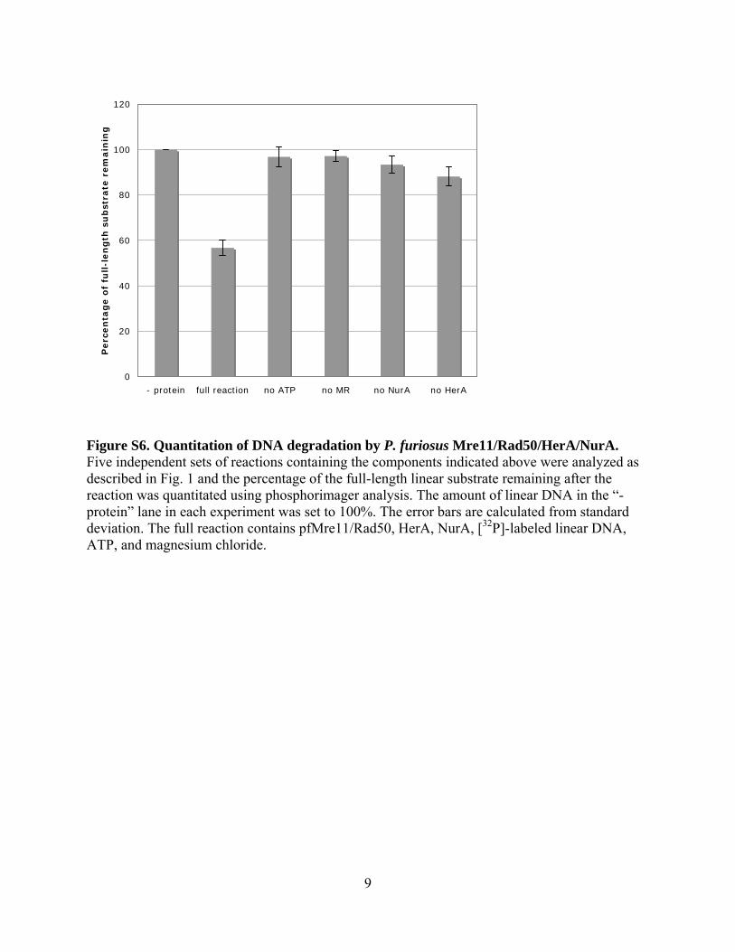
Figure S6. Quantitation of DNA degradation by P. furiosus Mre11/Rad50/HerA/NurA. Five independent sets of reactions containing the components indicated above were analyzed as described in Fig. 1 and the percentage of the full-length linear substrate remaining after the reaction was quantitated using phosphorimager analysis. The amount of linear DNA in the “- protein” lane in each experiment was set to 100%. The error bars are calculated from standard deviation. The full reaction contains pfMre11/Rad50, HerA, NurA, [32P]-labeled linear DNA, ATP, and magnesium chloride.

10

11
Figure S7. Primer extension to determine length of DNA resected by P. furiosus Mre11/Rad50. (A) Schematic of primer extension reaction. For simplicity, only the bottom strand of pTP163 is shown. The lengths of the full-length primer extension product and primer are indicated. The internally labeled primer extension product is indicated by the dashed line. Major MR resection sites are indicated as arrow heads and grouped into A, B, and C subgroups containing 2 or 3 cut sites. (B) P. furiosus Mre11/Rad50 resection reactions were performed with 2.3 nM pTP163 linearized with SacI and 165 nM Mre11/Rad50 at 65°C for 30 min or 1 hr as indicated. Reactions were then used in primer extension reactions and the primer extension products were analyzed on 10% polyacrylamide denaturing sequencing gels. The position of the 150 nt primer extension product and the various resected products are indicated. Single-stranded DNA markers are indicated.

12
Figure S8. P. furiosus Mre11/Rad50 performs endonucleolytic cleavage of double-strand DNA oligonucleotide duplexes. Substrate 3 from Figure 5 was [32P]-labeled on the 3’ end of the top strand or on the 5’ end of the top strand. Reactions were performed with pfMR wild-type, Mre11-H85L, or Rad50-S793R mutant complexes as indicated and analyzed as described for Fig. 5. All reactions were performed in the presence of 5 mM magnesium chloride. The positions of the nucleolytic cuts are shown with vertical arrows in the diagrams at left. The cleavage sites shown in gray arrows are not visible with the 3’ labeled-substrate, thus the internal cleavage events must occur first or always coincident with the cleavage events near the 5’ end of the top strand.
13
0
0.2
0.4
0.6
0.8
1
1.2
- pro
tein
full re
actio
n
no R
adA
no scD
NA
no A
TP
no M
R
no N
urA
no H
erA
RadA
only
% j
oin
t m
ole
cule
fo
rmati
on
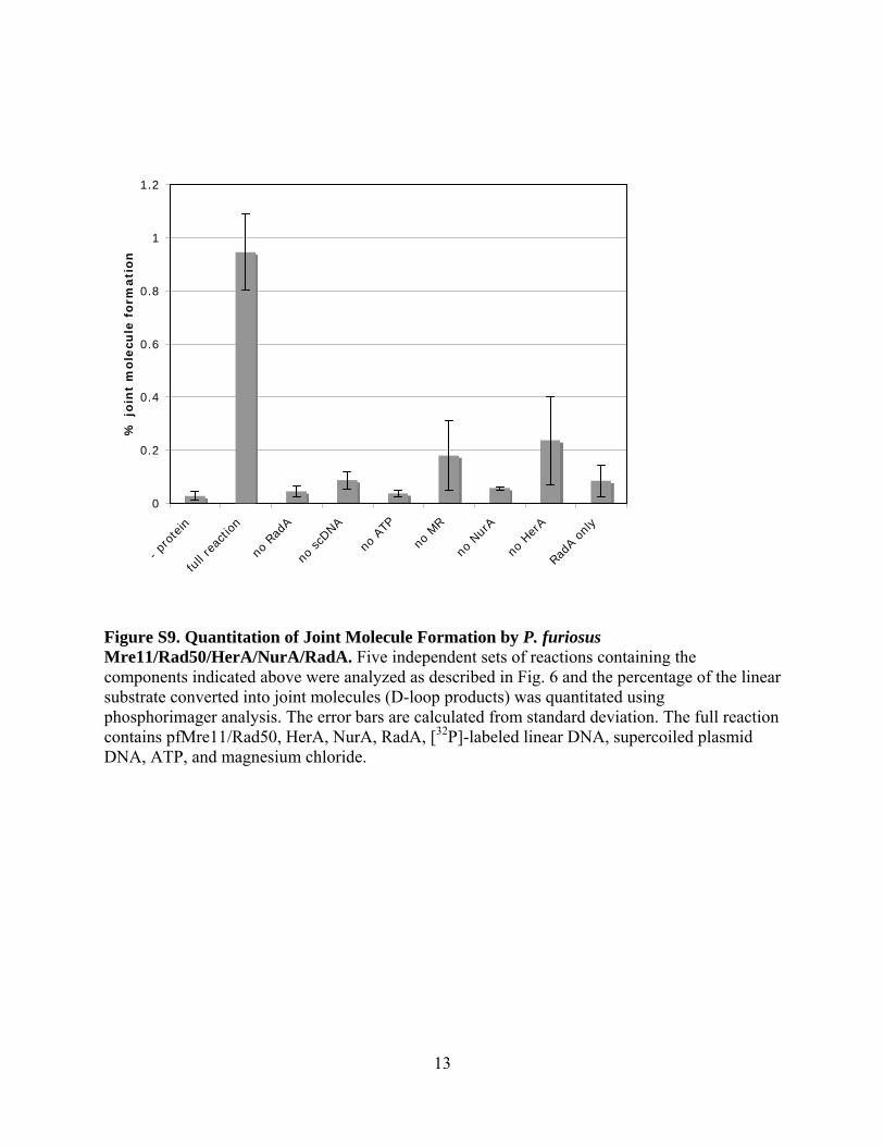
Figure S9. Quantitation of Joint Molecule Formation by P. furiosus Mre11/Rad50/HerA/NurA/RadA. Five independent sets of reactions containing the components indicated above were analyzed as described in Fig. 6 and the percentage of the linear substrate converted into joint molecules (D-loop products) was quantitated using phosphorimager analysis. The error bars are calculated from standard deviation. The full reaction contains pfMre11/Rad50, HerA, NurA, RadA, [32P]-labeled linear DNA, supercoiled plasmid DNA, ATP, and magnesium chloride.

14
Supplemental References
Bhaskara, V., Dupre, A., Lengsfeld, B., Hopkins, B.B., Chan, A., Lee, J.H., Zhang, X., Gautier, J., Zakian, V., and Paull, T.T. (2007). Rad50 adenylate kinase activity regulates DNA tethering by Mre11/Rad50 complexes. Mol Cell 25, 647-661.
Constantinesco, F., Forterre, P., Koonin, E.V., Aravind, L., and Elie, C. (2004). A bipolar DNA
helicase gene, herA, clusters with rad50, mre11 and nurA genes in thermophilic archaea. Nucleic Acids Res 32, 1439-1447.
Hopfner, K.P., Karcher, A., Shin, D., Fairley, C., Tainer, J.A., and Carney, J.P. (2000). Mre11
and rad50 from pyrococcus furiosus: cloning and biochemical characterization reveal an evolutionarily conserved multiprotein machine. J Bacteriol 182, 6036-6041.
Komori, K., Miyata, T., DiRuggiero, J., Holley-Shanks, R., Hayashi, I., Cann, I.K., Mayanagi,
K., Shinagawa, H., and Ishino, Y. (2000). Both RadA and RadB are involved in homologous recombination in Pyrococcus furiosus. J Biol Chem 275, 33782-33790.