cell-wall polysaccharides of developing flax plants’ · cell-wall polysaccharides of developing...
TRANSCRIPT

Plant Physiol. (1996) 110: 721-729
Cell-Wall Polysaccharides of Developing Flax Plants’
Tatyana A. Gorshkova, Sarah E. Wyatt, Vadim V. Salnikov, David M. Gibeaut, Marsel R. Ibragimov, Vera V. Lozovaya, and Nicholas C. Carpita*
Laboratory of Cell Wall Biochemistry, lnstitute of Biology, Russian Academy of Sciences, Kazan 420503, Russia (T.A.C., V.V.S., M.R.I., V.V.L.); and Department of Botany and Plant Pathology, Purdue University,
West Lafayette, Indiana 47907-1 155 (S.E.W., D.M.G., N.C.C.)
Flax (Linum usifafissimum 1.) fibers originate from procambial cells of the protophloem and develop in cortical bundles that en- circle the vascular cylinder. We determined the polysaccharide composition of the cell walls from various organs of the developing flax plant, from fiber-rich strips peeled from the stem, and from the xylem. Ammonium oxalate-soluble polysaccharides from all tissues contained 5-linked arabinans with low degrees of branching, rham- nogalacturonans, and polygalacturonic acid. The fiber-rich peels contained, in addition, substantial amounts of a buffer-soluble, 4-linked galactan branched at the 0 2 and 0 3 positions with nonreducing terminal-galactosyl units. l h e cross-linking glycans from all tissues were (fucogalacto)xyloglucan, typical of type-l cell walls, xylans containing (1 +4)-/3-~-xylosyl units branched exclu- sively at the xylosyl O 2 with f-(4-Omethyl)-glucosyluronic acid units, and (ga1acto)glucomannans. lissues containing predomi- nantly primary cell wall contained a larger proportion of xyloglu- can. The xylem cells were composed of about 60% 4-xylans, 32% cellulose, and small amounts of pectin and the other cross-linking polysaccharides. l h e noncellulosic polysaccharides of flax exhibit an uncommonly low degree of branching compared to similar polysaccharides from other flowering plants. Although the relative abundance of the various noncellulosic polysaccharides varies widely among the different cell types, the linkage structure and degree of branching of severa1 of the noncellulosic polysaccharides are invariant.
Flax (Linum usitatissimum L.) is an agronomically impor- tant source of natural fibers, especially in areas where cotton cannot be grown. The United States is a major producer of ”linseed” oil from seeds of the flax plants, whereas about 80% of the flax in eastern Europe is culti- vated for fiber, with two-thirds of that produced in Russia. In addition to their use in spinning and weaving into fabrics, these fine but durable fibers have many other com- mercial and environmental uses (Oosten, 1988). Studies of the chemical composition of the fiber cells have focused on investigations of the processed fibers and fibers in intact
Supported by grant No. DE-FG02-88ER13903 of the Division of Biological Energy Sciences, U.S. Department of Energy, to N.C.C., grant No. RH-8000 from the International Science Foun- dation to T.A.G., V.V.S., M.R.I., and V.V.L., and a grant from the Purdue University International Programs in Agriculture. Journal paper No. 14,938 of the Purdue University Agricultura1 Station.
* Corresponding author; e-mail [email protected]; fax 1-31 7-496 -1 823.
and “retted” plants, and attempts have been made to char- acterize some of the key alterations caused by an extensive processing that has an impact on fiber quality.
The fibers originate from procambial cells in the proto- phloem (Esau, 1977). Bundles of about a dozen cells each encircle the vascular cylinder and produce thick cellulosic walls that nearly fill the lumen at maturity. Mature fibers contain 60 to 70% cellulose (Sharma, 1986; Morvan et al., 1989; Lozovaya et al., 1990). Although the remaining ma- terial is rich in pectin, the noncellulosic polysaccharides have not been fully characterized (Davis et al., 1990; Mc- Dougall et al., 1993). After harvest, the plants are retted (by dew retting in the field or by warm-water retting in facto- ries), a process that helps to dissociate the bundles of fibers from other parts of the stem. Retting results in degradation of some pectins to free the fiber bundles from the pectin- rich sheaths of the cortical cells. Microbial endopolygalac- turonase (Chesson, 1979) or cultures of Erwinia carotovora (Morvan et al., 1989) can be used to mimic the retting process. Unlike cotton fibers, which grow as single epider- mal hair cells, the strength and integrity of the flax fibers rely to a certain extent on preservation of the cell-cell cementing during the retting process.
Morvan and colleagues (Morvan et al., 1989; Davis et al., 1990) provided data on the monosaccharide composition and the linkage structure of the chelator-soluble pectic polysaccharides of flax plants and unprocessed fibers. Mc- Dougall(l993) has examined the noncellulosic polysaccha- rides of processed fibers. Because of the limited and some- times contradictory information on the structure of the noncellulosic polysaccharides that accumulate during fiber development and remain after processing, we have exam- ined the soluble and pectic polysaccharides and cross- linking glycans from various organs of the developing flax plant. In addition, the cortical cells, phloem, and develop- ing fibers are easily peeled away from the vascular cylinder and analyzed separately. These studies permitted us to compare and contrast the specific changes in poly- saccharide constituents that accompany fiber and xylem development.
Abbreviations: AG(P), arabinogalactan(pr0tein); D (in chemical formulas), deuterium; GalA, galacturonic acid; GLC-EIMS, GLC- electron impact mass spectrometry; Mezh, cv Mezheumok; Novo, cv Novotorzhskii; Psko, cv Pskovskii kryazh; RG, rhamnogalactu- ronan; Rha, rhamnose; t, nonreducing terminal.
721 www.plantphysiol.orgon February 29, 2020 - Published by Downloaded from
Copyright © 1996 American Society of Plant Biologists. All rights reserved.

722 Corshkova et al. Plant Physiol. Vol. 11 O, 1996
MATERIALS A N D METHODS
Plant Material
Flax plants (Linum usitatissimum L.) were analyzed at two stages of development: ”fast growth” (1-month-old plants, which elongate several centimeters per day) and “bud- ding” (2-month-old plants, forming floral buds). Plants grown in the greenhouse were cut into the following parts: (a) total root; (b) hypocotyl; (c) mature leaves; (d) approx- imately 0.5 cm of the growing tip, including developing leaves and growing stem; and (e) remaining stem. In some experiments, the stem was further divided. A fiber-rich phloem sheet was peeled from the xylem, beginning at the base of the stem and proceeding up to a “snap point,” i.e. a point about 10 cm from the apex. Above the snap point the flax stem is easily sliced into small pieces, whereas below this point the stem is much firmer and can be sep- arated into fiber-rich peels and xylem tissues. The tissues were frozen in liquid nitrogen and lyophilized.
We also used four flax cultivars from the collection of the Flax Research Institute (Torzhok, Russia). Two of the cul- tivars, Novo and Svetoch, are the long-fibered flax grown widely, whereas Psko. is a high-quality but low-yielding cultivar obtained by an ancient selection that is in the background of many newly selected cultivars. Mezh is grown for both fibers and oil, although the fibers are of significantly lower quality. Data presented in tables are mean values of at least five independent analyses of cell- wall extracts.
Purification of the Cell Walls
Portions of the lyophilized plant materials (approximate- ly 100 mg each) were homogenized in 10 mL of ice-cold 50 mM’Tes (KOH), 10 mM ascorbate, pH 7.2, in a Duall glass- glass (Kontes, Vineland, NJ) motorized homogenizer, and the walls and cell debris were pelleted by centrifugation at 1200g for 5 min. The extract was brought to 80% (v/v) ethanol to precipitate the buffer-soluble polymers, and the resulting pellet was washed three times with 10 mL of 80% ethanol, dissolved in water, and lyophilized.
The cell walls and debris were washed sequentially five times with ice-cold 0.5 M potassium phosphate, pH 7, twice in water at ambient temperatures, twice in CHC1,:metha- no1 (Ll, v/v) at 45°C for 30 min, and twice in methanol and twice in water at ambient temperatures. Ten milliliters of DMSO were added to the pellet, and the suspension was stirred vigorously for 24 h to remove starch (Carpita and Kanabus, 1987). Any starch remaining in the pellet was then digested overnight with glucoamylase (Siekagaku Ko- gyo Co., Rockville, MD). The cell-wall material was washed twice with water and lyophilized.
The fiber-rich peels and xylem were cut into pieces smaller than 0.5 cm, rinsed with water, and homogenized in ice-cold 50 mM sodium acetate (pH 5.5) containing 50 mM NaCl and 30 mM sodium ascorbate. The wall material was collected on a nylon-cloth filter (Nitex; Tetko, Inc., Briarcliff Manor, NY) with 47-pm-wide pores supported by a sintered-glass funnel, and washed extensively with ad- ditional ice-cold homogenization buffer, 100 mM NaC1, and
water. The wall material was suspended in water, frozen, and lyophilized. The cell-wall material remaining after ei- ther extraction method was devoid of starch grains as judged by staining of a portion with potassium iodide- iodine solutions.
Fractionation of the Cell-Wall Material
Cell walls (15-40 mg) were extracted twice in 10 mL of aqueous 0.5% ammonium oxalate (pH 7) at 100°C for 1 h each. The cell-wall material was collected by centrifugation at 2500g. The supernatants were combined and dialyzed for at least 48 h against running deionized water (chelator- soluble material). To the cell-wall material was added 10 mL of 4 M KOH (supplemented with 3 mg/m.L NaBH,) under nitrogen, and the suspension was stirred -vigorously for 1 h. The unextracted material was pelleted as before and suspended in another 10 mL of 4 M KOH containing 3 mg/mL NaBH, and stirred vigorously overnight. The supernatants of KOH extractions were chilled, neutralized with glacial acetic acid, and dialyzed for 48 h against deionized water. Samples were lyophilized and weighed.
In an independent set of experiments, fiber-rich peels and xylem were separated and analyzed separately. The fiber-rich peels and xylem materials were extracted se- quentially with 0.5% ammonium oxalate, then 0.2 M NaOH (containing 3 mg/mL NaBH,), and then 4 M NaOH (con- taining NaBH,). Where noted, “weak“ alkali refers to ma- terial extracted with 0.2 M alkali, and ”strong” alkali refers to material extracted with 4 M alkali. 1nsolub:le material remaining after either. solvent was neutralized vvith glacial acetic acid and washed twice with water. Crystalline cel- lulose was determined after digestion of noncellulosic polymers in acetic-nitric acid for 1 h in a boiling water bath (Updegraff, 1969). The cellulose was washed several times in water, lyophilized, and weighed. To estimate the sugar composition of the acetic-nitric acid-soluble fraction, in some samples the a-cellulose was hydrolyzed with 2 M
TFA, and alditol acetates were prepared and separated as described below.
Column Chromatography
Lyophilized, buffer-soluble polymers were dissolved in 10 mL of 0.1 M Tris-HC1 (pH 7.2) containing 0.1 M NaC1. The solution was filtered through Whatman GF/C glass microfiber filters and loaded onto a 2.5-cm X 60-cm column of Sepharose 4B-200 (Sigma) equilibrated in the same buffer. The polymers were eluted at a rate of 30 mL/h, and 4-mL fractions were collected. Sugar content in each frac- tion was measured by the phenol-sulfuric acid assay (Dubois et al., 1956).
Determination of Sugar and Polymer Composition of the Pectic and Alkali-Soluble Polysaccharides
The uronic-acid units in polysaccharides in the ammo- nium oxalate extracts were activated by the water-soluble diimide l-cyclo-3-(2-morpholinoethyl)carbodiimide metho-p-toluenesulfonate and reduced with Nal3D, to their
www.plantphysiol.orgon February 29, 2020 - Published by Downloaded from Copyright © 1996 American Society of Plant Biologists. All rights reserved.
Cell-Wall Polysaccharides of Developing Flax Plants 723
respective 6,6-dideuterio neutra1 sugars as described pre- viously (Kim and Carpita, 1992). One to 2 mg of each fraction were hydrolyzed with 2 M TFA containing 1 pmol of myo-inositol (interna1 standard) for 90 min at 120°C. The TFA was evaporated under a stream of nitrogen, and the sugars were converted to alditol acetates (Carpita and Shea, 1989). The alditol acetates were separated by GLC on a 0.25-mm X 30-m vitreous silica capillary column of SP- 2330 (Supelco, Bellefonte, PA). Temperature was pro- grammed from 170 to 240°C at 5"C/min with a 6-min hold at the upper temperature.
Linkage Analyses
Polysaccharides were per-O-methylated with Li' meth- ylsulfinylmethanide and methyl iodide according to Gibeaut and Carpita (1991). Uronic acids in xylem extracts containing over 95% 4-xylan were activated with the water- soluble diimide and reduced with NaBD, to their respec- tive 6,6-dideuterio sugars, and the purified and lyophilized polymers were per-O-methylated with CD,I to reveal en- dogenous O-methylation (Carpita and Shea, 1989). The per-O-methylated polymers were recovered after addition of water to the mixture and partitioning into chloroform. The chloroform extracts were washed five times with a 3-fold excess of water, and the chloroform was evaporated. The permethylation step was repeated, and the methylated polymers were purified by chloroform partitioning and hydrolyzed in 2 M TFA for 90 min at 120°C. The sugars were then reduced with NaBD, and acetylated. GLC-EIMS analysis was used to verify a11 derivative structures (Car- pita and Shea, 1989).
Microscopy . Transverse sections (40-100 pm) of flax stem were
stained with 0.1% Cellufluor (Polysciences, Warrington, PA) in 1.25 mM sodium borate, pH 10, for 5 min. Fluores- cence micrographs were obtained using a Nikon UFX-11 microscope.
RESULTS
Fluorescence Microscopy
Like a11 plants, the flax plant exhibits characteristic growth stages. After germination and emergence, the stem undergoes a period of fast growth, with elongation of the stem from 20 cm to 70 to 80 cm over a period of 2 weeks (Lozovaya et al., 1990). Growth slows upon floral budding, and sequential phases of flowering, "green ripening," "yel- low ripening," and maturity occur over the next 6 weeks (Lozovaya et al., 1990). Fibers are retted at the green- to yellow-ripening stage. Fiber differentiation occurs at a11 growth stages, and in the study reported here, tissues and fiber strips from stems at the fast-growth stage of plant development were used. Leaves were stripped from the plant at the base of the petiole, and a fiber-rich sheet was peeled away from the xylem, beginning at the base of the stem and proceeding up to the snap point, about 10 cm
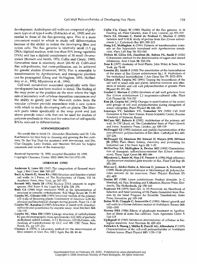
from the apex. Fiber development associated with this snap point is revealedby staining with Cellufluor (Fig. 1, A-C). In the uppermost portion of the stem, a11 of the cells had similar cell-wall thickness and intensity of fluorescence (Fig. 1A). Cells that were developing into fibers exhibited strong fluorescence a few millimeters above the snap point but with no apparent increase in wall thickness (Fig. 1B). The cross-section of the stem just below the snap point showed the intensive thickening of the cell wall (Fig. 1C). Stem elongation had ceased at this position, but the fiber cells continued intrusive growth (Esau, 1977). Sections through the mid-point of the fast-growing stem showed continued thickening (Fig. lD), and sections at this same position at the budding stage 4 weeks later showed that nearly the entire lumen was filled by secondary wall (Fig. 1E).
Cell-Wall Composition of Phloem Fiber-Rich Strips and Xylem
Current models of the primary cell wall depict cross- linking glycans interlacing microfibrils in a firm network embedded in a pectin gel (McCann and Roberts, 1992; Carpita and Gibeaut, 1993). We extracted pectins sequen- tially from primary walls with the Ca2+-chelator ammo- nium oxalate, followed by 0.2 M alkali. The weak alkali hydrolyzes ester linkages by which a subfraction of pectic substances may be attached to the wall matrix. The cross- linking glycans are then extracted with 4 M alkali, which disrupts primarily hydrogen bonding but may hydrolyze a small number of covalent linkages. Phloem fiber-rich peels and xylem cylinders of fast-growing and budding plants were compared with respect to their amounts of chelator- soluble and ester-linked pectic substances, strong alkali- soluble cross-linking glycans, and crystalline cellulose (Ta- ble I). From the fast-growth to budding stages, cellulose content in Novo phloem fibers rose from 39 to 52% of the cell wall, whereas in xylem the cellulose content remained constant. During subsequent growth cellulose content con- tinued to increase, and in mature fibers, it reached 70 to 80% of the total mass (Morvan et al., 1989; Lozovaya et al., 1990). .
During the fast-growth stage, the four cultivars exhibited some variation in cellulose content, from under 30% in Psko, to slightly higher in Mezh, to over 40% in Svetoch. Although not strictly proportional, the lower cellulose con- tent correlated with higher amounts of chelator- and weak- alkali-soluble pectin (Table I). The xylem contained consid- erably less pectic substances than the fiber peels, but cellulose content was slightly greater than 30%. In contrast to the fiber peels, the xylem contained a large proportion of alkali-soluble and acid-hydrolyzable cross-linking glycans (Table I). After exhaustive alkali extraction, a substantial amount of the remaining wall m a s was hydrolyzed by TFA. Glc constituted 32 and 25% of the hydrolyzable sugar from fiber-rich peels and xylem, respectively, indicating that a portion of the cellulose was susceptible to the hy- drolysis. Ara (26%) and Gal (19%) and smaller amounts of the other sugars were found in the TFA-soluble fraction
www.plantphysiol.orgon February 29, 2020 - Published by Downloaded from Copyright © 1996 American Society of Plant Biologists. All rights reserved.

724 Corshkova et al. Plant Physiol. Vol. 110, 1996
snappoint
Figure 1. (Legend appears on facing page.)
www.plantphysiol.orgon February 29, 2020 - Published by Downloaded from Copyright © 1996 American Society of Plant Biologists. All rights reserved.

Cell-Wall Polysaccharides of Developing Flax Plants 725
Table I . Cell-wall fraction and uronic acid content in phloem fiber peels and xvlem of the develoDina stems in severa/ flax varieties
Sample Acetic- Nitric Cellulose Acid
0.2 M 4 M Uronic Ammonium Acid” Oxalate NaOH NaOH
wt % Svetoch, fiber peel, fast-growth 8.2 11.9 14.9 11.1 19.0 43.1 Psko, fiber peel, fast-growth 9.6 14.9 20.1 12.4 26.0 26.6
Novo, fiber peel, fast-growth 7.2 15.7 8.8 8.7 27.8 39.0 Novo, fiber peel, budding 6.7 14.4 8.1 8.8 16.0 52.7 Novo, xylem, fast-growth 1 .o 3.6 11.7 17.9 34.8 32.0 Novo, xylem, budding 0.9 4.1 13.3 17.3 34.1 31.2
Mezh, fiber peel, fast-growth 10.4 18.3 14.9 11.1 21.8 33.9
a Total uronic acids were determined as wt % of the purified cell walls before extraction. Other samples of the cell walls were extracted sequentially with ammonium oxalate and NaOH to yield a-cellulose. The wt % of acetic-nitric fraction was determined by the difference between a-cellulose and cellulose.
from fiber-rich peels, whereas the fraction from xylem yielded Xyl (40%), Gal (l6%), and Ara (l lY0).
Sugar and Linkage Composition of Buffer-Soluble Polymers, Pectins, and Alkali-Soluble Glycans
Buffer-soluble polymers were fractionated on a Sepha- rose 4B-200 column, and sugar content was determined for each fraction; the elution profile varied with material from different tissues (Fig. 2). Phloem tissues contained high- molecular-weight polymers that almost voided the column. This fraction was 90% Gal, and linkage analysis showed that it was mainly 4-Gal branched at the 0-2 and 0-3 positions and with Gal the only detectable nonreducing terminal sugar (Table 11). The galactans are likely to be attached to the 0-4 of 2-linked Rha units of an almost fully branched RG, because only traces of unbranched 2-linked Rha were detected (Table 11). The 4-Glc and some of the 4,6-Glc (Table 11) could be from starch because the buffer- soluble polymers were not treated with DMSO and glu- coamylase. Because the 4,6-Glc is in much greater propor- tions than 4-Glc, it may be from another type of glycan or a constituent of the galactan. Neither the growing tip nor xylem contained appreciable amounts of these galactans (Fig. 2). The buffer-soluble material from a11 tissues con- tained two additional fractions (Fig. 2, I1 and 111) constitut- ing mostly type I1 AG(P)s and protein (data not shown).
The carboxyl groups of the glycosyluronic acids in the ammonium oxalate extracts were chemically reduced with NaBD, to generate 6,6-dideuterio sugars that could be distinguished from their respective neutral sugars by MS. By this method, GalA constituted almost 50% of the total sugar in the ammonium oxalate extract of the fiber-rich peels. Of the remaining neutral sugar, nearly half was Ara, with smaller amounts of Gal and Rha (Table 11). Methyl-
ation analysis verified that the GalA was virtually a11 4-linked, with only trace amounts of t-GalA (not shown). Linkage analyses of the neutral components revealed a 4-linked galactan and a relatively long and unbranched 5-linked arabinan in addition to f i a units typical of RG (Table 11). Simple linkage analysis cannot distinguish 4-linked arabinopyranose units from 5-linked arabino- furanose units, but NMR spectroscopy has verified the arabinan structure in flax and many other species (Bush, 1988; Davis et al., 1990). Although in smaller amounts, the 3,6-Gal indicates that the chelator-soluble pectins also con- tain type-I1 AGs. The xylem contained only about half as much pectin as the fiber peels, and a much greater propor- tion of the material required weak alkali for extraction. The chelator-soluble and weak alkali-soluble pectins of the fiber peels were similar in composition, differing primarily in the degree of branching of the rhamnosyl units of RG and the amount of 4-linked xylosyl units. In contrast, the small amount of chelator-soluble pectin from xylem cells con- tained substantial amounts of 4-linked xylan in addition to the arabinan and galactan, and the weak-alkali-soluble fraction was over 90% 4-linked xylan (Table 11).
Strong-alkali-extracted material from the fiber peels was also rich in Xyl (40 mo1 Yo) and contained considerable amounts of Glc, Gal, and Man (Table 11). From methylation analyses, 4-Xyl was the most abundant linkage. In addition to 2-Xyl, a11 other linked sugars expected from xyloglucan of type-I walls (Carpita and Gibeaut, 1993), namely nonre- ducing f-Xyl, 4- and 4,6-Glc, t- and 2-Gal, and t-Fuc, were found in ratios that indicated a decoration of a Xyl,Glc, heptasaccharide unit oligomer by t-Gal-, and t-Fuc-(1+2)- Gal side groups to form octa- and nonasaccharides.
Most of the Xyl in the fiber-rich peels was in the 4-linked xylan. The strong-alkali fractions from xylem walls, like
~~ ~ ~ ~~~
Figure 1 . (Figure appears on facing page.) Free-hand cross-sections of flax stems stained with Cellufluor and viewed with broad-wavelength fluorescence detection. A representative flax plant at the fast-growing stage from which sections were taken is shown at left. A, Section taken 5 m m above the snap point; B, section taken 1 m m above the snap point; C, section taken 1 m m below the snap point; D, section taken from the middle of the stem; E, section taken from the middle of the stem at budding stage 4 weeks later. The intense blue Cellufluor fluorescence is localized in phloem fiber walls. A representative bundle of fibers is encircled in white, and arrows indicate xylem. For A through D, bar = 100 p m ; for E, bar = 50 p m . The red color is from Chl autofluorescence, and yellow is the autofluorescence of lignin, primarily in xylem.
www.plantphysiol.orgon February 29, 2020 - Published by Downloaded from Copyright © 1996 American Society of Plant Biologists. All rights reserved.

726 Gorshkova et al. Plant Physiol. Vol. 1 1 O , 1996
0 0.3 o, d- a- : 0.2
e g 0.1
a
II a
10 20 30 40 50
Fraction Number Figure 2. Separation of buffer-soluble material from fiber peels (O), growing tip (O), and xylem ( O ) tissues by Sepharose 48-200 chro- matography. Polymers were eluted with 0.1 M Tris-HCI, pH 7.2, containing 0.1 M NaCI, and 4-mL fractions were collected. Relative amounts of total sugars were determined by the phenol-sulfuric acid method (Dubois et al., 1956). The void fraction and the three in- cluded fractions were collected, dialyred against water, and ana- lyzed for sugar composition.
weak-alkali fractions, was over 90% 4-linked xylan. This polymer is branched at every 12 to 13 residues soXely at the 0-2 position, as indicated by the relative amount of 2,4- linked Xyl (Table 11). No nonreducing terminal Ara or other neutral sugar was found in alkali-soluble fractions en- riched in xylan to account for the side group. Reduction of water-soluble carbodiimide-activated uronic acids in the xylan-rich fractions with NaBD, and subsequent methyl- ation with CDJ revealed that the only side group was t-(4-O-methyl)-glucuronic acid.
Substantial amounts of Man were found in the strong- alkali-extracted fractions of the fiber-rich peels, artd linkage analyses showed 4- and 4,6-Man in a ratio of about 3.5:l. After accounting for the appropriate amount of 4-Glc rel- ative to 4,6-Glc in xyloglucan, the remaining 4-Glc was in similar proportion to the mannosyl units. The t-Gal and remaining 4-Glc linkages indicated that (ga1acto)-gluco- mannans are components of flax fiber wallc;. Smaller amounts of a similar polymer were found in xylem as well.
Linkage analyses of neutral sugars from cell-wall frac- tions both in phloem and xylem parts of flax stems ob- tained at the budding stage of plant growth revealed no significant differences in distribution of matrix polysaccha- rides compared to the tissues from the fast-growth stage (data not shown).
Table II. Linkage analysis of neutral sugars froni cell-wall fractions o f fiber-rich peels and xylem from Novo flax stems taken at the fast- growth stage
n.d., Not detected; tr, trace.
Deduced Linkage"
Buffer Fibers
2-Rha 2,4-Rha
t-Ara 5-Ara 2,5-Ara 3,5-Ara
t-FUC
t-Xyl 2-Xyl 4-Xyl 2,4-Xyl 4-Man 4,6-Man t-Cal 2-Cal 3-Cal 4-Cal 6-Cal 2,4-Gal 3,4-Gal 3,6-Gal t-Glc 4-Glc 4,6-Glc
0.2 3.1
n.d. n.d. 0.7
n.d. n.d. tr
n.d. 1.8
n.d. tr tr
12.0 n.d. 4.6
2.2 2.0 3.5 tr tr 1.2 2.8
65.8
Ammonium Oxalate 0.2 M NaOH 4.0 M NaOti
Fibers
10.9 tr
n.d. tr
44.2 n.d. 2.6
n.d. n.d. 3.2
n.d. tr
n.d. 6.5
n.d. n.d. 19.4
tr n.d. n.d. 4.6
n.d. 7.1
n.d.
Xylem
5.8
0.8 2.5
17.2 n.d. 2.3 tr
n.d. 16.3
5.8
I .a i .a 0.6 9.2
n.d. n.d. 9.0
11.3 n.d. n.d. 9.0 0.2 6.2 0.2
Fibers
mo/ % 4.9 3.6 tr tr
31.2 2.4 3.4 tr
n.d. 7.2 tr 0.1 0.7 3.2
n.d. n.d. 26.1
1.3 n.d. n.d. 4.3 tr 9.8 0.3
Xylem
0.1 1.3 tr
n.d. 1.8
n.d. n.d. 1.1
n.d.
6.3 tr 0.3 0.6 0.3
n.d. 0.7 0.3
n.d. n.d. 0.4 tr 0.3 0.2
86.5
Fibers
0.4 1.6 1 .o 0.5 3.9
n.d. 0.8 . 5.6 3.7
28.9 2.2
2.3 4.7 6.0
n.d. 5.8 0.7
n.d. n.d. 1.3 0.4 9.5
1 1.4
8.5
Xylem
tr 0.5 tr
n.d. 0.5
n.d. n.d. 0.8
n.d. 86.5
6.7 1.4 0.4 0.3 0.2
n.d. 0.4 0.2
n.d. n.d. 0.2 tr 1.2 1 .I
The designation "2-Rha," for example, indicates attachment of another sugar at the 0 - 2 of a rhamnosyl unit itself linked to another sugar via the C- l position; the actual derivative deduced by CLC-EIMS was 1,2,5-tri-O-acetyI-(l -deuterio)-3,4-di-O-methyl-6-deoxymannitol. The 2-Xyl and 4-Xyl yields identical derivatives, but addition of a deuterium atom at C-1 upon reduction yields different primary and secondary fragments that are auantified bv ElMS (Caruita and Shea. 1989).
www.plantphysiol.orgon February 29, 2020 - Published by Downloaded from Copyright © 1996 American Society of Plant Biologists. All rights reserved.

Cell-Wall Polysaccharides of Developing Flax Plants 727
Table 111. Comparison of the major neutral sugar finkages found in the ammonium oxafate fractions o f cell walls from various flax tissues Linkages were deduced as descrihed in Table II.
Tissue 2-Rha 2,4-Rha 5-Ara 2.5-Ara 3,5-Ara 4-Xyl f-Cal 4-Cal 4-Clc 4,6-Clc
Root 9 9 29 3 Hypocotyl 8 12 30 2 Stem 8 8 34 3 Leaf 7 9 29 2 Crowing tip 6 11 30 4
mo/ % 5 10 9 16 5 4 3 9 9 13 9 5 5 10 9 12 8 1 3 1 1 9 9 11 9 3 14 8 10 7 6
Cell-Wall Analyses of Flax Plant Organs
Because the relative amounts of cell-wall polymers found in fiber-rich peels and xylem tissues varied markedly, we analyzed the cell-wall composition in the following parts from Novo plants taken at the fast-growth stage: (a) total root; (b) hypocotyl; (c) mature leaves; and (d) the terminal 0.5 cm of the apex, including developing leaves and stem. Various parts of the flax plant differed markedly in the proportion of cell-wall to total dry mass. The highest pro- portion of cell-wall to total dry mass was in the roots (43%), an amount almost 3 times higher than that in nonfibrous tissues such as leaves and the growing tip. Cellulose con- tent varied from l6YO of cell-wall mass in the growing tip to 40% in roots. In general, the higher the proportion of wall to total mass, the higher the crystalline cellulose content in the cell wall. Regardless of the cellulose content, the pro- portion of pectins in cell walls of various organs was fairly constant and constituted between 11 and 15% of the cell wall.
Ammonium oxalate-soluble fractions of the cell walls of various plant organs were similar in sugar composition (Table 111). However, quantitative composition of the alka- li-soluble fractions showed large differences between fi- brous and nonfibrous tissues: in root and hypocotyl the content of Xyl was about 60 to 70%, whereas in leaves and the growing tip it was about 30% (Table IV). Xyloglucan constituted the major portion of this fraction in leaves and the growing tip, whereas xylan was the main polysaccha- ride in fibrous tissues (Fig. 3). The degree of xylan branch- ing was the same in a11 plant organs, because the ratio between 4-Xyl and 2,4-Xyl did not vary significantly (Table IV).
Cell-Wall Analyses of the Fiber-Rich Peels of Severa1 Flax Varieties
After we obtained the sugar composition and linkage analyses, we roughly estimated the relative proportions of
the major polysaccharides in flax cell walls of four different cultivars. To obtain the proportion of polysaccharide in a cell-wall fraction, we calculated the mo1 % of sugar link- ages attributed to the specific polysaccharide. For xyloglu- can, we summed the t- and 2-Xy1, 2-Gal, and t-Fuc, 4,6-Glc and one-third of its proportion in 4-Glc. The rest of 4-Glc together with about equal amounts of 4- and 4,6-Man and t-Gal were attributed to gluco(ga1acto)mannan. The 4-Xyl and 2,4-Xyl were considered as xylan, and a11 5- and branched Ara residues were considered as arabinan. The 2-Rha and 2,4-Rha together with an equal amount of GalA were attributed to the RG backbone. Type-I galactan was estimated as 4-Gal plus 4,6-Gal, and type-I1 AG(P) was estimated as 6-Ga1, 3,6-Gal, and t-Ara.
Comparison of the polymer distributions in the three fractions from the four cultivars shows the consistency of the fractionations and subsequent methylation analyses (Fig. 4). The uronic acid content of both the chelator-soluble and weak-alkali-soluble fractions was about 50% of the total sugar (Table I), but the major neutral polymer was the 5-linked arabinan (Fig. 4). The pectic fractions constituted from 40 to 50% of this arabinan in each of the cultivars. There was also an enrichment of the gluco(ga1acto)mannan in the chelator-soluble fractions. Three major noncellulosic glycans were found in the strong-alkali extracts of a11 four cultivars. Most abundant was xyloglucan, followed by 4-0- methylglucuronoxylan and gluco(ga1acto)mannan. Slight variations in xylan, from about 22% in Psko to 30% in Mezh, were noted.
DlSCUSSlON
The composition of flax primary cell walls is typical of the type-I walls of most flowering plants (Carpita and Gibeaut, 1993). The major cross-linking glycan is a xyloglu- can with heptasaccharide units substituted primarily with t-Gal- and t-Fuc-(1-2)-Gal side groups at the 0-2 of the xylosyl units. The xyloglucan-cellulose framework is ap-
Table IV. Comparison of the major neutra/ sugar linkages found in the 4 M KOH fractions of cell walls from various flax tissues
Linkages were deduced as described in Table II.
Tissue t-Fuc 5-Ara t-Xyl 2-Xyl 4-Xyl 2,4-Xyl f-Cal 2-Cal 4-Man 4,6-Man 4-Clc 4 , 6 4 1 ~
mo/ X Root 1 a 3 6 56 4 4 2 2 1 6 5
Stem 2 9 4 6 40 3 7 6 3 1 7 11 Leaf 4 1 1 17 7 11 1 6 6 5 2 11 18 Crowina tip 3 14 12 5 15 1 8 3 7 3 13 14
H ypocotyl 1 6 5 5 46 4 6 3 3 3 6 10
www.plantphysiol.orgon February 29, 2020 - Published by Downloaded from Copyright © 1996 American Society of Plant Biologists. All rights reserved.

Corshkova et al. Plant Physiol. Vol. 'I 1 O, 1996
xylan xyloglucan
Organ Figure 3. Proportions of xylan and xyloglucan in alkali-soluble frac- tions of various flax organs. MOI % of each fraction was estimated based on the relative amount of 4- and 2,4-Xyl (4-xylans) and t- and 2-Xyl, 4- and 4,6-Glc, 2-Gal, and t-Fuc (xyloglucan).
parently embedded in a pectin matrix of both RG and homogalacturonan. Pectic substances of plants vary mostly in the composition of the neutra1 sugar side chains, which are attached to the 0-4 of the RG rhamnosyl units. Flax contains two principal pectic neutral-sugar polymers, a 5-arabinan and a 4-galactan, each with infrequent branch- ing. The appearance of large amounts of the high-molecu- lar-mass 4-galactan is coincident with phloem fiber devel- opment. Unlike type-I AGs, which contain t-Ara units at the 0-3 of 4-galactans (Bacic et al., 1988), the infrequent branching of the flax 4-galactan is at both the 0 - 2 and 0-3 positions, and the side chains terminate with galactosyl rather than arabinosyl units. Goubet and Morvan (1994) also reported these water-soluble galactans in flax cells in liquid culture, but linkage analyses of the galactan struc- tures were varied and inconsistent with our findings. Davis et al. (1990), using 'H- and 13C-NMR spectroscopy, showed that an RG from retted flax contains short side chains of (14)-l inked p-D-Gal units attached at the 0-4 of the rhamnosyl units of RG. In contrast, the flax-seed mucilage is rich in L-Gal, rather than D-Gal (Anderson and Lowe, 1947). The mucilage is also enriched in Rha, GalA, and Xyl,
- but linkage analyses have not been reported. The galactans found in our study are considerably longer than those reported by Davis et al. (1990), and they did not report the presence of a 5-arabinan. The arabinans and galactans may undergo considerable hydrolysis during further develop- ment or during the retting process.
The 3-, 6-, and 3,6-Gal units also found indicate that the ubiquitous type-I1 AG(P) is associated with the pectic frac- tion of a11 tissues. The walls from the fiber-rich peels con- tain the same pectic polysaccharides and cross-linking gly- cans as the growing tip, but they are enriched in 5-arabinan, 4-O-methyl-glucuronoxylan, glucomannan, and cellulose. Our findings of xylans, xyloglucan, and glu- comannan are consistent with those of McDougall (1993), who used an enzymological approach to yield diagnostic P-D-xylobiosyl, isoprimeverose, and P-D-manno-oligosac- charides from alkali- and boric-acid-soluble polysaccha- rides from retted flax fibers. In contrast to the fiber-rich peels, the xylem tracheary elements contain over 60% 4-0-
methylglucuronoxylan and only 30% cellulose at maturity. These findings are consistent with those of an early study reporting that 4-O-methylglucuronoxylan is the principal noncellulosic polysaccharide of xylem-enriched flax straw (Geerdes and Smith, 1955). The proportion of arabinan was similar in a11 organs, whereas the proportion of xylan was significantly higher in roots, hypocotyl, and stem than in leaves and the growing apex. Thus, different flax organs have distinctive cell-wall contents, proportions of pectic substances and cross-linking glycans, and individual polymers.
Although the relative abundance of the varioiis noncel- lulosic polysaccharides varies widely among tht: different cell types, the proportion of the linkage structure and the degree of branching of the individual polysaccharides are invariant. The 5-arabinans and 4-galactans have 'I very low degree of branching, the 4-xylans contain 4-O-rnethylglu- curonic acid side groups spaced about 12 to 1.3 residues apart and attached solely to the 0 -2 of xylosyl units, and (ga1acto)glucomannans have a small amount of branching t-Gal units at the mannosyl 0-6 position.
Reiter et al. (1993) isolated severa1 Arabidopsts mutants in polysaccharide composition of the primary cell wall, and these mutants prove the utility of genetic approaches to understanding the biogenesis of the primary wall. Flax is similar to Arabidopsis as a genetic model ancl provides special advantages for the study of cell-wall biogenesis and
1 5-arabinan
m
30
20
10
Novo Svetoch Psko Mezh O
Cultivar Figure 4. Comparison of the relative amounts of neutral-sugar poly- mers found in the ammonium oxalate and NaOH extracts from the cell walls from fiber-rich peels of four flax cultivars. Polymers char- acterized are shown in the key at the top of the figure. Linkage assignments were as described in the text.
www.plantphysiol.orgon February 29, 2020 - Published by Downloaded from Copyright © 1996 American Society of Plant Biologists. All rights reserved.
Cell-Wall Polysaccharides of Developing Flax Plants 729
development. Arabidopsis cell walls are composed of poly- mers typical of type-I walls (Zablackis et al., 1995) and are similar to those of the flax-growing apex. Flax is a more convenient model in which to study cell differentiation because of the ability to isolate the developing fiber and xylem cells. The flax genome is relatively small (1.5 pg DNA/diploid nucleus, with less than 50% being repetitive DNA) and has a diploid complement of 30 small chromo- somes (Bennett and Smith, 1976; Cullis and Cleary, 1985). Generation time is relatively short (60-90 d). Cultivated flax self-pollinates, but cross-pollination can be achieved for genetic studies (Pustovoit, 1973). Flax is amenable to transformation by Agrobacterium, and transgenic plantlets can be propagated (Dong and McHughen, 1991; McShef- frey et al., 1992; Mlynárová et al., 1994).
Cell-wall metabolism associated specifically with fiber development has not been studied in detail. The finding of the snap point as the position on the stem where the high rate of secondary wall cellulose deposition commences and the ability to gently peel the fiber-rich cortex from the vascular cylinder provide researchers with a new system with which to study developing cells ex planta. The fiber- rich peels taken specifically at the snap point and just above provide intact cells that can be used for studies of polymer synthesis in vitro and for extraction of cell-specific RNAs relevant to differentiation.
ACKNOWLEDGMENTS
We would like to thank Dr. Alexander Zhuchenko and Dr. Lilia Kurchakova for their help in choosing and growing the flax culti- vars, Dr. Jan Van Dam for assistance with microscopy, and Drs. Clint Chapple, Larry Dunkle, and Maureen McCann for helpful comments and review of the manuscript.
Received September 18, 1995; accepted December 18, 1995. Copyright Clearance Center: 0032-0889/96/110/0721/09.
LITERATURE CITED
Anderson E, Lowe HJ (1947) The composition of flaxseed muci- lage. J Biol Chem 168: 289-297
Bacic A, Harris PJ, Stone BA (1988) Structure and function of plant cell walls. In J Preiss, ed The Biochemistry of Plants, Vol 14. Academic Press, New York, pp 297-371
Bennett MD, Smith JB (1976) Nuclear DNA amounts in angio- sperms. Phil Trans R SOC Lond Ser B 274: 228-274
Bush CA (1988) High resolution NMR in the determination of structure in complex carbohydrates. Bull Magn Reson 10: 73-95
Carpita NC, Gibeaut DM (1993) Structural models of the primary cell walls of flowering plants. Consistency of structure with the physical and biochemical changes during growth. Plant J 3: 1-30
Carpita NC, Kanabus J (1987) Extraction of starch with dimethyl- sulfoxide and quantitation by enzymic assay. Anal Biochem 161:
Carpita NC, Shea EM (1989) Linkage structure of carbohydrates by gas chromatography-mass spectrometry (GC-MS) of partially methylated alditol acetates. rn CJ Biermann, GD McGinnis, eds, Analysis of Carbohydrates by GLC and MS. CRC Press, Boca Raton, FL, pp 157-216
Chesson A (1979) A laboratory method for the determination of fibre content of linen flax. NZ J Agric Res 22: 43-46
132-139
Cullis CA, Cleary W (1985) Fluidity of the flax genome. In M Freeling, ed, Plant Genetics, Alan R Liss, London, pp 303-310
Davis EA, Derouet C, Hervk du Penhoat C, Morvan C (1990) Isolation and N.M.R. study of pectins from flax (Linum usitatis- simum L.). Carbohydr Res 197: 205-215
Dong J-Z, McHughen A (1991) Patterns of transformation inten- sity on flax hypocotyls inoculated with Agrobacterium tumefa- ciens. Plant Cell Rep 10: 555-560
Dubois M, Gilles DA, Hamilton JK, Rebers PA, Smith F (1956) Colorimetric method for the determination of sugars and related substances. Anal Chem 28: 350-356
Esau K (1977) Anatomy of Seed Plants. John Wiley & Sons, New York, pp 74-81
Geerdes JD, Smith F (1955) The constitution of the hemicellulose of the straw of flax (Linum usitutissimum Sp.). 11. Hydrolysis of the methylated hemicellulose. J Am Chem SOC 77: 3572-3576
Gibeaut DM, Carpita NC (1991) Tracing the biosynthesis of the cell wall in intact cells and plants. Selective tumover and alter- ation of soluble and cell wall polysaccharides in grasses. Plant Physiol 97: 551-561
Goubet F, Morvan C (1994) Synthesis of cell wall galactans from flax (Linum usitatissimum L.) suspension-cultured cells. Plant Cell Physiol 35: 719-727
Kim JB, Carpita NC (1992) Changes in esterification of the uronic acid groups of cell wall polysaccharides during elongation of maize coleoptiles. Plant Physiol 98: 646-653
Lozovaya VV, Salnikov VV, Yumashev NV (1990) Cell Wall Formation in Flax Stem Tissues. Kazan Scientific Center, Russian Academy of Sciences, Kazan
McCann MC, Roberts K (1992). Architecture of the primary cell wall. In CW Lloyd, ed, The Cytoskeletal Basis of Plant Growth and Form. Academic Press, London, pp 109-129
McDougall GJ (1993) Isolation and partia1 characterization of the non-cellulosic polysaccharides of flax fibre. Carbohydr Res 241:
McDougall GJ, Morrison IM, Stewart D, Weyers JDB, Hillman JR (1993) Plant fibers: botany, chemistry and processing for industrial use. J Sci Food Agric 6 2 1-20
McSheffrey SA, McHughen A, Devine MD (1992) Characteriza- tion of transgenic sulfonylurea-resistant flax (Linum usitatissi- mum). Theor Appl Genet 8 4 480486
Mlynárová L, Bauer M, Nap J-P, Pretová A (1994) High efficiency Agrobacterium-mediated gene transfer to flax. Plant Cell Rep 13:
Morvan C, Abdul-Hafez A, Morvan O, Jauneau A, Demarty M (1989) fitude physicochimique et biochimique de polysaccha- rides extraits de lin sous-roui. Plant Physiol Biochem 27:
Oosten BT (1988) Linum usitatissimum: Product diversity. In G Marshall, ed, Flax: Breeding and Utilization, Kluwer Press, Dor- drecht, The Netherlands, pp 158-160
Pustovoit VS (1973) Seed flax. In VS Pustovoit, ed, Handbook of Selection and Seed Growing of Oil Plants (translated from Rus- sian by the Israel Program for Scientific Translations). Keter Press, Jerusalem, pp 103-148
Reiter W-D, Chapple C, Somerville C (1993) Altered growth and cell walls in a fucose-deficient mutant of Arubidopsis. Science 261:
Sharma HSS (1986) Effects of glyphosate treatment on lignifica- tion of fibres of some flax cultivars. Tests Agrochem Cultiv 7
Updegraff D (1969) Semimicro determination of cellulose in bio- logical materials. Anal Biochem 3 2 420-424
Zablackis E, Huang J, Miiller B, Darvill AG, Albersheim P (1995) Characterization of the cell-wall polysaccharides of Arabidopsis thaliana leaves. Plant Physiol 107: 1129-1138
227-236
282-285
451-459
1032-1035
114-1 15
www.plantphysiol.orgon February 29, 2020 - Published by Downloaded from Copyright © 1996 American Society of Plant Biologists. All rights reserved.