cellscapes - university of guelph - home pagejmathur/pdf/cellscapes.pdf · sareen, elyse roach,...
TRANSCRIPT
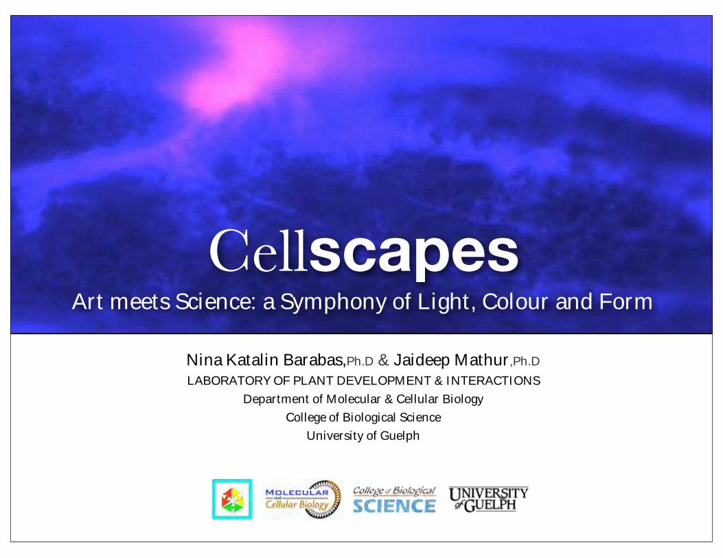
CellscapesArt meets Science: a Symphony of Light, Colour and Form
Nina Katalin Barabas,Ph.D & Jaideep Mathur,Ph.D
LABORATORY OF PLANT DEVELOPMENT & INTERACTIONSDepartment of Molecular & Cellular Biology
College of Biological ScienceUniversity of Guelph
CellscapesArt meets Science: a Symphony
of Light, Colour and Form
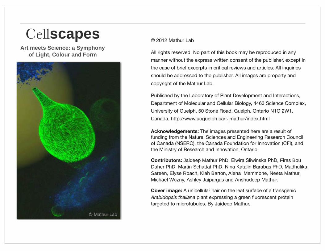
© 2012 Mathur Lab
All rights reserved. No part of this book may be reproduced in any manner without the express written consent of the publisher, except in the case of brief excerpts in critical reviews and articles. All inquiries should be addressed to the publisher. All images are property and copyright of the Mathur Lab.
Published by the Laboratory of Plant Development and Interactions, Department of Molecular and Cellular Biology, 4463 Science Complex, University of Guelph, 50 Stone Road, Guelph, Ontario N1G 2W1, Canada, http://www.uoguelph.ca/~jmathur/index.html
Acknowledgements: The images presented here are a result of funding from the Natural Sciences and Engineering Research Council of Canada (NSERC), the Canada Foundation for Innovation (CFI), and the Ministry of Research and Innovation, Ontario,
Contributors: Jaideep Mathur PhD, Elwira Sliwinska PhD, Firas Bou Daher PhD, Martin Schattat PhD, Nina Katalin Barabas PhD, Madhulika Sareen, Elyse Roach, Kiah Barton, Alena Mammone, Neeta Mathur, Michael Wozny, Ashley Jaipargas and Anshudeep Mathur.
Cover image: A unicellular hair on the leaf surface of a transgenic Arabidopsis thaliana plant expressing a green fluorescent protein targeted to microtubules. By Jaideep Mathur.
6
A unicellular hair on the leaf surface of a transgenic Arabidopsis plant expressing a green fluorescent protein (GFP) targeted to microtubules.
Image credit: Jaideep Mathur: Mathur and Chua, 2000, PLANT CELL Vol 12(4)
CONFOCAL MICROSCOPY 2
‘Timeless Elegance’
© Mathur Lab
CellscapesArt meets Science: a Symphony
of Light, Colour and Form
Dedicated to all lovers of Nature and her
beautiful creations.CONTACT
Please contact us for information regarding educational materials and multimedia exhibitions featuring similar scientific images and movies.
Nina Katalin Barabas, [email protected]
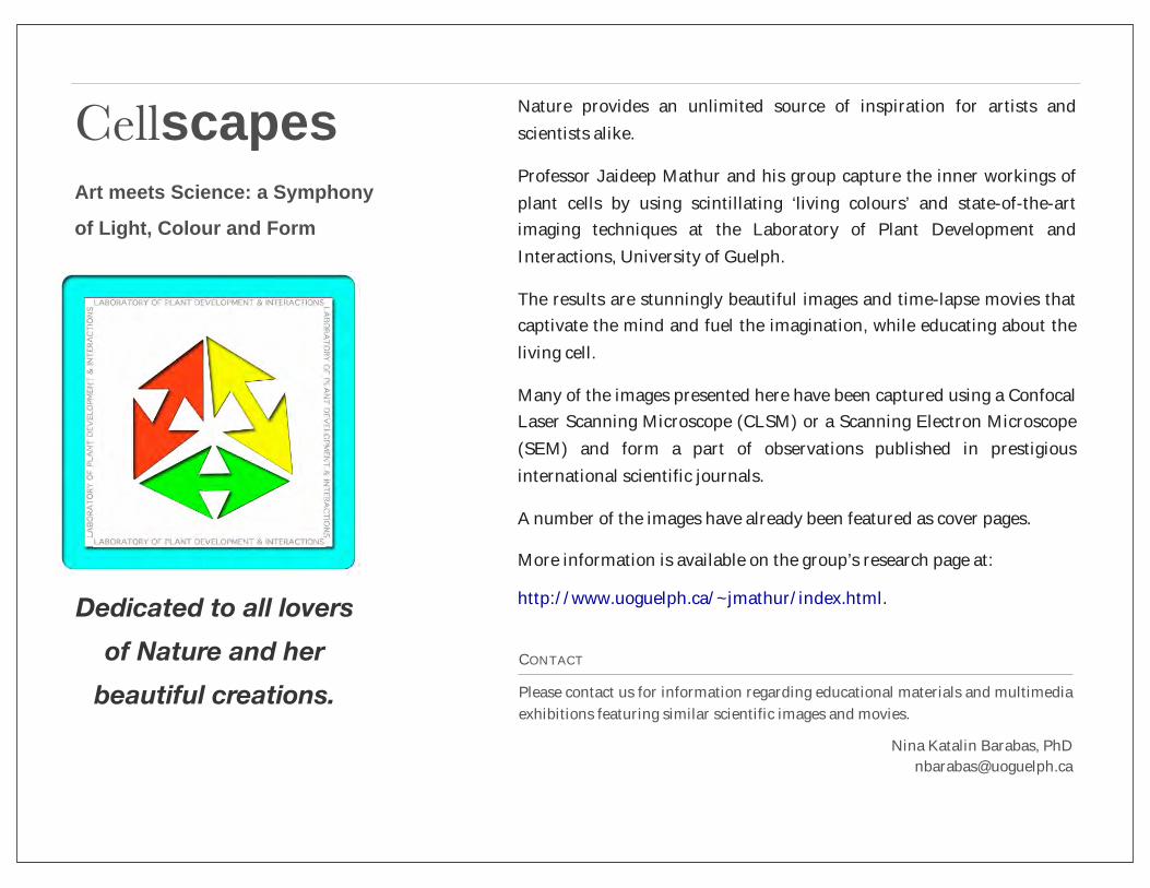
Nature provides an unlimited source of inspiration for artists and scientists alike.
Professor Jaideep Mathur and his group capture the inner workings of plant cells by using scintillating ‘living colours’ and state-of-the-art imaging techniques at the Laboratory of Plant Development and Interactions, University of Guelph.
The results are stunningly beautiful images and time-lapse movies that captivate the mind and fuel the imagination, while educating about the living cell.
Many of the images presented here have been captured using a Confocal Laser Scanning Microscope (CLSM) or a Scanning Electron Microscope (SEM) and form a part of observations published in prestigious international scientific journals.
A number of the images have already been featured as cover pages.
More information is available on the group’s research page at:
http://www.uoguelph.ca/~jmathur/index.html.

8
Microtubule ends labeled with a red fluorescent protein (RFP, originating from a radiating mushroom coral), and actin filaments highlighted by a green fluorescent protein (GFP, isolated from jellyfish), observed in a single onion epidermal cell.
Image captured with a Leica TCS-SP5 Confocal Laser Scanning Microscope.
CONFOCAL MICROSCOPY 1
‘Sunrise’
Image credit: Jaideep Mathur
CONFOCAL MICROSCOPY 2
‘Timeless Elegance’A unicellular hair on the leaf surface of a transgenic Arabidopsis plant expressing a green fluorescent protein (GFP) targeted to microtubules.
Image captured with a Leica TCS-SP5 Confocal Laser Scanning Microscope.
Image credit: Jaideep Mathur
Mathur and Chua, 2000, PLANT CELL Vol 12(4)
Stomata on the hypocotyl of an Arabidopsis thaliana seedling expressing a green to red photo-convertible Eos fluorescent protein targeted to chloroplasts. Differential colouring of plastids is brought about through photo-conversion.
Image captured with a Leica TCS-SP5 Confocal Laser Scanning Microscope.
CONFOCAL MICROSCOPY 3
‘Gemstones’
Image credit: Jaideep Mathur
Schattat et al. 2012. Plant Cell 24(4):1465-77.
CONFOCAL MICROSCOPY 4
‘Fearless’A germinated seedling of thale cress (Arabidopsis thaliana) still carrying its protective seed coat. The blue dots are nuclei and the seedling was stained with propidium iodide to show cellular profiles.
Image captured with a Leica TCS-SP5 Confocal Laser Scanning Microscope.
Image credit: Martin Schattat
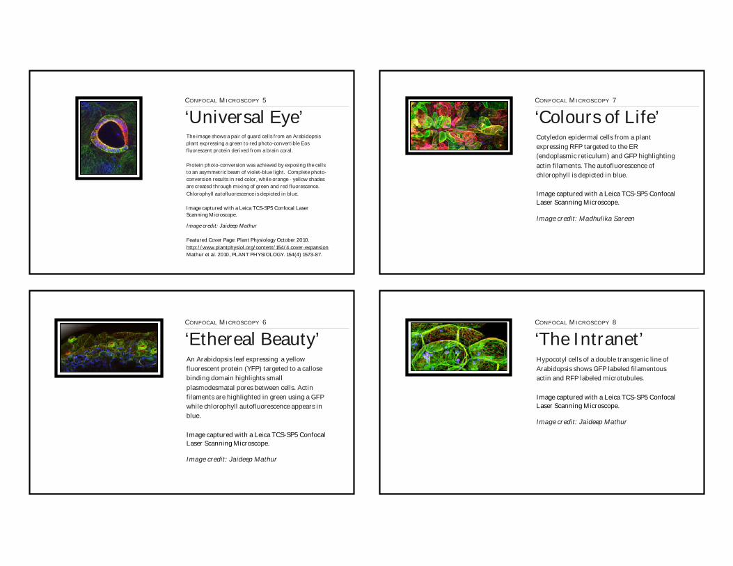
The image shows a pair of guard cells from an Arabidopsis plant expressing a green to red photo-convertible Eos fluorescent protein derived from a brain coral.
Protein photo-conversion was achieved by exposing the cells to an asymmetric beam of violet-blue light. Complete photo-conversion results in red color, while orange - yellow shades are created through mixing of green and red fluorescence. Chlorophyll autofluorescence is depicted in blue.
Image captured with a Leica TCS-SP5 Confocal Laser Scanning Microscope.
CONFOCAL MICROSCOPY 5
‘Universal Eye’
CONFOCAL MICROSCOPY 6
‘Ethereal Beauty’An Arabidopsis leaf expressing a yellow fluorescent protein (YFP) targeted to a callose binding domain highlights small plasmodesmatal pores between cells. Actin filaments are highlighted in green using a GFP while chlorophyll autofluorescence appears in blue.
Image captured with a Leica TCS-SP5 Confocal Laser Scanning Microscope.
Image credit: Jaideep Mathur
Image credit: Jaideep Mathur
Featured Cover Page: Plant Physiology October 2010.http://www.plantphysiol.org/content/154/4.cover-expansion Mathur et al. 2010, PLANT PHYSIOLOGY. 154(4) 1573-87.
Cotyledon epidermal cells from a plant expressing RFP targeted to the ER (endoplasmic reticulum) and GFP highlighting actin filaments. The autofluorescence of chlorophyll is depicted in blue.
Image captured with a Leica TCS-SP5 Confocal Laser Scanning Microscope.
CONFOCAL MICROSCOPY 7
‘Colours of Life’
Image credit: Madhulika Sareen
CONFOCAL MICROSCOPY 8
‘The Intranet’Hypocotyl cells of a double transgenic line of Arabidopsis shows GFP labeled filamentous actin and RFP labeled microtubules.
Image captured with a Leica TCS-SP5 Confocal Laser Scanning Microscope.
Image credit: Jaideep Mathur

GFP labelled endoplasmic reticulum in hair cells of a mutant with defective actin organization.
Image captured with a Leica TCS-SP5 Confocal Laser Scanning Microscope.
CONFOCAL MICROSCOPY 9
‘Toddlers’
Image credit: Jaideep Mathur
CONFOCAL MICROSCOPY 10
‘The winner takes it all’A hyphal network on the surface of a plant expressing GFP targeted to microtubules before the fungus finds an entry point and invades the cells.
Image captured with a Leica TCS-SP5 Confocal Laser Scanning Microscope.
Image credit: Jaideep Mathur
8
Every plant cell differs slightly from its neighbours. This independent nature is seen here in cells expressing either RFP targeted to the ER, GFP targeted to actin filaments or both.
Image captured with a Leica TCS-SP5 Confocal Laser Scanning Microscope.
CONFOCAL MICROSCOPY 11
‘Freedom of expression’
Image credit: Jaideep Mathur
CONFOCAL MICROSCOPY 12
‘The Hub’The circular base of a leaf trichome (hair cell) acts as an information hub for the numerous cells surrounding it. Cytosolic GFP and chlorophyll autoflorescence (depicted in blue).
Image captured with a Leica TCS-SP5 Confocal Laser Scanning Microscope.
Image credit: Jaideep Mathur
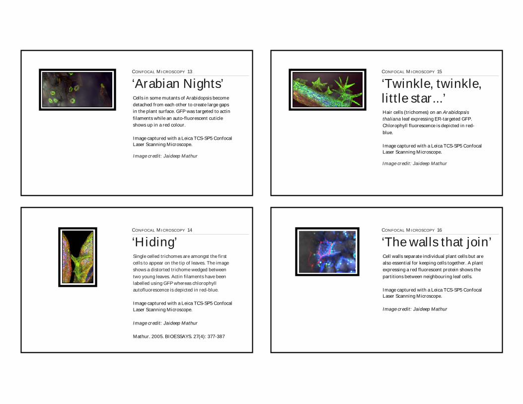
Cells in some mutants of Arabidopsis become detached from each other to create large gaps in the plant surface. GFP was targeted to actin filaments while an auto-fluorescent cuticle shows up in a red colour.
Image captured with a Leica TCS-SP5 Confocal Laser Scanning Microscope.
CONFOCAL MICROSCOPY 13
‘Arabian Nights’
Image credit: Jaideep Mathur
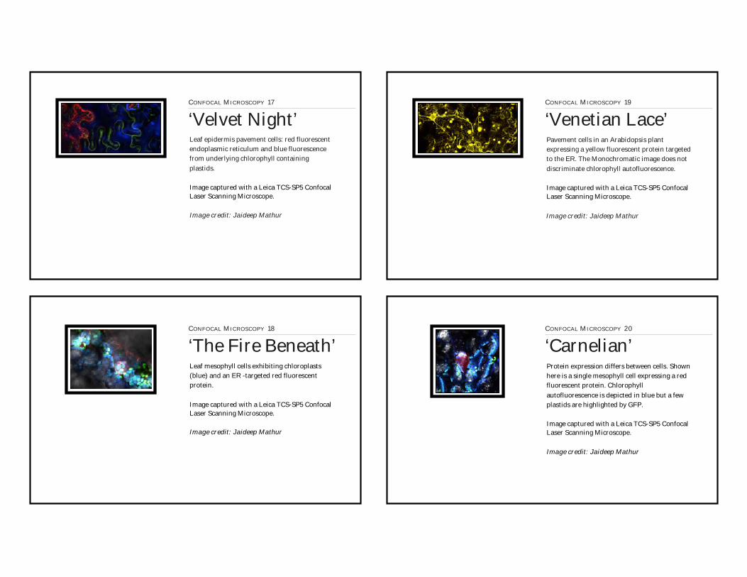
CONFOCAL MICROSCOPY 14
‘Hiding’Single celled trichomes are amongst the first cells to appear on the tip of leaves. The image shows a distorted trichome wedged between two young leaves. Actin filaments have been labelled using GFP whereas chlorophyll autofluorescence is depicted in red-blue.
Image captured with a Leica TCS-SP5 Confocal Laser Scanning Microscope.
Image credit: Jaideep Mathur
Mathur. 2005. BIOESSAYS. 27(4): 377-387
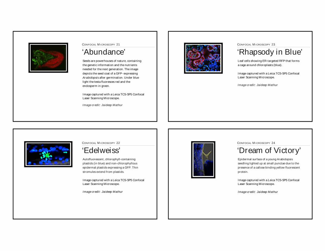
Hair cells (trichomes) on an Arabidopsis thaliana leaf expressing ER-targeted GFP. Chlorophyll fluorescence is depicted in red-blue.
Image captured with a Leica TCS-SP5 Confocal Laser Scanning Microscope.
CONFOCAL MICROSCOPY 15
‘Twinkle, twinkle,little star...’
Image credit: Jaideep Mathur
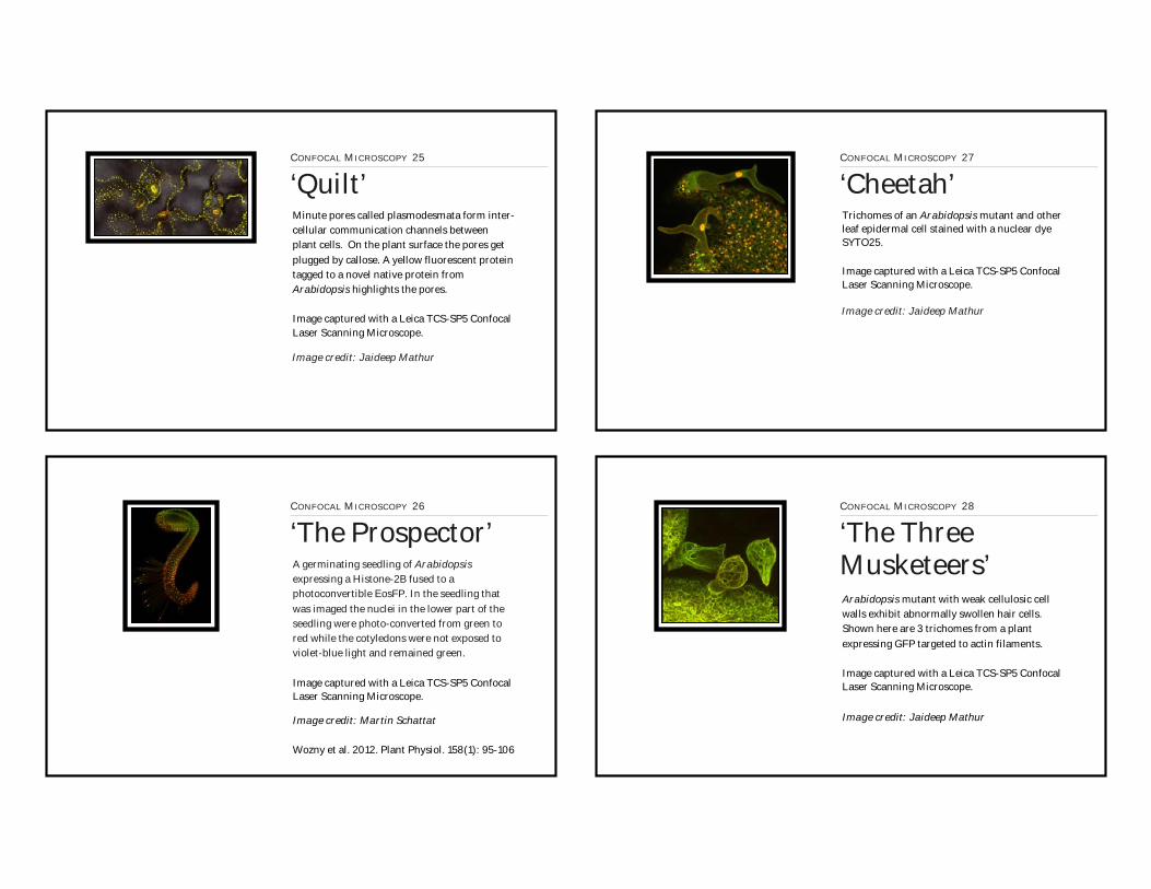
CONFOCAL MICROSCOPY 16
‘The walls that join’Cell walls separate individual plant cells but are also essential for keeping cells together. A plant expressing a red fluorescent protein shows the partitions between neighbouring leaf cells.
Image captured with a Leica TCS-SP5 Confocal Laser Scanning Microscope.
Image credit: Jaideep Mathur
Leaf epidermis pavement cells: red fluorescent endoplasmic reticulum and blue fluorescence from underlying chlorophyll containing plastids.
Image captured with a Leica TCS-SP5 Confocal Laser Scanning Microscope.
CONFOCAL MICROSCOPY 17
‘Velvet Night’
Image credit: Jaideep Mathur
CONFOCAL MICROSCOPY 18
‘The Fire Beneath’Leaf mesophyll cells exhibiting chloroplasts (blue) and an ER -targeted red fluorescent protein.
Image captured with a Leica TCS-SP5 Confocal Laser Scanning Microscope.
Image credit: Jaideep Mathur
Pavement cells in an Arabidopsis plant expressing a yellow fluorescent protein targeted to the ER. The Monochromatic image does not discriminate chlorophyll autofluorescence.
Image captured with a Leica TCS-SP5 Confocal Laser Scanning Microscope.
CONFOCAL MICROSCOPY 19
‘Venetian Lace’
Image credit: Jaideep Mathur
CONFOCAL MICROSCOPY 20
‘Carnelian’Protein expression differs between cells. Shown here is a single mesophyll cell expressing a red fluorescent protein. Chlorophyll autofluorescence is depicted in blue but a few plastids are highlighted by GFP.
Image captured with a Leica TCS-SP5 Confocal Laser Scanning Microscope.
Image credit: Jaideep Mathur
Seeds are powerhouses of nature, containing the genetic information and the nutrients needed for the next generation. The image depicts the seed coat of a GFP- expressing Arabidopsis after germination. Under blue light the testa fluoresces red and the endosperm in green.
Image captured with a Leica TCS-SP5 Confocal Laser Scanning Microscope.
CONFOCAL MICROSCOPY 21
‘Abundance’
Image credit: Jaideep Mathur
CONFOCAL MICROSCOPY 22
‘Edelweiss’Autofluorescent, chlorophyll-containing plastids (in blue) and non-chlorophyllous epidermal plastids expressing a GFP. Thin stromules extend from plastids.
Image captured with a Leica TCS-SP5 Confocal Laser Scanning Microscope.
Image credit: Jaideep Mathur
Leaf cells showing ER-targeted RFP that forms a cage around chloroplasts (blue).
Image captured with a Leica TCS-SP5 Confocal Laser Scanning Microscope.
CONFOCAL MICROSCOPY 23
‘Rhapsody in Blue’
Image credit: Jaideep Mathur
CONFOCAL MICROSCOPY 24
‘Dream of Victory’Epidermal surface of a young Arabidopsis seedling lighted up at small punctae due to the presence of a callose binding yellow fluorescent protein.
Image captured with a Leica TCS-SP5 Confocal Laser Scanning Microscope.
Image credit: Jaideep Mathur
Minute pores called plasmodesmata form inter-cellular communication channels between plant cells. On the plant surface the pores get plugged by callose. A yellow fluorescent protein tagged to a novel native protein from Arabidopsis highlights the pores.
Image captured with a Leica TCS-SP5 Confocal Laser Scanning Microscope.
CONFOCAL MICROSCOPY 25
‘Quilt’
Image credit: Jaideep Mathur
CONFOCAL MICROSCOPY 26
‘The Prospector’A germinating seedling of Arabidopsis expressing a Histone-2B fused to a photoconvertible EosFP. In the seedling that was imaged the nuclei in the lower part of the seedling were photo-converted from green to red while the cotyledons were not exposed to violet-blue light and remained green.
Image captured with a Leica TCS-SP5 Confocal Laser Scanning Microscope.
Image credit: Martin Schattat
Wozny et al. 2012. Plant Physiol. 158(1): 95-106
Trichomes of an Arabidopsis mutant and other leaf epidermal cell stained with a nuclear dye SYTO25.
Image captured with a Leica TCS-SP5 Confocal Laser Scanning Microscope.
CONFOCAL MICROSCOPY 27
‘Cheetah’
Image credit: Jaideep Mathur
CONFOCAL MICROSCOPY 28
‘The Three Musketeers’Arabidopsis mutant with weak cellulosic cell walls exhibit abnormally swollen hair cells. Shown here are 3 trichomes from a plant expressing GFP targeted to actin filaments.
Image captured with a Leica TCS-SP5 Confocal Laser Scanning Microscope.
Image credit: Jaideep Mathur

A seemingly tangled mesh of microtubules lies just beneath the cell surface. Shown here are GFP highlighted cortical microtubules in leaf epidermal cells.
Image captured with a Leica TCS-SP5 Confocal Laser Scanning Microscope.
CONFOCAL MICROSCOPY 29
‘Irish Moss Path’
Image credit: Jaideep Mathur
CONFOCAL MICROSCOPY 30
‘Whispering’Trichomes on an Arabidopsis seedling defective in cellulosic content of the cell wall expressing a GFP targeted to actin filaments. The blue colour of the trichome surface is due to an artificial depiction of propidium iodide staining.
Image captured with a Leica TCS-SP5 Confocal Laser Scanning Microscope.
Image credit: Jaideep Mathur
Bloated trichome cells develop when cell walls are weakened. Shown here are cells from an Arabidopsis mutant that expresses GFP targeted to fine actin filaments. The red colour is due to staining with FM4-64 a membrane permeant dye.
Image captured with a Leica TCS-SP5 Confocal Laser Scanning Microscope.
CONFOCAL MICROSCOPY 31
‘Curious’
Image credit: Jaideep Mathur
CONFOCAL MICROSCOPY 32
‘Luxuriance’Hypocotyl cells of an Arabidopsis seedling expressing a GFP and stained with rhodamine-6-G stain.
Image captured with a Leica TCS-SP5 Confocal Laser Scanning Microscope.
Image credit: Jaideep Mathur
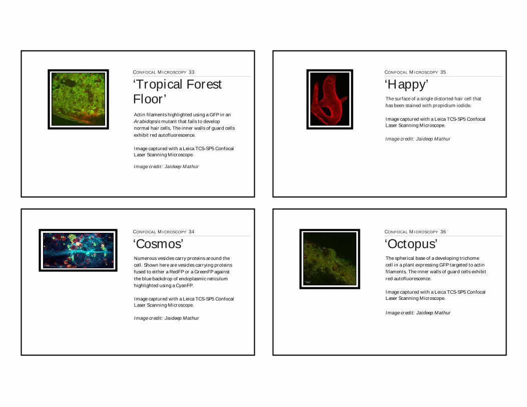
Actin filaments highlighted using a GFP in an Arabidopsis mutant that fails to develop normal hair cells. The inner walls of guard cells exhibit red autofluorescence.
Image captured with a Leica TCS-SP5 Confocal Laser Scanning Microscope.
CONFOCAL MICROSCOPY 33
‘Tropical ForestFloor’
Image credit: Jaideep Mathur
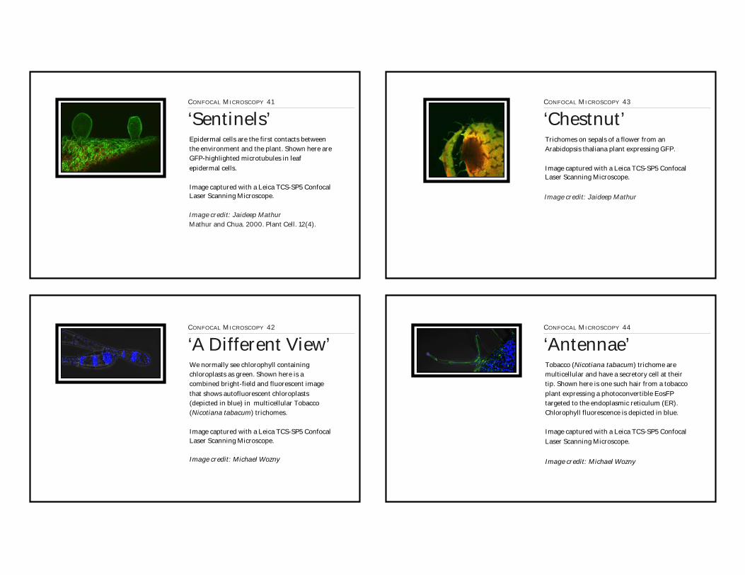
CONFOCAL MICROSCOPY 34
‘Cosmos’Numerous vesicles carry proteins around the cell. Shown here are vesicles carrying proteins fused to either a RedFP or a GreenFP against the blue backdrop of endoplasmic reticulum highlighted using a CyanFP.
Image captured with a Leica TCS-SP5 Confocal Laser Scanning Microscope.
Image credit: Jaideep Mathur
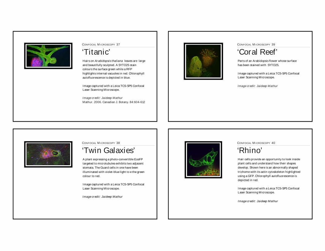
The surface of a single distorted hair cell that has been stained with propidium iodide.
Image captured with a Leica TCS-SP5 Confocal Laser Scanning Microscope.
CONFOCAL MICROSCOPY 35
‘Happy’
Image credit: Jaideep Mathur
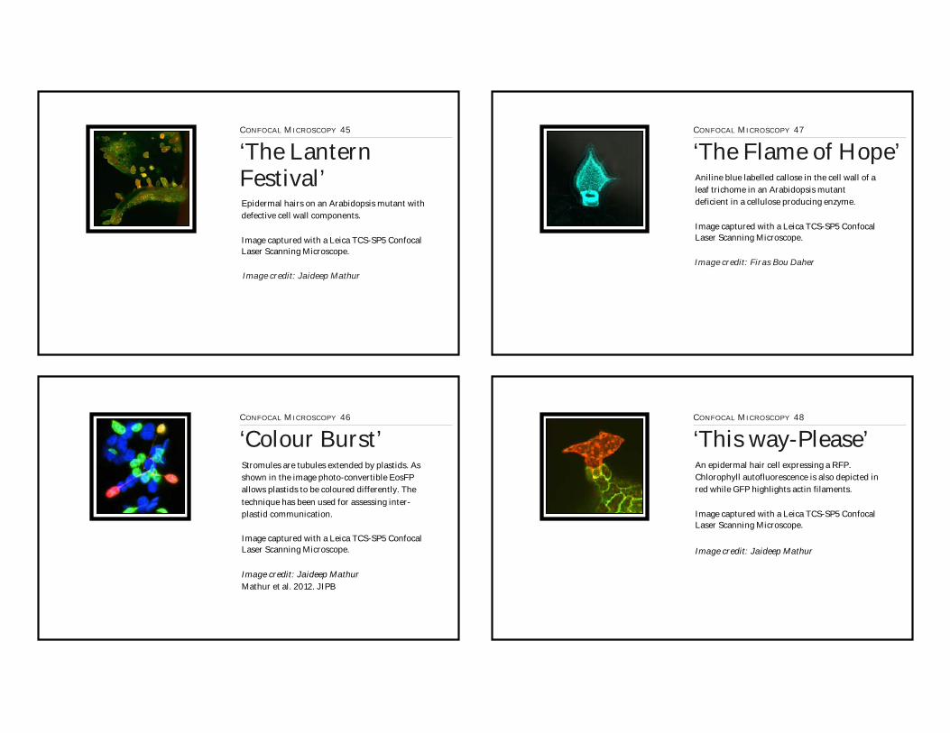
CONFOCAL MICROSCOPY 36
‘Octopus’The spherical base of a developing trichome cell in a plant expressing GFP targeted to actin filaments. The inner walls of guard cells exhibit red autofluorescence.
Image captured with a Leica TCS-SP5 Confocal Laser Scanning Microscope.
Image credit: Jaideep Mathur
Hairs on Arabidopsis thaliana leaves are large and beautifully sculpted. A SYTO25 stain colours the surface green while a RFP highlights internal vacuoles in red. Chlorophyll autofluorescence is depicted in blue.
Image captured with a Leica TCS-SP5 Confocal Laser Scanning Microscope.
CONFOCAL MICROSCOPY 37
‘Titanic’
Image credit: Jaideep MathurMathur. 2006. Canadian J. Botany. 84:604-612
CONFOCAL MICROSCOPY 38
‘Twin Galaxies’A plant expressing a photo-convertible EosFP targeted to microtubules exhibits two adjacent stomata. The Guard cells in one have been illuminated with violet-blue light to e the green colour to red.
Image captured with a Leica TCS-SP5 Confocal Laser Scanning Microscope.
Image credit: Jaideep Mathur
Parts of an Arabidopsis flower whose surface has been stained with SYTO25.
Image captured with a Leica TCS-SP5 Confocal Laser Scanning Microscope.
CONFOCAL MICROSCOPY 39
‘Coral Reef’
Image credit: Jaideep Mathur
CONFOCAL MICROSCOPY 40
‘Rhino’Hair cells provide an opportunity to look inside plant cells and understand how their shapes develop. Shown here is an abnormally shaped trichome with its actin cytoskeleton highlighted using a GFP. Chlorophyll autofluorescence is depicted in red.
Image captured with a Leica TCS-SP5 Confocal Laser Scanning Microscope.
Image credit: Jaideep Mathur
Epidermal cells are the first contacts between the environment and the plant. Shown here are GFP-highlighted microtubules in leaf epidermal cells.
Image captured with a Leica TCS-SP5 Confocal Laser Scanning Microscope.
CONFOCAL MICROSCOPY 41
‘Sentinels’
Image credit: Jaideep MathurMathur and Chua. 2000. Plant Cell. 12(4).
CONFOCAL MICROSCOPY 42
‘A Different View’We normally see chlorophyll containing chloroplasts as green. Shown here is a combined bright-field and fluorescent image that shows autofluorescent chloroplasts (depicted in blue) in multicellular Tobacco (Nicotiana tabacum) trichomes.
Image captured with a Leica TCS-SP5 Confocal Laser Scanning Microscope.
Image credit: Michael Wozny
Trichomes on sepals of a flower from an Arabidopsis thaliana plant expressing GFP.
Image captured with a Leica TCS-SP5 Confocal Laser Scanning Microscope.
CONFOCAL MICROSCOPY 43
‘Chestnut’
Image credit: Jaideep Mathur
CONFOCAL MICROSCOPY 44
‘Antennae’Tobacco (Nicotiana tabacum) trichome are multicellular and have a secretory cell at their tip. Shown here is one such hair from a tobacco plant expressing a photoconvertible EosFP targeted to the endoplasmic reticulum (ER). Chlorophyll fluorescence is depicted in blue.
Image captured with a Leica TCS-SP5 Confocal Laser Scanning Microscope.
Image credit: Michael Wozny
Epidermal hairs on an Arabidopsis mutant with defective cell wall components.
Image captured with a Leica TCS-SP5 Confocal Laser Scanning Microscope.
CONFOCAL MICROSCOPY 45
‘The Lantern Festival’
Image credit: Jaideep Mathur
CONFOCAL MICROSCOPY 46
‘Colour Burst’Stromules are tubules extended by plastids. As shown in the image photo-convertible EosFP allows plastids to be coloured differently. The technique has been used for assessing inter-plastid communication.
Image captured with a Leica TCS-SP5 Confocal Laser Scanning Microscope.
Image credit: Jaideep MathurMathur et al. 2012. JIPB
Aniline blue labelled callose in the cell wall of a leaf trichome in an Arabidopsis mutant deficient in a cellulose producing enzyme.
Image captured with a Leica TCS-SP5 Confocal Laser Scanning Microscope.
CONFOCAL MICROSCOPY 47
‘The Flame of Hope’
Image credit: Firas Bou Daher
CONFOCAL MICROSCOPY 48
‘This way-Please’An epidermal hair cell expressing a RFP. Chlorophyll autofluorescence is also depicted in red while GFP highlights actin filaments.
Image captured with a Leica TCS-SP5 Confocal Laser Scanning Microscope.
Image credit: Jaideep Mathur
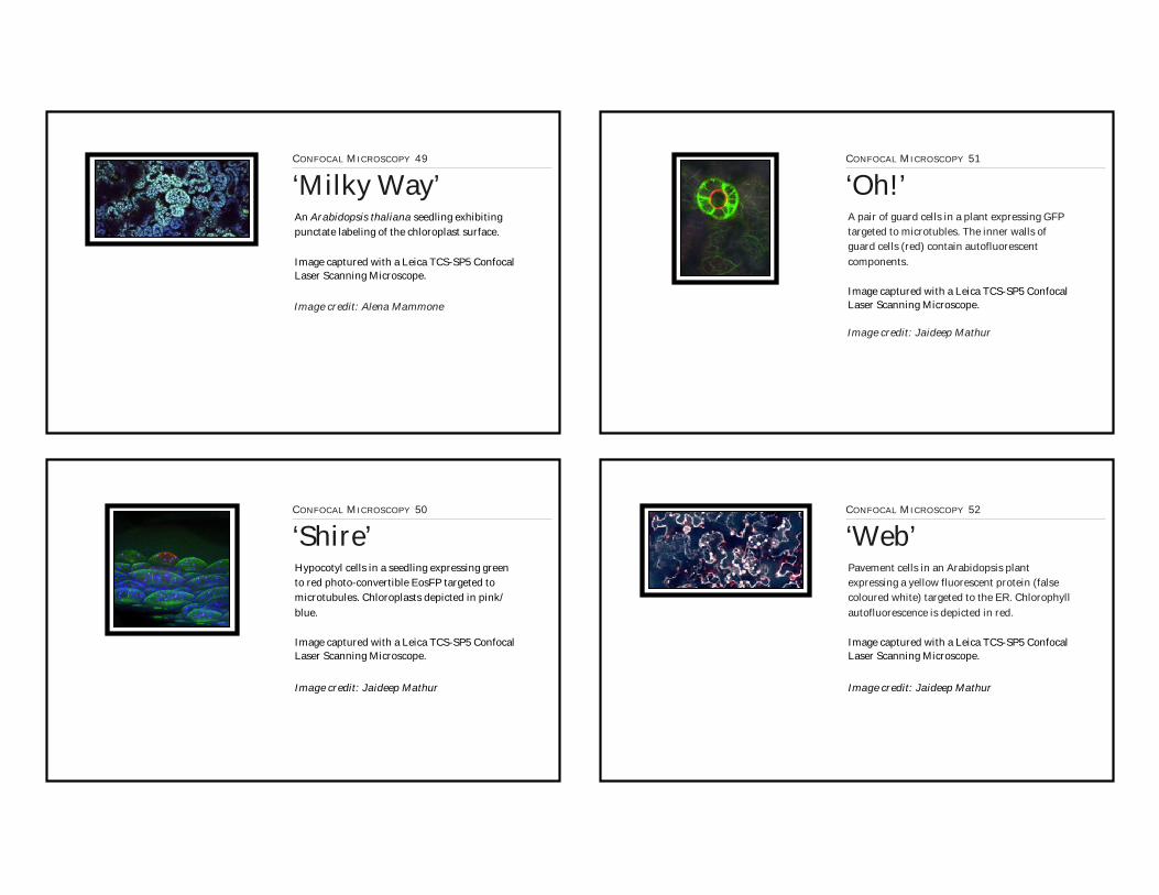
An Arabidopsis thaliana seedling exhibiting punctate labeling of the chloroplast surface.
Image captured with a Leica TCS-SP5 Confocal Laser Scanning Microscope.
CONFOCAL MICROSCOPY 49
‘Milky Way’
Image credit: Alena Mammone
CONFOCAL MICROSCOPY 50
‘Shire’Hypocotyl cells in a seedling expressing green to red photo-convertible EosFP targeted to microtubules. Chloroplasts depicted in pink/blue.
Image captured with a Leica TCS-SP5 Confocal Laser Scanning Microscope.
Image credit: Jaideep Mathur
A pair of guard cells in a plant expressing GFP targeted to microtubles. The inner walls of guard cells (red) contain autofluorescent components.
Image captured with a Leica TCS-SP5 Confocal Laser Scanning Microscope.
CONFOCAL MICROSCOPY 51
‘Oh!’
Image credit: Jaideep Mathur
CONFOCAL MICROSCOPY 52
‘Web’Pavement cells in an Arabidopsis plant expressing a yellow fluorescent protein (false coloured white) targeted to the ER. Chlorophyll autofluorescence is depicted in red.
Image captured with a Leica TCS-SP5 Confocal Laser Scanning Microscope.
Image credit: Jaideep Mathur

Chloroplasts (depicted in blue) are surrounded by a loose ER mesh, highlighted here using GFP.
Image captured with a Leica TCS-SP5 Confocal Laser Scanning Microscope.
CONFOCAL MICROSCOPY 53
‘Forget-me-not’
Image credit: Jaideep Mathur
CONFOCAL MICROSCOPY 54
‘Hot-and-Cold’Epidermis of an Arabidopsis plant expressing photo-convertible EosFP targeted to microtubules. Chlorophyll autofluorescence is depicted in red.
Image captured with a Leica TCS-SP5 Confocal Laser Scanning Microscope.
Image credit: Jaideep Mathur
Cell walls of leaf hair on an Arabidopsis mutant stained with SYTO25 .
Image captured with a Leica TCS-SP5 Confocal Laser Scanning Microscope.
CONFOCAL MICROSCOPY 55
‘Lanterns’
Image credit: Jaideep Mathur
CONFOCAL MICROSCOPY 56
‘River’Section of a leaf expressing YFP that highlights ‘callose’ a cell wall component. Chlorophyll autofluorescence is depicted in blue.
Image captured with a Leica TCS-SP5 Confocal Laser Scanning Microscope.
Image credit: Jaideep Mathur

Unicellular hairs on the leaf surface are large cells that tower above the relatively flat cells of the epidermis. Shown here is a single trichome of an Arabidopsis plant expressing GFP targeted to peroxisomes with SYTO25-stained walls.
Image captured with a Leica TCS-SP5 Confocal Laser Scanning Microscope.
CONFOCAL MICROSCOPY 57
‘Rio de Janeiro’
Image credit: Jaideep Mathur
CONFOCAL MICROSCOPY 58
‘Variations’A powerful colour discrimination based technique has been developed by our lab through the use of a green to red photo-convertible EosFP. Shown here are tobacco cells expressing EosFP targeted to the nucleus. An upper cell shows individual chromosomes painted in green to red.
Image captured with a Leica TCS-SP5 Confocal Laser Scanning Microscope.
Image credit: Jaideep Mathur
Wozny et al. 2012. Plant Physiol. 158: 95-106
False-colour coding of callus cells for depth. Red is farthest from the viewer.
Image captured with a Leica TCS-SP5 Confocal Laser Scanning Microscope.
CONFOCAL MICROSCOPY 59
‘Painted Silk’
Image credit: Jaideep Mathur
CONFOCAL MICROSCOPY 60
‘Legend of SleepyHollow’A distorted hair cell on an Arabidopsis mutant plant expressing GFP targeted to cytoskeletal elements.
Image captured with a Leica TCS-SP5 Confocal Laser Scanning Microscope.
Image credit: Jaideep Mathur
Mathur et al. 1999. Development. 130: 3137
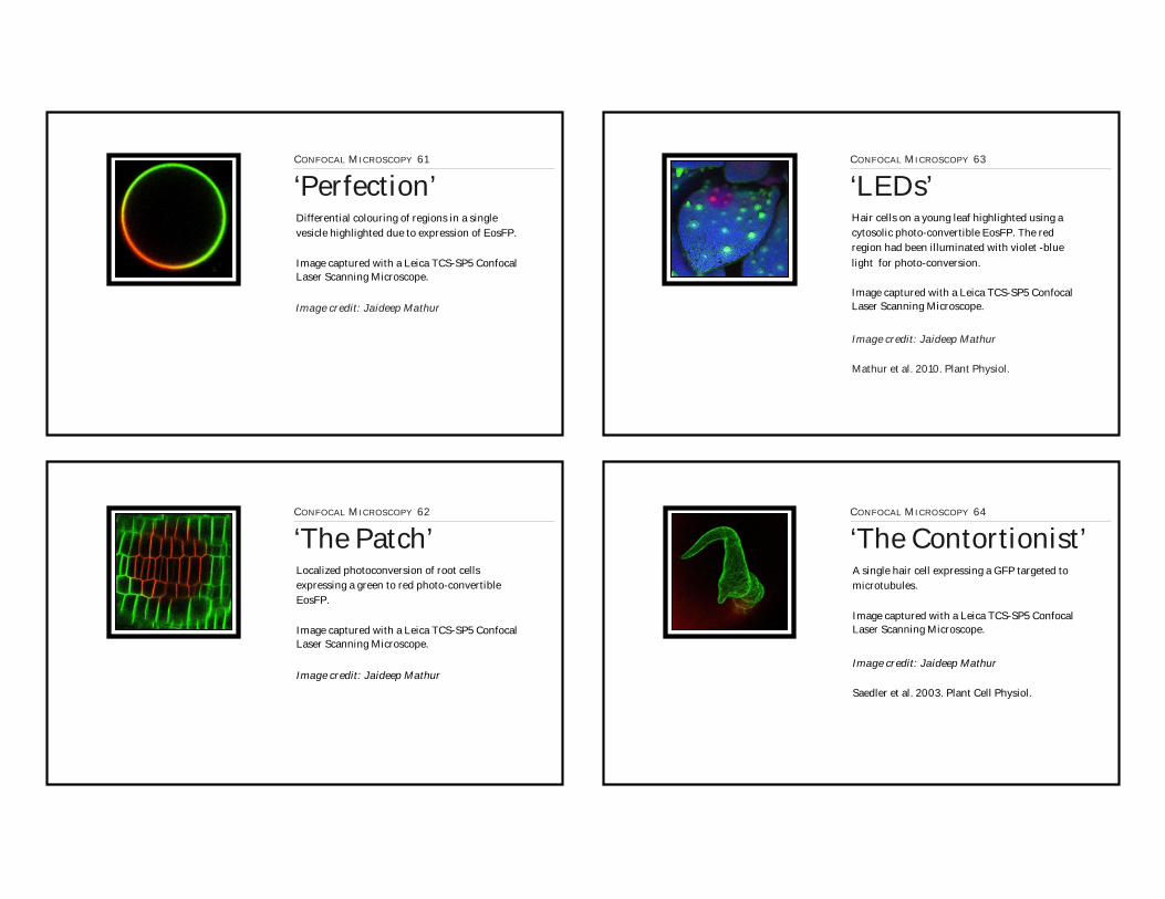
Differential colouring of regions in a single vesicle highlighted due to expression of EosFP.
Image captured with a Leica TCS-SP5 Confocal Laser Scanning Microscope.
CONFOCAL MICROSCOPY 61
‘Perfection’
Image credit: Jaideep Mathur
CONFOCAL MICROSCOPY 62
‘The Patch’Localized photoconversion of root cells expressing a green to red photo-convertible EosFP.
Image captured with a Leica TCS-SP5 Confocal Laser Scanning Microscope.
Image credit: Jaideep Mathur
Hair cells on a young leaf highlighted using a cytosolic photo-convertible EosFP. The red region had been illuminated with violet -blue light for photo-conversion.
Image captured with a Leica TCS-SP5 Confocal Laser Scanning Microscope.
CONFOCAL MICROSCOPY 63
‘LEDs’
Image credit: Jaideep Mathur
Mathur et al. 2010. Plant Physiol.
CONFOCAL MICROSCOPY 64
‘The Contortionist’A single hair cell expressing a GFP targeted to microtubules.
Image captured with a Leica TCS-SP5 Confocal Laser Scanning Microscope.
Image credit: Jaideep Mathur
Saedler et al. 2003. Plant Cell Physiol.
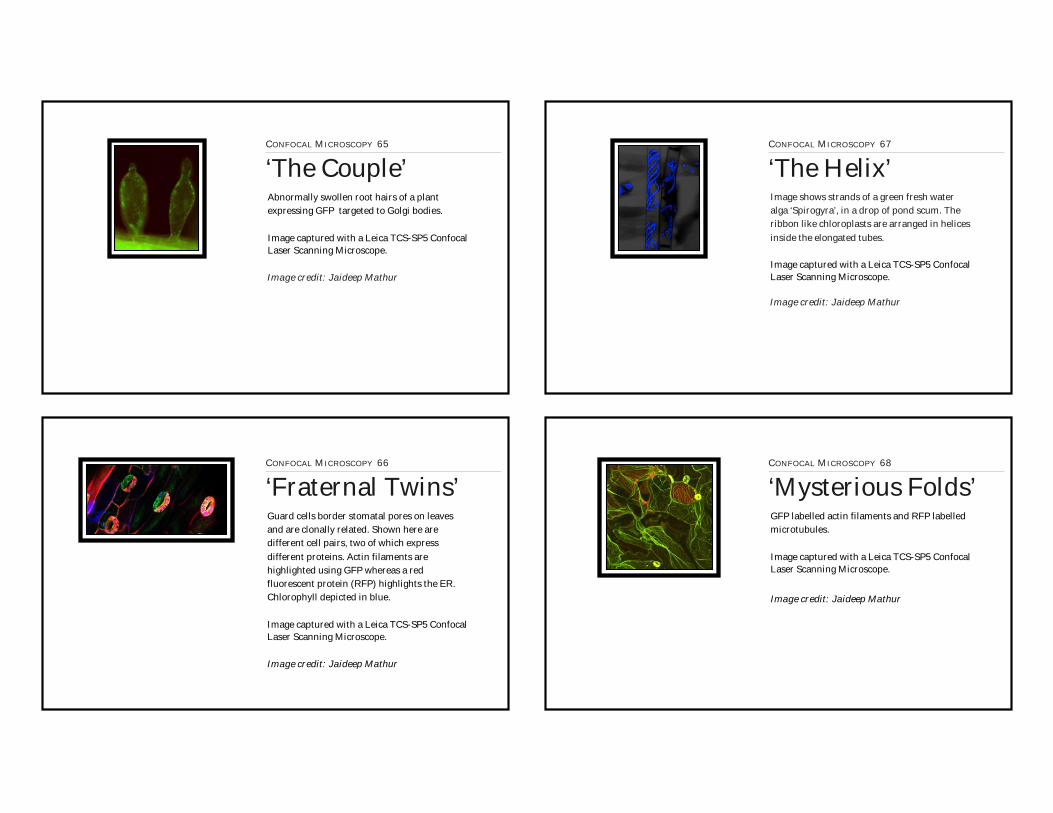
Abnormally swollen root hairs of a plant expressing GFP targeted to Golgi bodies.
Image captured with a Leica TCS-SP5 Confocal Laser Scanning Microscope.
CONFOCAL MICROSCOPY 65
‘The Couple’
Image credit: Jaideep Mathur
CONFOCAL MICROSCOPY 66
‘Fraternal Twins’Guard cells border stomatal pores on leaves and are clonally related. Shown here are different cell pairs, two of which express different proteins. Actin filaments are highlighted using GFP whereas a red fluorescent protein (RFP) highlights the ER. Chlorophyll depicted in blue.
Image captured with a Leica TCS-SP5 Confocal Laser Scanning Microscope.
Image credit: Jaideep Mathur
Image shows strands of a green fresh water alga ‘Spirogyra’, in a drop of pond scum. The ribbon like chloroplasts are arranged in helices inside the elongated tubes.
Image captured with a Leica TCS-SP5 Confocal Laser Scanning Microscope.
CONFOCAL MICROSCOPY 67
‘The Helix’
Image credit: Jaideep Mathur
CONFOCAL MICROSCOPY 68
‘Mysterious Folds’GFP labelled actin filaments and RFP labelled microtubules.
Image captured with a Leica TCS-SP5 Confocal Laser Scanning Microscope.
Image credit: Jaideep Mathur

The 3D volume rendered image shows a green fluorescent protein (GFP) labeled chloroplast with an extended stromule against a backdrop of endoplasmic reticulum (ER) highlighted using red fluorescent protein (RFP).
Image captured with a Leica TCS SP5 Confocal Laser Scanning Microscope and 3D volume rendered using Imaris 6.4.0 software.
RENDERED IMAGES 1
‘Life Seed’
Image credit: Martin Schattat & Kiah Barton
Featured Cover Page: Plant Physiology April 2011.http://www.plantphysiol.org/content/155/4.cover-expansion
Schattat et al. PLANT PHYSIOLOGY. 2011. Apr 155(4):1667-1677.
RENDERED IMAGES 2
‘Colour Splashes’
Differential colouring of plastids and their stromules achieved through expression of plastid-targeted EosFP.
Image captured with a Leica TCS SP5 Confocal Laser Scanning Microscope and 3D volume rendered using Imaris 6.4.0 software.
Image credit: Jaideep Mathur
Featured Cover Page: Plant Cell 2012http://www.plantcell.org/content/24/4.cover-expansion
Schattat et al. 2012. Plant Cell.
Chloroplast depicted in blue and endoplasmic reticulum (ER) in green in a plant cell.
Image captured with a Leica TCS SP5 Confocal Laser Scanning Microscope and 3D volume rendered using Imaris 6.4.0 software.
RENDERED IMAGES 3
‘In a Galaxy Far,Far Away...’
Image credit: Kiah Barton
RENDERED IMAGES 4
‘Reaching Out’Chloroplast with extended stromule (green), embedded in a network of endoplasmic reticulum (ER; in red).
Image captured with a Leica TCS SP5 Confocal Laser Scanning Microscope and 3D volume rendered using Imaris 6.4.0 software.
Image credit: Kiah Barton
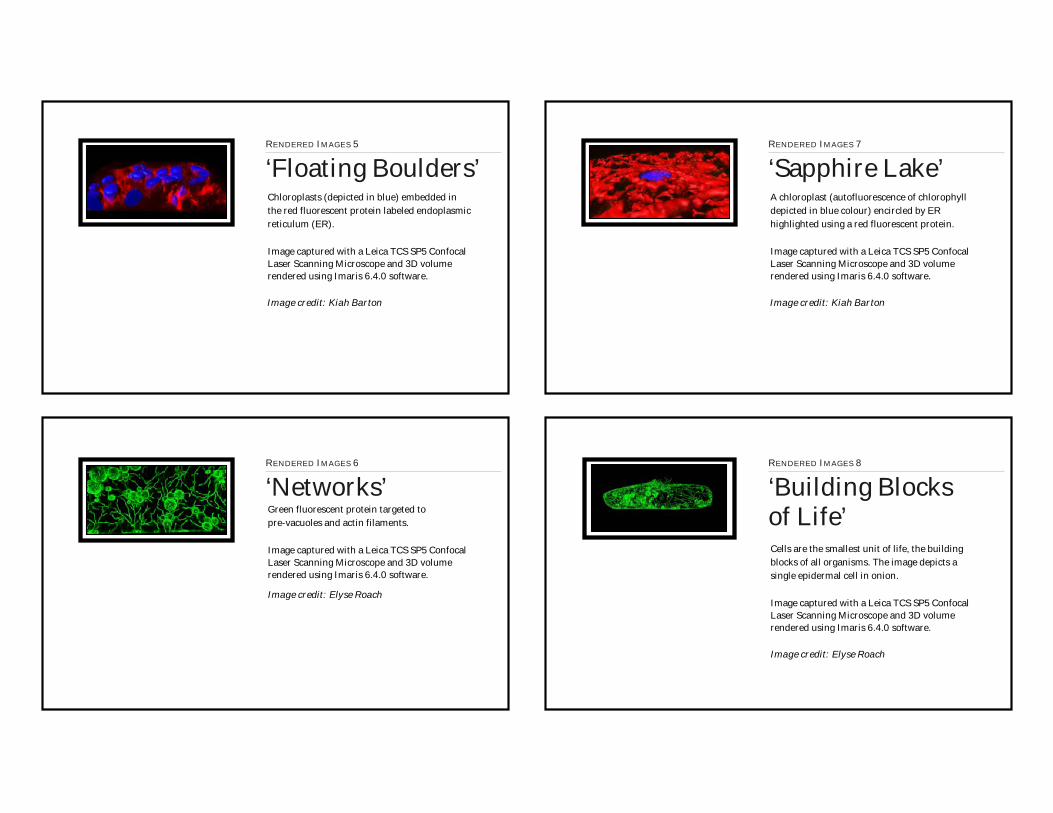
Chloroplasts (depicted in blue) embedded in the red fluorescent protein labeled endoplasmic reticulum (ER).
Image captured with a Leica TCS SP5 Confocal Laser Scanning Microscope and 3D volume rendered using Imaris 6.4.0 software.
RENDERED IMAGES 5
‘Floating Boulders’
Image credit: Kiah Barton
RENDERED IMAGES 6
‘Networks’Green fluorescent protein targeted to pre-vacuoles and actin filaments.
Image captured with a Leica TCS SP5 Confocal Laser Scanning Microscope and 3D volume rendered using Imaris 6.4.0 software.
Image credit: Elyse Roach
A chloroplast (autofluorescence of chlorophyll depicted in blue colour) encircled by ER highlighted using a red fluorescent protein.
Image captured with a Leica TCS SP5 Confocal Laser Scanning Microscope and 3D volume rendered using Imaris 6.4.0 software.
RENDERED IMAGES 7
‘Sapphire Lake’
Image credit: Kiah Barton
RENDERED IMAGES 8
‘Building Blocksof Life’Cells are the smallest unit of life, the building blocks of all organisms. The image depicts a single epidermal cell in onion.
Image captured with a Leica TCS SP5 Confocal Laser Scanning Microscope and 3D volume rendered using Imaris 6.4.0 software.
Image credit: Elyse Roach

Stigmatic papillae of an Arabidopsis thaliana flower.
Image captured with a Hitachi Tabletop TM-1000 Scanning Electron Microscope.
SCANNING ELECTRON MICROGRAPH 1
‘Grace’
Image credit: Jaideep Mathur
SCANNING ELECTRON MICROGRAPH 2
‘Postures’Scanning electron micrograph showing distorted unicellular hair on the leaf surface of an Arabidopsis mutant.
Image captured with a Hitachi Tabletop TM-1000 Scanning Electron Microscope.
Image credit: Jaideep Mathur
Pollen grains from commercially available bee pollen granules.
Image captured with a Hitachi Tabletop TM-1000 Scanning Electron Microscope.
SCANNING ELECTRON MICROGRAPH 3
‘Sticky & Sweet’
Image credit: Firas Bou Daher
SCANNING ELECTRON MICROGRAPH 4
‘Guarded Future’Scanning electron micrograph showing the flower buds of an Arabidopsis axillary inflorescence.
Image captured with a Hitachi Tabletop TM-1000 Scanning Electron Microscope.
Image credit: Jaideep Mathur
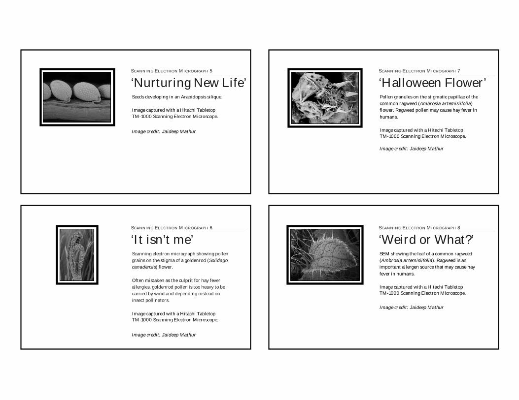
Seeds developing in an Arabidopsis silique.
Image captured with a Hitachi Tabletop TM-1000 Scanning Electron Microscope.
SCANNING ELECTRON MICROGRAPH 5
‘Nurturing New Life’
Image credit: Jaideep Mathur
SCANNING ELECTRON MICROGRAPH 6
‘It isn’t me’Scanning electron micrograph showing pollen grains on the stigma of a goldenrod (Solidago canadensis) flower.
Often mistaken as the culprit for hay fever allergies, goldenrod pollen is too heavy to be carried by wind and depending instead on insect pollinators.
Image captured with a Hitachi Tabletop TM-1000 Scanning Electron Microscope.
Image credit: Jaideep Mathur
Pollen granules on the stigmatic papillae of the common ragweed (Ambrosia artemisiifolia) flower. Ragweed pollen may cause hay fever in humans.
Image captured with a Hitachi Tabletop TM-1000 Scanning Electron Microscope.
SCANNING ELECTRON MICROGRAPH 7
‘Halloween Flower’
Image credit: Jaideep Mathur
SCANNING ELECTRON MICROGRAPH 8
‘Weird or What?’SEM showing the leaf of a common ragweed (Ambrosia artemisiifolia). Ragweed is an important allergen source that may cause hay fever in humans.
Image captured with a Hitachi Tabletop TM-1000 Scanning Electron Microscope.
Image credit: Jaideep Mathur

SEM of epidermal hair on a leaf treated with the microtubule stabilizing drug Taxol.
Image captured with a Hitachi Tabletop TM-1000 Scanning Electron Microscope.
SCANNING ELECTRON MICROGRAPH 9
‘Puffball’
Image credit: Jaideep Mathur
SCANNING ELECTRON MICROGRAPH 10
‘Together We Can’Garlic mustard (Alliaria petiolata) has emerged as a rapidly growing invasive species. The false-coloured SEM of an unopened inflorescence symbolizes the packed potential of this plant.
Image captured with a Hitachi Tabletop TM-1000 Scanning Electron Microscope.
Image credit: Firas Bou Daher
Trichomes on the leaf of a tobacco (Nicotiana tabacum) plant.
Image captured with a Hitachi Tabletop TM-1000 Scanning Electron Microscope.
SCANNING ELECTRON MICROGRAPH 11
‘Lollipops’
Image credit: Michael Wozny
SCANNING ELECTRON MICROGRAPH 12
‘Don’t Push!’Swollen hair cells on both sides of a leaf surface.
Image captured with a Hitachi Tabletop TM-1000 Scanning Electron Microscope
Image credit: Michael Wozny

SEM showing seeds from a Fall Anemone (Anemone sp.) plant.
Image captured with a Hitachi Tabletop TM-1000 Scanning Electron Microscope.
SCANNING ELECTRON MICROGRAPH 13
‘Mandala’
Image credit: Firas Bou Daher
SCANNING ELECTRON MICROGRAPH 14
‘Budding Beauty’Unicellular hair cells on buds of an Arabidopsis thaliana mutant plant.
Image captured with a Hitachi Tabletop TM-1000 Scanning Electron Microscope.
Image credit: Ashley Jaipargas
Scanning electron micrograph of an Arabidopsis flower; petals in the background, stigmatic papillae and anthers in the front.
Image captured with a Hitachi Tabletop TM-1000 Scanning Electron Microscope.
SCANNING ELECTRON MICROGRAPH 15
‘Lady’
Image credit: Jaideep Mathur
SCANNING ELECTRON MICROGRAPH 16
‘Squeezeballs’Pseudo-colored SEM depicting pollen grains of the Common Hibiscus or China rose (Hibiscus rosa-sinensis).
Image captured with a Hitachi Tabletop TM-1000 Scanning Electron Microscope.
Image credit: Michael Wozny

10010010010010010000010000000000000000000000 101101101101101101101101101101101101
1021020211020202102102021021022101010210 1031030303030303330303030330303033033333

10410404100404104044444444444444444 1051051051110510101100500510505111001110510500555
106106 107107777777

108108 109109109109
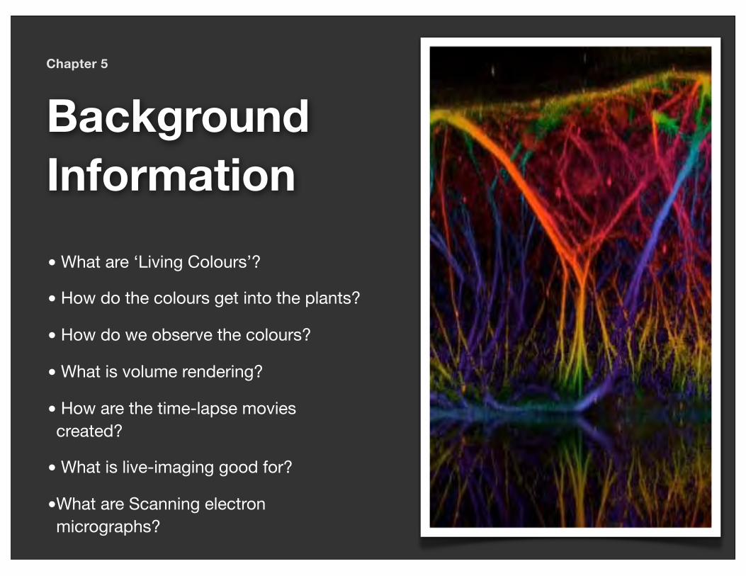
Chapter 5
BackgroundInformation• What are ‘Living Colours’?
• How do the colours get into the plants?
• How do we observe the colours?
• What is volume rendering?
• How are the time-lapse movies created?
• What is live-imaging good for?
•What are Scanning electron micrographs?
LIVING COLOURS for painting plantsKiah Barton, Firas Bou Daher, Ashley Jaipargas, Alena Mammone, Neeta Mathur, Madhulika Sareen, Michael Wozny, Nina K. Barabas & Jaideep Mathur
What are living colours ?
Living colours, are genetically encoded proteins, found naturally in diverse living organisms, that fluoresce when illuminated with light of a specific colour. The term was introduced following the cloning of the Green Fluorescent Prote in (GFP), produced by the bioluminescent jellyfish Aequorea victoria. GFP’s small size makes it a useful fluorescent tag, as it can be fused to other proteins without disrupting their function [1, 2].
Did you know?The advances that GFP has allowed in the field of cell biology are so substantial that the 2008 Nobel Prize in Chemistry was awarded for its cloning and development [4].
Are there many living colours ?
Since its initial discovery [1], many colour mutants of GFP have been created [2]. They range from blue to yellow. Many variants are significantly brighter and more tolerant to cellular
conditions and a range of temperatures in comparison to the wild-type protein. GFP variants also display reduced photobleaching, or loss of fluorescence due to damage by light. One variant called HyPer-GFP has even been engineered to exhibit an increase in fluorescence intensity in response to hydrogen peroxide. All these properties have allowed GFP and its derivatives to be expressed in a wide range of organisms. Genes from other marine organisms such as corals and anemones have contributed to the growth of the ‘living colours’.
A commonly used red fluorescent protein (RFP) is derived from the Discosoma coral [5]. In addition, a new class of inducible FPs dubbed ‘optical highlighters’ has become available from stony corals.
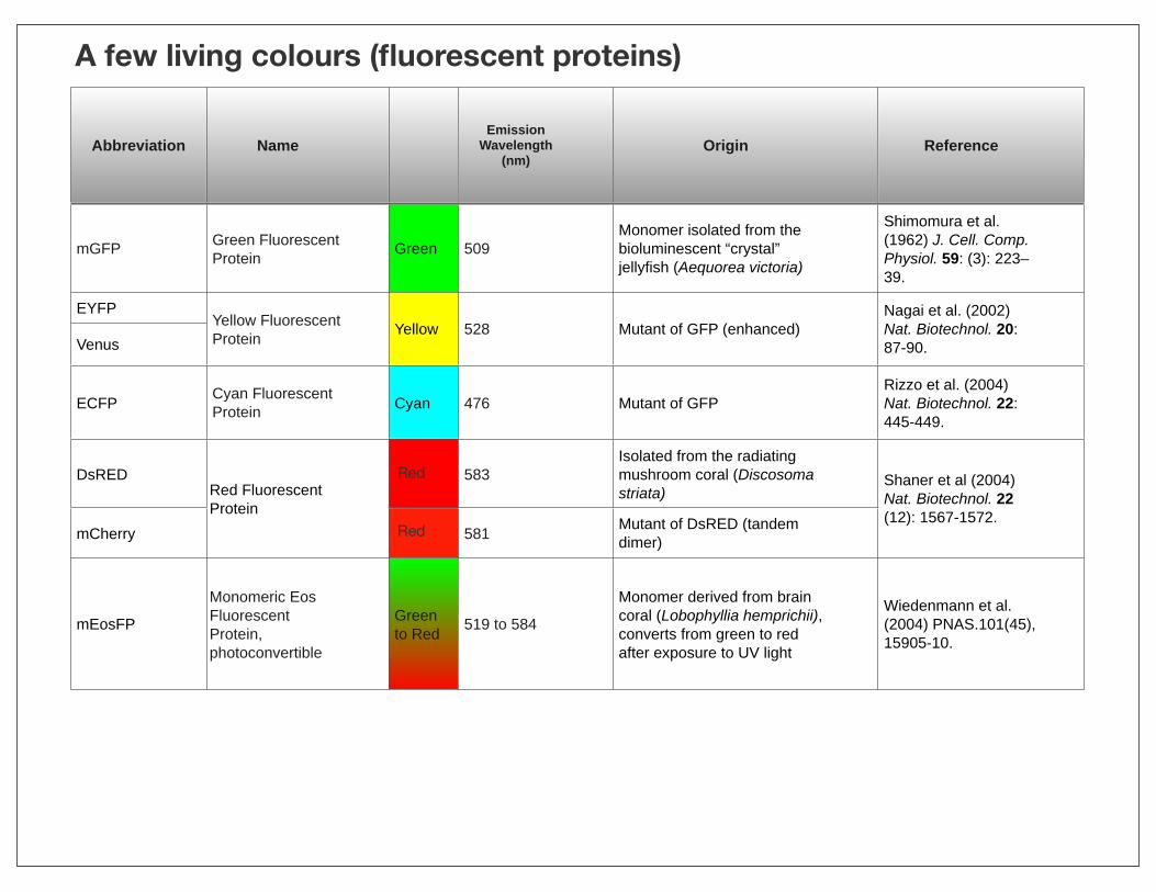
Abbreviation NameEmission
Wavelength (nm)
Origin Reference
mGFP Green Fluorescent Protein Green 509
Monomer isolated from the bioluminescent “crystal” jellyfish (Aequorea victoria)
Shimomura et al. (1962) J. Cell. Comp. Physiol. 59: (3): 223–39.
EYFPYellow Fluorescent Protein Yellow 528 Mutant of GFP (enhanced)
Nagai et al. (2002) Nat. Biotechnol. 20: 87-90.Venus
Yellow Fluorescent Protein Yellow 528 Mutant of GFP (enhanced)
Nagai et al. (2002) Nat. Biotechnol. 20: 87-90.
ECFP Cyan Fluorescent Protein Cyan 476 Mutant of GFP
Rizzo et al. (2004) Nat. Biotechnol. 22: 445-449.
DsREDRed Fluorescent Protein
583Isolated from the radiating mushroom coral (Discosoma striata)
Shaner et al (2004) Nat. Biotechnol. 22 (12): 1567-1572.
mCherry
Red Fluorescent Protein
581 Mutant of DsRED (tandem dimer)
Shaner et al (2004) Nat. Biotechnol. 22 (12): 1567-1572.
mEosFPMonomeric Eos Fluorescent Protein, photoconvertible
Green to Red 519 to 584
Monomer derived from brain coral (Lobophyllia hemprichii), converts from green to red after exposure to UV light
Wiedenmann et al. (2004) PNAS.101(45), 15905-10.
A few living colours (fluorescent proteins)
Red
Red

Key References: [1] Prasher et al.(1992). Primary structure of the Aequorea victoria green-fluorescent protein. Gene 111: 229-233. [2] Tsien, R.Y. (1998). The green fluorescent protein. Annu. Rev. Biochem. 67: 509-544. [3] Zhang et al. (2002). Creating new fluorescent probes for cell biology. Nat. Rev. Mol. Biol. 3:, 906-918. [4] “The Nobel Prize in Chemistry 2008 - Press Release”. Nobelprize.org.http://www.nobelprize.org/nobel_prizes/chemistry/laureates/2008/press.html [5] Shaner et al. (2007). Advances in fluorescent protein technology. J. Cell Sci. 120: 4247-4260. [6] Wiedenmann et al. (2004). EosFP, a fluorescent marker protein with UV-inducible green-to-red fluorescence conversion. Proc. Natl. Acad. Sci. 101: 15905-15910. [7] Mathur et al. (2010) Plant Physiol. 154: 1573-87.
These newly discovered ‘optical highlighters’ change their fluorescent properties in response to specific wavelengths of light, most often in the violet-blue range. As shown in the accompanying figure these include the photo-activatable, photo-switchable and photo-convertible fluorescent proteins [5].Our lab has been developing Eos fluorescent protein [6] as a precise cell biological tool [7].
LIVING COLOURS for painting plants

How do the colours get into plants?by Alena Mammone
We create new fluorescent plants by first manipulating pieces of DNA through the general process known as gene cloning. While the end product is a transgenic plant carrying fluorescent labels, all the molecular work is carried out using bacteria and mobile DNA elements called plasmids.
In bacteria, plasmids are very common and are used to spread new traits such as drug resistance to other cells. They are circular and tiny compared to the bacterial chromosome. To cut the DNA molecular scissors known as restriction enzymes (RE) are used.
In nature, RE are used by bacteria as defense mechanisms, to protect against foreign DNA by chopping it into tiny pieces.
Bacteriologists have characterized thousands of these enzymes and many are available for use in cloning DNA. Other proteins, called ligases are able to join cut fragments of DNA.
A gene for a fluorescent protein, such as the Green fluorescent Protein (GFP) gene from the jellyfish is placed in a plasmid. Using a ligase it may be joined to another gene or a signal sequence from another organism. Eventually expression of such a stitched gene will produce a chimeric fusion protein in the cell.
The assembled gene is now introduced into a natural plant pathogen, Agrobacterium tumefaciens that is able to transfer it into a plant’s genome. Thus a transgenic plant is created.
When the introduced novel gene is expressed we see the targeted structures in a cell become highlighted in bright fluorescent colours. Transgenic plants carrying different coloured proteins are crossed to produce multicolour plants that allow visualization of different structures in different colours.

Creating transgenic plants that express targeted fluorescent proteins
Schematic: by Kiah Barton
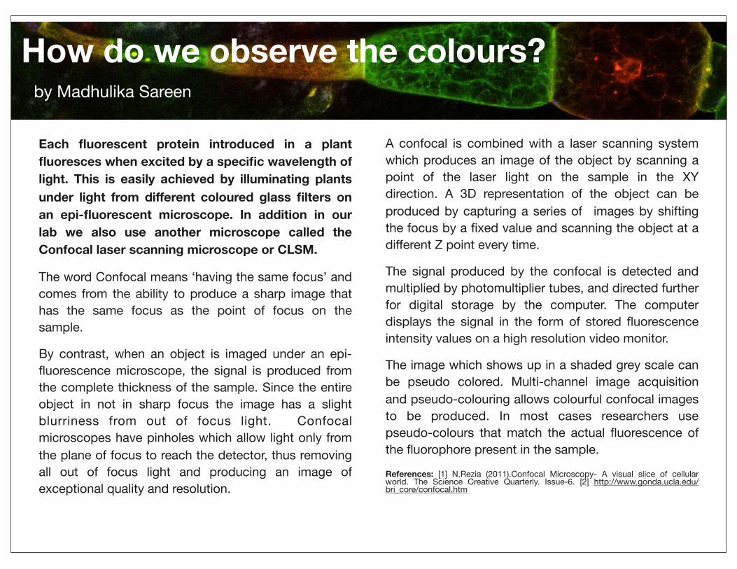
How do we observe the colours?by Madhulika Sareen
Each fluorescent protein introduced in a plant fluoresces when excited by a specific wavelength of light. This is easily achieved by illuminating plants under light from different coloured glass filters on an epi-fluorescent microscope. In addition in our lab we also use another microscope called the Confocal laser scanning microscope or CLSM.
The word Confocal means ‘having the same focus’ and comes from the ability to produce a sharp image that has the same focus as the point of focus on the sample.
By contrast, when an object is imaged under an epi- fluorescence microscope, the signal is produced from the complete thickness of the sample. Since the entire object in not in sharp focus the image has a slight blurriness from out of focus light. Confocal microscopes have pinholes which allow light only from the plane of focus to reach the detector, thus removing all out of focus light and producing an image of exceptional quality and resolution.
A confocal is combined with a laser scanning system which produces an image of the object by scanning a point of the laser light on the sample in the XY direction. A 3D representation of the object can be produced by capturing a series of images by shifting the focus by a fixed value and scanning the object at a different Z point every time.
The signal produced by the confocal is detected and multiplied by photomultiplier tubes, and directed further for digital storage by the computer. The computer displays the signal in the form of stored fluorescence intensity values on a high resolution video monitor.
The image which shows up in a shaded grey scale can be pseudo colored. Multi-channel image acquisition and pseudo-colouring allows colourful confocal images to be produced. In most cases researchers use pseudo-colours that match the actual fluorescence of the fluorophore present in the sample.
References: [1] N.Rezia (2011).Confocal Microscopy- A visual slice of cellular world. The Science Creative Quarterly. Issue-6. [2] http://www.gonda.ucla.edu/bri_core/confocal.htm

What is volume rendering?by Kiah Barton
Volume rendering is a technique used as digital aid for visualizing an image in three dimensions.
In the lab we use fluorescent proteins to observe the behaviour of organelles or compartments within a living cell by taking single snapshots or time-lapse images using a confocal microscope. With this microscope it is possible to image the cell in three dimensions by taking a series of single-plane images, each of which is of the same area but is shifted through the cell by a fixed distance in the z-direction. This stack of images can then be either compressed to form a single two-dimensional image representing the entire section, or it can be volume rendered so that it can be observed from any angle.
The digital stack of images acquired can itself be rotated in three dimensions, but there are gaps between the slices that can make it appear hazy or blurred when not viewed directly from the top or bottom. This can make it difficult to truly get a sense
of the relative positions of the organelles being observed. For example if one organelle was weaving around or through another in the z-direction this might not be readily apparent. They may appear to blur together in the stack, while in the compressed image it can only be seen that they occupied the same x-y position.
The Imaris image rendering software being used in our lab takes the stack of images and puts a surface on the fluorescence based on its intensity. By filling in the spaces between slices and giving the fluorescence an easily visualized surface, a single cohesive three dimensional image is obtained that can be rotated, zoomed and made selectively transparent.
This technique can thus be used to bring out relationships between compartments in the cell that might not be apparent in non-rendered images.
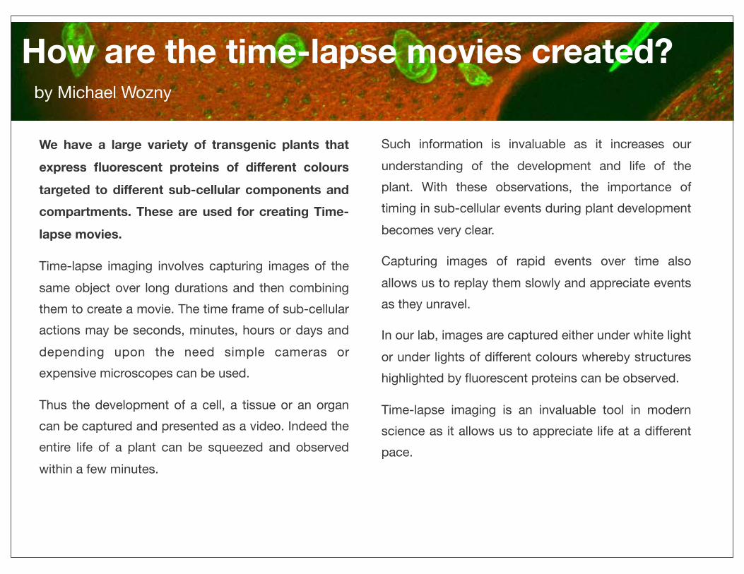
How are the time-lapse movies created?by Michael Wozny
We have a large variety of transgenic plants that express fluorescent proteins of different colours targeted to different sub-cellular components and compartments. These are used for creating Time-lapse movies.
Time-lapse imaging involves capturing images of the same object over long durations and then combining them to create a movie. The time frame of sub-cellular actions may be seconds, minutes, hours or days and depending upon the need simple cameras or expensive microscopes can be used.
Thus the development of a cell, a tissue or an organ can be captured and presented as a video. Indeed the entire life of a plant can be squeezed and observed within a few minutes.
Such information is invaluable as it increases our understanding of the development and life of the plant. With these observations, the importance of timing in sub-cellular events during plant development becomes very clear.
Capturing images of rapid events over time also allows us to replay them slowly and appreciate events as they unravel.
In our lab, images are captured either under white light or under lights of different colours whereby structures highlighted by fluorescent proteins can be observed.
Time-lapse imaging is an invaluable tool in modern science as it allows us to appreciate life at a different pace.

What is live-imaging good for?
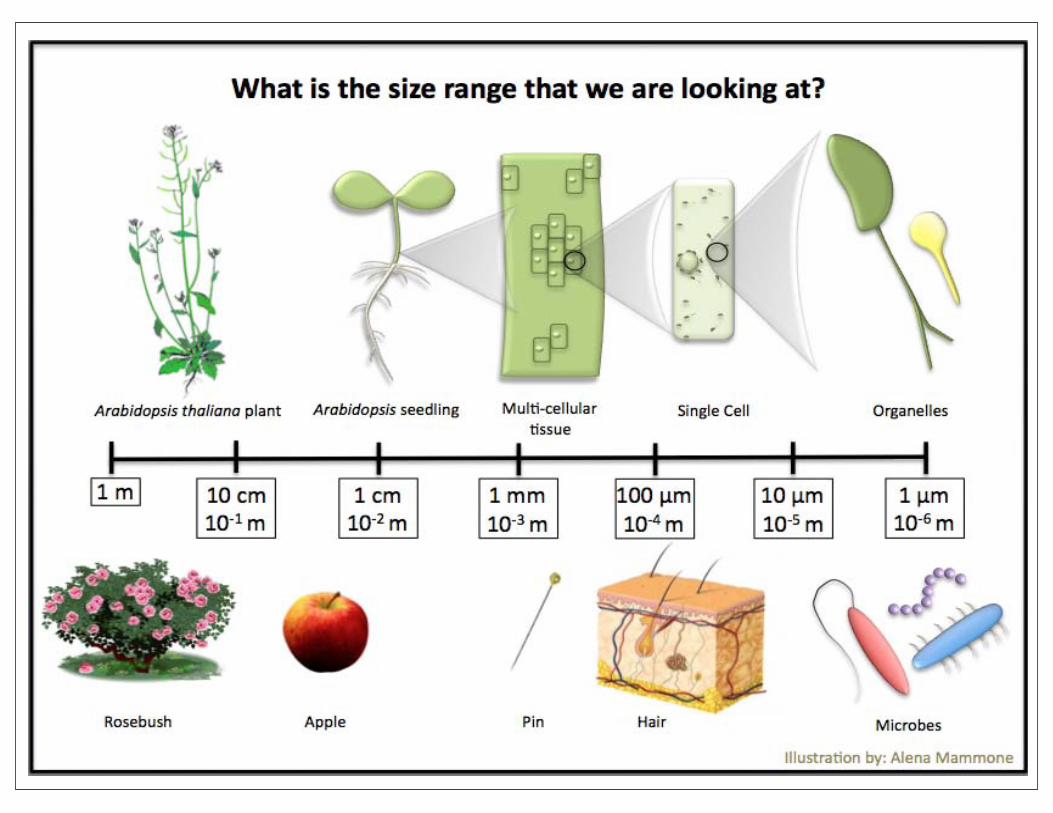
Visualization: By introducing a targeted fluorescent protein into plants we are now able to visualize most sub-cellular compartments and proteins in the plant cell. Under normal conditions (A), these extremely small components (in the range of 1000th to 10,000th of a millimetre) are generally transparent or translucent, not fluorescent, making them almost impossible to visualize.
Understanding: By making the sub-cellular level visible in multicolour, it is easier to follow different organelles, the cytoskeleton, protein sorting and trafficking, the localization, interactions, intracellular fate and extracellular movement of new proteins in the living organism in 4D (x,y,z,t dimensions) and at high resolution, especially with the aid of laser-based microscopes. For plants this brings new understanding about the mechanisms involved in their growth and development under various conditions.
Education: Observing the basic processes as they happen in the cell brings a new level of knowledge concerning life. Consequently, these ‘ colourful ’ transgenic plants are an invaluable educational tool for
answering fundamental questions about the modus operandi of the ‘building blocks of life’.
Fascination: Observing living processes taking place right before our eyes is awe-inspiring. A glimpse into the innermost workings of a cell elicits a sense of wonder and fascination for the living world, to which we belong. Live-imaging provides an opportunity to formulate new concepts, make discoveries and perhaps even cause paradigm shifts in our ideas about life and its numerous forms.
Bright field (A) Blue light (B) Green light (C) Merged (D)
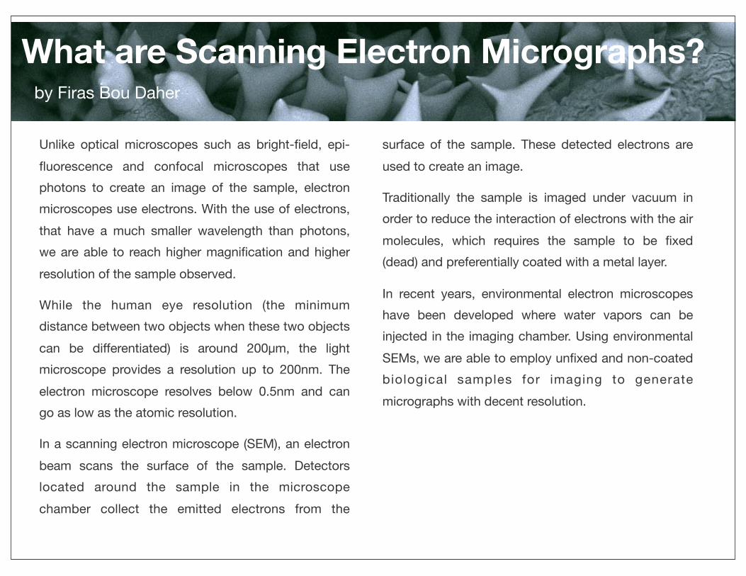
What are Scanning Electron Micrographs?by Firas Bou Daher
Unlike optical microscopes such as bright-field, epi-fluorescence and confocal microscopes that use photons to create an image of the sample, electron microscopes use electrons. With the use of electrons, that have a much smaller wavelength than photons, we are able to reach higher magnification and higher resolution of the sample observed.
While the human eye resolution (the minimum distance between two objects when these two objects can be differentiated) is around 200μm, the light microscope provides a resolution up to 200nm. The electron microscope resolves below 0.5nm and can go as low as the atomic resolution.
In a scanning electron microscope (SEM), an electron beam scans the surface of the sample. Detectors located around the sample in the microscope chamber collect the emitted electrons from the
surface of the sample. These detected electrons are used to create an image.
Traditionally the sample is imaged under vacuum in order to reduce the interaction of electrons with the air molecules, which requires the sample to be fixed (dead) and preferentially coated with a metal layer.
In recent years, environmental electron microscopes have been developed where water vapors can be injected in the imaging chamber. Using environmental SEMs, we are able to employ unfixed and non-coated biological samples for imaging to generate micrographs with decent resolution.

Welcome to many more great images in our collection!
•Numerous featured cover pages reflect publishers’ high interest
•Beautiful ‘living’ colours and graceful forms captivate the imagination
•A glimpse into the innermost workings of life elicit a sense of wonder
Fascinating. Educating. Inspiring.

Cellscapes available soon in full page electronic book
format via the iTunes stores and other venues.
Check our website for updates.
CONTACT
Please contact us for information regarding educational
materials and multimedia exhibitions featuring similar
scientific images and movies.
Jaideep Mathur, PhD Nina Katalin Barabas, PhD
[email protected] [email protected]
http://www.uoguelph.ca/~jmathur/