cellular actions of the insulin-like growth factor binding proteins
TRANSCRIPT
Cellular Actions of the Insulin-Like Growth FactorBinding Proteins
SUE M. FIRTH AND ROBERT C. BAXTER
Kolling Institute of Medical Research, University of Sydney, Royal North Shore Hospital, St. Leonards, New South Wales2065, Australia
In addition to their roles in IGF transport, the six IGF-bindingproteins (IGFBPs) regulate cell activity in various ways. Bysequestering IGFs away from the type I IGF receptor, theymay inhibit mitogenesis, differentiation, survival, and otherIGF-stimulated events. IGFBP proteolysis can reverse thisinhibition or generate IGFBP fragments with novel bioactiv-ity. Alternatively, IGFBP interaction with cell or matrix com-ponents may concentrate IGFs near their receptor, enhancingIGF activity. IGF receptor-independent IGFBP actions arealso increasingly recognized. IGFBP-1 interacts with �5�1 in-tegrin, influencing cell adhesion and migration. IGFBP-2, -3,-5, and -6 have heparin-binding domains and can bind glycos-aminoglycans. IGFBP-3 and -5 have carboxyl-terminal basicmotifs incorporating heparin-binding and additional basic
residues that interact with the cell surface and matrix, thenuclear transporter importin-�, and other proteins. Serine/threonine kinase receptors are proposed for IGFBP-3 and -5,but their signaling functions are poorly understood. Othercell surface IGFBP-interacting proteins are uncharacterizedas functional receptors. However, IGFBP-3 binds and modu-lates the retinoid X receptor-�, interacts with TGF� signalingthrough Smad proteins, and influences other signaling path-ways. These interactions can modulate cell cycle and apo-ptosis. Because IGFBPs regulate cell functions by diversemechanisms, manipulation of IGFBP-regulated pathways isspeculated to offer therapeutic opportunities in cancer andother diseases. (Endocrine Reviews 23: 824–854, 2002)
I. Introductory OverviewA. IGFBP structureB. Relationship between serum and tissue IGFBPs
II. Modulation of IGF Activity by IGFBPs and TheirProteasesA. IGFBP-4, -5, and -6B. IGFBP-1, -2, and -3
III. IGF and IGFRI-Independent Effects of IGFBPsIV. Adhesion and MigrationV. Cell-Binding Sites and Putative Receptors
A. Glycosaminoglycan-binding domains on IGFBPsB. The role of posttranslational modifications in cell
surface associationC. Other putative IGFBP receptors
VI. Interactions of IGFBP-3 with Known SignalingPathwaysA. IGFBP-3 and TGF� signalingB. IGFBP-3, retinoids, and nuclear signalingC. Other pathways
VII. Cell Cycle, Apoptosis, and SurvivalVIII. Implications for Animal Physiology
IX. Implications for Human DiseaseX. Concluding Comment
I. Introductory Overview
A. IGFBP structure
THE IGF BINDING PROTEIN (IGFBP) gene family con-sists of six well characterized members that encode a
family of homologous multifunctional proteins, IGFBP-1 toIGFBP-6. The genes share a common structural organization,in which four conserved exons are located within genesranging from 5 kb (IGFBP-1) to more than 30 kb (IGFBP-2 andIGFBP-5) (1). The IGFBP genes, like those of the IGFs them-selves, are believed to have emerged early in vertebrate evo-lution (2). Exon 1 of the IGFBP gene family is shared byseveral other genes encoding a variety of proteins, leading toproposals that they might be members of a larger gene su-perfamily (3, 4).
The precursor forms of all six IGFBPs have secretory signalpeptides of between 20 and 39 amino acids, and the matureproteins are all found extracellularly. They share a highlyconserved structure that is generally described as consistingof three domains of approximately equal size. In addition,important subdomains, or functional motifs, within each do-main are now recognized as contributing to their diverseactions. Structural aspects of the IGFBPs have been exten-sively reviewed (3, 5–7). The mature proteins have between216 and 289 amino acids, giving core molecular masses ofbetween 22.8 and 31.3 kDa.
The conserved amino-terminal domain contains six disul-fide bonds in all but IGFBP-6, which has five. The organi-zation of these disulfides has been determined for severalIGFBPs, revealing that, despite differences in the pairing ofcysteines among IGFBP-1, -4, and -6 (8, 9), all form disulfidebonds within the domain. As recently reviewed (6), impor-tant IGF-binding residues are found in the amino-terminal
Abbreviations: ALS, Acid-labile subunit; CHO, Chinese hamster ova-ry; ECM, extracellular matrix; FAK, focal adhesion kinase; HBD,heparin-binding domain; IGFBP, IGF binding protein; IGFRI and IGF-RII, type I and II IGF receptors; NSCLC, non-small-cell lung carcinoma;PAPP-A, pregnancy-associated plasma protein A; PI, phosphatidylino-sitol; PKB, protein kinase B; RA, retinoic acid; RAR, RA receptor; RGD,Arg-Gly-Asp; RGE, Arg-Gly-Glu; RXR, retinoid X receptor; STAT1, sig-nal transducer and activator of transcription-1; T�RI, T�RII, and T�RV,type I, II, and V receptor for TGF�; vitD, 1,25-dihydroxyvitamin D3.
0163-769X/02/$20.00/0 Endocrine Reviews 23(6):824–854Printed in U.S.A. Copyright © 2002 by The Endocrine Society
doi: 10.1210/er.2001-0033
824
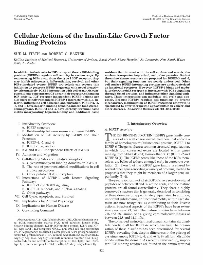
domain (Table 1), predicted by nuclear magnetic resonancestudies on IGFBP-5 (10) and confirmed for IGFBP-3 andIGFBP-5 by mutagenesis studies (11–13). Although no othermajor functional motifs have been identified in the amino-terminal domain, the observation that amino-terminal pro-teolytic fragments of IGFBP-3 cause IGF-independent inhi-bition of mitogenesis (14, 15) implies the presence of anotheractive subdomain in this region.
The conserved carboxyl-terminal domain is also cysteinerich, with three disulfide bonds in all IGFBPs, formed by thepairing of adjacent cysteines within the domain (8, 9, 16).IGF-binding residues are also present in this domain (Table1), demonstrated by the binding activity of natural carboxyl-terminal fragments of IGFBP-2 (17, 18) and recombinantcarboxyl-terminal IGFBP-3 fragments (19, 20), and mutagen-esis of IGFBP-5 residues (21). The observation that residuesinvolved in IGF binding occur in both amino- and carboxyl-terminal domains implies the existence of an IGF-bindingpocket involving both domains. As shown in Fig. 1, otherimportant subdomains have also been identified within thecarboxyl-terminal region of various IGFBPs; for example,Arg-Gly-Asp (RGD) integrin-binding motifs are located atresidues 221–223 of IGFBP-1 (22) and residues 265–267 ofIGFBP-2 (23). Functionally important 18-residue basic motifswith heparin-binding activity have also been identified atresidues 215–232 of IGFBP-3 and residues 201–218 ofIGFBP-5 and are involved in interaction with the serumglycoprotein ALS (acid-labile subunit) (24–26) and other li-gands such as plasminogen activator inhibitor-1 (27) andtransferrin (28), cell and matrix binding (24, 29), and nucleartransport (30), as discussed in detail later.
The central domain of the IGFBPs shows essentially nostructural conservation among any members of the family. It
contains no disulfide bonds apart from an intradomain bondin IGFBP-4 (9). Three sites of N-linked glycosylation inIGFBP-3 (31) and one in IGFBP-4 (32) are found in this region.Other sites of posttranslational modification are also foundin the central domain: potential phosphoacceptor sites on allIGFBPs, some of which are phosphorylated in IGFBP-1, -3,and -5 (33), and proteolytic cleavage sites in some of theproteins (15, 34, 35). Secondary IGFBP-5 binding sites for ALS(36) and heparin (37), and a potential cell-association domainof IGFBP-3 (38), are also found in this region (Fig. 1).
FIG. 1. Generalized diagram of IGFBP structure showing proposedinteraction with IGF-I through both N and C domains. Functionaldomains and sites of posttranslational modification are indicated.
TABLE 1. Functional domains of IGFBPs
Domain Function IGFBP Ref.
Amino terminal IGF binding IGFBP-1 375IGFBP-2 145, 376IGFBP-3 11, 19, 377IGFBP-5 10, 11
Insulin binding IGFBP-3 377Inhibition of insulin receptor autophosphorylation IGFBP-3 377Inhibition of mitogenesis IGFBP-3 14, 15
Central Heparin bindinga IGFBP-2 242IGFBP-3 199IGFBP-5 213
ALS bindinga IGFBP-5 36Cell binding IGFBP-3 38
Carboxyl terminal IGF binding IGFBP-1 378IGFBP-2 17, 18, 145IGFBP-3 19IGFBP-5 21
Nuclear localization signalb IGFBP-3IGFBP-5
30, 275
Heparin-bindingb IGFBP-3IGFBP-5
29
ALS bindingb IGFBP-3 24IGFBP-5 25, 26
Cell bindingb IGFBP-3 24, 29IGFBP-5 29
Integrin binding IGFBP-1 22a Central domain ALS and heparin-binding sites may only be unmasked when carboxyl-terminal domain has been deleted.b Carboxyl-terminal nuclear localization signal encompasses heparin-binding domain, which interacts with numerous other ligands.
Firth and Baxter • Cellular Actions of IGFBPs Endocrine Reviews, December 2002, 23(6):824–854 825
B. Relationship between serum and tissue IGFBPs
The term “multifunctional” is very aptly applied to theIGFBPs. Originally described as passive circulating transportproteins for IGF-I and IGF-II, IGFBPs are now recognized asplaying a variety of roles in the circulation, the extracellularenvironment, and inside the cell. The regulation and actionsof circulating IGFBPs have been addressed in several reviews(39–43). In brief, the major IGF transport function can beattributed to IGFBP-3, the most abundant circulating IGFBP.It carries 75% or more of serum IGF-I and IGF-II in hetero-trimeric complexes that also contain ALS, a leucine-rich gly-coprotein of approximately 85 kDa (44). IGFBP-5, present atabout 10% of the molar concentration of IGFBP-3, can formsimilar ternary complexes (45). Approximately 90% ofIGFBP-3 and 55% of IGFBP-5 circulate in these complexes inhealthy adults (46). All six IGFBPs are also found in thecirculation in the free form or in binary complexes with IGFs.Free or binary-complexed IGFBPs are believed to exit thecirculation rapidly, whereas ternary complexes appear to beessentially confined to the vascular compartment (47–49).
IGFBPs exert a complex array of functions at the cellularlevel. There is little information on the exact relationshipbetween IGFBPs in the circulation and those in the cellularenvironment, but it appears that the IGFBPs may be differ-entially targeted to different tissues depending on both theirprimary structure and their posttranslational modifications.In some situations, endogenous IGFBPs from circulating ter-nary complexes may be found at low concentration in thetissues, as first implied by comparison of IGFBPs in serumand lymph (50). Using exogenous IGFBPs, Boes et al. (51)demonstrated in an isolated perfused heart model thatIGFBP-4, after crossing the capillary endothelium, preferen-tially localizes to connective tissue rather than cardiac mus-cle, the exact distribution depending on the glycosylationstate. In contrast, IGFBP-1, -2, and -3 are preferentially lo-calized to cardiac muscle. IGFBP-3 injected iv appears ini-tially in the liver (40% of injected dose) and kidney (4%),within 5 min of administration (52). Uptake by muscle wasnot examined in this study.
IGFBP-4 administered systemically to mice has also beenshown to act on bone, stimulating bone alkaline phosphataseactivity and serum osteocalcin by a mechanism that appearsto involve IGFBP-4 proteolysis and increased IGF-I avail-ability (53). The influence of circulating IGFBPs at the tissuelevel is further indicated by the observation that IGFBP-1administered iv in rats inhibits IGF-I-stimulated 2-deoxy-glucose uptake in cardiac and skeletal muscle (54). Similarly,exogenous IGFBP-3 administered to rats blocks the hypo-glycemic effect of coadministered IGF-I, an effect that de-pends explicitly on its ability to form complexes with ALS inthe circulation (55).
In addition to these effects of IGFBPs derived from thecirculation, there are undoubtedly important local actions ofIGFBPs, both autocrine and paracrine. As well as modulatingactivation of the type I IGF receptor (IGFRI) by IGFs (20, 56,57), IGFBPs are documented to affect cell motility and ad-hesion (22, 58), apoptosis and survival, and cell cycle (59–61).They interact with diverse previously characterized signal-ing pathways (22, 62–64) and may have unique signaling
pathways of their own (65). Their cellular effects are likely tobe influenced by posttranslational modifications, e.g., glyco-sylation affecting cell interaction (31, 66), phosphorylationaffecting IGF binding affinity (67) and susceptibility to pro-teases (68), and proteolysis affecting both IGF-independentand IGF-dependent actions (15, 69). The purpose of this re-view is to document and provide a critical discussion ofcurrent knowledge on these and related topics.
II. Modulation of IGF Activity by IGFBPs andTheir Proteases
Two known receptors present on most cell types specifi-cally recognize the IGFs. IGFRI, a heterotetrameric tyrosinekinase that is homologous to the insulin receptor, has beenshown definitively to mediate the effects of both IGF-I andIGF-II (70). The type II IGF receptor (IGFRII), which is struc-turally distinct from IGFRI and also binds glycoproteins con-taining mannose 6-phosphate moieties, has been reported tointeract with G protein pathways (71) but its role in IGF-IIsignal transduction remains controversial. By virtue of itshigh specificity for IGF-II, it is thought to regulate the levelof extracellular IGF-II by targeting it for degradation, andhence inhibit the autocrine/paracrine actions of IGF-II me-diated through IGFRI (72). In its soluble form, the receptormay sequester IGF-II, thus inhibiting its cellular actions (73).
It is well established, from in vitro systems, that the IGFsacting through the IGFRI have acute anabolic effects on me-tabolism as well as longer term effects on cell replication anddifferentiation (40). Apart from their mitogenic activity, theIGFs also have potent inhibitory effects on apoptosis (74, 75).However, the bioactivity of IGFs is not only dependent ontheir interaction with IGFRI but is also influenced by thefamily of IGFBPs in the local cellular environment, which canpotentially either inhibit or enhance IGF actions dependingon the complement of IGFBPs present. Because most cellsexpress more than one IGFBP, it is clear that the regulationof each IGFBP plays an important role in regulating thecellular effects of IGFs. Furthermore, a wide range of pro-teolytic enzymes can catalyze the limited hydrolysis ofIGFBPs. Documented cleavage sites in IGFBP-3, -4, and -5are shown in Table 2. Some of the resulting fragments havebeen reported to retain biological activity (76, 77). In cer-tain cell types, IGF-I itself regulates the expression of specificIGFBPs (78) or their proteases (79, 80), thus adding furthercomplexity.
A. IGFBP-4, -5, and -6
Vascular smooth muscle cells express both IGF-I andIGFRI, and IGF-I is a potent regulator of migration, prolif-eration, and apoptosis in these cells (81–85). Duan and Clem-mons (86) showed that IGFBP-4 and IGFBP-5 expression wasregulated differentially by IGF-I in porcine vascular smoothmuscle cells. IGF-I decreased IGFBP-4 levels by activating anIGFBP-4-specific protease and increased IGFBP-5 levels bystimulating gene expression (86, 87). In addition, exogenousIGFBP-4 and IGFBP-5 have opposing effects on IGF-I-induced DNA synthesis in these cells; IGFBP-4 inhibitswhereas IGFBP-5 potentiates IGF-I effects. Therefore, the
826 Endocrine Reviews, December 2002, 23(6):824–854 Firth and Baxter • Cellular Actions of IGFBPs
balance between levels of IGFBP-4 and IGFBP-5 regulated byIGF-I has a direct impact on cellular proliferation.
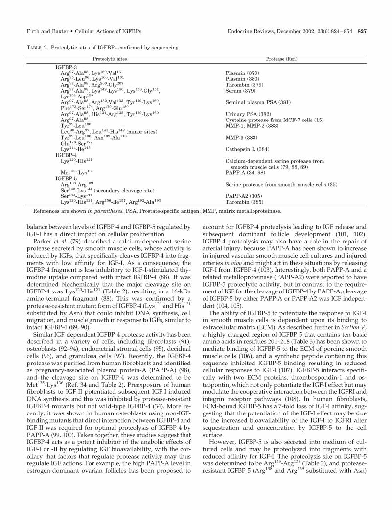
Parker et al. (79) described a calcium-dependent serineprotease secreted by smooth muscle cells, whose activity isinduced by IGFs, that specifically cleaves IGFBP-4 into frag-ments with low affinity for IGF-I. As a consequence, theIGFBP-4 fragment is less inhibitory to IGF-I-stimulated thy-midine uptake compared with intact IGFBP-4 (88). It wasdetermined biochemically that the major cleavage site onIGFBP-4 was Lys120-His121 (Table 2), resulting in a 16-kDaamino-terminal fragment (88). This was confirmed by aprotease-resistant mutant form of IGFBP-4 (Lys120 and His121
substituted by Asn) that could inhibit DNA synthesis, cellmigration, and muscle growth in response to IGFs, similar tointact IGFBP-4 (89, 90).
Similar IGF-dependent IGFBP-4 protease activity has beendescribed in a variety of cells, including fibroblasts (91),osteoblasts (92–94), endometrial stromal cells (95), decidualcells (96), and granulosa cells (97). Recently, the IGFBP-4protease was purified from human fibroblasts and identifiedas pregnancy-associated plasma protein-A (PAPP-A) (98),and the cleavage site on IGFBP-4 was determined to beMet135-Lys136 (Ref. 34 and Table 2). Preexposure of humanfibroblasts to IGF-II potentiated subsequent IGF-I-inducedDNA synthesis, and this was inhibited by protease-resistantIGFBP-4 mutants but not wild-type IGFBP-4 (34). More re-cently, it was shown in human osteoblasts using non-IGF-binding mutants that direct interaction between IGFBP-4 andIGF-II was required for optimal proteolysis of IGFBP-4 byPAPP-A (99, 100). Taken together, these studies suggest thatIGFBP-4 acts as a potent inhibitor of the anabolic effects ofIGF-I or -II by regulating IGF bioavailability, with the cor-ollary that factors that regulate protease activity may thusregulate IGF actions. For example, the high PAPP-A level inestrogen-dominant ovarian follicles has been proposed to
account for IGFBP-4 proteolysis leading to IGF release andsubsequent dominant follicle development (101, 102).IGFBP-4 proteolysis may also have a role in the repair ofarterial injury, because PAPP-A has been shown to increasein injured vascular smooth muscle cell cultures and injuredarteries in vivo and might act in these situations by releasingIGF-I from IGFBP-4 (103). Interestingly, both PAPP-A and arelated metalloproteinase (PAPP-A2) were reported to haveIGFBP-5 proteolytic activity, but in contrast to the require-ment of IGF for the cleavage of IGFBP-4 by PAPP-A, cleavageof IGFBP-5 by either PAPP-A or PAPP-A2 was IGF indepen-dent (104, 105).
The ability of IGFBP-5 to potentiate the response to IGF-Iin smooth muscle cells is dependent upon its binding toextracellular matrix (ECM). As described further in Section V,a highly charged region of IGFBP-5 that contains ten basicamino acids in residues 201–218 (Table 3) has been shown tomediate binding of IGFBP-5 to the ECM of porcine smoothmuscle cells (106), and a synthetic peptide containing thissequence inhibited IGFBP-5 binding resulting in reducedcellular responses to IGF-I (107). IGFBP-5 interacts specifi-cally with two ECM proteins, thrombospondin-1 and os-teopontin, which not only potentiate the IGF-I effect but maymodulate the cooperative interaction between the IGFRI andintegrin receptor pathways (108). In human fibroblasts,ECM-bound IGFBP-5 has a 7-fold loss of IGF-I affinity, sug-gesting that the potentiation of the IGF-I effect may be dueto the increased bioavailability of the IGF-I to IGFRI aftersequestration and concentration by IGFBP-5 to the cellsurface.
However, IGFBP-5 is also secreted into medium of cul-tured cells and may be proteolyzed into fragments withreduced affinity for IGF-I. The proteolysis site on IGFBP-5was determined to be Arg138-Arg139 (Table 2), and protease-resistant IGFBP-5 (Arg138 and Arg139 substituted with Asn)
TABLE 2. Proteolytic sites of IGFBPs confirmed by sequencing
Proteolytic sites Protease (Ref.)
IGFBP-3Arg97-Ala98, Lys160-Val161 Plasmin (379)Arg95-Leu96, Lys160-Val161 Plasmin (380)Arg97-Ala98, Arg206-Gly207 Thrombin (379)Arg97-Ala98, Lys149-Lys150, Lys150-Gly151,Lys154-Asp155
Serum (379)
Arg97-Ala98, Arg132-Val133, Tyr159-Lys160,Phe173-Ser174, Arg179-Glu180
Seminal plasma PSA (381)
Arg97-Ala98, His131-Arg132, Tyr159-Lys160 Urinary PSA (382)Arg97-Ala98 Cysteine protease from MCF-7 cells (15)Tyr99-Leu100 MMP-1, MMP-2 (383)Leu96-Arg97, Leu141-His142 (minor sites)Tyr99-Leu100, Asn109-Ala110 MMP-3 (383)Glu176-Ser177
Lys144-Ile145 Cathepsin L (384)IGFBP-4
Lys120-His121 Calcium-dependent serine protease fromsmooth muscle cells (79, 88, 89)
Met135-Lys136 PAPP-A (34, 98)IGFBP-5
Arg138-Arg139 Serine protease from smooth muscle cells (35)Ser143-Lys144 (secondary cleavage site)Ser143-Lys144 PAPP-A2 (105)Lys120-His121, Arg156-Ile157, Arg192-Ala193 Thrombin (385)
References are shown in parentheses. PSA, Prostate-specific antigen; MMP, matrix metalloproteinase.
Firth and Baxter • Cellular Actions of IGFBPs Endocrine Reviews, December 2002, 23(6):824–854 827
inhibited IGF-I-stimulated DNA and protein synthesis andmigration of porcine smooth muscle cells (35). This is con-sistent with the finding that proteolyzed IGFBP-5 in theconditioned medium had no effect on the IGF-I stimulationof growth in cultured fibroblasts (109) and suggests that, incontrast to ECM-bound IGFBP-5, soluble IGFBP-5 acts as aninhibitor of IGF-I-stimulatory effects and that proteolysis ofIGFBP-5 may serve as an important regulatory mechanism ofthis function. Although the IGFBP-5 protease in mediumconditioned by smooth muscle cells has not been identified,similar proteolytic activity against IGFBP-5 in fibroblast-conditioned medium has been attributed to the complementcomponents C1s and/or C1r (110). The biological signifi-cance of IGFBP-5 cleavage by these enzymes is as yetunknown.
Analogous to the situation in fibroblasts and smooth mus-cle cells, the IGF-dependent actions of IGFBP-5 in bone cellsappear to be dependent on its location. IGFBP-5 is thoughtto act as a depot for IGF-II in bone via its high affinity forECM proteins and hydroxyapatite, the mineral constituent ofbone, and potentiates the proliferative actions of IGF-II onosteoblastic cells (111, 112). In addition, the stimulation ofosteoclastic activity by IGFBP-5 can be blocked by IGF-Iantibody (113). These results have led to the proposal that theIGFs, which are sequestered and concentrated in bone byIGFBP-5, may be released during osteoclastic resorption,thus leading to stimulation of osteoblastic activity duringbone remodeling. In contrast, IGF-I- and IGF-II-stimulatedDNA and glycogen synthesis in a human osteoblastic cell linewas inhibited by soluble recombinant IGFBP-5 (114). Like-wise, the relative insensitivity of U2 osteosarcoma cellsto IGF-I compared with the non-IGFBP-binding analog,des(1–3)IGF-I, was attributed to the inhibitory effect of en-dogenously secreted intact IGFBP-5 (115). However, proteo-lyzed IGFBP-5 derived from the medium of U2 cells en-hanced IGF-I-stimulated osteoblast mitogenesis (116).Intriguingly, the amount of intact IGFBP-5 was increasedsignificantly in the medium of these cells treated with IGF-Iwithout a concomitant increase in mRNA levels or reciprocaldecrease in proteolytic fragments (115), suggesting that IGF-Inot only has a protective effect on IGFBP-5 proteolysis, butmay also affect the compartmentalization of IGFBP-5.
It is also well established that IGFs can stimulate bothproliferation and differentiation of skeletal muscle cells andthese actions are mediated through IGFRI. However, theactions of IGFs are modulated by the expression of IGFBP-4,-5, and -6. As described for other cell types, IGFBP-4 is mainlyinhibitory (117, 118) whereas IGFBP-5 could be either inhib-itory or stimulatory to IGF actions (119, 120). By employingIGF-II analogs, Bach et al. (121) demonstrated that the inhi-
bition of IGF-II-induced proliferation and differentiation ofL6A1 rat myoblasts by IGFBP-6 was correlated to its affinityfor the analogs. It would appear that the IGFBPs, on balance,are generally inhibitory to IGF actions in myoblasts.
There are relatively few studies on the function of IGFBP-6and generally, IGFBP-6 appears to inhibit the actions ofIGF-II with some selectivity because it has 20- to 100-foldhigher affinity for IGF-II than IGF-I (122, 123). Cell systemsin which IGFBP-6 has been shown to inhibit IGF-II-inducedeffects such as proliferation, differentiation, cell adhesion,and colony formation include osteoblasts, keratinocytes,myoblasts, and colon cancer cells (121, 124–128).
B. IGFBP-1, -2, and -3
Several studies have described both the inhibition andpotentiation of IGF actions by IGFBP-1 in a variety of cells(40). The positive or negative modulation by IGFBP-1 isthought to be related to its phosphorylation state becausedephosphorylation of human IGFBP-1 reduces its affinity forIGF-I by 6-fold (129). Curiously, phosphorylation appears tohave no effect on the affinity of rat IGFBP-1 (130), raising thequestion whether IGFBP-1 regulatory mechanisms describedin humans are relevant to other species. DephosphorylatedIGFBP-1 has been shown to enhance IGF-I-induced DNAsynthesis (131–133), whereas phosphorylated IGFBP-1 in-hibits IGF-I effects (131, 133, 134). Although high-affinityphospho-IGFBP-1 is assumed to act by blocking IGF accessto the IGFRI, no experimental evidence has yet explainedhow the low-affinity dephosphorylated form could enhanceIGF action (rather than simply not inhibiting it). Polymer-ization of IGFBP-1 by tissue transglutaminase has also re-cently been shown to ablate its inhibition of IGF-I-stimulatedprotein synthesis (135), but the contribution of this effect tonet IGFBP-1 cellular activity is not clear.
In the extensively studied paracrine interactions at thematernal-fetal interface during pregnancy, it is thought thatIGFBP-1 secreted by the maternal decidua inhibits placentaltrophoblast invasiveness (further discussed in Section IV),but this inhibitory effect is repressed by the down-regulationof IGFBP-1 production by placental trophoblast-derivedIGF-II (136, 137). An alternative mechanism was proposed byGibson et al. (138), who identified both phosphorylated andnonphosphorylated isoforms of IGFBP-1 secreted by decidu-alized endometrium under basal conditions. In the presenceof trophoblast-derived IGF-II, the nonphosphorylated formof IGFBP-1—noted above to have lower IGF affinity thanphospho-IGFBP-1 (67)—was predominantly produced bydecidual cells. Placental phosphatases could also generatethis form by dephosphorylating phospho-IGFBP-1. In addi-
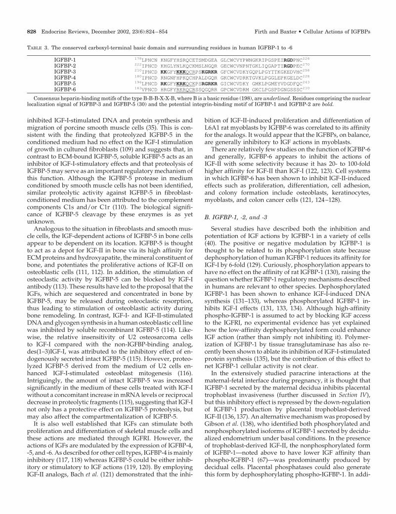
TABLE 3. The conserved carboxyl-terminal basic domain and surrounding residues in human IGFBP-1 to -6
IGFBP-1 178LPNCN KNGFYHSRQCETSMDGEA GLCWCVYPWNGKRIPGSPEIRGDPNC226
IGFBP-2 222IPNCD KHGLYNLKQCKMSLNGQR GECWCVNPNTGKLIQGAPTIRGDPEC270
IGFBP-3 210IPNCD KKGFYKKKQCRPSKGRKR GFCWCVDKYGQPLPGYTTKGKEDVHC258
IGFBP-4 180IPNCD RNGNFHPKQCHPALDGQR GKCWCVDRKTGVKLPGGLEPKGELDC228
IGFBP-5 196LPNCD RKGFYKRKQCKPSRGRKR GICWCVDKY GMKLPGMEYVDGDFQC243
IGFBP-6 163VPNCD HRGFYRKRQCRSSQGQRR GPCWCVDRM GKCLPGSPDGNGSSSC210
Consensus heparin-binding motifs of the type B-B-B-X-X-B, where B is a basic residue (198), are underlined. Residues comprising the nuclearlocalization signal of IGFBP-3 and IGFBP-5 (30) and the potential integrin-binding motif of IGFBP-1 and IGFBP-2 are bold.
828 Endocrine Reviews, December 2002, 23(6):824–854 Firth and Baxter • Cellular Actions of IGFBPs
tion, a protease was found to act specifically on nonphospho-IGFBP-1, which would be expected to further decrease its IGFaffinity. The net result of these posttranslational modifica-tions would be IGFBP-1 forms with reduced affinity forIGF-I, thus increasing IGF-I bioavailability to enhance tissuegrowth (138).
IGF-I is actively involved in the process of dermal woundhealing by stimulating reepithelialization of the wounds, andthis action is potentiated by IGFBP-1 (139–143), although thecontribution of its phosphorylation state to this action is notknown. The enhancement of IGF-I actions appears to berelated to the ability of IGFBP-1 to bind to �5�1 integrin(discussed further in Sections III and IV) because a non-integrin-binding IGFBP-1 mutant had no effect (144).IGFBP-2, which has the integrin-binding motif RGD, likeIGFBP-1, was ineffective in augmenting the IGF-I enhance-ment of wound repair (144).
In general, IGFBP-2 appears to inhibit IGF actions, in par-ticular those of IGF-II, possibly related to its higher affinityfor IGF-II (40), although this affinity difference is in fact only2-fold (145). Overexpression of IGFBP-2 in human embryonickidney fibroblasts results in inhibition of cell proliferation,which can be reversed by the addition of exogenous IGFs,thus suggesting that IGFBP-2 has an inhibitory effect on IGFaction (146). This is supported by a previous study thatshowed growth stimulation of intestinal epithelial cells trans-fected with an antisense IGFBP-2 construct (147). In addition,Hoflich et al. (148) recently reported that giant GH transgenicmice, which had 2- to 3-fold increased expression of serumIGF-I levels, had a significant reduction in growth parame-ters when crossed with IGFBP-2 transgenic mice, suggestingthat IGFBP-2 is also inhibitory to IGF-I actions in vivo.
Addition of equimolar concentrations of IGFBP-2 com-pletely inhibited IGF-II-stimulated DNA synthesis in non-small-cell lung carcinoma (NSCLC) cells but had no signif-icant effect on IGF-I-stimulated DNA synthesis (149, 150).This is not easily explained because, as noted above, therelative affinities for IGF-I and IGF-II do not differ greatly.Interestingly, IGFs bind predominantly to IGF receptors inNSCLC cells, which have relatively low levels of membrane-associated IGFBP-2. In contrast, IGFs bind to high levels ofmembrane-associated IGFBP-2 in small-cell lung carcinoma(SCLC), which do not respond to IGFs even though IGFRI ispresent (149). This suggests that both soluble and membrane-associated IGFBP-2 may be competing with the IGF receptorsfor ligand and may therefore be regulating IGF responsive-ness in lung carcinoma.
Proteolyzed IGFBP-2 has been detected in serum (151),milk (18), and cerebrospinal fluid (152) and has decreasedaffinity for IGFs compared with intact IGFBP-2. Serum with-drawal from porcine aortic smooth muscle cells inducesthe secretion of a calcium-dependent serine protease forIGFBP-2, the activity of which is relatively more enhanced byIGF-II than IGF-I (153, 154). Menouny et al. (155) reported thatthe interaction between the plasmin system and IGFBP-2 canmodulate the bioavailability of IGF-II, which mediates au-tocrine proliferation in neuroblastoma cells. However, in themajority of cell studies in which IGFBP-2 is detected byimmunoblot, it appears in culture medium at its intact size
of approximately 34 kDa. Thus, IGFBP-2 proteolysis may notbe a very widespread regulatory mechanism.
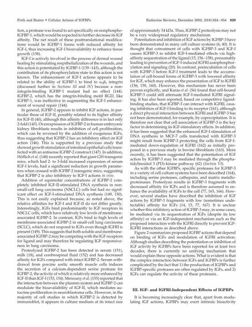
Potentiation and inhibition of IGF actions by IGFBP-3 havebeen demonstrated in many cell culture systems (6, 40). It isthought that cotreatment of cells with IGFBP-3 and IGF-Icauses IGFBP-3 to inhibit IGF-I-mediated effects via high-affinity sequestration of the ligand (15, 156–158), presumablyleading to prevention of IGF-I-induced IGFRI autophosphor-ylation and signaling (20). In contrast, preincubation of cellswith IGFBP-3 before IGF-I treatment leads to the accumu-lation of cell-bound forms of IGFBP-3 with lowered affinityfor IGF, which may enhance the presentation of IGF to IGFRI(156, 159, 160). However, this mechanism has never beenproven explicitly, and Karas et al. (56) found that cell-boundIGFBP-3 could still attenuate IGF-I-mediated IGFRI signal-ing. It has also been reported, based on competitive ligand-binding studies, that IGFBP-3 can interact with IGFRI, caus-ing inhibition of IGF-I binding to its receptor (161), althougha direct physical interaction between IGFRI and IGFBP-3 hasnot been demonstrated, for example, by coprecipitation. It istherefore not clear that cell association of IGFBP-3 is the keyfactor in determining its IGF-stimulatory effects. In addition,it has been suggested that the enhanced IGF-I stimulation ofDNA synthesis in MCF-7 cells transfected with IGFBP-3might result from IGFBP-3 protecting the cells from IGF-I-mediated down-regulation of IGFRI (162) as initially pro-posed in a previous study in bovine fibroblasts (163). Morerecently, it has been suggested that the potentiation of IGFaction by IGFBP-3 may be mediated through the phospha-tidylinositol 3 (PI3)-kinase pathway (62) (Section VI).
As with the other IGFBPs, specific proteases for IGFBP-3in a variety of cell culture systems have been described (164),including serine proteases, cathepsins, and matrix metallo-proteinases. Proteolysis results in IGFBP-3 fragments withdecreased affinity for IGFs and is therefore assumed to en-hance the availability of IGFs to the cell (77, 165, 166). How-ever, several studies have described the inhibition of IGFactions by IGFBP-3 fragments with low (sometimes unde-tectable) affinity for IGFs (14, 15, 77, 167). It is unclearwhether this inhibitory action of IGFBP-3 may, in some cases,be mediated via its sequestration of IGFs (despite its lowaffinity) or via an IGF-independent mechanism such as theproposed interaction with the IGFRI directly to prevent IGF-IGFRI interactions as described above.
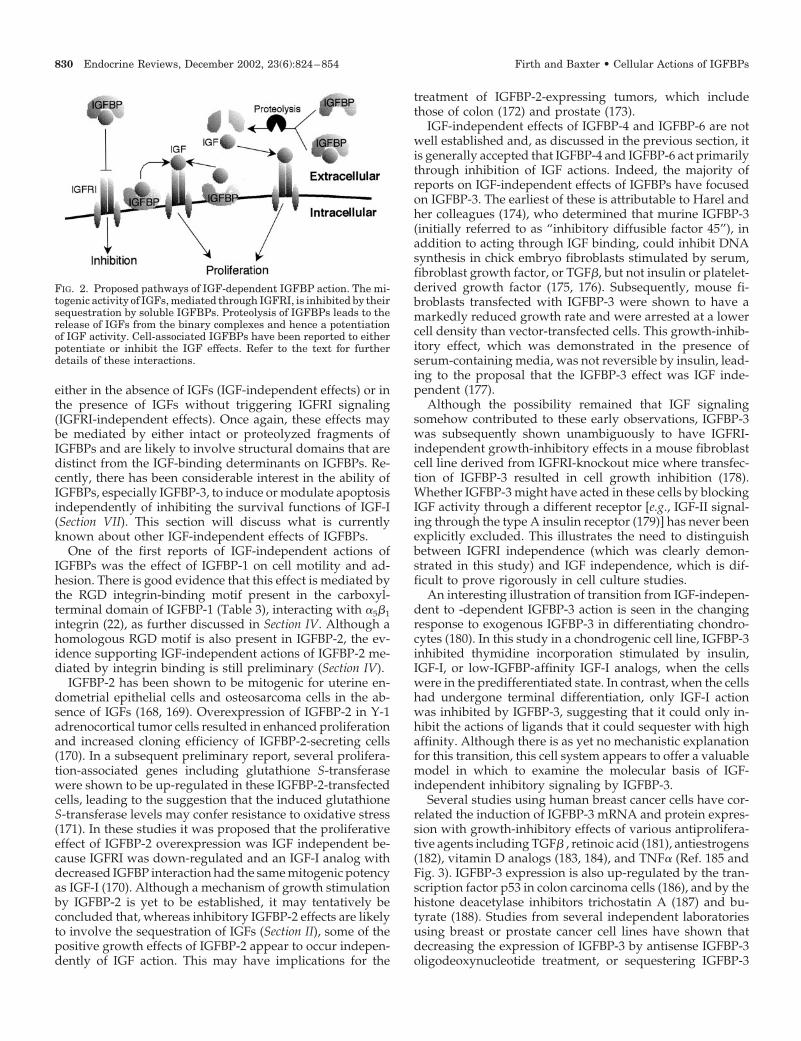
Figure 2 summarizes proposed IGFBP actions that dependon binding of IGFs and modulation of IGFRI activation.Although studies describing the potentiation or inhibition ofIGF activity by IGFBPs have been reported for at least twodecades, there is currently no unifying mechanism thatwould explain these opposite actions. What is evident is thatthe complex interaction between IGFs and IGFBPs is furthercomplicated by the fact that 1) the production of IGFBPs andIGFBP-specific proteases are often regulated by IGFs, and 2)IGFs can regulate the activity of these proteases.
III. IGF- and IGFRI-Independent Effects of IGFBPs
It is becoming increasingly clear that, apart from modu-lating IGF actions, IGFBPs may exert intrinsic bioactivity
Firth and Baxter • Cellular Actions of IGFBPs Endocrine Reviews, December 2002, 23(6):824–854 829
either in the absence of IGFs (IGF-independent effects) or inthe presence of IGFs without triggering IGFRI signaling(IGFRI-independent effects). Once again, these effects maybe mediated by either intact or proteolyzed fragments ofIGFBPs and are likely to involve structural domains that aredistinct from the IGF-binding determinants on IGFBPs. Re-cently, there has been considerable interest in the ability ofIGFBPs, especially IGFBP-3, to induce or modulate apoptosisindependently of inhibiting the survival functions of IGF-I(Section VII). This section will discuss what is currentlyknown about other IGF-independent effects of IGFBPs.
One of the first reports of IGF-independent actions ofIGFBPs was the effect of IGFBP-1 on cell motility and ad-hesion. There is good evidence that this effect is mediated bythe RGD integrin-binding motif present in the carboxyl-terminal domain of IGFBP-1 (Table 3), interacting with �5�1integrin (22), as further discussed in Section IV. Although ahomologous RGD motif is also present in IGFBP-2, the ev-idence supporting IGF-independent actions of IGFBP-2 me-diated by integrin binding is still preliminary (Section IV).
IGFBP-2 has been shown to be mitogenic for uterine en-dometrial epithelial cells and osteosarcoma cells in the ab-sence of IGFs (168, 169). Overexpression of IGFBP-2 in Y-1adrenocortical tumor cells resulted in enhanced proliferationand increased cloning efficiency of IGFBP-2-secreting cells(170). In a subsequent preliminary report, several prolifera-tion-associated genes including glutathione S-transferasewere shown to be up-regulated in these IGFBP-2-transfectedcells, leading to the suggestion that the induced glutathioneS-transferase levels may confer resistance to oxidative stress(171). In these studies it was proposed that the proliferativeeffect of IGFBP-2 overexpression was IGF independent be-cause IGFRI was down-regulated and an IGF-I analog withdecreased IGFBP interaction had the same mitogenic potencyas IGF-I (170). Although a mechanism of growth stimulationby IGFBP-2 is yet to be established, it may tentatively beconcluded that, whereas inhibitory IGFBP-2 effects are likelyto involve the sequestration of IGFs (Section II), some of thepositive growth effects of IGFBP-2 appear to occur indepen-dently of IGF action. This may have implications for the
treatment of IGFBP-2-expressing tumors, which includethose of colon (172) and prostate (173).
IGF-independent effects of IGFBP-4 and IGFBP-6 are notwell established and, as discussed in the previous section, itis generally accepted that IGFBP-4 and IGFBP-6 act primarilythrough inhibition of IGF actions. Indeed, the majority ofreports on IGF-independent effects of IGFBPs have focusedon IGFBP-3. The earliest of these is attributable to Harel andher colleagues (174), who determined that murine IGFBP-3(initially referred to as “inhibitory diffusible factor 45”), inaddition to acting through IGF binding, could inhibit DNAsynthesis in chick embryo fibroblasts stimulated by serum,fibroblast growth factor, or TGF�, but not insulin or platelet-derived growth factor (175, 176). Subsequently, mouse fi-broblasts transfected with IGFBP-3 were shown to have amarkedly reduced growth rate and were arrested at a lowercell density than vector-transfected cells. This growth-inhib-itory effect, which was demonstrated in the presence ofserum-containing media, was not reversible by insulin, lead-ing to the proposal that the IGFBP-3 effect was IGF inde-pendent (177).
Although the possibility remained that IGF signalingsomehow contributed to these early observations, IGFBP-3was subsequently shown unambiguously to have IGFRI-independent growth-inhibitory effects in a mouse fibroblastcell line derived from IGFRI-knockout mice where transfec-tion of IGFBP-3 resulted in cell growth inhibition (178).Whether IGFBP-3 might have acted in these cells by blockingIGF activity through a different receptor [e.g., IGF-II signal-ing through the type A insulin receptor (179)] has never beenexplicitly excluded. This illustrates the need to distinguishbetween IGFRI independence (which was clearly demon-strated in this study) and IGF independence, which is dif-ficult to prove rigorously in cell culture studies.
An interesting illustration of transition from IGF-indepen-dent to -dependent IGFBP-3 action is seen in the changingresponse to exogenous IGFBP-3 in differentiating chondro-cytes (180). In this study in a chondrogenic cell line, IGFBP-3inhibited thymidine incorporation stimulated by insulin,IGF-I, or low-IGFBP-affinity IGF-I analogs, when the cellswere in the predifferentiated state. In contrast, when the cellshad undergone terminal differentiation, only IGF-I actionwas inhibited by IGFBP-3, suggesting that it could only in-hibit the actions of ligands that it could sequester with highaffinity. Although there is as yet no mechanistic explanationfor this transition, this cell system appears to offer a valuablemodel in which to examine the molecular basis of IGF-independent inhibitory signaling by IGFBP-3.
Several studies using human breast cancer cells have cor-related the induction of IGFBP-3 mRNA and protein expres-sion with growth-inhibitory effects of various antiprolifera-tive agents including TGF� , retinoic acid (181), antiestrogens(182), vitamin D analogs (183, 184), and TNF� (Ref. 185 andFig. 3). IGFBP-3 expression is also up-regulated by the tran-scription factor p53 in colon carcinoma cells (186), and by thehistone deacetylase inhibitors trichostatin A (187) and bu-tyrate (188). Studies from several independent laboratoriesusing breast or prostate cancer cell lines have shown thatdecreasing the expression of IGFBP-3 by antisense IGFBP-3oligodeoxynucleotide treatment, or sequestering IGFBP-3
FIG. 2. Proposed pathways of IGF-dependent IGFBP action. The mi-togenic activity of IGFs, mediated through IGFRI, is inhibited by theirsequestration by soluble IGFBPs. Proteolysis of IGFBPs leads to therelease of IGFs from the binary complexes and hence a potentiationof IGF activity. Cell-associated IGFBPs have been reported to eitherpotentiate or inhibit the IGF effects. Refer to the text for furtherdetails of these interactions.
830 Endocrine Reviews, December 2002, 23(6):824–854 Firth and Baxter • Cellular Actions of IGFBPs
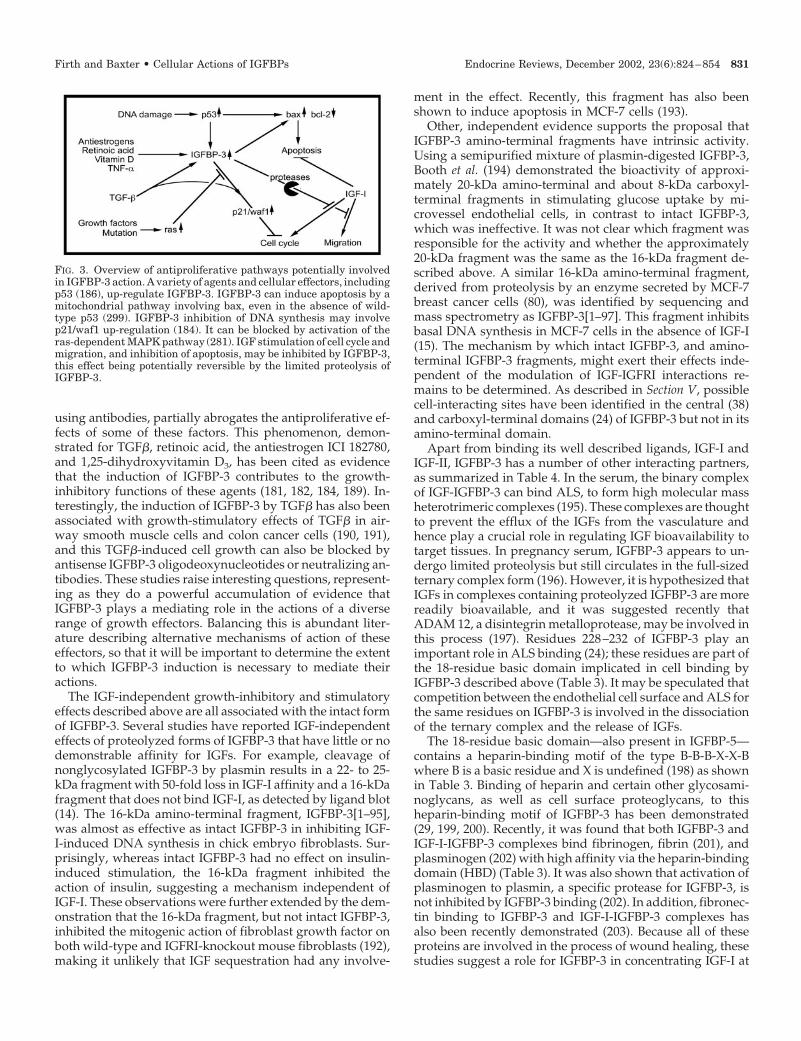
using antibodies, partially abrogates the antiproliferative ef-fects of some of these factors. This phenomenon, demon-strated for TGF�, retinoic acid, the antiestrogen ICI 182780,and 1,25-dihydroxyvitamin D3, has been cited as evidencethat the induction of IGFBP-3 contributes to the growth-inhibitory functions of these agents (181, 182, 184, 189). In-terestingly, the induction of IGFBP-3 by TGF� has also beenassociated with growth-stimulatory effects of TGF� in air-way smooth muscle cells and colon cancer cells (190, 191),and this TGF�-induced cell growth can also be blocked byantisense IGFBP-3 oligodeoxynucleotides or neutralizing an-tibodies. These studies raise interesting questions, represent-ing as they do a powerful accumulation of evidence thatIGFBP-3 plays a mediating role in the actions of a diverserange of growth effectors. Balancing this is abundant liter-ature describing alternative mechanisms of action of theseeffectors, so that it will be important to determine the extentto which IGFBP-3 induction is necessary to mediate theiractions.
The IGF-independent growth-inhibitory and stimulatoryeffects described above are all associated with the intact formof IGFBP-3. Several studies have reported IGF-independenteffects of proteolyzed forms of IGFBP-3 that have little or nodemonstrable affinity for IGFs. For example, cleavage ofnonglycosylated IGFBP-3 by plasmin results in a 22- to 25-kDa fragment with 50-fold loss in IGF-I affinity and a 16-kDafragment that does not bind IGF-I, as detected by ligand blot(14). The 16-kDa amino-terminal fragment, IGFBP-3[1–95],was almost as effective as intact IGFBP-3 in inhibiting IGF-I-induced DNA synthesis in chick embryo fibroblasts. Sur-prisingly, whereas intact IGFBP-3 had no effect on insulin-induced stimulation, the 16-kDa fragment inhibited theaction of insulin, suggesting a mechanism independent ofIGF-I. These observations were further extended by the dem-onstration that the 16-kDa fragment, but not intact IGFBP-3,inhibited the mitogenic action of fibroblast growth factor onboth wild-type and IGFRI-knockout mouse fibroblasts (192),making it unlikely that IGF sequestration had any involve-
ment in the effect. Recently, this fragment has also beenshown to induce apoptosis in MCF-7 cells (193).
Other, independent evidence supports the proposal thatIGFBP-3 amino-terminal fragments have intrinsic activity.Using a semipurified mixture of plasmin-digested IGFBP-3,Booth et al. (194) demonstrated the bioactivity of approxi-mately 20-kDa amino-terminal and about 8-kDa carboxyl-terminal fragments in stimulating glucose uptake by mi-crovessel endothelial cells, in contrast to intact IGFBP-3,which was ineffective. It was not clear which fragment wasresponsible for the activity and whether the approximately20-kDa fragment was the same as the 16-kDa fragment de-scribed above. A similar 16-kDa amino-terminal fragment,derived from proteolysis by an enzyme secreted by MCF-7breast cancer cells (80), was identified by sequencing andmass spectrometry as IGFBP-3[1–97]. This fragment inhibitsbasal DNA synthesis in MCF-7 cells in the absence of IGF-I(15). The mechanism by which intact IGFBP-3, and amino-terminal IGFBP-3 fragments, might exert their effects inde-pendent of the modulation of IGF-IGFRI interactions re-mains to be determined. As described in Section V, possiblecell-interacting sites have been identified in the central (38)and carboxyl-terminal domains (24) of IGFBP-3 but not in itsamino-terminal domain.
Apart from binding its well described ligands, IGF-I andIGF-II, IGFBP-3 has a number of other interacting partners,as summarized in Table 4. In the serum, the binary complexof IGF-IGFBP-3 can bind ALS, to form high molecular massheterotrimeric complexes (195). These complexes are thoughtto prevent the efflux of the IGFs from the vasculature andhence play a crucial role in regulating IGF bioavailability totarget tissues. In pregnancy serum, IGFBP-3 appears to un-dergo limited proteolysis but still circulates in the full-sizedternary complex form (196). However, it is hypothesized thatIGFs in complexes containing proteolyzed IGFBP-3 are morereadily bioavailable, and it was suggested recently thatADAM 12, a disintegrin metalloprotease, may be involved inthis process (197). Residues 228–232 of IGFBP-3 play animportant role in ALS binding (24); these residues are part ofthe 18-residue basic domain implicated in cell binding byIGFBP-3 described above (Table 3). It may be speculated thatcompetition between the endothelial cell surface and ALS forthe same residues on IGFBP-3 is involved in the dissociationof the ternary complex and the release of IGFs.
The 18-residue basic domain—also present in IGFBP-5—contains a heparin-binding motif of the type B-B-B-X-X-Bwhere B is a basic residue and X is undefined (198) as shownin Table 3. Binding of heparin and certain other glycosami-noglycans, as well as cell surface proteoglycans, to thisheparin-binding motif of IGFBP-3 has been demonstrated(29, 199, 200). Recently, it was found that both IGFBP-3 andIGF-I-IGFBP-3 complexes bind fibrinogen, fibrin (201), andplasminogen (202) with high affinity via the heparin-bindingdomain (HBD) (Table 3). It was also shown that activation ofplasminogen to plasmin, a specific protease for IGFBP-3, isnot inhibited by IGFBP-3 binding (202). In addition, fibronec-tin binding to IGFBP-3 and IGF-I-IGFBP-3 complexes hasalso been recently demonstrated (203). Because all of theseproteins are involved in the process of wound healing, thesestudies suggest a role for IGFBP-3 in concentrating IGF-I at
FIG. 3. Overview of antiproliferative pathways potentially involvedin IGFBP-3 action. A variety of agents and cellular effectors, includingp53 (186), up-regulate IGFBP-3. IGFBP-3 can induce apoptosis by amitochondrial pathway involving bax, even in the absence of wild-type p53 (299). IGFBP-3 inhibition of DNA synthesis may involvep21/waf1 up-regulation (184). It can be blocked by activation of theras-dependent MAPK pathway (281). IGF stimulation of cell cycle andmigration, and inhibition of apoptosis, may be inhibited by IGFBP-3,this effect being potentially reversible by the limited proteolysis ofIGFBP-3.
Firth and Baxter • Cellular Actions of IGFBPs Endocrine Reviews, December 2002, 23(6):824–854 831
wound sites, and, conceivably, after proteolysis of IGFBP-3by plasmin, IGF-I is released to exert its mitogenic effects.
IGFBP-3 has also been shown to bind to human serumtransferrin, the effect depending on the degree of iron sat-uration (28). In an independent study, transferrin itself wasshown to bind IGFs, although with an affinity 200-fold lowerthan the IGF-IGFBP-3 affinity (204). Interestingly, the affinityof IGF-II for IGFBP-3 was increased 5-fold in the presence oftransferrin. Transferrin, a key component of iron transportand metabolism, is involved in various aspects of cell-cellinteractions and cell viability (205) and reported to regulateprogrammed cell death (206). IGFBP-3 induced cell prolif-eration in bladder smooth muscle cells, and apoptosis inprostate cancer cells was blocked by transferrin cotreatment(28). However, it is not clear whether these effects were dueto the modulation of IGFBP-3 or transferrin (or both) inter-acting with their respective receptors, or whether IGF bind-ing by either protein was involved. The related iron-bindingprotein, lactoferrin, has also been described as bindingIGFBP-3, competing with IGF-binding, and affectingIGFBP-3 nuclear entry in mammary cells (207). Because IGFsmay modulate the cellular interactions between IGFBP-3 andtransferrin or lactoferrin, or may even be central to theireffects, these studies do not necessarily represent IGF-inde-pendent actions of IGFBP-3. They do, however, raise thepossibility of cellular entry of IGFBP-3 through transferrinreceptors.
The first description of IGF-independent effects ofIGFBP-5 was reported in 1992 when a 23-kDa IGFBP-5 frag-ment was shown to stimulate normal osteoblast mitogenesisin the absence of IGF-I (116). Using the recombinant amino-
terminal fragment IGFBP-5[1–169], Andress et al. (208) con-firmed the initial study and suggested that the IGFBP-5 frag-ment-stimulated mitogenesis may be mediated by low-affinity binding of the fragment to the osteoblast surface. Thisis further supported by reports that IGFBP-5 binds to cellsurfaces via its basic carboxyl-terminal region (residues 201–218; Refs. 29 and 209), shown in Table 3. In contrast, IGFBP-5[1–169] did not display intrinsic bioactivity in mesangialcells but inhibited IGF-I-stimulated migration (210). It is cur-rently unexplained how IGFBP-5[1–169], shown to have de-creased affinity for IGF-I (208), could be a more potent in-hibitor of IGF-I than intact IGFBP-5.
Analogous to IGFBP-3, the HBD region of IGFBP-5 hasbeen implicated as the binding region for several differentmolecules including ALS (25, 26), heparin, and various gly-cosaminoglycans (211, 212), IGF-I (21, 213), plasminogenactivator-1 (27), osteopontin (108), thrombospondin (108),hydroxyapatite (214), and importin � (30) as well as to theECM (Refs. 29 and 209 and Table 4). At present, it is notentirely clear if the same specific residues within the 201–218region of IGFBP-5 (Table 3) are required for interacting withthis diverse group of molecules. This issue is addressed in moredetail in a recent review of IGFBP mutagenesis studies (7).
As described above, IGFBP-5 binds to ECM (215) andhydroxyapatite (214), indicating that it may accumulate inbone and sequester IGFs to bone. IGFs are important regu-lators of bone formation because IGF-I knockout mice showsevere impairment of bone growth (216). Administration ofrecombinant human IGFBP-5 to mice increased bone forma-tion parameters and decreased bone resorption parameters.The increase in bone formation was not mediated by in-
TABLE 4. Proteins known to interact with IGFBPs
IGFBP Cell-surface and extracellular binding partners Intracellular binding partners
IGFBP-1 IGF-I and IGF-II�5�1 integrin (22)
IGFBP-2 IGF-I and IGF-II�5�1 integrin (233)Glycosaminoglycans (242, 243)
IGFBP-3 IGF-I and IGF-II Importin � (30)ALS (195) RXR (64)Latent TGF� binding protein-1 (386) E7 oncoprotein (311)Type 1� collagen (387)IGFRI (161)TGF� type V receptor (257)Transferrin (28)Lactoferrin (207)Glycosaminoglycans (29, 199, 200)Fibrin and fibrinogen (201)Plasminogen (202)Fibronectin (203)
IGFBP-4 IGF-I and IGF-IIIGFBP-5 IGF-I and IGF-II
Importin � (30)ALS (25) FHL2 (312)Plasminogen activator inhibitor-1 (27)Glycosaminoglycans (211, 212)Osteopontin (108)Thrombospondin (108)Vitronectin (388)Hydroxyapatite (214)IGFBP-5 receptor (65)
IGFBP-6 IGF-I and IGF-II
References are shown in parentheses.
832 Endocrine Reviews, December 2002, 23(6):824–854 Firth and Baxter • Cellular Actions of IGFBPs
creases in circulating levels of IGF-I, providing indirect ev-idence of an IGF-independent effect (217). This finding wasfurther extended by a similar study in IGF-I knockout mice.Treatment of osteoblast cells, derived from IGF-I knockoutmice, with recombinant human IGFBP-5 significantly in-creased proliferation and alkaline phosphatase activity, amarker of osteoblast differentiation. When injected in vivointo IGF-I knockout mice, recombinant human IGFBP-5 in-creased local levels of alkaline phosphatase activity and os-teocalcin, markers of bone formation, whereas an equimolaradministration of IGF-I did not have a significant effect (112).A second study using ovariectomized mice also showed thatadministration of recombinant human IGFBP-5 stimulatedosteoblast activity and bone accretion in the femur and spine(218). Although the mechanism involved in this IGF-inde-pendent effect of IGFBP-5 is yet to be established, IGFBP-5has been shown to stimulate the binding of GH to GH re-ceptors, resulting in the potentiation of GH-stimulated mi-togenesis in rat osteoblasts (219), an intriguing result ifconfirmed.
The concept of IGF-independent effects by IGFBPs on cel-lular growth has gained wide acceptance in recent years, yetthe mechanisms underlying these activities are still poorlyunderstood. Although many questions remain unansweredregarding specific receptors and intracellular signaling (Sec-tions V and VI), the complexity of having several IGFBPsexpressed in the same cell system, each possibly existing indifferent isoforms due to posttranslational modifications,and potentially having opposing effects that may be IGFdependent or independent, adds to the challenge faced byresearchers. Recent studies using overexpression systemshave shown such complexity of IGFBP regulation and thenecessity for caution in interpreting the data. Transfection ofIGFBP-2 into an epidermoid carcinoma cell line, which nor-mally does not secrete IGFBP-2, resulted in increased tumorgrowth. However, concomitant with the expression ofIGFBP-2, there was a decrease in IGFBP-1 expression andan increase in IGFBP-3 proteolysis (220). When IGFBP-4was transfected into prostate carcinoma cells, the delayedonset of tumorigenesis was accompanied by a decrease inIGFBP-2 expression (221). However, when an antisenseIGFBP-4 construct was transfected into the same cell line,tumor growth was also decreased but in this instance, thiswas accompanied by an increase in IGFBP-3 and IGFBP-6expression (222). Thus, it may well be that changes in cellactivity attributed to a single IGFBP are, in reality, the resultof alterations in several proteins.
IV. Adhesion and Migration
The regulation of cell adhesion to, and release from, theECM is recognized as an active process involving complexsignaling events that can influence cytoskeletal rearrange-ment, cell motility, and tumor invasiveness (223, 224). IGFsare well known to increase cell migration (225, 226), whereasIGF-increased cell adhesion to matrix proteins has also beendescribed, effects that can be blocked by IGFBPs (35, 126,227). The possibility that IGFBPs might have an effect on celladhesion and motility independent of their IGF-binding
function was first suggested by the observation that IGFBP-1and IGFBP-2 contain an RGD integrin-binding motif in theircarboxyl-terminal domain (Ref. 5 and Table 3). Integrinsfunction as cell adhesion receptors, transducing extracellularsignals both through phosphorylation cascades and throughdirect connection with cytoskeletal elements (228).
Jones et al. (22) first reported the increased migration ofChinese hamster ovary (CHO) cells transfected to expresshuman IGFBP-1. Cells expressing a mutated form of IGFBP-1containing WGD in place of the 221RGD motif failed to showincreased migration, and the stimulatory effect of wild-typeIGFBP-I could be blocked by the addition of a syntheticpeptide containing the RGD sequence. The interacting cellsurface protein, isolated by affinity chromatography on im-mobilized IGFBP-1, was identified as �5�1 integrin, the fi-bronectin receptor (22). Despite its RGD motif, IGFBP-2 wasunable to stimulate smooth muscle cell migration under con-ditions where IGFBP-1 was stimulatory (229). However, bothIGFBP-1 and IGFBP-2 were found to inhibit IGF-stimulatedmigration of smooth muscle cells, in contrast to the stimu-latory effect of IGFBP-1 seen in the absence of IGFs.
IGF-independent actions of IGFBP-1 mediated by inte-grins have been demonstrated in several systems, includingthe stimulation of healing in a dermal wound model (144)and the stimulation of cell detachment and apoptosis inbreast cancer cells (58). Although focal adhesion kinase(FAK) has been implicated in the IGFBP-1-induced changesin cellular adhesion and migration, the mechanism is un-clear, with both dephosphorylation (58) and increasedphosphorylation (230) of FAK reported as a consequenceof IGFBP-1 action. The most extensively studied system forIGFBP-1 signaling through �5�1 integrin is that of humantrophoblast cell migration. Gleeson et al. (230) reported thatIGFBP-1 stimulated the migration of extravillous trophoblastcells, the effect again being dependent on RGD interactionwith �5�1 integrin. In vivo, the IGFBP-1 is assumed to comefrom the decidua, an abundant source of this protein (231).In contrast, decidua-derived IGFBP-1 has been shown byothers to prevent cytotrophoblast attachment to fibronectinand was inhibitory to cytotrophoblast invasiveness (137). Asnoted earlier, trophoblast-derived IGF-II, by inhibiting de-cidual IGFBP-1 production, has been proposed to overcomethe inhibitory effect, thus allowing the trophoblast to invade(136).
There is considerably less evidence that the RGD motif inIGFBP-2 can initiate IGF-independent signaling. Indeed,only preliminary data in published abstracts currently sup-port this hypothesis. Mutation to Arg-Gly-Glu (RGE) de-creased IGFBP-2 cell association in ovine choroid plexus cells(232); in a more recent report, IGFBP-2 was shown to cellassociate through its RGD to �5�1 integrin (233), similarly toIGFBP-1. In contrast, IGFBP-2 with the RGE mutation wasidentical with the wild-type protein in cell association andgrowth inhibition when expressed in a transgenic mousemodel (234). In two adhesive tumor cell lines (Ewing sarcomaA673 and Hs578T breast cancer) IGFBP-2 was reported toinduce FAK dephosphorylation and affect cell adhesion andmigration, suggesting that its cell interaction is functional(233). Overall, the lack of published data on an IGFBP-2-integrin interaction, together with cell migration studies
Firth and Baxter • Cellular Actions of IGFBPs Endocrine Reviews, December 2002, 23(6):824–854 833
where IGFBP-2 does not mimic IGFBP-1 (229), makes this anarea where further investigation is needed.
The effect of IGFBP-5 on the migration of glomerular mes-angial cells has been studied by Abrass et al. (210). AlthoughIGFBP-5 was inhibitory to IGF-I-stimulated migration, it wasstimulatory when added alone. At high concentration, the18-residue carboxyl-terminal fragment IGFBP-5[201–218]also showed potent stimulatory activity. This peptide rep-resents the basic motif (Table 3) known to be involved in ALSbinding, cell and matrix interaction, and nuclear transloca-tion of IGFBP-3 and IGFBP-5. IGF-I-induced cell migrationinvolves �V�3 integrin and is blocked by the disintegrinkistrin (235); however, migration induced by IGFBP-5[201–218] was not inhibited by kistrin (210), indicating a differentmechanism. IGFBP-5 also induced marked morphologicalchanges in mesangial cells, with multiple filopodia devel-oping (236). The small GTPase Cdc42, known to be involvedin filopodia formation, was shown to be activated by theIGFBP-5 peptide, and early addition of staurosporine inhib-ited the IGFBP-5 effect, consistent with signaling through aserine-threonine protein kinase. The possible role of a putativeIGFBP-5 receptor in this function is discussed in Section V.
V. Cell-Binding Sites and Putative Receptors
Beyond regulation of cell adhesion and migration, IGFBPshave major effects in regulating cell cycle and apoptosis, asdiscussed in Section VII. Identification of the signaling path-ways that mediate these effects on cell proliferation, and thereceptors that initiate signaling, has been among the majorgoals in IGFBP research in recent years. In the previoussection, �5�1 integrin, the fibronectin receptor, was discussedas a cell surface protein complex of known structure thatbinds IGFBP-1 through an identified domain, initiating adefinable response of cellular events. Despite intensive in-vestigation and the reporting of numerous putative receptorproteins, there are, to date, no other examples in the literatureof a fully characterized cell surface protein that would satisfythe usual criteria for a signaling receptor for any IGFBP,namely reversible and saturable ligand binding, and the ini-tiation of a definable intracellular signaling pathway.
Although the characterization of cell surface IGFBP re-ceptors has proved elusive, cell binding of IGFBPs has beenreported in many systems, resulting in the partial descriptionof several interacting proteins. In some of the early literature,before the identification of the six IGFBPs and the establish-ment of specific analytical reagents, it was unclear whichIGFBP was being studied. Clemmons et al. (239) used affinitylabeling to demonstrate the association of a 35- to 40-kDaIGFBP with fibroblast monolayers. Although no cell bindingsite was identified, the interaction was proposed to modifycell responsiveness to IGFs (237). By using [Q3A4Y15L16]IGF-I,which has greatly reduced affinity for IGFBPs but near-normal affinity for the IGFRI, it was estimated that surface-bound IGFBPs could contribute up to 80% of the total IGF-Ibinding sites in human glioblastoma cells and fetal fibro-blasts (238). Data from the laboratories of Clemmons (239)and Conover (240) further showed, using IGF-I analogs, thatfibroblasts exposed to IGF-I released an unidentified IGFBP
of approximately 40 kDa from the cell surface, independentlyof IGFRI interaction. Martin et al. (241) extended theseobservations using specific immunological detection ofIGFBP-3 to show IGF-I-dependent release of IGFBP-3 fromthe fibroblast cell surface, with reciprocal appearance in theculture medium. Receptor-inactive IGF-I analogs were fullyactive in this process. It thus appears that, whereas IGFs canbind to cell surface IGFBP-3 and other IGFBPs, the IGF-IGFBP interaction may, paradoxically, also act to releaseIGFBP-3 from the cell.
A. Glycosaminoglycan-binding domains on IGFBPs
In addition to the integrin system discussed earlier forIGFBP-1 and IGFBP-2, other mechanisms of IGFBP-2 inter-action with cells have been reported. In the rat olfactory bulb,IGFBP-2 has been shown to interact with cell surface pro-teoglycan binding sites. In vitro, IGFBP-2 bound to chon-droitin-4 and -6-sulfate, keratan sulfate, and the proteogly-can aggrecan (242). Arai et al. (243) also demonstratedIGFBP-2 interaction with glycosaminoglycans, but only ifIGF-I or IGF-II was present. IGFs were similarly requiredfor IGFBP-2 interaction with cell matrix in fibroblast cultures.Residues 180KKLR in the central domain of human IGFBP-2represent a consensus short HBD of the form B-B-X-B, whereB is a basic residue (198), although the role of these residuesin IGFBP-2 cell binding has not been demonstrated. Theconsequence of IGFBP-2 binding for cell function is un-known, but it may serve to concentrate IGFs near type I IGFreceptors as it can increase IGF-stimulated proliferation insome cell types (170, 220), although paradoxically it is growthinhibitory in other cells (146), and when overexpressed invivo (244).
IGFBP-4 has no consensus HBD, whereas a putative longHBD of the form B-B-B-X-X-B (198) is found in humanIGFBP-6 residues 173RKRQCR (Table 3). IGFBP-4 is notknown to associate with cell surface-binding sites, but asingle report describes nonglycosylated IGFBP-6 binding toheparan sulfate, chondroitin sulfate, and other glycosami-noglycans (66), as further discussed below. In contrast, thereare numerous reports of the cell association of IGFBP-3 andIGFBP-5, and evidence for growth-regulatory signaling bythese proteins. The observation that heparin, like IGF-I, waseffective in releasing IGFBP-3 from the fibroblast cell surfaceinto the culture medium led to the suggestion that its bindingsites might involve proteoglycans (241). A variety of sulfatedglycosaminoglycans in addition to heparin are partially ef-fective in competing with radioiodinated IGFBP-3 orIGFBP-5 for binding to endothelial cell monolayers, as is an18-residue basic peptide corresponding to IGFBP-3 [215–232](29). This sequence, like the corresponding motif in IGFBP-5(and IGFBP-6), contains a consensus long HBD at residues220KKKQCR (Table 3). However, removal of sulfated pro-teoglycans by growing various cell types in 5 mm sodiumchlorate, or treating them with a mixture of heparinases, wasunable to prevent IGFBP-3 or IGFBP-5 binding (29, 245),leading to the conclusion that the inhibitory effect of heparinprobably resulted from a direct interaction between heparinand the binding protein. The interaction of heparin with
834 Endocrine Reviews, December 2002, 23(6):824–854 Firth and Baxter • Cellular Actions of IGFBPs
these basic residues presumably also accounts for its abilityto block IGFBP-3 binding to ALS (246).
Site-directed mutagenesis of IGFBP-3 residues 228KGRKRto the corresponding IGFBP-1 residues MDGEA (Table 3)substantially abolished binding to Chinese hamster ovarycells (24, 247) and other cell types (our unpublished data).This region is adjacent to, but not overlapping, the carboxyl-terminal consensus HBD, and the mutant protein retainedconsiderable affinity for a heparin-agarose column (24), de-spite the loss of cell binding. A contribution to the residualheparin binding may come from a secondary short consensusHBD at residues 149KKGH, which has been shown to befunctional (199), and provides further evidence against gly-cosaminoglycans as IGFBP-3 cell surface-binding sites.
Although IGFBP-3[1–184], representing the amino-termi-nal and central domains, shows no binding to CHO cells, incontrast to the full-length protein (24), a central domain cell-binding site on IGFBP-3 has been proposed on the basis ofcompetition studies between nonglycosylated IGFBP-3 andthe central domain peptides IGFBP-3[88–148] and IGFBP-3[88–183] for binding to Hs578T human breast cancer cells(38). This result, contrary to the observation of Firth et al. (24),is possibly explained by cell-specific differences in IGFBP-3binding sites. It remains unclear whether a central domaincell-binding site for IGFBP-3 would involve the HBD resi-dues 149KKGH, because a peptide containing these residueshad extremely low activity in competing for bound IGFBP-3(38). A central domain heparin binding site, involving con-sensus short HBD residues in this region, has also beendescribed for IGFBP-5 (37). This appears to be masked by thecarboxyl-terminal domain in the intact protein, but is activein a carboxyl-terminally truncated form of IGFBP-5. Whetherit has a role in cell association of IGFBP-5 is unknown.
B. The role of posttranslational modifications in cellsurface association
Of the four IGFBPs with well documented cell and matrixassociation, only IGFBP-3 has consensus sites for N-glyco-sylation (248), whereas IGFBP-5 is reported to be O-glyco-sylated (249), and the low level of carbohydrate on IGFBP-1is presumably also O-linked (Ref. 250 and Table 5). In con-trast to IGFBP-1 and IGFBP-5, the primary structures ofIGFBP-2 and IGFBP-3 predict no O-glycosylation sites (Ref.31 and Table 5). In IGFBP-3, carbohydrate increases the coreprotein size of 29 kDa to forms estimated to be 40–43 kDa.Of the three potential glycosylation sites at Asn89, Asn109, andAsn172, the first two are always used, carrying an estimated4 kDa and 4.5 kDa of carbohydrate, respectively, whereas thethird site alternatively contains either undetectable or about5 kDa of carbohydrate, accounting for the characteristic dou-blet form of the protein (31). Comparison of Escherichia coli-derived and CHO cell-derived IGFBP-3 indicates that gly-cosylation has no significant effect on the binding of IGF-I(251) or ALS (31).
Although nonglycosylated E. coli IGFBP-3 appearedsimilar to the glycosylated protein in its effect onIGF-I-stimulated aminoisobutyrate uptake by fibroblasts, nodirect comparison of their ability to cell associate was re-
ported (159). However, IGFBP-3 forms in which various N-glycosylation sites have been altered by mutagenesis revealthat decreasing glycosylation tends to increase cell surfaceassociation, so that nonglycosylated IGFBP-3 shows approx-imately 3-fold higher binding to both CHO cells and T47Dbreast cancer cells compared with the fully glycosylated pro-tein (Refs. 31 and 252 and Table 6). This suggests that thecarbohydrate present in natural IGFBP-3 might mask poten-tial cell-association sites and raises the question whether cellbinding studied using E. coli IGFBP-3 reflects the binding ofthe native protein.
Interestingly, cell association by IGFBP-6 also appears tobe inhibited by carbohydrate (Table 6). Binding to glycos-aminoglycans is greatly inhibited by O-glycosylation, andthe nonglycosylated protein, which is not known to occur innature, has been shown to bind to PC12 cell membranes,whereas the natural, O-glycosylated form shows no binding(66). This suggests that, as seen to a smaller degree inIGFBP-3, cell binding sites of IGFBP-6 are permanentlymasked by carbohydrate.
IGFBP-1, IGFBP-3, and IGFBP-5 are all secreted as phos-phoproteins (Ref. 33 and Table 5). IGFBP-1 phosphorylationincreases IGF-I affinity 6-fold for the human protein (67) but,as noted in Section II, has no effect on the rat protein (130).Phosphorylation differences in IGFBP-1 appear to accountfor two distinct species isolated from human amniotic fluid,the more weakly anionic of which was found to enhanceIGF-I-stimulated DNA synthesis (131), whereas the morestrongly anionic was inhibitory. Only the less anionic form,presumably in a lower phosphorylation state, bound tosmooth muscle cells (131), suggesting that phosphorylationmay be inhibitory to cell surface interaction of IGFBP-1. Al-though, as described earlier, IGFBP-1 can interact with cellsthrough �5�1 integrin, it is not clear how this interaction ismodulated by phosphorylation.
There is also evidence that phosphorylation inhibitsIGFBP-3 cell binding (Table 6). Human skin fibroblasts se-crete IGFBP-3 into the culture medium as a phosphoprotein,but release of surface-bound IGFBP-3 from fibroblasts usingan IGFRI-inactive IGF-I analog was found to increase totalIGFBP-3 but not phospho-IGFBP-3 in the culture medium,implying that surface-bound IGFBP-3 was nonphosphory-lated (253). More recently, phosphorylation of IGFBP-3 invitro by protein kinase CK2 has been shown by direct bindingstudies to be inhibitory to cell surface association (68).
The functional implications of the effects of phosphory-lation and glycosylation on cell binding, especially in the caseof IGFBP-3, are uncertain. Clearly, if cell signaling byIGFBP-3 can be initiated by cell surface binding, this processmay be modulated by phosphorylation, possibly in a dy-namic way. Modification of signaling by changes in glyco-sylation is less likely to be biologically relevant, at least as aform of acute regulation. A key limitation in interpretingthese studies is the unknown relationship between generalcell surface binding of IGFBPs, e.g., the heparin-displaceablebinding of IGFBP-3 by fibroblasts (241), and binding to truefunctional receptors. For example, if phosphorylatedIGFBP-3 shows decreased cell surface binding (Table 6), doesthis imply decreased signaling through a cell surface recep-tor? In the case of IGFBP-3 and IGFBP-5 it would be pre-
Firth and Baxter • Cellular Actions of IGFBPs Endocrine Reviews, December 2002, 23(6):824–854 835
mature to conclude that the extensively studied binding in-volving carboxyl-terminal basic residues, which appears toaccount for the majority of cell-binding sites (29), representsreceptor binding. As discussed elsewhere, IGFBP-3 mutated
in these residues, and truncated IGFBP-5 lacking these residues,both elicit biological effects. Therefore it may be surmised thatfunctional, low-abundance receptors do not necessarily make amajor contribution to overall cell binding of IGFBPs.
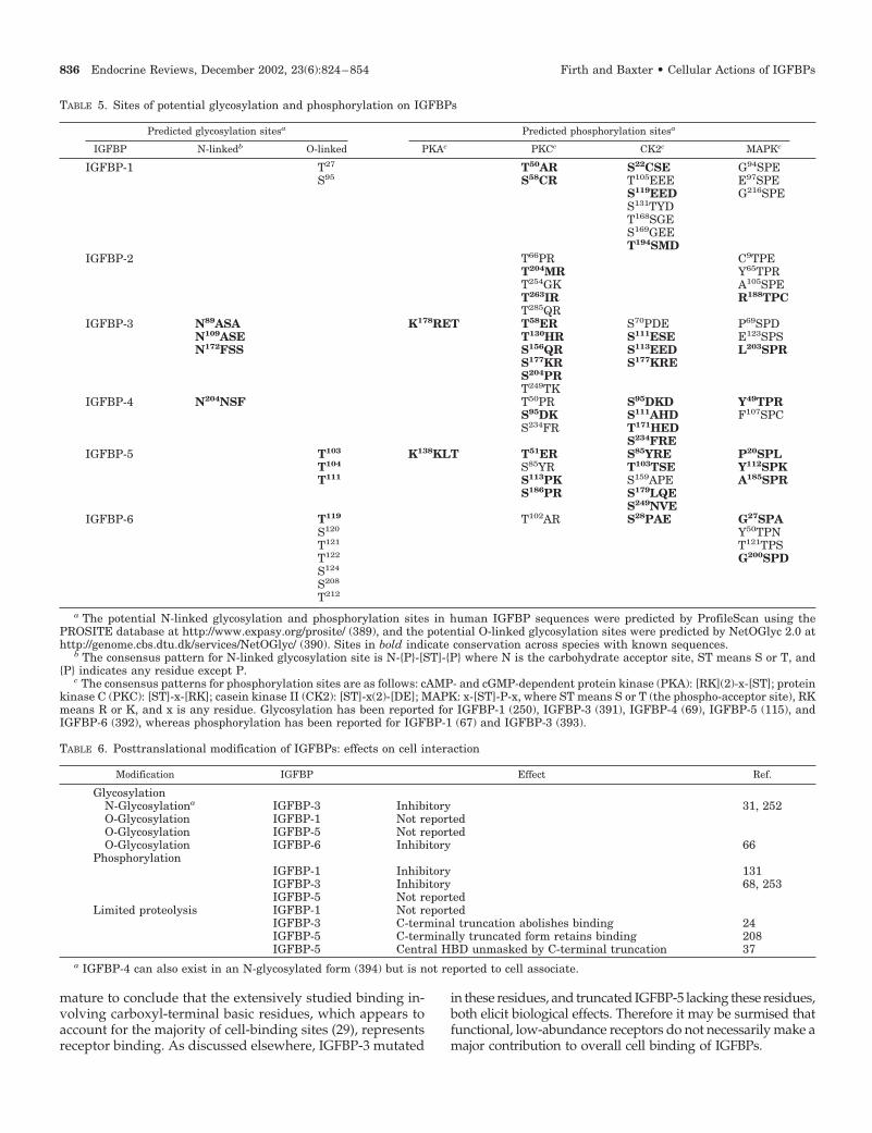
TABLE 5. Sites of potential glycosylation and phosphorylation on IGFBPs
Predicted glycosylation sitesa Predicted phosphorylation sitesa
IGFBP N-linkedb O-linked PKAc PKCc CK2c MAPKc
IGFBP-1 T27 T50AR S22CSE G94SPES95 S58CR T105EEE E97SPE
S119EED G216SPES131TYDT168SGES169GEET194SMD
IGFBP-2 T66PR C9TPET204MR Y65TPRT254GK A105SPET263IR R188TPCT285QR
IGFBP-3 N89ASA K178RET T58ER S70PDE P69SPDN109ASE T130HR S111ESE E123SPSN172FSS S156QR S113EED L203SPR
S177KR S177KRES204PRT249TK
IGFBP-4 N204NSF T50PR S95DKD Y49TPRS95DK S111AHD F107SPCS234FR T171HED
S234FREIGFBP-5 T103 K138KLT T51ER S85YRE P20SPL
T104 S85YR T103TSE Y112SPKT111 S113PK S159APE A185SPR
S186PR S179LQES249NVE
IGFBP-6 T119 T102AR S28PAE G27SPAS120 Y50TPNT121 T121TPST122 G200SPDS124
S208
T212
a The potential N-linked glycosylation and phosphorylation sites in human IGFBP sequences were predicted by ProfileScan using thePROSITE database at http://www.expasy.org/prosite/ (389), and the potential O-linked glycosylation sites were predicted by NetOGlyc 2.0 athttp://genome.cbs.dtu.dk/services/NetOGlyc/ (390). Sites in bold indicate conservation across species with known sequences.
b The consensus pattern for N-linked glycosylation site is N-{P}-[ST]-{P} where N is the carbohydrate acceptor site, ST means S or T, and{P} indicates any residue except P.
c The consensus patterns for phosphorylation sites are as follows: cAMP- and cGMP-dependent protein kinase (PKA): [RK](2)-x-[ST]; proteinkinase C (PKC): [ST]-x-[RK]; casein kinase II (CK2): [ST]-x(2)-[DE]; MAPK: x-[ST]-P-x, where ST means S or T (the phospho-acceptor site), RKmeans R or K, and x is any residue. Glycosylation has been reported for IGFBP-1 (250), IGFBP-3 (391), IGFBP-4 (69), IGFBP-5 (115), andIGFBP-6 (392), whereas phosphorylation has been reported for IGFBP-1 (67) and IGFBP-3 (393).
TABLE 6. Posttranslational modification of IGFBPs: effects on cell interaction
Modification IGFBP Effect Ref.
GlycosylationN-Glycosylationa IGFBP-3 Inhibitory 31, 252O-Glycosylation IGFBP-1 Not reportedO-Glycosylation IGFBP-5 Not reportedO-Glycosylation IGFBP-6 Inhibitory 66
PhosphorylationIGFBP-1 Inhibitory 131IGFBP-3 Inhibitory 68, 253IGFBP-5 Not reported
Limited proteolysis IGFBP-1 Not reportedIGFBP-3 C-terminal truncation abolishes binding 24IGFBP-5 C-terminally truncated form retains binding 208IGFBP-5 Central HBD unmasked by C-terminal truncation 37
a IGFBP-4 can also exist in an N-glycosylated form (394) but is not reported to cell associate.
836 Endocrine Reviews, December 2002, 23(6):824–854 Firth and Baxter • Cellular Actions of IGFBPs
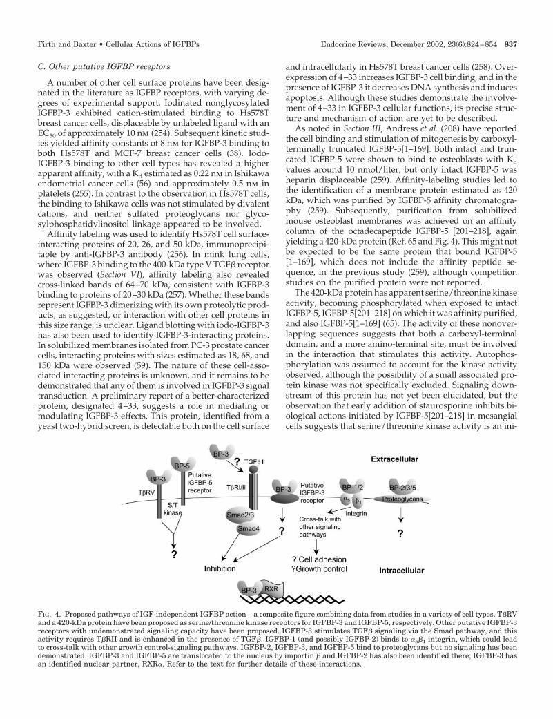
C. Other putative IGFBP receptors
A number of other cell surface proteins have been desig-nated in the literature as IGFBP receptors, with varying de-grees of experimental support. Iodinated nonglycosylatedIGFBP-3 exhibited cation-stimulated binding to Hs578Tbreast cancer cells, displaceable by unlabeled ligand with anEC50 of approximately 10 nm (254). Subsequent kinetic stud-ies yielded affinity constants of 8 nm for IGFBP-3 binding toboth Hs578T and MCF-7 breast cancer cells (38). Iodo-IGFBP-3 binding to other cell types has revealed a higherapparent affinity, with a Kd estimated as 0.22 nm in Ishikawaendometrial cancer cells (56) and approximately 0.5 nm inplatelets (255). In contrast to the observation in Hs578T cells,the binding to Ishikawa cells was not stimulated by divalentcations, and neither sulfated proteoglycans nor glyco-sylphosphatidylinositol linkage appeared to be involved.
Affinity labeling was used to identify Hs578T cell surface-interacting proteins of 20, 26, and 50 kDa, immunoprecipi-table by anti-IGFBP-3 antibody (256). In mink lung cells,where IGFBP-3 binding to the 400-kDa type V TGF� receptorwas observed (Section VI), affinity labeling also revealedcross-linked bands of 64–70 kDa, consistent with IGFBP-3binding to proteins of 20–30 kDa (257). Whether these bandsrepresent IGFBP-3 dimerizing with its own proteolytic prod-ucts, as suggested, or interaction with other cell proteins inthis size range, is unclear. Ligand blotting with iodo-IGFBP-3has also been used to identify IGFBP-3-interacting proteins.In solubilized membranes isolated from PC-3 prostate cancercells, interacting proteins with sizes estimated as 18, 68, and150 kDa were observed (59). The nature of these cell-asso-ciated interacting proteins is unknown, and it remains to bedemonstrated that any of them is involved in IGFBP-3 signaltransduction. A preliminary report of a better-characterizedprotein, designated 4–33, suggests a role in mediating ormodulating IGFBP-3 effects. This protein, identified from ayeast two-hybrid screen, is detectable both on the cell surface
and intracellularly in Hs578T breast cancer cells (258). Over-expression of 4–33 increases IGFBP-3 cell binding, and in thepresence of IGFBP-3 it decreases DNA synthesis and inducesapoptosis. Although these studies demonstrate the involve-ment of 4–33 in IGFBP-3 cellular functions, its precise struc-ture and mechanism of action are yet to be described.
As noted in Section III, Andress et al. (208) have reportedthe cell binding and stimulation of mitogenesis by carboxyl-terminally truncated IGFBP-5[1–169]. Both intact and trun-cated IGFBP-5 were shown to bind to osteoblasts with Kd
values around 10 nmol/liter, but only intact IGFBP-5 washeparin displaceable (259). Affinity-labeling studies led tothe identification of a membrane protein estimated as 420kDa, which was purified by IGFBP-5 affinity chromatogra-phy (259). Subsequently, purification from solubilizedmouse osteoblast membranes was achieved on an affinitycolumn of the octadecapeptide IGFBP-5 [201–218], againyielding a 420-kDa protein (Ref. 65 and Fig. 4). This might notbe expected to be the same protein that bound IGFBP-5[1–169], which does not include the affinity peptide se-quence, in the previous study (259), although competitionstudies on the purified protein were not reported.
The 420-kDa protein has apparent serine/threonine kinaseactivity, becoming phosphorylated when exposed to intactIGFBP-5, IGFBP-5[201–218] on which it was affinity purified,and also IGFBP-5[1–169] (65). The activity of these nonover-lapping sequences suggests that both a carboxyl-terminaldomain, and a more amino-terminal site, must be involvedin the interaction that stimulates this activity. Autophos-phorylation was assumed to account for the kinase activityobserved, although the possibility of a small associated pro-tein kinase was not specifically excluded. Signaling down-stream of this protein has not yet been elucidated, but theobservation that early addition of staurosporine inhibits bi-ological actions initiated by IGFBP-5[201–218] in mesangialcells suggests that serine/threonine kinase activity is an ini-
FIG. 4. Proposed pathways of IGF-independent IGFBP action—a composite figure combining data from studies in a variety of cell types. T�RVand a 420-kDa protein have been proposed as serine/threonine kinase receptors for IGFBP-3 and IGFBP-5, respectively. Other putative IGFBP-3receptors with undemonstrated signaling capacity have been proposed. IGFBP-3 stimulates TGF� signaling via the Smad pathway, and thisactivity requires T�RII and is enhanced in the presence of TGF�. IGFBP-1 (and possibly IGFBP-2) binds to �5�1 integrin, which could leadto cross-talk with other growth control-signaling pathways. IGFBP-2, IGFBP-3, and IGFBP-5 bind to proteoglycans but no signaling has beendemonstrated. IGFBP-3 and IGFBP-5 are translocated to the nucleus by importin � and IGFBP-2 has also been identified there; IGFBP-3 hasan identified nuclear partner, RXR�. Refer to the text for further details of these interactions.
Firth and Baxter • Cellular Actions of IGFBPs Endocrine Reviews, December 2002, 23(6):824–854 837

tiating event in the action of IGFBP-5 (236). A very recentreport also implicates serine phosphorylation of Ras inIGFBP-5 signaling (260).
VI. Interactions of IGFBP-3 with KnownSignaling Pathways
A. IGFBP-3 and TGF� signaling
A protein of similar high molecular mass to the putativeIGFBP-5 receptor has been designated a receptor for IGFBP-3in mink lung epithelial cells (Ref. 257 and Fig. 4). The typeV receptor for TGF� (T�RV) is an incompletely characterizedserine/threonine kinase of approximately 400 kDa, first de-scribed a decade ago (261). Although its relative importancein TGF� signaling, compared with the type I and type IITGF� receptor (T�RI and T�RII) system (262), is unclear, itis reported to mediate growth inhibition by TGF� in cellslacking T�RI and T�RII (263). Recombinant nonglycosylatedIGFBP-3 was shown to bind to this protein and to inhibitTGF� binding, and IGFBP-3-induced growth inhibition inT�R-deficient mink lung cells could be blocked by a TGF�1peptide antagonist (257). The interaction of IGFBP-3 with a400-kDa protein in mink lung cells has been confirmed in-dependently using a partially glycosylated IGFBP-3 mutant,IGFBP-3[N109D, N172D] (264). Judged by its comigrationwith a similar protein that binds TGF�, the 400-kDa proteincould be identical with the T�RV. In this study, Leu60-IGF-I,an analog with decreased IGFRI binding activity, inhibitedboth the interaction of IGFBP-3 with the 400-kDa protein, andthe inhibition of DNA synthesis seen with IGFBP-3 alone,leading to the suggestion that the 400-kDa protein might beinvolved in IGFBP-3 inhibitory signaling (264).
In addition to IGFBP-3, IGFBP-4 and -5 bind to the T�RV(265), raising the possibility of a similarity between this pro-tein and the IGFBP-5-binding 420-kDa serine/threonine ki-nase described by Andress (65). However, IGFBP-4 and -5 areweak receptor ligands, and weak inhibitors of DNA synthe-sis in mink lung cells, compared with IGFBP-3 (265). It istherefore possible that T�RV is a receptor in mink lung cellswith relative specificity for IGFBP-3, but its role in IGFBP-3signal transduction, its presence in other cell types, and itscomplete structural characterization remain to be determined.
The possibility that IGFBP-3 signals through a TGF�-related pathway is of particular interest because IGFBP-3 isbelieved to mediate some of the growth effects of TGF� invarious cell types. TGF� was initially shown to stimulateIGFBP-3 production by skin fibroblasts and was suggested tomodulate IGF action by this mechanism (266). More recently,both growth-inhibitory (59, 189) and growth-stimulatory(190, 191) actions of TGF� have been shown to involve theinduction of IGFBP-3, the effects being blocked whenIGFBP-3 induction is abrogated. These and similar studies,described more fully in Section III, have implicated IGFBP-3in TGF� action but did not address the concept that IGFBP-3might interact with a TGF� signaling pathway, as implied bythe studies with T�RV.
Recently, the T�RII has been shown to play an importantrole in IGFBP-3 action in T47D breast cancer cells. ManyT47D cell lines lack T�RII and are TGF� resistant. Restoration
of the receptor by cDNA transfection restored sensitivity toTGF� and rendered the cells sensitive to synergistic growthinhibition by IGFBP-3 and TGF� (63). IGFBP-3 has subse-quently been shown to stimulate the phosphorylation of theT�RI and of the signaling intermediates Smad2 and Smad3(63, 267) in both T47D and MCF-7 breast cancer cells. Thisdistinguishes IGFBP-3 action involving T�RII from effectsmediated by T�RV, which do not involve Smad phosphor-ylation (265). IGFBP-3 has reduced activity in stimulatingthis pathway after immunoneutralization of endogenousTGF�1, suggesting that IGFBP-3 can act independently, butits effects are enhanced by endogenous TGF� (267).
Interestingly, IGFBP-3 mutated in its nuclear localizationdomain (228KGRKR3MDGEA), which has greatly reducedcell surface binding (24) and fails to translocate to the nucleus(268), is able to induce T�RI and Smad phosphorylation andto increase plasminogen activator inhibitor-1 transcriptionalactivity, a Smad-dependent process (267). These observa-tions distinguish IGFBP-3 actions through the Smad pathwayfrom IGFBP-3 antiproliferative effects mediated through nu-clear interactions, described below. It is therefore now pos-sible to define an intracellular signaling cascade initiated byIGFBP-3 (Fig. 4) and dependent upon the presence of a de-fined cell surface receptor system. However, it is not yetknown whether IGFBP-3 is a direct ligand for T�RII or ini-tiates signaling in an indirect manner; therefore, this studydoes not yet define an IGFBP-3 receptor. Moreover, it re-mains to be shown whether this is a ubiquitous pathway forIGFBP-3 action or is confined to the breast cancer cell linesin which it was first demonstrated.
B. IGFBP-3, retinoids, and nuclear signaling
All-trans-retinoic acid (RA) is a potent inducer of IGFBP-3in breast and other cancer cells (269–272). This effect, whichis believed to contribute to the growth-inhibitory effect ofRA, requires the presence of RA receptor (RAR)-� (273) andcan be blocked by retinoid X receptor (RXR)-specific retin-oids (274). Although signaling through RAR stimulatesIGFBP-3 production, experiments using a RA response ele-ment reporter system suggest that IGFBP-3 actually inhibitsRA signaling (64). If these in vitro data can be extrapolatedto RA action in vivo, this observation could have importantimplications for the regulation of sensitivity to retinoids astherapeutic agents.
In contrast to possible effects on RA signaling, IGFBP-3 hasbeen shown in one study to enhance signaling through RXR,as determined using an retinoid X response element reporter,and has been observed by a variety of techniques to interactdirectly with RXR�. IGFBP-3 and RXR� have been colocal-ized by confocal microscopy in the cytoplasm and nucleus ofLAPC-4 prostate cancer cells, with nuclear localization moreevident after exposure to an RXR-specific ligand. TheIGFBP-3 residues involved in the RXR interaction appear tobe located within the 18-residue basic domain sequence 215–232 (64). As previously noted, IGFBP-3 and IGFBP-5 sharethis carboxyl-terminal domain, which resembles a consensusbipartite nuclear localization sequence (Table 3 and Ref. 275).
A variety of studies have identified IGFBP-3 in cell nucleior directly demonstrated its nuclear transport (172, 268, 276–
838 Endocrine Reviews, December 2002, 23(6):824–854 Firth and Baxter • Cellular Actions of IGFBPs
278); competition studies suggest that IGFBP-5 uses the sametransport system as IGFBP-3 (268). Nuclear transport ofIGFBP-3 appears to be favored in dividing cells and can serveto cotransport IGF-I to the nucleus (277, 278). The plasmamembrane may present a major barrier to the transport ofextracellular IGFBP-3 to the nucleus, because extracellularIGFBP-3 translocates to a relatively small percentage of cellnuclei, but in cells with a permeabilized plasma membrane,IGFBP-3 can be found in almost all nuclei, demonstratingthat transport from the cytoplasm to the nucleus occursreadily (268). Its retention within the nucleus when the nu-clear membrane is permeabilized suggests that it interactswith insoluble elements there (30, 268). The nuclear transportprotein importin-� has been shown to mediate translocationof both IGFBP-3 and IGFBP-5 (30). This interaction appearspredominantly to require residues 228–232 of IGFBP-3, and214–218 of IGFBP-5, although other basic residues within the18-residue basic domain are also involved (Table 7).
IGFBP-3 interacts with an RXR-retinoid X response ele-ment complex, as determined by EMSA, and in F9 embryonalcarcinoma cells appears to require this interaction to induceapoptosis, as the viability of RXR�-deficient F9 cells is un-affected by IGFBP-3 whereas a control cell line expressingRXR� has reduced viability when exposed to IGFBP-3. Fur-thermore, an RXR-specific ligand has been reported to en-hance IGFBP-3-induced apoptosis (64). However, a recentstudy (279) shows that the mutant form of IGFBP-3 that failsto translocate to the nucleus (IGFBP-3[228MDGEA]) can stillinduce apoptosis in breast cancer cells. These data imply that,in these cells, either RXR-IGFBP-3 interaction is not requiredfor apoptosis, or interaction between these proteins in thecytoplasm may be sufficient. RXR can dimerize with numer-ous nuclear receptors including RAR, vitamin D receptor,thyroid receptor, peroxisome proliferator-activated recep-tors, and others, as well as itself (280); thus, it may be spec-ulated that IGFBP-3, by binding RXR, could also influencesignaling by these other receptors. Because this could havewide implications for understanding nuclear actions of
IGFBPs, it will be important to confirm this study and dem-onstrate other cell systems in which the IGFBP-3-RXR inter-action occurs.
C. Other pathways
The mechanism underlying the phenomenon of enhance-ment of IGF-I activity by IGFBP-3, discussed in Section II, isnot fully understood. A recent study by Conover et al. (62)demonstrated that LY294002, an inhibitor of PI3-kinase ac-tivity, could block the ability of IGFBP-3, when preincubatedwith bovine fibroblasts, to stimulate IGF-I-stimulated ami-noisobutyrate uptake, implying that this pathway was re-quired to mediate the IGFBP-3 effect. Although IGFBP-3alone did not directly stimulate PI3-kinase, as assessed byimmunoblot for the active phosphoform of its downstreameffector protein kinase B (PKB)/Akt, it enhanced the effect ofIGF-I on this phosphorylation (62). IGFBP-5 is also reportedto activate signaling through PI3-kinase. LNCaP prostatecancer cells stably transfected to overexpress IGFBP-5 werefound to have an increased proliferation rate, concomitantwith increased PKB/Akt phosphorylation (61). PI3-kinaseblockade by LY294002 induced apoptosis independent ofIGFBP-5 expression, but IGF-I rescued the cells from deathonly if IGFBP-5 was overexpressed. This effect parallels theeffect of IGFBP-3 described above, to the extent that bothIGFBPs appear capable of sensitizing cells to IGF-I action. Inthe study of Conover et al. (62), a decrease in PKB/Aktphosphorylated at a Thr-Pro site in cells preincubated withIGFBP-3 alone suggested that dephosphorylation of this sitemight be involved in the heightened IGF-I sensitivity; con-sistent with this observation, okadaic acid, a serine/threo-nine phosphatase inhibitor, blocked the potentiating effect ofIGFBP-3 on IGF-I. These data provide evidence that IGFBP-3might influence growth factor signaling by activating a spe-cific protein phosphatase system; whether IGFBP-5 actsthrough a similar mechanism is unknown.
An interaction between MAPK signaling and IGFBP-3 ac-tivity has also been established, with the demonstration thatthe inhibitory effect of IGFBP-3 on DNA synthesis in breastepithelial cells is blocked in cells expressing oncogenic ras,and restored by the MAPK/ERK pathway inhibitor PD98059(Ref. 281 and Fig. 3). The sensitivity to IGFBP-3 of Hs578Tbreast cancer cells, which express oncogenic ras, can also beenhanced by treatment with PD98059. Changes in IGFBP-3sensitivity were unrelated to the extent of IGFBP-3 cell as-sociation (281), suggesting that the regulation of cellular sen-sitivity to IGFBP-3 does not involve a mechanism as straight-forward as modulating the interaction between IGFBP-3 andan abundant cell surface receptor.
Very recently, another signaling intermediate, STAT1 (sig-nal transducer and activator of transcription-1) has beenimplicated in IGFBP-3 action during the process of chon-drocyte differentiation (282). Predifferentiated chondrocyteswere previously shown to provide a convincing example ofIGF-independent IGFBP-3 action (180), as discussed in Sec-tion III. IGFBP-3 strongly up-regulated STAT1 mRNA in thissystem, and phospho-STAT1 protein was shown to increaseand translocate to the nucleus. Furthermore, in the presenceof a STAT1 antisense oligonucleotide, IGFBP-3 action was
TABLE 7. Effect of mutations within the nuclear localizationsignals of IGFBP-3 and IGFBP-5 on their nuclear accumulation
Nuclear localization sequence Nuclearaccumulation
IGFBP-3K K G F Y K K K Q C R P S K G R K R � �– N – – – – – – – – – – – – – – – – � �– – – – – H S R – – – – – – – – – – �– – – – – – – – – – – – – M D G E A �– N – – – H S R – – – – – – – – – – �– N – – – – – – – – – – – M D G E A �A A – – – – – – – – – – – – – – – – �
IGFBP-5R K G F Y K R K Q C K P S R G R K R � �– N – – – – – – – – – – – – – – – – � �– – – – – H S R – – – – – – – – – – �– – – – – – – – – – – – – M D G E A �A A – – – – – – – – – – – – – – – – �
The bipartite nuclear localization signal sequences of IGFBP-3 andIGFBP-5 are shown with the basic residues indicated in bold.Changes in amino acid residues are indicated for the differentIGFBP-3 and IGFBP-5 mutants. The effect of each mutation on nu-clear accumulation was reported by Schedlich et al. (30).
Firth and Baxter • Cellular Actions of IGFBPs Endocrine Reviews, December 2002, 23(6):824–854 839

ablated, strongly implicating STAT1 induction and phos-phorylation in the antiproliferative action of IGFBP-3 in thesecells (282). This is yet another example of an IGFBP-3-activated signaling mechanism observed in a single cellline. It should now be relatively easy to determine whetherSTAT1 induction by IGFBP-3 occurs in other systems.
Is it possible to integrate these reports of IGFBP-3 inter-action with diverse signaling pathways into a single unifyingmechanism of action? At present there is insufficient infor-mation available to do so, because each of the above-described studies has only been performed in a limited num-ber of laboratories (often only one) and cell types. One wellestablished fact is the translocation of IGFBP-3 to the cellnucleus, although the cell signals that initiate this are un-known, and the function of IGFBP-3, once inside the nucleus,is still unclear. As noted above, by mutating the nuclearlocalization signal of IGFBP-3, it is now possible to distin-guish between cellular effects that require nuclear translo-cation and those that do not. A similar mutagenesis approachinvolving other key functional residues may eventually fur-ther delineate distinct pathways of IGFBP-3 action, e.g., an-tiproliferative effects seen with non-IGF-binding forms ofIGFBP-3 (282) add further weight to the concept of trulyIGF-independent actions.
The relationship between MAPK activity and IGFBP-3 sig-naling, if confirmed in other laboratories, may help to inte-grate some of the pathways described earlier. Phosphoryla-tion of effector proteins by MAPK can interrupt pathwaysinvolving both Smads (283) and RXR� (284), both reportedto mediate some IGFBP-3 effects (63, 64). This suggests thatcell sensitivity to growth-inhibitory effects of IGFBP-3 maybe regulated by a variety of intersecting pathways. Under-standing the mechanisms underlying IGFBP-3 sensitivityand resistance may be important in developing therapeuticapproaches to cancer cell proliferation because, despite itsantiproliferative and proapoptotic activity in vitro (SectionVII), a high level of tumor IGFBP-3 has been associated withpoor prognosis in breast malignancy (285). This apparentdiscrepancy highlights the fact that few of the regulatorymechanisms demonstrated in cell culture models have yetbeen confirmed in vivo.
VII. Cell Cycle, Apoptosis, and Survival
IGFs stimulate cell proliferation as well as inhibiting ap-optosis, thus acting as cell survival factors. The antiapoptoticfunctions of IGF-I are mediated by IGFRI, independent of thereceptor’s ability to transduce mitogenic signals (70). Similarto their modulation of IGF-I’s cellular proliferative effects,IGFBPs can also modulate the antiapoptotic effects of IGF-Iby regulating the IGF-I-IGFRI interaction. Perturbations ateach level of the IGF axis have been implicated in cancerdevelopment and progression in several cell types (75). Forexample, treatment of MCF-7 cells with either exogenousIGFBP-3 or an antiestrogen that stimulates the endogenousproduction of IGFBP-3 resulted in increased apoptosis. Thiseffect could be inhibited by a non-IGFBP-binding IGF-I an-alog but not by IGF-I, suggesting, but not proving, thatIGFBP-3 induced apoptosis by sequestering IGF-I from
IGFRI (286). Similarly, serum withdrawal from human ter-atocarcinoma cells induced apoptosis that could be reversedby simultaneous treatment with IGF-I or IGF-II. However,the survival effect by IGF-II was abrogated by cotreatmentwith either IGFBP-2 or IGFRI blocking antibody, again sug-gesting that interference of the IGF-IGFRI interaction canabolish the antiapoptotic effect (287).
Modulation of IGFBPs by proteolysis also plays a role inregulating their antiproliferative effects. For example, theproliferation of DU145 prostate adenocarcinoma cells inserum-free medium is thought to occur through an autocrineIGF loop because these cells express IGF-I, IGF-II, IGFRI, andIGFBP-2, -3, and -4, although the IGFBP-3 is proteolyzed.However, addition of anti-IGFRI antibody or intact IGFBP-3can inhibit this serum-free proliferation (288). Concurrentwith the inhibition of DNA synthesis, exogenous IGFBP-3promotes a decline in the number of cells accumulating in theG2/M phase and a reduction in the levels of p34(cdc2), aprotein required for G2 transition to mitosis (289). The cellswere shown to produce matrix metalloproteinase 9, a pro-tease for IGFBP-3, and it was proposed that the equilibriumbetween proteolysis of IGFBP-3 by matrix metalloproteinase9, resulting in low-affinity IGF binding fragments, and se-questration of IGF by intact IGFBP-3 may determine theoutcome of cellular proliferation or inhibition (288).
Studies of this kind illustrate clearly that IGFBPs can affectcell cycle progression and apoptosis by preventing IGFRIactivation. IGFBPs can also exert IGF-independent effects oncell cycle arrest and apoptosis (290, 291). As noted in SectionIII, previous studies have shown 1,25-dihydroxyvitamin D3(vitD) to be a potent growth inhibitor of some cancer cells andthat it up-regulates IGFBP-3 (183, 292). Whereas in PC-3prostate cancer cells, IGFBP-3-mediated growth inhibition byvitD appears to involve IGF sequestration (292), in Hs578Tbreast cancer cells the effects of vitD analogs are thought tobe IGF independent because the cells are IGF unresponsive(183). The mechanism of inhibition by vitD may involve theaccumulation of cells in the G1 phase of the cell cycle. Insupport of this, it was recently reported that the inhibition ofgrowth in LNCaP prostate cancer cells in an IGF-free systemby vitD is mediated by the up-regulation of the cyclin-dependent kinase inhibitor p21/WAF1/CIP1 via the induc-tion of IGFBP-3 (184) (Fig. 3). This effect can be abrogated byusing IGFBP-3 immunoneutralizing antibodies. The inhibi-tion of growth of T47D cells by overexpression of IGFBP-3was also associated with an accumulation of cells in G1 phase(60). However, because IGFs stimulate cell cycle progressionat the G1-S transition (293), the possibility of IGF involvementin these IGFBP-3 actions still needs to be considered.
Rajah et al. (59) provided the first unambiguous demon-stration of the IGFRI-independent proapototic effects ofIGFBP-3 in an IGFRI-negative mouse fibroblast cell line, bytreating the cells with exogenous IGFBP-3 and by transfect-ing IGFBP-3 into the cells. In both instances, the basal levelof apoptosis was significantly increased, a key observationthat will be important to confirm in other laboratories. UsingHs578T breast cancer cells, which do not respond to IGF-I, asa model to determine IGF-independent effects of exogenousIGFBPs on apoptosis, Perks et al. (294) found that none of theIGFBPs alone had a significant inhibitory effect on cell
840 Endocrine Reviews, December 2002, 23(6):824–854 Firth and Baxter • Cellular Actions of IGFBPs
growth. However, when apoptosis was induced by a cer-amide analog, C2, in these cells, IGFBP-3 accentuated theapoptotic effect whereas IGFBP-4 and IGFBP-5 reduced C2-induced apoptosis, and IGFBP-1, IGFBP-2, and IGFBP-6 hadno significant effect. In addition, although IGFBP-3 had noeffect on cell death induced by an RGD peptide throughintegrin detachment, IGFBP-5 inhibited integrin detach-ment-induced apoptosis (294, 295). These findings indicatethat the IGFBPs have distinctive functional effects on differ-ent apoptotic signaling pathways even though they sharestructural homology.
A further study has suggested that the enhancement ofC2-induced apoptosis in Hs578T by IGFBP-3 may involvecell association and subsequent proteolysis of the bindingprotein, and that this effect can be abrogated by cotreatmentwith IGF-I (296). IGFBP-3 has also been shown to enhance theapoptotic effect of paclitaxel, a cytotoxic drug that acts viaTNF� and subsequent ceramide generation (297).
There is also evidence that IGFBP-3 can induce apoptosisby itself, as well as potentiate the apoptotic effects of otherDNA-damaging stimuli such as ionizing and UV irradiationand chemotherapeutic drugs (298–301). Transfection ofIGFBP-3 into breast cancer cells expressing either wild-type(MCF-7) or mutant (T47D) p53 resulted in an induction ofapoptosis (299), suggesting that the IGFBP-3 effect can occurindependently of functional p53. Furthermore, transfectionof IGFBP-3 into T47D cells restored the sensitivity of theseradiation-resistant cells to ionizing radiation. This IGFBP-3action in MCF-7 and T47D cells is one of few cases where ithas been shown to be apoptotic by itself: in most other stud-ies, it enhances the apoptotic effect of other agents. Becauseall cell culture studies involve a degree of cell apoptosis,presumably due to limitations of the culture conditions, it isnot clear what contributory role other factors may play incells that appear to respond to IGFBP-3 alone. This may, infact, be a semantic argument because all tissue and tumorgrowth is a balance between proliferative and apoptotic ac-tivities, and studies of xenograft tumors expressing IGFBP-3show inhibition of tumor growth (302, 303).
The inhibition of protein kinase C�, the expression ofwhich is correlated to tumor progression in glioblastomamultiforme, by either antisense oligonucleotide (304) or aprotein kinase C�-specific inhibitor (305), was reported to beassociated with the induction of p53 and IGFBP-3 concom-itant with apoptotic cell death. A tumor suppressor, p53regulates the transcription of many cellular genes that areinvolved in mediating its effects on cell cycle arrest andapoptosis (306). IGFBP-3 was identified in 1995 as one of thep53-inducible genes and a mediator of p53-dependent apo-ptosis in EB1 colon carcinoma cells in response to cellularstress (186). Consistent with this, Williams et al. (298) re-ported that IGFBP-3 potentiation of apoptosis after ionizingradiation in colonic adenoma cells was dependent on func-tional p53. In an esophageal carcinoma cell line, apoptosisafter UV irradiation was accompanied by an increase in p53levels, which was enhanced and prolonged by exogenousIGFBP-3 treatment (300). Although the level of endogenousIGFBP-3 was not examined, this study raises the possibilitythat IGFBP-3 may also act upstream or independently of p53
in an autocrine fashion as well as downstream of p53 as themediator of its actions (Fig. 3).
The intracellular mechanisms that mediate the IGF- andIGFRI-independent antiproliferative and proapoptotic func-tions of IGFBP-3 are still being elucidated. The induction ofapoptosis in breast cancer cells overexpressing IGFBP-3 wasassociated with an increase in the proapoptotic Bax and Badand a decrease in the antiapoptotic Bcl-2 and Bcl-xL (Ref. 299and Fig. 3). This led to the suggestion that IGFBP-3 may exertits apoptotic effect by modulating the Bax-to-Bcl-2 ratio. Bcl-2activity may be further decreased by its serine phosphory-lation in response to IGFBP-3 (307). The ratio of proapoptoticand antiapoptotic members of the Bcl-2 family of cytoplasmicproteins is an important determinant of survival or death incells (308). These proteins reside or assemble on the surfaceof the mitochondria during apoptosis. Indeed it has beenshown that apoptosis induced by a mitochondrial respi-ratory chain inhibitor, antimycin A, was accentuated byIGFBP-3. Together with the observation that both antimycinA and IGFBP-3 can also enhance ceramide-induced apopto-sis (295, 309), it would appear that IGFBP-3 accentuates apo-ptosis induced via pathways involving the mitochondria.
Caspases, a family of cysteine aspartic acid-specific pro-teases, occupy a central position as intracellular effectors ofapoptotic signals. When activated by various apoptotic stim-uli, caspases then activate numerous cellular substrates byrestricted proteolysis, and may involve the release of mito-chondrial proteins, resulting in cell death (310). Apoptosisinduced by IGFBP-3 in PC-3 cells could be inhibited by acaspase inhibitor, thus implicating the involvement of thiskey pathway (59). However, it remains to be shown whichcaspases are involved and whether there is a direct or indirectinteraction with IGFBP-3.
The E7 oncoprotein, encoded by the human papillomavi-rus type 16, has recently been identified as an IGFBP-3 in-teracting partner in a yeast two-hybrid screen (311). Co-expression of E7 with IGFBP-3 in PC-3 cells reduced thenumber of apoptotic cells compared with PC-3 cells trans-fected with IGFBP-3 alone, thus suggesting that E7 can in-hibit IGFBP-3-induced apoptosis. Furthermore, it was shownthat E7 and IGFBP-3 colocalize intracellularly, and degrada-tion of intracellular IGFBP-3 was enhanced in E7 coexpress-ing cells, the effect being reversible by a proteasome inhib-itor. E7 mutants with reduced oncogenic potential were notas effective in inhibiting IGFBP-3-induced apoptosis. Thisstudy suggests that the degradation of intracellular IGFBP-3,and hence the abrogation of the proapoptotic function ofIGFBP-3, could potentially contribute toward the transform-ing capacity of E7. Functional inactivation of IGFBP-3 by anoncogene was also demonstrated in breast epithelial cellsthat were transformed by constitutively activated Ha-rasoncogene. However, in contrast to the E7 study, the cellulargrowth resistance to IGFBP-3 was not due to degradation ofthe protein but was associated with an increase in both se-creted and cell-associated IGFBP-3 (281).
It remains unclear whether the proapoptotic function ofIGFBP-3 is effected by secreted extracellular IGFBP-3, whichis then internalized, or by intracellular IGFBP-3. The abilityof exogenous IGFBP-3 to induce apoptosis in several cellsystems (59, 294), and of IGFBP-3 neutralizing antibodies to
Firth and Baxter • Cellular Actions of IGFBPs Endocrine Reviews, December 2002, 23(6):824–854 841
inhibit IGFBP-3-induced apoptosis in PC-3 cells (59), pro-vides some evidence that the apoptotic signal may initiate atthe cell surface. This raises questions about the identity of thesignaling receptor for IGFBP-3, as discussed in Section V.Questions also remain about how IGFBP-3 exerts its apo-ptotic function. However, as discussed in Section VI, IGFBP-3can translocate to the nucleus by binding to the importin �nuclear transport factor (30) and was recently shown to in-teract with the nuclear receptor, RXR� (64). Butt et al. (299)reported that IGFBP-3 modulated the mRNA levels of bcl-2in IGFBP-3-transfected MCF-7 cells, raising the possibilitythat the regulation of gene expression by nuclear IGFBP-3may effect its apoptotic function.
Like IGFBP-3, IGFBP-5 also translocates to the nucleus bybinding to the importin � nuclear transport factor (30). Al-though the full spectrum of intracellular binding partners forIGFBP-5 (like IGFBP-3) is far from understood, the recentidentification by two-hybrid screening, and confirmation bycoprecipitation, of FHL2 as an IGFBP-5-interacting proteinraises interesting questions (312). FHL2 interacts with a va-riety of cellular proteins including the androgen receptor,suggesting the possibility of a direct role for IGFBP-5 intranscriptional regulation, but the true significance of thisinteraction will require further extensive experimentation.
The role of IGFBP-5 in apoptosis and cell survival has beenstudied in models of mammary gland and prostate involu-tion. GH and prolactin enhance mammary epithelial cellsurvival by increasing IGF-I production and decreasingIGFBP-5 production, respectively (313). Involution of themammary gland involves apoptosis of the epithelial cells aswell as extensive remodeling and degradation of the ECM.It has been proposed that IGF-dependent and -independentfunctions of IGFBP-5 may be involved in these processes.During mammary gland involution, there is a decrease inprolactin levels concomitant with a 50-fold increase inIGFBP-5 concentration in milk, which is thought to inhibitIGF-I-mediated cell survival, thus releasing the cells fromsuppression of apoptosis (314). Furthermore, IGFBP-5 bindsto PAI-1, effectively increasing the activation of plasminogento plasmin, which then initiates degradation and remodelingof the ECM (315), processes involved in mammary glandinvolution.
The IGF axis plays an important role in the maintenanceof the normal prostate as well as in the regression of bothnormal prostate and prostate tumors after androgen with-drawal or castration. Increased expression of IGFBP-5 incastration-induced and androgen withdrawal- or ablation-induced apoptosis in the prostate gland or in the involutingprostate is associated with reduced IGF-I activity (316–319),suggesting that IGFBP-5 was inhibiting the antiapo-ptotic function of IGF-I. Miyake et al. (320) recently demon-strated that although there was no difference in prostate-specific antigen levels, tumor incidence, or growth ratesbetween control and IGFBP-5 overexpressing LNCaP cellsgrown in intact mice, prostate-specific antigen levels andtumor growth rates were increased in mice bearing IGFBP-5overexpressing LnCaP cells after castration. This indicatesthat after castration, IGFBP-5 can accelerate the rate of pro-gression of prostate cancer cells to androgen independence.This was supported by the observation that there was a delay
in the recurrence of androgen-independent Shionogi tumorsin mice treated with IGFBP-5 antisense oligonucleotide com-pared with mice treated with control oligonucleotide. In cellculture studies in vitro, the treatment of Shionogi tumor cellswith IGFBP-5 antisense oligonucleotide resulted in inhibitionof cell growth that was reversed in the presence of IGF-I,suggesting that the IGFBP-5 effect was IGF dependent.
It was previously shown that IGF-II promotes differenti-ation of myoblasts to myotubes and acts as a survival factor(321, 322) whereas IGFBP-5 blocked IGF-stimulated myo-genesis (120). Treatment of C2 myoblasts with TNF� down-regulated the secretion of IGF-II and IGFBP-5 and was as-sociated with the blockade of myoblast differentiation andinduction of apoptosis (323). However, TNF� was unable toinduce apoptosis in C2 cells transfected with IGFBP-5, thusindicating that IGFBP-5 may have an antiapoptotic role in thesurvival of these cells during differentiation.
The regulation of ovarian follicle selection and atresia isanother biological process that is thought to depend on thebalance between IGF-I and IGFBPs. Studies in a variety ofspecies indicate that follicle atresia involves an apoptoticprocess that may be blocked by IGF-I, whereas follicle de-velopment and selection requires IGF-I (324–326). IGFBP-2,-4, and -5 have all been implicated in the regulation of ovar-ian IGF-I availability, and there is some evidence thatIGFBP-4 plays a key role. As noted in Section II, IGFBP-4degradation is IGF-stimulated, and in the human ovarianfollicle has been shown to be due to PAPP-A activity (101).Other IGFBPs might modulate this process by regulatingIGF-I availability (327). IGFBP-4 gene expression appearsconfined to apoptotic and atretic follicles (325), whereasdominant follicles acquire an IGFBP-4 protease, whichwould enhance IGF-I action (326). In the rat, IGFBP-5 pro-teolysis may play a parallel role to that of IGFBP-4 proteolysisin other species (328, 329). Although the details appear todiffer between species, these and similar studies empha-size the central role of IGFBPs in regulating ovariandevelopment.
IGFBP-1 has been shown to stimulate cell detachment andapoptosis in breast cancer cells through dephosphorylationof FAK (Ref. 58; see Section IV). The expression of IGFBP-1in the endometrium during the secretory phase of the men-strual cycle (330) and the regulation of cytotrophoblast in-vasion by IGFBP-1 (136, 137) suggests that IGFBP-1 may playa role in apoptosis and remodeling. However, this awaitsfurther investigation.
Exposure of lung epithelial cells to hyperoxia leads toinhibition of cell division and subsequently induction ofapoptosis via the Fas pathway which is associated with anincreased expression of IGFBP-2 and IGFBP-3 (331). Al-though previous studies have shown that IGFBP-2 is up-regulated in growth-arrested cells (332–334), it remains to beshown that IGFBP-2 plays a role in the induction of apopto-sis. Interestingly, the increased expression of IGFBP-2 waspredominantly found intracellularly and in the nucleuswhereas IGFBP-3 was found in the extracellular compart-ment (331). There are few reports of nuclear localization ofIGFBP-2, and it will be important to confirm this observationin other cell systems.
IGFBP-4, known to be IGF inhibitory, when overexpressed
842 Endocrine Reviews, December 2002, 23(6):824–854 Firth and Baxter • Cellular Actions of IGFBPs
in M12 prostate epithelial cells inhibits IGF-I-induced pro-liferation. Apoptosis induced by 6-hydroxyurea is increasedin the IGFBP-4 transfectants, but IGFBP-2 levels are de-creased concomitantly. When injected into nude mice thereis a delay in the onset of tumor formation by IGFBP-4 trans-fectants compared with controls (221). Surprisingly, whenthe same cell line was transfected with antisense IGFBP-4, theeffects were decreased cellular proliferation, decreased col-ony formation in soft agar, decreased tumor formation, andincreased apoptosis induced by etoposide. This was accom-panied by an increase in IGFBP-3 and IGFBP-6 expression(222). Therefore, the role of IGFBP-4 in apoptosis remainscontroversial and will require further study in isolation of theother IGFBPs.
Infection of NSCLC cell lines with an adenovirus express-ing human IGFBP-6 reduced cell numbers by inducing apo-ptosis, the effects of which were not reversible by IGF-I orIGF-II (335). Moreover, treatment of the cells with exogenousIGFBP-6 did not result in inhibition of cell growth, suggest-ing that the IGFBP-6 effect is mediated by an intracellularform of IGFBP-6 through an IGF-independent mechanism. Incontrast, IGFBP-6 stimulated cell growth and decreased apo-ptosis in the Saos-2/B-10 cell line (336).
Because of the central position occupied by the IGF-IGFBPsystem in cell growth regulation, it is inevitable that the fullextent of the specific functions of IGFBPs in the determina-tion of cell survival or death remains to be revealed. It is,however, clear that these proteins interact with proliferativeand apoptotic processes at multiple levels and in differenttissues.
VIII. Implications for Animal Physiology
The previous sections have highlighted many of the im-portant studies, mostly conducted at the level of cell biology,that shed light on the complex array of cellular actions of theIGFBPs. Transposing knowledge of these cellular actions tothe areas of animal and human physiology will present manychallenges. Gene deletion experiments in mice have the po-tential to provide unique information pointing to cellularactions of IGFBPs, but to date IGFBP-2 is the only IGFBP forwhich a mouse knockout model has been published. In thismodel there was no overall growth phenotype, althoughspleen size was reduced and liver size increased (337). No-tably, altered levels of other IGFBPs pointed to the possibilitythat the deletion of one IGFBP could cause compensatorychanges in others, making the interpretation of such exper-iments, in terms of normal physiology, extremely complex.
A report of other IGFBP gene deletions has also appearedin abstract form. Although still preliminary, these data sug-gest that IGFBP-4 is the only IGFBP that, when deleted, altersgrowth. Surprisingly, the phenotype was a reduction ingrowth (338), even though IGFBP-4 is generally inhibitory tocell proliferation. No cellular mechanism for the growth in-hibition is currently available, although this animal modelappears to offer a novel opportunity to study previouslyunrecognized effects of IGFBP-4. Another preliminary ob-servation was the delayed mammary gland involution inIGFBP-5 knockout mice, consistent with the known role of
IGFBP-5 in mammary epithelial cell apoptosis (Section VII).These are intriguing observations, but unpublished at thetime of writing, and clearly just the tip of the iceberg in theuse of gene deletions to study IGFBP cellular function.
Gene overexpression in vivo may also provide informationon cellular IGFBP actions, although ectopic expression hasthe potential to yield misleading results. Transgenic mousemodels including overexpression of IGFBP-1 (339–342), -2(148, 244), -3 (343–345), and -4 (346) have been reported.IGFBP-1 overexpression was found to have no effect (339), orin another study to cause up to 20% reduction (340), insomatic growth, whereas IGFBP-2 and its RGD-to-RGE mu-tant led to growth reduction (234, 244); and IGFBP-3 causedno overall growth phenotype in one study (343) and modestgrowth reduction in another (345). The variation in pheno-type for IGFBP overexpression models in different labora-tories, and the contrast between lack of a growth effectin IGFBP-2 knockout mice and the growth reduction inIGFBP-2-overexpressing mice, highlights the difficultyin drawing simple conclusions from such studies.
In general, transgenic studies published to date have notprovided major new insights into cellular actions of theIGFBPs. An interesting exception is the decreased apoptosisin the involuting mammary gland described in a tissue-specific IGFBP-3-overexpressing model (344). This observa-tion certainly raises questions about the proapoptotic effectsof IGFBP-3 consistently noted in cultured breast cells (SectionVII). Other studies have supported earlier hypotheses con-cerning IGFBP actions; for example, the observation thatseveral IGFBP-1-overexpressing models show fasting hyper-glycemia (342, 347) is consistent with the hypothesis thatIGFBP-1 has a counterregulatory role in glucose homeostasis(348), although it is unclear whether this is primarily anendocrine or a cellular action of IGFBP-1.
In other cases, tissue-specific changes resulting fromIGFBP overexpression may be interpreted as indicating un-expected roles for IGFBPs, such as the consistently seen neg-ative effect of IGFBP-1 overexpression on brain growth anddevelopment (349–351). However, there may be insufficientinformation available to distinguish true physiologicalIGFBP actions from the more general consequences of high-level IGF sequestration. For example, the hypoplastic actionof smooth muscle-specific IGFBP-4 overexpression (346) hasinteresting implications for muscle IGF action but does notnecessarily delineate the role of IGFBP-4 in physiologicalmuscle growth regulation. Further refinement in the tempo-ral and spatial regulation of IGFBP expression, and in trans-gene responsiveness to physiological influences, as at-tempted in a recent IGFBP-1 transgenic model (342), mayallow more definitive conclusions on IGFBP action to bedrawn in the future.
IX. Implications for Human Disease
The use of serum measurements of IGFBPs is well estab-lished as an aid to the diagnosis or monitoring of growthdisorders (352, 353) and is developing in relation to someother diseases (354, 355). In the context of predicting cancerrisk, there has been considerable recent interest in reports
Firth and Baxter • Cellular Actions of IGFBPs Endocrine Reviews, December 2002, 23(6):824–854 843
that a high serum IGFBP-3 level reduces the relative risk ofdeveloping breast cancer predicted by high IGF-I levels inpremenopausal women (356) and also tends to attenuateprostate and lung cancer risk in patients with high IGF-I (357,358). Low serum IGFBP-3 may also be associated with in-creased risk of colorectal cancer (359), although another pro-spective study of colorectal cancer found that high IGFBP-3did not modify the risk related to high IGF-I, and patients inthe highest quintile for IGFBP-3 actually had an increasedrisk (360). Other studies of breast (361), lung (362), and pros-tate (363) cancer risk also challenge the predictive value ofserum IGFBP-3 levels. Correlative studies of IGFBP-3 levelsin women with breast cancer show variable results, withdecreased (364, 365), increased (366), and unchanged (367)IGFBP-3 levels reported.
There has also been some interest in the measurement ofother serum IGFBPs in relation to cancer status or develop-ment. Increasing IGFBP-1 levels have been associated with adecrease in colorectal cancer risk (360), and in correlativestudies, breast cancer patients have been shown in one studyto have lower serum levels of IGFBP-1 and IGFBP-6 thanwomen with benign breast disease (365). Elevated serumIGFBP-2 levels are also associated with malignancies of theprostate (368–370) and ovary (371).
It is not at all clear how changes in serum IGFBP levelsreflect changes at the level of specific tissues, and, in contrastto the growing use of serum IGFBP measurement in cancerand other disease states, there are currently no accepteddiagnostic or therapeutic applications that exploit knowl-edge of changes in IGFBPs at the cellular level. This is largelydue to the relative paucity of data on the measurement andinterpretation of IGFBP protein levels in tumor samples.
In general, high levels of IGFBP-3 in breast tumor speci-mens are associated with unfavorable prognosis (285, 372),despite the antiproliferative and proapoptotic activity of thisprotein in many in vitro studies. This suggests that somemalignancies can escape from the growth-inhibitory effectsof IGFBP-3, or even become stimulated by it, as demon-strated in vitro (60). If this transition, which is not yet un-derstood mechanistically, occurs preferentially in vivo, due tothe presence of factors not studied in cell culture systems, itis possible that some mechanisms delineated in vitro mayhave limited relevance to tumor growth in vivo. Therefore theassociation of tissue IGFBP-3 levels with relevant signalingintermediates and cell growth markers in patient tumor sam-ples will be an important area for study.
Similarly, the high tissue IGFBP-2 levels associated withsome cancerous tissues, such as ovarian (373) and colorectal(374), are consistent with its actions being growth stimula-tory rather than inhibitory in these tumor types, but whethertumor IGFBP-2 measurement will provide any prognosticinformation is not known.
X. Concluding Comment
How might the limited clinical information on IGFBPs intumors and in other diseased tissues be reconciled with thevast accumulation of in vitro studies on cellular actions ofIGFBPs, and exploited to clinical advantage? It needs to be
stressed that many of the potential “breakthrough” discov-eries on IGFBP signaling pathways, and other cellular phe-nomena involving IGFBPs, still await confirmation in otherlaboratories and other cell systems. Even when confirmed invitro, the links between cell culture studies and in vivo IGFBPactions are likely to be complex. A primary aim should be toincrease our understanding of the relationship between in-hibitory and stimulatory actions of IGFBPs on cell prolifer-ation and migration, because these are known in some casesto differ between cell culture and in vivo situations. Delin-eation of the signaling pathways involved in these dichot-omous effects may allow the development of targeted ther-apeutics to activate growth inhibition (e.g., in cancer) orstimulation (e.g., in tissue engineering). IGFBPs themselvesmight be exploited as active agents in tissue-directed genetherapy approaches, or IGFBP mimetics may be developedwhen their receptor systems are more fully understood. Be-cause the IGF-IGFBP system is already recognized as centralto processes of cell growth, differentiation, and migration,and the understanding of the cellular pathways mediatingthese effects is progressing, the prospect of clinical applica-tions based on the IGFBPs seems increasingly likely.
Acknowledgments
Address all correspondence and requests for reprints to: Robert C.Baxter, Ph.D., D.Sc., Kolling Institute of Medical Research, Royal NorthShore Hospital, St. Leonards, New South Wales 2065, Australia. E-mail:[email protected]
References
1. Baxter RC 1997 Molecular aspects of insulin-like growth factorbinding proteins. Adv Mol Cell Endocrinol 1:123–159
2. Upton Z, Chan SJ, Steiner DF, Wallace JC, Ballard FJ 1993 Evo-lution of insulin-like growth factor binding proteins. Growth Regul3:29–32
3. Hwa V, Oh Y, Rosenfeld RG 1999 The insulin-like growth factor-binding protein (IGFBP) superfamily. Endocr Rev 20:761–787
4. Vilmos P, Gaudenz K, Hegedus Z, Marsh JL 2001 The Twistedgastrulation family of proteins, together with the IGFBP and CCNfamilies, comprise the TIC superfamily of cysteine rich secretedfactors. Mol Pathol 54:317–323
5. Drop SL, Schuller AG, Lindenbergh-Kortleve DJ, Groffen C,Brinkman A, Zwarthoff EC 1992 Structural aspects of the IGFBPfamily. Growth Regul 2:69–79
6. Baxter RC 2000 Insulin-like growth factor (IGF)-binding proteins:interactions with IGFs and intrinsic bioactivities. Am J Physiol278:E967–E976
7. Clemmons DR 2001 Use of mutagenesis to probe IGF-bindingprotein structure/function relationships. Endocr Rev 22:800–817
8. Neumann GM, Bach LA 1999 The N-terminal disulfide linkages ofhuman insulin-like growth factor-binding protein-6 (hIGFBP-6)and hIGFBP-1 are different as determined by mass spectrometry.J Biol Chem 274:14587–14594
9. Chelius D, Baldwin MA, Lu X, Spencer EM 2001 Expression,purification and characterization of the structure and disulfidelinkages of insulin-like growth factor binding protein-4. J Endo-crinol 168:283–296
10. Kalus W, Zweckstetter M, Renner C, Sanchez Y, Georgescu J,Grol M, Demuth D, Schumacher R, Dony C, Lang K, Holak TA1998 Structure of the IGF-binding domain of the insulin-like growthfactor-binding protein-5 (IGFBP-5): implications for IGF and IGF-Ireceptor interactions. EMBO J 17:6558–6572
11. Imai Y, Moralez A, Andag U, Clarke JB, Busby WH, ClemmonsDR 2000 Substitutions for hydrophobic amino acids in the N-
844 Endocrine Reviews, December 2002, 23(6):824–854 Firth and Baxter • Cellular Actions of IGFBPs
terminal domains of IGFBP-3 and -5 markedly reduce IGF-I bind-ing and alter their biologic actions. J Biol Chem 275:18188–18194
12. Buckway CK, Wilson EM, Ahlsen M, Bang P, Oh Y, Rosenfeld RG2001 Mutation of three critical amino acids of the N-terminal do-main of IGF-binding protein-3 essential for high affinity IGF bind-ing. J Clin Endocrinol Metab 86:4943–4950
13. Hong J, Zhang G, Dong F, Rechler MM 2002 Insulin-like growthfactor binding protein-3 (IGFBP-3) mutants that do not bind IGF-Ior IGF-II stimulate apoptosis in human prostate cancer cells. J BiolChem 277:10489–10497
14. Lalou C, Lassarre C, Binoux M 1996 A proteolytic fragment ofinsulin-like growth factor (IGF) binding protein-3 that fails to bindIGFs inhibits the mitogenic effects of IGF-I and insulin. Endocri-nology 137:3206–3212
15. Salahifar H, Firth SM, Baxter RC, Martin JL 2000 Characterizationof an amino-terminal fragment of insulin-like growth factor bind-ing protein-3 and its effects in MCF-7 breast cancer cells. GrowthHorm IGF Res 10:367–377
16. Forbes BE, Turner D, Hodge SJ, McNeil KA, Forsberg G, WallaceJC 1998 Localization of an insulin-like growth factor (IGF) bindingsite of bovine IGF binding protein-2 using disulfide mapping anddeletion mutation analysis of the C-terminal domain. J Biol Chem273:4647–4652
17. Wang J-F, Hampton B, Mehlman T, Burgess WH, Rechler MM1988 Isolation of a biologically active fragment from the carboxyterminus of the fetal rat binding protein for insulin-like growthfactors. Biochem Biophys Res Commun 157:718–726
18. Ho PJ, Baxter RC 1997 Characterization of truncated insulin-likegrowth factor-binding protein-2 in human milk. Endocrinology138:3811–3818
19. Galanis M, Firth SM, Bond J, Nathanielsz A, Kortt AA, HudsonPJ, Baxter RC 2001 Ligand-binding characteristics of recombinantamino- and carboxyl-terminal fragments of human insulin-likegrowth factor-binding protein-3. J Endocrinol 169:123–133
20. Devi GR, Yang DH, Rosenfeld RG, Oh Y 2000 Differential effectsof insulin-like growth factor (IGF)-binding protein-3 and its pro-teolytic fragments on ligand binding, cell surface association, andIGF-I receptor signaling. Endocrinology 141:4171–4179
21. Bramani S, Song H, Beattie J, Tonner E, Flint DJ, Allan GJ 1999Amino acids within the extracellular matrix (ECM) binding region(201–218) of rat insulin-like growth factor binding protein(IGFBP)-5 are important determinants in binding IGF-I. J Mol En-docrinol 23:117–123
22. Jones JI, Gockerman A, Busby WH, Wright G, Clemmons DR1993 Insulin-like growth factor binding protein 1 stimulates cellmigration and binds to the �5�1 integrin by means of its Arg-Gly-Asp sequence. Proc Natl Acad Sci USA 90:10553–10557
23. Binkert C, Landwehr J, Mary JL, Schwander J, Heinrich G 1989Cloning, sequence analysis and expression of a cDNA encoding anovel insulin-like growth factor binding protein (IGFBP-2). EMBOJ 8:2497–2502
24. Firth SM, Ganeshprasad U, Baxter RC 1998 Structural determi-nants of ligand and cell surface binding of insulin-like growthfactor binding protein-3. J Biol Chem 273:2631–2638
25. Twigg SM, Kiefer MC, Zapf J, Baxter RC 1998 Insulin-like growthfactor-binding protein 5 complexes with the acid-labile subunit:role of the carboxyl-terminal domain. J Biol Chem 273:28791–28798
26. Firth SM, Clemmons DR, Baxter RC 2001 Mutagenesis of basicamino acids in the carboxyl-terminal region of insulin-like growthfactor binding protein-5 affects acid-labile subunit binding. Endo-crinology 142:2147–2150
27. Nam TJ, Busby W, Clemmons DR 1997 Insulin-like growth factorbinding protein-5 binds to plasminogen activator inhibitor-I. En-docrinology 138:2972–2978
28. Weinzimer SA, Gibson TB, Collett-Solberg PF, Khare A, Liu B,Cohen P 2001 Transferrin is an insulin-like growth factor-bindingprotein-3 binding protein. J Clin Endocrinol Metab 86:1806–1813
29. Booth BA, Boes M, Andress DL, Dake BL, Kiefer MC, Maack C,Linhardt RJ, Bar K, Caldwell EE, Weiler J, Bar RS 1995 IGFBP-3and IGFBP-5 association with endothelial cells: role of C-terminalheparin binding domain. Growth Regul 5:1–17
30. Schedlich LJ, Le Page SL, Firth SM, Briggs LJ, Jans DA, Baxter RC2000 Nuclear import of insulin-like growth factor-binding
protein-3 and -5 is mediated by the importin � subunit. J Biol Chem275:23462–23470
31. Firth SM, Baxter RC 1999 Characterisation of recombinant glyco-sylation variants of insulin-like growth factor binding protein-3. JEndocrinol 160:379–387
32. Cheung PT, Smith EP, Shimasaki S, Ling N, Chernausek SD 1991Characterization of an insulin-like growth factor binding protein(IGFBP-4) produced by the B104 rat neuronal cell line: chemical andbiological properties and differential synthesis by sublines. Endo-crinology 129:1006–1015
33. Coverley JA, Baxter RC 1997 Phosphorylation of insulin-likegrowth factor binding proteins. Mol Cell Endocrinol 128:1–5
34. Conover CA, Durham SK, Zapf J, Masiarz FR, Kiefer MC 1995Cleavage analysis of insulin-like growth factor (IGF)-dependentIGF-binding protein-4 proteolysis and expression of protease-resistant IGF-binding protein-4 mutants. J Biol Chem 270:4395–4400
35. Imai Y, Busby WH, Smith CE, Clarke JB, Garmong AJ, HorwitzGD, Rees C, Clemmons DR 1997 Protease-resistant form of insu-lin-like growth factor-binding protein 5 is an inhibitor of insulin-like growth factor-I actions on porcine smooth muscle cells inculture. J Clin Invest 100:2596–2605
36. Twigg SM, Kiefer MC, Zapf J, Baxter RC 2000 A central domainbinding site in insulin-like growth factor-binding protein 5 for theacid-labile subunit. Endocrinology 141:454–457
37. Song H, Shand JH, Beattie J, Flint DJ, Allan GJ 2001 The carboxy-terminal domain of IGF-binding protein-5 inhibits heparin bindingto a site in the central domain. J Mol Endocrinol 26:229–239
38. Yamanaka Y, Fowlkes JL, Wilson EM, Rosenfeld RG, Oh Y 1999Characterization of insulin-like growth factor binding protein-3(IGFBP-3) binding to human breast cancer cells: kinetics of IGFBP-3binding and identification of receptor binding domain on theIGFBP-3 molecule. Endocrinology 140:1319–1328
39. Baxter RC 1993 Circulating binding proteins for the insulinlikegrowth factors. Trends Endocrinol Metab 4:91–96
40. Jones JI, Clemmons DR 1995 Insulin-like growth factors and theirbinding proteins: biological actions. Endocr Rev 16:3–34
41. Baxter RC 1995 Insulin-like growth factor binding proteins as glu-coregulators. Metabolism Clin Exp 44(Suppl 4):12–17
42. Rajaram S, Baylink DJ, Mohan S 199 7 Insulin-like growth factorbinding proteins in serum and other biological fluids: Regulationand functions. Endocr Rev 18:801–831
43. Murphy LJ 1998 Insulin-like growth factor binding proteins: func-tional diversity or redundancy? J Mol Endocrinol 21:97–107
44. Baxter RC, Martin JL, Beniac VA 1989 High molecular weightinsulin-like growth factor binding protein complex. Purificationand properties of the acid-labile subunit from human serum. J BiolChem 264:11843–11848
45. Twigg SM, Baxter RC 1998 Insulin-like growth factor (IGF)-binding protein 5 forms an alternative ternary complex with IGFsand the acid-labile subunit. J Biol Chem 273:6074–6079
46. Baxter RC, Meka S, Firth SM 2002 Molecular distribution of IGF-binding protein-5 in human serum. J Clin Endocrinol Metab 87:271–276
47. Guler H-P, Zapf J, Schmid C, Froesch ER 1989 Insulin-like growthfactors I and II in healthy man. Estimations of half-lives and pro-duction rates. Acta Endocrinol (Copenh) 121:753–758
48. Young SCJ, Miles MV, Clemmons DR 1992 Determination ofthe pharmacokinetic profiles of insulin-like growth factor bindingproteins-1 and -2 in rats. Endocrinology 131:1867–1873
49. Lewitt MS, Saunders H, Phuyal JL, Baxter RC 1994 Complexformation by human insulin-like growth factor-binding protein-3and human acid-labile subunit in growth hormone-deficient rats.Endocrinology 134:2404–2409
50. Binoux M, Hossenlopp P 1988 Insulin-like growth factor (IGF) andIGF-binding proteins: comparison of human serum and lymph.J Clin Endocrinol Metab 67:509–514
51. Boes M, Booth BA, Sandra A, Dake BL, Bergold A, Bar RS 1992Insulin-like growth factor binding protein (IGFBP)4 accounts forthe connective tissue distribution of endothelial cell IGFBPs per-fused through the isolated heart. Endocrinology 131:327–330
52. Arany E, Zabel P, Hill DJ 1996 Rapid clearance of human insulin-like growth factor binding protein-3 from the rat circulation and
Firth and Baxter • Cellular Actions of IGFBPs Endocrine Reviews, December 2002, 23(6):824–854 845
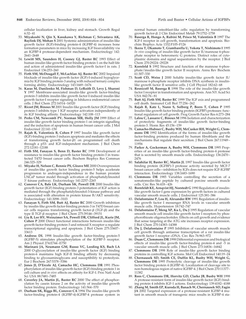
cellular localization in liver, kidney and stomach. Growth Regul6:32–41
53. Miyakoshi N, Qin X, Kasukawa Y, Richman C, Srivastava AK,Baylink DJ, Mohan S 2001 Systemic administration of insulin-likegrowth factor (IGF)-binding protein-4 (IGFBP-4) increases boneformation parameters in mice by increasing IGF bioavailability viaan IGFBP-4 protease-dependent mechanism. Endocrinology 142:2641–2648
54. Lewitt MS, Saunders H, Cooney GJ, Baxter RC 1993 Effect ofhuman insulin-like growth factor-binding protein-1 on the half-lifeand action of administered insulin-like growth factor-I in rats. JEndocrinol 136:253–260
55. Firth SM, McDougall F, McLachlan AJ, Baxter RC 2002 Impairedblockade of insulin-like growth factor (IGF)-I-induced hypoglyce-mia by IGF binding protein-3 analog with reduced ternary complexforming ability. Endocrinology 143:1669–1676
56. Karas M, Danilenko M, Fishman D, LeRoith D, Levy J, SharoniY 1997 Membrane-associated insulin-like growth factor-bindingprotein-3 inhibits insulin-like growth factor-I-induced insulin-likegrowth factor-I receptor signaling in Ishikawa endometrial cancercells. J Biol Chem 272:16514–16520
57. Ricort JM, Binoux M 2001 Insulin-like growth factor (IGF) bindingprotein-3 inhibits type 1 IGF receptor activation independently ofits IGF binding affinity. Endocrinology 142:108–113
58. Perks CM, Newcomb PV, Norman MR, Holly JM 1999 Effect ofinsulin-like growth factor binding protein-1 on integrin signallingand the induction of apoptosis in human breast cancer cells. J MolEndocrinol 22:141–150
59. Rajah R, Valentinis B, Cohen P 1997 Insulin like growth factor(IGF)-binding protein-3 induces apoptosis and mediates the effectsof transforming growth factor-�1 on programmed cell deaththrough a p53- and IGF-independent mechanism. J Biol Chem272:12181–12188
60. Firth SM, Fanayan S, Benn D, Baxter RC 1998 Development ofresistance to insulin-like growth factor binding protein-3 in trans-fected T47D breast cancer cells. Biochem Biophys Res Commun246:325–329
61. Miyake H, Nelson C, Rennie PS, Gleave ME 2000 Overexpressionof insulin-like growth factor binding protein-5 helps accelerateprogression to androgen-independence in the human prostateLNCaP tumor model through activation of phosphatidylinositol3�-kinase pathway. Endocrinology 141:2257–2265
62. Conover CA, Bale LK, Durham SK, Powell DR 2000 Insulin-likegrowth factor (IGF) binding protein-3 potentiation of IGF action ismediated through the phosphatidylinositol-3-kinase pathway andis associated with alteration in protein kinase B/AKT sensitivity.Endocrinology 141:3098–3103
63. Fanayan S, Firth SM, Butt AJ, Baxter RC 2000 Growth inhibitionby insulin-like growth factor-binding protein-3 in T47D breast can-cer cells requires transforming growth factor-� (TGF-�) and thetype II TGF-� receptor. J Biol Chem 275:39146–39151
64. Liu B, Lee HY, Weinzimer SA, Powell DR, Clifford JL, Kurie JM,Cohen P 2000 Direct functional interactions between insulin-likegrowth factor-binding protein-3 and retinoid X receptor-� regulatetranscriptional signaling and apoptosis. J Biol Chem 275:33607–33613
65. Andress DL 1998 Insulin-like growth factor-binding protein-5(IGFBP-5) stimulates phosphorylation of the IGFBP-5 receptor.Am J Physiol 274:E744–E750
66. Marinaro JA, Neumann GM, Russo VC, Leeding KS, Bach LA2000 O-glycosylation of insulin-like growth factor (IGF) bindingprotein-6 maintains high IGF-II binding affinity by decreasingbinding to glycosaminoglycans and susceptibility to proteolysis.Eur J Biochem 267:5378–5386
67. Jones JI, D’Ercole AJ, Camacho HC, Clemmons DR 1991 Phos-phorylation of insulin-like growth factor (IGF)-binding protein 1 incell culture and in vivo: effects on affinity for IGF-I. Proc Natl AcadSci USA 88:7481–7485
68. Coverley JA, Martin JL, Baxter RC 2000 The effect of phosphor-ylation by casein kinase 2 on the activity of insulin-like growthfactor binding protein. Endocrinology 141:564–570
69. Durham SK, Riggs BL, Conover CA 1994 The insulin-like growthfactor-binding protein-4 (IGFBP-4)-IGFBP-4 protease system in
normal human osteoblast-like cells: regulation by transforminggrowth factor-�. J Clin Endocrinol Metab 79:1752–1758
70. Baserga R, Hongo A, Rubini M, Prisco M, Valentinis B 1997 TheIGF-I receptor in cell growth, transformation and apoptosis. Bio-chim Biophys Acta 1332:F105–F126
71. Ikezu T, Okamoto T, Giambarella U, Yokota T, Nishimoto I 1995In vivo coupling of insulin-like growth factor II/mannose 6-phos-phate receptor to heteromeric G proteins. Distinct roles of cyto-plasmic domains and signal sequestration by the receptor. J BiolChem 270:29224–29228
72. Kornfeld S 1992 Structure and function of the mannose 6-phos-phate/insulin-like growth factor II receptors. Annu Rev Biochem61:307–330
73. Scott CD, Weiss J 2000 Soluble insulin-like growth factor II/mannose 6-phosphate receptor inhibits DNA synthesis in insulin-like growth factor II sensitive cells. J Cell Physiol 182:62–68
74. Resnicoff M, Baserga R 1998 The role of the insulin-like growthfactor I receptor in transformation and apoptosis. Ann NY Acad SciUSA 842:76–81
75. Butt AJ, Firth SM, Baxter RC 1999 The IGF axis and programmedcell death. Immunol Cell Biol 77:256–262
76. Rajah R, Katz L, Nunn S, Solberg P, Beers T, Cohen P 1995Insulin-like growth factor binding protein (IGFBP) proteases: func-tional regulators of cell growth. Prog Growth Factor Res 6:273–284
77. Lalou C, Lassarre C, Binoux M 1996 Isolation and characterizationof proteolytic fragments of insulin-like growth factor-bindingprotein-3. Horm Res 45:156–159
78. Camacho-Hubner C, Busby WH, McCusker RH, Wright G, Clem-mons DR 1992 Identification of the forms of insulin-like growthfactor-binding proteins produced by human fibroblasts and themechanisms that regulate their secretion. J Biol Chem 267:11949–11956
79. Parker A, Gockerman A, Busby WH, Clemmons DR 1995 Prop-erties of an insulin-like growth factor-binding protein-4 proteasethat is secreted by smooth muscle cells. Endocrinology 136:2470–2476
80. Salahifar H, Baxter RC, Martin JL 1997 Insulin-like growth factorbinding protein (IGFBP)-3 protease activity secreted by MCF-7breast cancer cells—inhibition by IGFs does not require IGF-IGFBPinteraction. Endocrinology 138:1683–1690
81. Clemmons DR 1985 Variables controlling the secretion of asomatomedin-like peptide by cultured porcine smooth musclecells. Circ Res 56:418–426
82. Bornfeldt KE, Arnqvist HJ, Norstedt G 1990 Regulation of insulin-like growth factor-I gene expression by growth factors in culturedvascular smooth muscle cells. J Endocrinol 125:381–386
83. Delafontaine P, Lou H, Alexander RW 1991 Regulation of insulin-like growth factor I messenger RNA levels in vascular smoothmuscle cells. Hypertension 18:742–747
84. Delafontaine P, Meng XP, Ku L, Du J 1995 Regulation of vascularsmooth muscle cell insulin-like growth factor I receptors by phos-phorothioate oligonucleotides. Effects on cell growth and evidencethat sense targeting at the ATG site increases receptor expression.J Biol Chem 270:14383–14388
85. Du J, Delafontaine P 1995 Inhibition of vascular smooth musclecell growth through antisense transcription of a rat insulin-likegrowth factor I receptor cDNA. Circ Res 76:963–972
86. Duan C, Clemmons DR 1998 Differential expression and biologicaleffects of insulin-like growth factor-binding protein-4 and -5 invascular smooth muscle cells. J Biol Chem 273:16836–16842
87. Clemmons DR 1998 Role of insulin-like growth factor bindingproteins in controlling IGF actions. Mol Cell Endocrinol 140:19–24
88. Chernausek SD, Smith CE, Duffin KL, Busby WH, Wright G,Clemmons DR 1995 Proteolytic cleavage of insulin-like growthfactor binding protein 4 (IGFBP-4). Localization of cleavage site tonon-homologous region of native IGFBP-4. J Biol Chem 270:11377–11382
89. Rees C, Clemmons DR, Horvitz GD, Clarke JB, Busby WH 1998A protease-resistant form of insulin-like growth factor (IGF) bind-ing protein 4 inhibits IGF-1 actions. Endocrinology 139:4182–4188
90. Zhang M, Smith EP, Kuroda H, Banach W, Chernausek SD, FaginJA 2002 Targeted expression of a protease-resistant IGFBP-4 mu-tant in smooth muscle of transgenic mice results in IGFBP-4 sta-
846 Endocrine Reviews, December 2002, 23(6):824–854 Firth and Baxter • Cellular Actions of IGFBPs
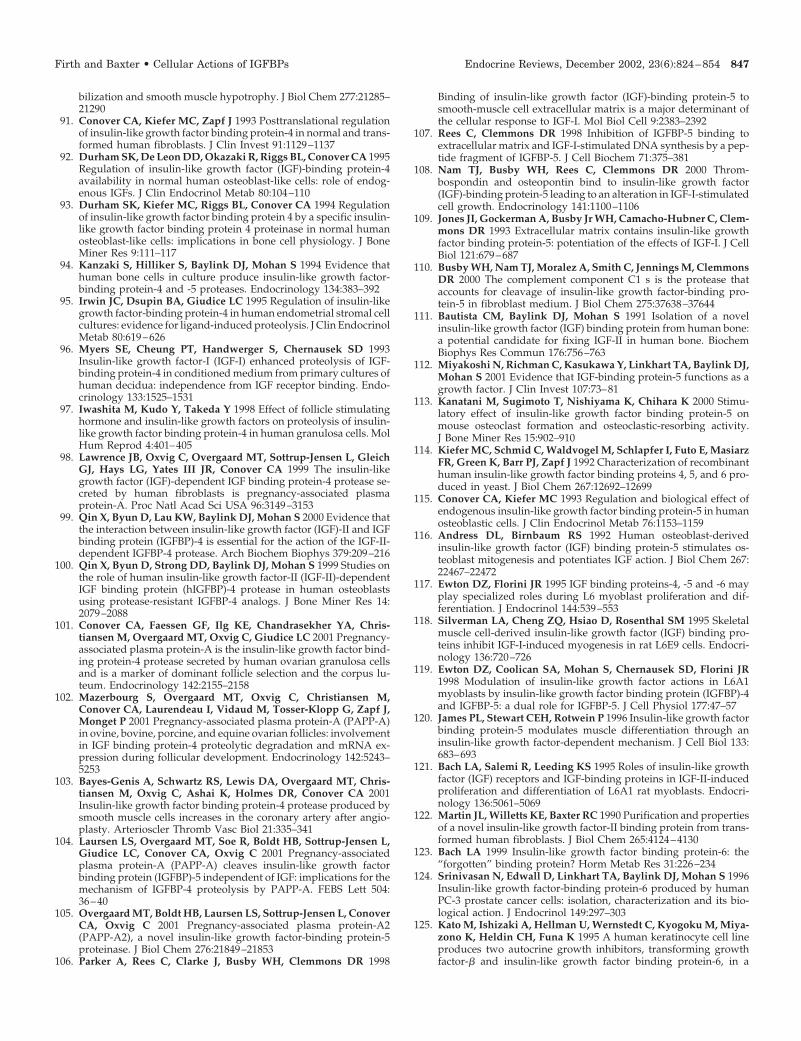
bilization and smooth muscle hypotrophy. J Biol Chem 277:21285–21290
91. Conover CA, Kiefer MC, Zapf J 1993 Posttranslational regulationof insulin-like growth factor binding protein-4 in normal and trans-formed human fibroblasts. J Clin Invest 91:1129–1137
92. Durham SK, De Leon DD, Okazaki R, Riggs BL, Conover CA 1995Regulation of insulin-like growth factor (IGF)-binding protein-4availability in normal human osteoblast-like cells: role of endog-enous IGFs. J Clin Endocrinol Metab 80:104–110
93. Durham SK, Kiefer MC, Riggs BL, Conover CA 1994 Regulationof insulin-like growth factor binding protein 4 by a specific insulin-like growth factor binding protein 4 proteinase in normal humanosteoblast-like cells: implications in bone cell physiology. J BoneMiner Res 9:111–117
94. Kanzaki S, Hilliker S, Baylink DJ, Mohan S 1994 Evidence thathuman bone cells in culture produce insulin-like growth factor-binding protein-4 and -5 proteases. Endocrinology 134:383–392
95. Irwin JC, Dsupin BA, Giudice LC 1995 Regulation of insulin-likegrowth factor-binding protein-4 in human endometrial stromal cellcultures: evidence for ligand-induced proteolysis. J Clin EndocrinolMetab 80:619–626
96. Myers SE, Cheung PT, Handwerger S, Chernausek SD 1993Insulin-like growth factor-I (IGF-I) enhanced proteolysis of IGF-binding protein-4 in conditioned medium from primary cultures ofhuman decidua: independence from IGF receptor binding. Endo-crinology 133:1525–1531
97. Iwashita M, Kudo Y, Takeda Y 1998 Effect of follicle stimulatinghormone and insulin-like growth factors on proteolysis of insulin-like growth factor binding protein-4 in human granulosa cells. MolHum Reprod 4:401–405
98. Lawrence JB, Oxvig C, Overgaard MT, Sottrup-Jensen L, GleichGJ, Hays LG, Yates III JR, Conover CA 1999 The insulin-likegrowth factor (IGF)-dependent IGF binding protein-4 protease se-creted by human fibroblasts is pregnancy-associated plasmaprotein-A. Proc Natl Acad Sci USA 96:3149–3153
99. Qin X, Byun D, Lau KW, Baylink DJ, Mohan S 2000 Evidence thatthe interaction between insulin-like growth factor (IGF)-II and IGFbinding protein (IGFBP)-4 is essential for the action of the IGF-II-dependent IGFBP-4 protease. Arch Biochem Biophys 379:209–216
100. Qin X, Byun D, Strong DD, Baylink DJ, Mohan S 1999 Studies onthe role of human insulin-like growth factor-II (IGF-II)-dependentIGF binding protein (hIGFBP)-4 protease in human osteoblastsusing protease-resistant IGFBP-4 analogs. J Bone Miner Res 14:2079–2088
101. Conover CA, Faessen GF, Ilg KE, Chandrasekher YA, Chris-tiansen M, Overgaard MT, Oxvig C, Giudice LC 2001 Pregnancy-associated plasma protein-A is the insulin-like growth factor bind-ing protein-4 protease secreted by human ovarian granulosa cellsand is a marker of dominant follicle selection and the corpus lu-teum. Endocrinology 142:2155–2158
102. Mazerbourg S, Overgaard MT, Oxvig C, Christiansen M,Conover CA, Laurendeau I, Vidaud M, Tosser-Klopp G, Zapf J,Monget P 2001 Pregnancy-associated plasma protein-A (PAPP-A)in ovine, bovine, porcine, and equine ovarian follicles: involvementin IGF binding protein-4 proteolytic degradation and mRNA ex-pression during follicular development. Endocrinology 142:5243–5253
103. Bayes-Genis A, Schwartz RS, Lewis DA, Overgaard MT, Chris-tiansen M, Oxvig C, Ashai K, Holmes DR, Conover CA 2001Insulin-like growth factor binding protein-4 protease produced bysmooth muscle cells increases in the coronary artery after angio-plasty. Arterioscler Thromb Vasc Biol 21:335–341
104. Laursen LS, Overgaard MT, Soe R, Boldt HB, Sottrup-Jensen L,Giudice LC, Conover CA, Oxvig C 2001 Pregnancy-associatedplasma protein-A (PAPP-A) cleaves insulin-like growth factorbinding protein (IGFBP)-5 independent of IGF: implications for themechanism of IGFBP-4 proteolysis by PAPP-A. FEBS Lett 504:36–40
105. Overgaard MT, Boldt HB, Laursen LS, Sottrup-Jensen L, ConoverCA, Oxvig C 2001 Pregnancy-associated plasma protein-A2(PAPP-A2), a novel insulin-like growth factor-binding protein-5proteinase. J Biol Chem 276:21849–21853
106. Parker A, Rees C, Clarke J, Busby WH, Clemmons DR 1998
Binding of insulin-like growth factor (IGF)-binding protein-5 tosmooth-muscle cell extracellular matrix is a major determinant ofthe cellular response to IGF-I. Mol Biol Cell 9:2383–2392
107. Rees C, Clemmons DR 1998 Inhibition of IGFBP-5 binding toextracellular matrix and IGF-I-stimulated DNA synthesis by a pep-tide fragment of IGFBP-5. J Cell Biochem 71:375–381
108. Nam TJ, Busby WH, Rees C, Clemmons DR 2000 Throm-bospondin and osteopontin bind to insulin-like growth factor(IGF)-binding protein-5 leading to an alteration in IGF-I-stimulatedcell growth. Endocrinology 141:1100–1106
109. Jones JI, Gockerman A, Busby Jr WH, Camacho-Hubner C, Clem-mons DR 1993 Extracellular matrix contains insulin-like growthfactor binding protein-5: potentiation of the effects of IGF-I. J CellBiol 121:679–687
110. Busby WH, Nam TJ, Moralez A, Smith C, Jennings M, ClemmonsDR 2000 The complement component C1 s is the protease thataccounts for cleavage of insulin-like growth factor-binding pro-tein-5 in fibroblast medium. J Biol Chem 275:37638–37644
111. Bautista CM, Baylink DJ, Mohan S 1991 Isolation of a novelinsulin-like growth factor (IGF) binding protein from human bone:a potential candidate for fixing IGF-II in human bone. BiochemBiophys Res Commun 176:756–763
112. Miyakoshi N, Richman C, Kasukawa Y, Linkhart TA, Baylink DJ,Mohan S 2001 Evidence that IGF-binding protein-5 functions as agrowth factor. J Clin Invest 107:73–81
113. Kanatani M, Sugimoto T, Nishiyama K, Chihara K 2000 Stimu-latory effect of insulin-like growth factor binding protein-5 onmouse osteoclast formation and osteoclastic-resorbing activity.J Bone Miner Res 15:902–910
114. Kiefer MC, Schmid C, Waldvogel M, Schlapfer I, Futo E, MasiarzFR, Green K, Barr PJ, Zapf J 1992 Characterization of recombinanthuman insulin-like growth factor binding proteins 4, 5, and 6 pro-duced in yeast. J Biol Chem 267:12692–12699
115. Conover CA, Kiefer MC 1993 Regulation and biological effect ofendogenous insulin-like growth factor binding protein-5 in humanosteoblastic cells. J Clin Endocrinol Metab 76:1153–1159
116. Andress DL, Birnbaum RS 1992 Human osteoblast-derivedinsulin-like growth factor (IGF) binding protein-5 stimulates os-teoblast mitogenesis and potentiates IGF action. J Biol Chem 267:22467–22472
117. Ewton DZ, Florini JR 1995 IGF binding proteins-4, -5 and -6 mayplay specialized roles during L6 myoblast proliferation and dif-ferentiation. J Endocrinol 144:539–553
118. Silverman LA, Cheng ZQ, Hsiao D, Rosenthal SM 1995 Skeletalmuscle cell-derived insulin-like growth factor (IGF) binding pro-teins inhibit IGF-I-induced myogenesis in rat L6E9 cells. Endocri-nology 136:720–726
119. Ewton DZ, Coolican SA, Mohan S, Chernausek SD, Florini JR1998 Modulation of insulin-like growth factor actions in L6A1myoblasts by insulin-like growth factor binding protein (IGFBP)-4and IGFBP-5: a dual role for IGFBP-5. J Cell Physiol 177:47–57
120. James PL, Stewart CEH, Rotwein P 1996 Insulin-like growth factorbinding protein-5 modulates muscle differentiation through aninsulin-like growth factor-dependent mechanism. J Cell Biol 133:683–693
121. Bach LA, Salemi R, Leeding KS 1995 Roles of insulin-like growthfactor (IGF) receptors and IGF-binding proteins in IGF-II-inducedproliferation and differentiation of L6A1 rat myoblasts. Endocri-nology 136:5061–5069
122. Martin JL, Willetts KE, Baxter RC 1990 Purification and propertiesof a novel insulin-like growth factor-II binding protein from trans-formed human fibroblasts. J Biol Chem 265:4124–4130
123. Bach LA 1999 Insulin-like growth factor binding protein-6: the“forgotten” binding protein? Horm Metab Res 31:226–234
124. Srinivasan N, Edwall D, Linkhart TA, Baylink DJ, Mohan S 1996Insulin-like growth factor-binding protein-6 produced by humanPC-3 prostate cancer cells: isolation, characterization and its bio-logical action. J Endocrinol 149:297–303
125. Kato M, Ishizaki A, Hellman U, Wernstedt C, Kyogoku M, Miya-zono K, Heldin CH, Funa K 1995 A human keratinocyte cell lineproduces two autocrine growth inhibitors, transforming growthfactor-� and insulin-like growth factor binding protein-6, in a
Firth and Baxter • Cellular Actions of IGFBPs Endocrine Reviews, December 2002, 23(6):824–854 847
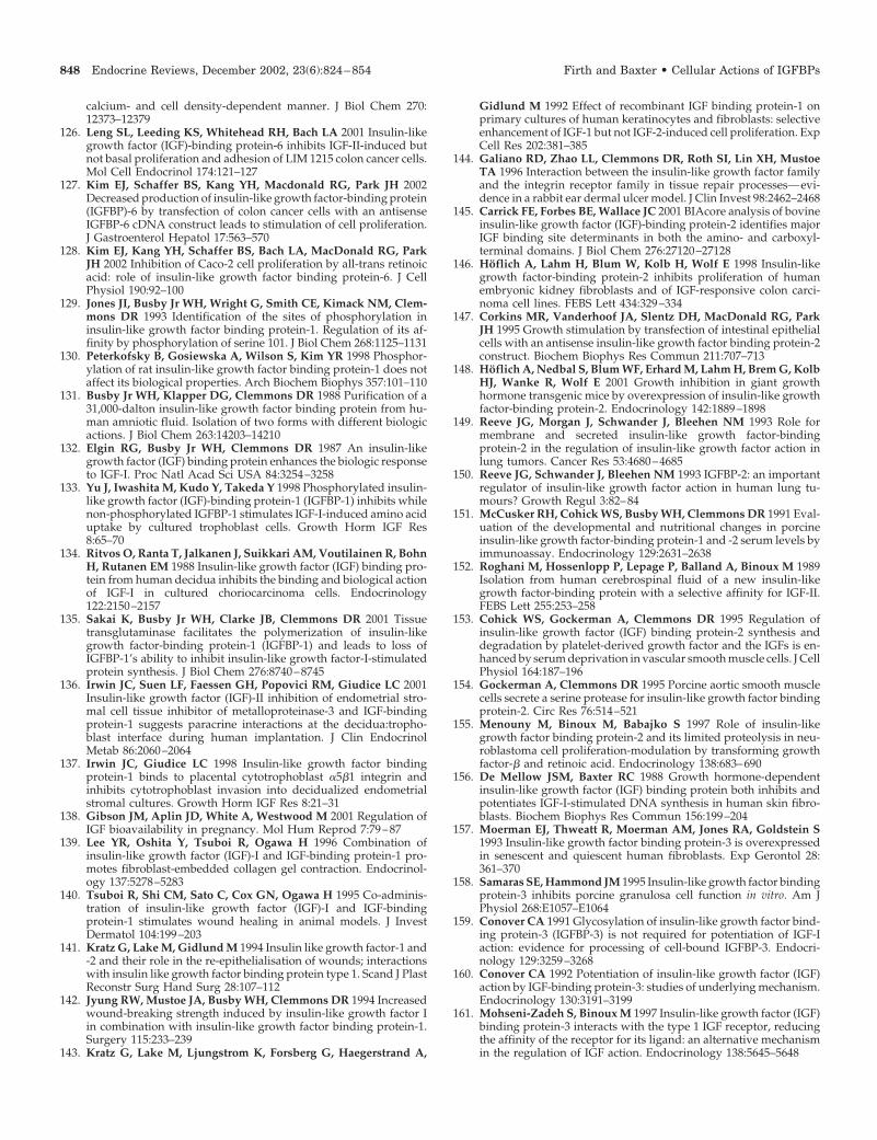
calcium- and cell density-dependent manner. J Biol Chem 270:12373–12379
126. Leng SL, Leeding KS, Whitehead RH, Bach LA 2001 Insulin-likegrowth factor (IGF)-binding protein-6 inhibits IGF-II-induced butnot basal proliferation and adhesion of LIM 1215 colon cancer cells.Mol Cell Endocrinol 174:121–127
127. Kim EJ, Schaffer BS, Kang YH, Macdonald RG, Park JH 2002Decreased production of insulin-like growth factor-binding protein(IGFBP)-6 by transfection of colon cancer cells with an antisenseIGFBP-6 cDNA construct leads to stimulation of cell proliferation.J Gastroenterol Hepatol 17:563–570
128. Kim EJ, Kang YH, Schaffer BS, Bach LA, MacDonald RG, ParkJH 2002 Inhibition of Caco-2 cell proliferation by all-trans retinoicacid: role of insulin-like growth factor binding protein-6. J CellPhysiol 190:92–100
129. Jones JI, Busby Jr WH, Wright G, Smith CE, Kimack NM, Clem-mons DR 1993 Identification of the sites of phosphorylation ininsulin-like growth factor binding protein-1. Regulation of its af-finity by phosphorylation of serine 101. J Biol Chem 268:1125–1131
130. Peterkofsky B, Gosiewska A, Wilson S, Kim YR 1998 Phosphor-ylation of rat insulin-like growth factor binding protein-1 does notaffect its biological properties. Arch Biochem Biophys 357:101–110
131. Busby Jr WH, Klapper DG, Clemmons DR 1988 Purification of a31,000-dalton insulin-like growth factor binding protein from hu-man amniotic fluid. Isolation of two forms with different biologicactions. J Biol Chem 263:14203–14210
132. Elgin RG, Busby Jr WH, Clemmons DR 1987 An insulin-likegrowth factor (IGF) binding protein enhances the biologic responseto IGF-I. Proc Natl Acad Sci USA 84:3254–3258
133. Yu J, Iwashita M, Kudo Y, Takeda Y 1998 Phosphorylated insulin-like growth factor (IGF)-binding protein-1 (IGFBP-1) inhibits whilenon-phosphorylated IGFBP-1 stimulates IGF-I-induced amino aciduptake by cultured trophoblast cells. Growth Horm IGF Res8:65–70
134. Ritvos O, Ranta T, Jalkanen J, Suikkari AM, Voutilainen R, BohnH, Rutanen EM 1988 Insulin-like growth factor (IGF) binding pro-tein from human decidua inhibits the binding and biological actionof IGF-I in cultured choriocarcinoma cells. Endocrinology122:2150–2157
135. Sakai K, Busby Jr WH, Clarke JB, Clemmons DR 2001 Tissuetransglutaminase facilitates the polymerization of insulin-likegrowth factor-binding protein-1 (IGFBP-1) and leads to loss ofIGFBP-1’s ability to inhibit insulin-like growth factor-I-stimulatedprotein synthesis. J Biol Chem 276:8740–8745
136. Irwin JC, Suen LF, Faessen GH, Popovici RM, Giudice LC 2001Insulin-like growth factor (IGF)-II inhibition of endometrial stro-mal cell tissue inhibitor of metalloproteinase-3 and IGF-bindingprotein-1 suggests paracrine interactions at the decidua:tropho-blast interface during human implantation. J Clin EndocrinolMetab 86:2060–2064
137. Irwin JC, Giudice LC 1998 Insulin-like growth factor bindingprotein-1 binds to placental cytotrophoblast �5�1 integrin andinhibits cytotrophoblast invasion into decidualized endometrialstromal cultures. Growth Horm IGF Res 8:21–31
138. Gibson JM, Aplin JD, White A, Westwood M 2001 Regulation ofIGF bioavailability in pregnancy. Mol Hum Reprod 7:79–87
139. Lee YR, Oshita Y, Tsuboi R, Ogawa H 1996 Combination ofinsulin-like growth factor (IGF)-I and IGF-binding protein-1 pro-motes fibroblast-embedded collagen gel contraction. Endocrinol-ogy 137:5278–5283
140. Tsuboi R, Shi CM, Sato C, Cox GN, Ogawa H 1995 Co-adminis-tration of insulin-like growth factor (IGF)-I and IGF-bindingprotein-1 stimulates wound healing in animal models. J InvestDermatol 104:199–203
141. Kratz G, Lake M, Gidlund M 1994 Insulin like growth factor-1 and-2 and their role in the re-epithelialisation of wounds; interactionswith insulin like growth factor binding protein type 1. Scand J PlastReconstr Surg Hand Surg 28:107–112
142. Jyung RW, Mustoe JA, Busby WH, Clemmons DR 1994 Increasedwound-breaking strength induced by insulin-like growth factor Iin combination with insulin-like growth factor binding protein-1.Surgery 115:233–239
143. Kratz G, Lake M, Ljungstrom K, Forsberg G, Haegerstrand A,
Gidlund M 1992 Effect of recombinant IGF binding protein-1 onprimary cultures of human keratinocytes and fibroblasts: selectiveenhancement of IGF-1 but not IGF-2-induced cell proliferation. ExpCell Res 202:381–385
144. Galiano RD, Zhao LL, Clemmons DR, Roth SI, Lin XH, MustoeTA 1996 Interaction between the insulin-like growth factor familyand the integrin receptor family in tissue repair processes—evi-dence in a rabbit ear dermal ulcer model. J Clin Invest 98:2462–2468
145. Carrick FE, Forbes BE, Wallace JC 2001 BIAcore analysis of bovineinsulin-like growth factor (IGF)-binding protein-2 identifies majorIGF binding site determinants in both the amino- and carboxyl-terminal domains. J Biol Chem 276:27120–27128
146. Hoflich A, Lahm H, Blum W, Kolb H, Wolf E 1998 Insulin-likegrowth factor-binding protein-2 inhibits proliferation of humanembryonic kidney fibroblasts and of IGF-responsive colon carci-noma cell lines. FEBS Lett 434:329–334
147. Corkins MR, Vanderhoof JA, Slentz DH, MacDonald RG, ParkJH 1995 Growth stimulation by transfection of intestinal epithelialcells with an antisense insulin-like growth factor binding protein-2construct. Biochem Biophys Res Commun 211:707–713
148. Hoflich A, Nedbal S, Blum WF, Erhard M, Lahm H, Brem G, KolbHJ, Wanke R, Wolf E 2001 Growth inhibition in giant growthhormone transgenic mice by overexpression of insulin-like growthfactor-binding protein-2. Endocrinology 142:1889–1898
149. Reeve JG, Morgan J, Schwander J, Bleehen NM 1993 Role formembrane and secreted insulin-like growth factor-bindingprotein-2 in the regulation of insulin-like growth factor action inlung tumors. Cancer Res 53:4680–4685
150. Reeve JG, Schwander J, Bleehen NM 1993 IGFBP-2: an importantregulator of insulin-like growth factor action in human lung tu-mours? Growth Regul 3:82–84
151. McCusker RH, Cohick WS, Busby WH, Clemmons DR 1991 Eval-uation of the developmental and nutritional changes in porcineinsulin-like growth factor-binding protein-1 and -2 serum levels byimmunoassay. Endocrinology 129:2631–2638
152. Roghani M, Hossenlopp P, Lepage P, Balland A, Binoux M 1989Isolation from human cerebrospinal fluid of a new insulin-likegrowth factor-binding protein with a selective affinity for IGF-II.FEBS Lett 255:253–258
153. Cohick WS, Gockerman A, Clemmons DR 1995 Regulation ofinsulin-like growth factor (IGF) binding protein-2 synthesis anddegradation by platelet-derived growth factor and the IGFs is en-hanced by serum deprivation in vascular smooth muscle cells. J CellPhysiol 164:187–196
154. Gockerman A, Clemmons DR 1995 Porcine aortic smooth musclecells secrete a serine protease for insulin-like growth factor bindingprotein-2. Circ Res 76:514–521
155. Menouny M, Binoux M, Babajko S 1997 Role of insulin-likegrowth factor binding protein-2 and its limited proteolysis in neu-roblastoma cell proliferation-modulation by transforming growthfactor-� and retinoic acid. Endocrinology 138:683–690
156. De Mellow JSM, Baxter RC 1988 Growth hormone-dependentinsulin-like growth factor (IGF) binding protein both inhibits andpotentiates IGF-I-stimulated DNA synthesis in human skin fibro-blasts. Biochem Biophys Res Commun 156:199–204
157. Moerman EJ, Thweatt R, Moerman AM, Jones RA, Goldstein S1993 Insulin-like growth factor binding protein-3 is overexpressedin senescent and quiescent human fibroblasts. Exp Gerontol 28:361–370
158. Samaras SE, Hammond JM 1995 Insulin-like growth factor bindingprotein-3 inhibits porcine granulosa cell function in vitro. Am JPhysiol 268:E1057–E1064
159. Conover CA 1991 Glycosylation of insulin-like growth factor bind-ing protein-3 (IGFBP-3) is not required for potentiation of IGF-Iaction: evidence for processing of cell-bound IGFBP-3. Endocri-nology 129:3259–3268
160. Conover CA 1992 Potentiation of insulin-like growth factor (IGF)action by IGF-binding protein-3: studies of underlying mechanism.Endocrinology 130:3191–3199
161. Mohseni-Zadeh S, Binoux M 1997 Insulin-like growth factor (IGF)binding protein-3 interacts with the type 1 IGF receptor, reducingthe affinity of the receptor for its ligand: an alternative mechanismin the regulation of IGF action. Endocrinology 138:5645–5648
848 Endocrine Reviews, December 2002, 23(6):824–854 Firth and Baxter • Cellular Actions of IGFBPs
162. Chen JC, Shao ZM, Sheikh MS, Hussain A, LeRoith D, RobertsJr CT, Fontana JA 1994 Insulin-like growth factor-binding proteinenhancement of insulin-like growth factor-I (IGF-I)-mediated DNAsynthesis and IGF-I binding in a human breast carcinoma cell line.J Cell Physiol 158:69–78
163. Conover CA, Powell DR 1991 Insulin-like growth factor (IGF)-binding protein-3 blocks IGF-I-induced receptor down-regulationand cell desensitization in cultured bovine fibroblasts. Endocrinol-ogy 129:710–716
164. Wetterau LA, Moore MG, Lee K-W, Shim ML, Cohen P 1999Novel aspects of the insulin-like growth factor binding proteins.Mol Genet Metab 68:161–181
165. Cohen P, Peehl DM, Graves HC, Rosenfeld RG 1994 Biologicaleffects of prostate specific antigen as an insulin-like growth factorbinding protein-3 protease. J Endocrinol 142:407–415
166. Angelloz-Nicoud P, Binoux M 1995 Autocrine regulation of cellproliferation by the insulin-like growth factor (IGF) and IGF bind-ing protein-3 protease system in a human prostate carcinoma cellline (PC-3). Endocrinology 136:5485–5492
167. Angelloz-Nicoud P, Lalou C, Binoux M 1998 Prostate carcinoma(PC-3) cell proliferation is stimulated by the 22–25-kDa proteolyticfragment (1–160) and inhibited by the 16-kDa fragment (1–95) ofrecombinant human insulin-like growth factor binding protein-3.Growth Horm IGF Res 8:71–75
168. Badinga L, Song S, Simmen RC, Clarke JB, Clemmons DR, Sim-men FA 1999 Complex mediation of uterine endometrial epithelialcell growth by insulin-like growth factor-II (IGF-II) and IGF-bind-ing protein-2. J Mol Endocrinol 23:277–285
169. Slootweg MC, Ohlsson C, Salles JP, de Vries CP, Netelenbos JC1995 Insulin-like growth factor binding proteins-2 and -3 stimulategrowth hormone receptor binding and mitogenesis in rat osteo-sarcoma cells. Endocrinology 136:4210–4217
170. Hoflich A, Fettscher O, Lahm H, Blum WF, Kolb HJ, EngelhardtD, Wolf E, Weber MM 2000 Overexpression of insulin-like growthfactor-binding protein-2 results in increased tumorigenic potentialin Y-1 adrenocortical tumor cells. Cancer Res 60:834–838
171. Hoflich A, Fettscher O, Lahm H, Wolf E, Weber MM 2000 IGFBP-2induces proliferation-associated genes in transfected tumor cells:evidence for a role as a malignancy factor. Growth Horm IGF Res10:A27 (Abstract)
172. Miraki-Moud F, Jenkins PJ, Fairclough PD, Jordan S, Bustin SA,Jones AM, Lowe DG, Monson JP, Grossman AB, Besser GM,Camacho-Hubner C 2001 Increased levels of insulin-like growthfactor binding protein-2 in sera and tumours from patients withcolonic neoplasia with and without acromegaly. Clin Endocrinol(Oxf) 54:499–508
173. Tennant MK, Thrasher JB, Twomey PA, Birnbaum RS, PlymateSR 1996 Insulin-like growth factor-binding protein-2 and -3 ex-pression in benign human prostate epithelium, prostate intraepi-thelial neoplasia, and adenocarcinoma of the prostate. J Clin En-docrinol Metab 81:411–420
174. Blat C, Delbe J, Villaudy J, Chatelain G, Golde A, Harel L 1989Inhibitory diffusible factor 45 bifunctional activity. As a cell growthinhibitor and as an insulin-like growth factor I-binding protein.J Biol Chem 264:12449–12454
175. Villaudy J, Delbe J, Blat C, Desauty G, Golde A, Harel L 1991 AnIGF binding protein is an inhibitor of FGF stimulation. J CellPhysiol 149:492–496
176. Imbenotte J, Liu L, Desauty G, Harel L 1992 Stimulation by TGF�of chick embryo fibroblasts-inhibition by an IGFBP-3. Exp Cell Res199:229–233
177. Cohen P, Lamson G, Okajima T, Rosenfeld RG 1993 Transfectionof the human IGFBP-3 gene into BALB/c fibroblasts—a model forthe cellular functions of IGFBPs. Growth Regul 3:23–26
178. Valentinis B, Bhala A, DeAngelis T, Baserga R, Cohen P 1995 Thehuman insulin-like growth factor (IGF) binding protein-3 inhibitsthe growth of fibroblasts with a targeted disruption of the IGF-Ireceptor gene. Mol Endocrinol 9:361–367
179. Frasca F, Pandini G, Scalia P, Sciacca L, Mineo R, Costantino A,Goldfine ID, Belfiore A, Vigneri R 1999 Insulin receptor isoformA, a newly recognized, high-affinity insulin-like growth factor IIreceptor in fetal and cancer cells. Mol Cell Biol 19:3278–3288
180. Spagnoli A, Hwa V, Horton WA, Lunstrum GP, Roberts CT,
Chiarelli F, Torello M, Rosenfeld RG 2001 Antiproliferative ef-fects of insulin-like growth factor-binding protein-3 in mesenchy-mal chondrogenic cell line RCJ3.1C5.18. Relationship to differen-tiation stage. J Biol Chem 276:5533–5540
181. Gucev ZS, Oh Y, Kelley KM, Rosenfeld RG 1996 Insulin-likegrowth factor binding protein 3 mediates retinoic acid- and trans-forming growth factor �2-induced growth inhibition in humanbreast cancer cells. Cancer Res 56:1545–1550
182. Huynh H, Yang XF, Pollak M 1996 Estradiol and antiestrogensregulate a growth inhibitory insulin like growth factor bindingprotein 3 autocrine loop in human breast cancer cells. J Biol Chem271:1016–1021
183. Colston KW, Perks CM, Xie SP, Holly JM 1998 Growth inhibitionof both MCF-7 and Hs578T human breast cancer cell lines byvitamin D analogues is associated with increased expression ofinsulin-like growth factor binding protein-3. J Mol Endocrinol 20:157–162
184. Boyle BJ, Zhao XY, Cohen P, Feldman D 2001 Insulin-like growthfactor binding protein-3 mediates 1�,25-dihydroxyvitamin D3growth inhibition in the LNCaP prostate cancer cell line throughp21/WAF1. J Urol 165:1319–1324
185. Rozen F, Zhang J, Pollak M 1998 Antiproliferative action of tumornecrosis factor-� on MCF-7 breast cancer cells is associated withincreased insulin-like growth factor binding protein-3 accumula-tion. Int J Oncol 13:865–869
186. Buckbinder L, Talbott R, Velasco-Miguel S, Takenaka I, Faha B,Seizinger BR, Kley N 1995 Induction of the growth inhibitor IGF-binding protein 3 by p53. Nature 377:646–649
187. Gray SG, Kytola S, Lui WO, Larsson C, Ekstrom TJ 2000 Mod-ulating IGFBP-3 expression by trichostatin A: potential therapeuticrole in the treatment of hepatocellular carcinoma. Int J Mol Med5:33–41
188. Walker GE, Wilson EM, Powell D, Oh Y 2001 Butyrate, a histonedeacetylase inhibitor, activates the human IGF binding protein-3promoter in breast cancer cells: molecular mechanism involves anSp1/Sp3 multiprotein complex. Endocrinology 142:3817–3827
189. Oh Y, Muller HL, Ng L, Rosenfeld RG 1995 Transforming growthfactor-�-induced cell growth inhibition in human breast cancercells is mediated through insulin-like growth factor-bindingprotein-3 action. J Biol Chem 270:13589–13592
190. Cohen P, Rajah R, Rosenbloom J, Herrick DJ 2000 IGFBP-3 me-diates TGF-�1-induced cell growth in human airway smooth mus-cle cells. Am J Physiol 278:L545–L551
191. Kansra S, Ewton DZ, Wang J, Friedman E 2000 IGFBP-3 mediatesTGF�1 proliferative response in colon cancer cells. Int J Cancer87:373–378
192. Zadeh SM, Binoux M 1997 The 16-kDa proteolytic fragment ofinsulin-like growth factor (IGF) binding protein-3 inhibits the mi-togenic action of fibroblast growth factor on mouse fibroblasts witha targeted disruption of the type 1 IGF receptor gene. Endocrinol-ogy 138:3069–3072
193. Bernard L, Babajko S, Binoux M, Ricort JM 2002 The amino-terminal region of insulin-like growth factor binding protein-3,(1–95)IGFBP-3, induces apoptosis of MCF-7 breast carcinoma cells.Biochem Biophys Res Commun 293:55–60
194. Booth BA, Boes M, Dake BL, Bar RS 1999 Isolation and charac-terization of plasmin-generated bioactive fragments of IGFBP-3.Am J Physiol 39:E450–E454
195. Baxter RC, Martin JL 1989 Structure of the Mr 140,000 growthhormone-dependent insulin-like growth factor binding proteincomplex: determination by reconstitution and affinity-labeling.Proc Natl Acad Sci USA 86:6898–6902
196. Suikkari AM, Baxter RC 1992 Insulin-like growth factor-bindingprotein-3 is functionally normal in pregnancy serum. J Clin Endo-crinol Metab 74:177–183
197. Shi Z, Xu W, Loechel F, Wewer UM, Murphy LJ 2000 ADAM 12,a disintegrin metalloprotease, interacts with insulin-like growthfactor-binding protein-3. J Biol Chem 275:18574–18580
198. Cardin AD, Weintraub HJ 1989 Molecular modeling of protein-glycosaminoglycan interactions. Arteriosclerosis 9:21–32
199. Fowlkes JL, Serra DM 1996 Characterization of glycosamino-glycan-binding domains present in insulin-like growth factor-binding protein-3. J Biol Chem 271:14676–14679
Firth and Baxter • Cellular Actions of IGFBPs Endocrine Reviews, December 2002, 23(6):824–854 849
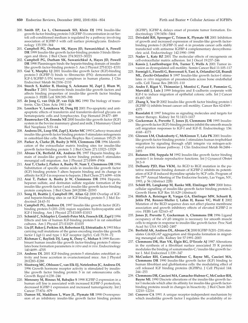
200. Smith EP, Lu L, Chernausek SD, Klein DJ 1994 Insulin-likegrowth factor-binding protein-3 (IGFBP-3) concentration in rat Ser-toli cell-conditioned medium is regulated by a pathway involvingassociation of IGFBP-3 with cell surface proteoglycans. Endocri-nology 135:359–364
201. Campbell PG, Durham SK, Hayes JD, Suwanichkul A, PowellDR 1999 Insulin-like growth factor-binding protein-3 binds fibrin-ogen and fibrin. J Biol Chem 274:30215–30221
202. Campbell PG, Durham SK, Suwanichkul A, Hayes JD, PowellDR 1998 Plasminogen binds the heparin-binding domain of insulin-like growth factor-binding protein-3. Am J Physiol 275:E321–E331
203. Gui Y, Murphy LJ 2001 Insulin-like growth factor (IGF)-bindingprotein-3 (IGFBP-3) binds to fibronectin (FN): demonstration ofIGF-I/IGFBP-3/FN ternary complexes in human plasma. J ClinEndocrinol Metab 86:2104–2110
204. Storch S, Kubler B, Honing S, Ackmann M, Zapf J, Blum W,Braulke T 2001 Transferrin binds insulin-like growth factors andaffects binding properties of insulin-like growth factor bindingprotein-3. FEBS Lett 509:395–398
205. de Jong G, van Dijk JP, van Eijk HG 1990 The biology of trans-ferrin. Clin Chim Acta 190:1–46
206. Lesnikov V, Lesnikova M, Deeg HJ 2001 Pro-apoptotic and anti-apoptotic effects of transferrin and transferrin-derived glycans onhematopoietic cells and lymphocytes. Exp Hematol 29:477–489
207. Baumrucker CR, Erondu NE 2000 Insulin-like growth factor (IGF)system in the bovine mammary gland and milk. J Mammary GlandBiol Neoplasia 5:53–64
208. Andress DL, Loop SM, Zapf J, Kiefer MC 1993 Carboxy-truncatedinsulin-like growth factor binding protein-5 stimulates mitogenesisin osteoblast-like cells. Biochem Biophys Res Commun 195:25–30
209. Parker A, Clarke JB, Busby Jr WH, Clemmons DR 1996 Identifi-cation of the extracellular matrix binding sites for insulin-likegrowth factor-binding protein 5. J Biol Chem 271:13523–13529
210. Abrass CK, Berfield AK, Andress DL 1997 Heparin binding do-main of insulin-like growth factor binding protein-5 stimulatesmesangial cell migration. Am J Physiol 273:F899–F906
211. Arai T, Clarke J, Parker A, Busby W, Nam T, Clemmons DR 1996Substitution of specific amino acids in insulin-like growth factor(IGF) binding protein 5 alters heparin binding and its change inaffinity for IGF-I in response to heparin. J Biol Chem 271:6099–6106
212. Arai T, Parker A, Busby Jr W, Clemmons DR 1994 Heparin,heparan sulfate, and dermatan sulfate regulate formation of theinsulin-like growth factor-I and insulin-like growth factor-bindingprotein complexes. J Biol Chem 269:20388–20393
213. Song H, Beattie J, Campbell IW, Allan GJ 2000 Overlap of IGF-and heparin-binding sites in rat IGF-binding protein-5. J Mol En-docrinol 24:43–51
214. Campbell PG, Andress DL 1997 Insulin-like growth factor (IGF)-binding protein-5-(201–218) region regulates hydroxyapatite andIGF-I binding. Am J Physiol 273:E1005–E1013
215. Schmid C, Schlapfer I, Gosteli-Peter MA, Froesch ER, Zapf J 1996Effects and fate of human IGF-binding protein-5 in rat osteoblastcultures. Am J Physiol 271:E1029–E1035
216. Liu JP, Baker J, Perkins AS, Robertson EJ, Efstratiadis A 1993 Micecarrying null mutations of the genes encoding insulin-like growthfactor I (igf-1) and type 1 IGF receptor (igf1r). Cell 75:59–72
217. Richman C, Baylink DJ, Lang K, Dony C, Mohan S 1999 Recom-binant human insulin-like growth factor-binding protein-5 stimu-lates bone formation parameters in vitro and in vivo. Endocrinology140:4699–4705
218. Andress DL 2001 IGF-binding protein-5 stimulates osteoblast ac-tivity and bone accretion in ovariectomized mice. Am J Physiol28:E283–E288
219. Slootweg MC, Ohlsson C, van Elk EJ, Netelenbos JC, Andress DL1996 Growth hormone receptor activity is stimulated by insulin-like growth factor binding protein 5 in rat osteosarcoma cells.Growth Regul 6:238–246
220. Menouny M, Binoux M, Babajko S 1998 IGFBP-2 expression in ahuman cell line is associated with increased IGFBP-3 proteolysis,decreased IGFBP-1 expression and increased tumorigenicity. Int JCancer 77:874–879
221. Damon SE, Maddison L, Ware JL, Plymate SR 1998 Overexpres-sion of an inhibitory insulin-like growth factor binding protein
(IGFBP), IGFBP-4, delays onset of prostate tumor formation. En-docrinology 139:3456–3464
222. Drivdahl RH, Sprenger C, Trimm K, Plymate SR 2001 Inhibitionof growth and increased expression of insulin-like growth factor-binding protein-3 (IGFBP-3) and -6 in prostate cancer cells stablytransfected with antisense IGFBP-4 complementary deoxyribonu-cleic acid. Endocrinology 142:1990–1998
223. Nadav L, Katz BZ 2001 The molecular effects of oncogenesis oncell-extracellular matrix adhesion. Int J Oncol 19:237–246
224. Kassis J, Lauffenburger DA, Turner T, Wells A 2001 Tumor in-vasion as dysregulated cell motility. Semin Cancer Biol 11:105–117
225. Formigli L, Fiorelli G, Benvenuti S, Tani A, Orlandini GE, BrandiML, Zecchi-Orlandini S 1997 Insulin-like growth factor-I stimu-lates in vitro migration of preosteoclasts across bone endothelialcells. Cell Tissue Res 288:101–110
226. Andre F, Rigot V, Thimonier J, Montixi C, Parat F, Pommier G,Marvaldi J, Luis J 1999 Integrins and E-cadherin cooperate withIGF-I to induce migration of epithelial colonic cells. Int J Cancer83:497–505
227. Zhang X, Yee D 2002 Insulin-like growth factor binding protein-1(IGFBP-1) inhibits breast cancer cell motility. Cancer Res 62:4369–4375
228. Ruoslahti E 1997 Integrins as signaling molecules and targets fortumor therapy. Kidney Int 51:1413–1417
229. Gockerman A, Prevette T, Jones JI, Clemmons DR 1995 Insulin-like growth factor (IGF)-binding proteins inhibit the smooth musclecell migration responses to IGF-I and IGF-II. Endocrinology 136:4168–4173
230. Gleeson LM, Chakraborty C, McKinnon T, Lala PK 2001 Insulin-like growth factor-binding protein 1 stimulates human trophoblastmigration by signaling through �5�1 integrin via mitogen-acti-vated protein kinase pathway. J Clin Endocrinol Metab 86:2484–2493
231. Rutanen EM, Seppala M 1992 Insulin-like growth factor bindingprotein-1 in female reproductive functions. Int J Gynaecol Obstet39:3–9
232. Delhanty PJD, Han VKM, An RGD to RGE mutation in the pu-tative membrane binding domain of IGFBP-2 inhibits its potenti-ation of IGF-II induced thymidine uptake by SCP cells. Program ofthe 75th Annual Meeting of The Endocrine Society, Las Vegas, NV,1993, p 56 (Abstract 22B)
233. Schutt BS, Langkamp M, Ranke MB, Elmlinger MW 2000 Intra-cellular signalling of insulin-like growth factor binding protein-2.Growth Horm IGF Res 10:A29 (Abstract)
234. Hoflich A, Reisinger R, Vargas GA, Elmlinger MW, Schuett B,Jehle PM, Renner-Muller I, Lahm H, Russo VC, Wolf E 2002Mutation of the RGD sequence does not affect plasma membraneassociation and growth inhibitory effects of elevated IGFBP-2 invivo. FEBS Lett 523:63–67
235. Jones JI, Prevette T, Gockerman A, Clemmons DR 1996 Ligandoccupancy of the �V-�3 integrin is necessary for smooth musclecells to migrate in response to insulin-like growth factor. Proc NatlAcad Sci USA 93:2482–2487
236. Berfield AK, Andress DL, Abrass CK 2000 IGFBP-5(201–218) stim-ulates Cdc42GAP aggregation and filopodia formation in migrat-ing mesangial cells. Kidney Int 57:1991–2003
237. Clemmons DR, Han VK, Elgin RG, D’Ercole AJ 1987 Alterationsin the synthesis of a fibroblast surface associated 35 K proteinmodulates the binding of somatomedin-C/insulin-like growth fac-tor I. Mol Endocrinol 1:339–347
238. McCusker RH, Camacho-Hubner C, Bayne ML, Cascieri MA,Clemmons DR 1990 Insulin-like growth factor (IGF) binding tohuman fibroblast and glioblastoma cells: the modulating effect ofcell released IGF binding proteins (IGFBPs). J Cell Physiol 144:244–253
239. Clemmons DR, Cascieri MA, Camacho-Hubner C, McCusker RH,Bayne ML 1990 Discrete alterations of the insulin-like growth fac-tor I molecule which alter its affinity for insulin-like growth factor-binding proteins result in changes in bioactivity. J Biol Chem 265:12210–12216
240. Conover CA 1991 A unique receptor-independent mechanism bywhich insulinlike growth factor I regulates the availability of in-
850 Endocrine Reviews, December 2002, 23(6):824–854 Firth and Baxter • Cellular Actions of IGFBPs
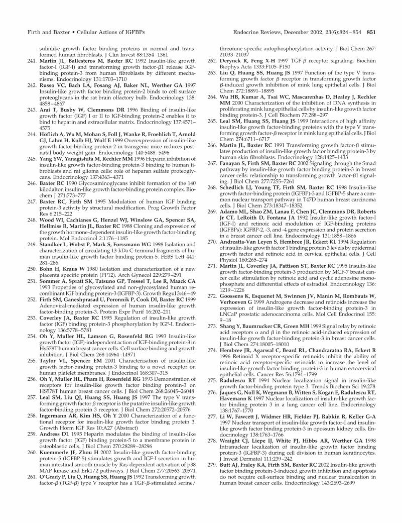
sulinlike growth factor binding proteins in normal and trans-formed human fibroblasts. J Clin Invest 88:1354–1361
241. Martin JL, Ballesteros M, Baxter RC 1992 Insulin-like growthfactor-I (IGF-I) and transforming growth factor-�1 release IGF-binding protein-3 from human fibroblasts by different mecha-nisms. Endocrinology 131:1703–1710
242. Russo VC, Bach LA, Fosang AJ, Baker NL, Werther GA 1997Insulin-like growth factor binding protein-2 binds to cell surfaceproteoglycans in the rat brain olfactory bulb. Endocrinology 138:4858–4867
243. Arai T, Busby W, Clemmons DR 1996 Binding of insulin-likegrowth factor (IGF) I or II to IGF-binding protein-2 enables it tobind to heparin and extracellular matrix. Endocrinology 137:4571–4575
244. Hoflich A, Wu M, Mohan S, Foll J, Wanke R, Froehlich T, ArnoldGJ, Lahm H, Kolb HJ, Wolf E 1999 Overexpression of insulin-likegrowth factor-binding protein-2 in transgenic mice reduces post-natal body weight gain. Endocrinology 140:5488–5496
245. Yang YW, Yanagishita M, Rechler MM 1996 Heparin inhibition ofinsulin-like growth factor-binding protein-3 binding to human fi-broblasts and rat glioma cells: role of heparan sulfate proteogly-cans. Endocrinology 137:4363–4371
246. Baxter RC 1990 Glycosaminoglycans inhibit formation of the 140kilodalton insulin-like growth factor-binding protein complex. Bio-chem J 271:773–777
247. Baxter RC, Firth SM 1995 Modulation of human IGF bindingprotein-3 activity by structural modification. Prog Growth FactorRes 6:215–222
248. Wood WI, Cachianes G, Henzel WJ, Winslow GA, Spencer SA,Hellmiss R, Martin JL, Baxter RC 1988 Cloning and expression ofthe growth hormone-dependent insulin-like growth factor-bindingprotein. Mol Endocrinol 2:1176–1185
249. Standker L, Wobst P, Mark S, Forssmann WG 1998 Isolation andcharacterization of circulating 13-kDa C-terminal fragments of hu-man insulin-like growth factor binding protein-5. FEBS Lett 441:281–286
250. Bohn H, Kraus W 1980 Isolation and characterization of a newplacenta specific protein (PP12). Arch Gynecol 229:279–291
251. Sommer A, Spratt SK, Tatsuno GP, Tressel T, Lee R, Maack CA1993 Properties of glycosylated and non-glycosylated human re-combinant IGF binding protein-3 (IGFBP-3). Growth Regul 3:46–49
252. Firth SM, Ganeshprasad U, Poronnik P, Cook DI, Baxter RC 1999Adenoviral-mediated expression of human insulin-like growthfactor-binding protein-3. Protein Expr Purif 16:202–211
253. Coverley JA, Baxter RC 1995 Regulation of insulin-like growthfactor (IGF) binding protein-3 phosphorylation by IGF-I. Endocri-nology 136:5778–5781
254. Oh Y, Muller HL, Lamson G, Rosenfeld RG 1993 Insulin-likegrowth factor (IGF)-independent action of IGF-binding protein-3 inHs578T human breast cancer cells. Cell surface binding and growthinhibition. J Biol Chem 268:14964–14971
255. Taylor VL, Spencer EM 2001 Characterisation of insulin-likegrowth factor-binding protein-3 binding to a novel receptor onhuman platelet membranes. J Endocrinol 168:307–315
256. Oh Y, Muller HL, Pham H, Rosenfeld RG 1993 Demonstration ofreceptors for insulin-like growth factor binding protein-3 onHS578T human breast cancer cells. J Biol Chem 268:26045–26048
257. Leal SM, Liu QJ, Huang SS, Huang JS 1997 The type V trans-forming growth factor � receptor is the putative insulin-like growthfactor-binding protein 3 receptor. J Biol Chem 272:20572–20576
258. Ingermann AR, Kim HS, Oh Y 2000 Characterization of a func-tional receptor for insulin-like growth factor binding protein 3.Growth Horm IGF Res 10:A27 (Abstract)
259. Andress DL 1995 Heparin modulates the binding of insulin-likegrowth factor (IGF) binding protein-5 to a membrane protein inosteoblastic cells. J Biol Chem 270:28289–28296
260. Kuemmerle JF, Zhou H 2002 Insulin-like growth factor-bindingprotein-5 (IGFBP-5) stimulates growth and IGF-I secretion in hu-man intestinal smooth muscle by Ras-dependent activation of p38MAP kinase and Erk1/2 pathways. J Biol Chem 277:20563–20571
261. O’Grady P, Liu Q, Huang SS, Huang JS 1992 Transforming growthfactor-� (TGF-�) type V receptor has a TGF-�-stimulated serine/
threonine-specific autophosphorylation activity. J Biol Chem 267:21033–21037
262. Derynck R, Feng X-H 1997 TGF-� receptor signaling. BiochimBiophys Acta 1333:F105–F150
263. Liu Q, Huang SS, Huang JS 1997 Function of the type V trans-forming growth factor � receptor in transforming growth factor�-induced growth inhibition of mink lung epithelial cells. J BiolChem 272:18891–18895
264. Wu HB, Kumar A, Tsai WC, Mascarenhas D, Healey J, RechlerMM 2000 Characterization of the inhibition of DNA synthesis inproliferating mink lung epithelial cells by insulin-like growth factorbinding protein-3. J Cell Biochem 77:288–297
265. Leal SM, Huang SS, Huang JS 1999 Interactions of high affinityinsulin-like growth factor-binding proteins with the type V trans-forming growth factor-� receptor in mink lung epithelial cells. J BiolChem 274:6711–6717
266. Martin JL, Baxter RC 1991 Transforming growth factor-� stimu-lates production of insulin-like growth factor binding protein-3 byhuman skin fibroblasts. Endocrinology 128:1425–1433
267. Fanayan S, Firth SM, Baxter RC 2002 Signaling through the Smadpathway by insulin-like growth factor binding protein-3 in breastcancer cells: relationship to transforming growth factor-�1 signal-ing. J Biol Chem 277:7255–7261
268. Schedlich LJ, Young TF, Firth SM, Baxter RC 1998 Insulin-likegrowth factor-binding protein (IGFBP)-3 and IGFBP-5 share a com-mon nuclear transport pathway in T47D human breast carcinomacells. J Biol Chem 273:18347–18352
269. Adamo ML, Shao ZM, Lanau F, Chen JC, Clemmons DR, RobertsJr CT, LeRoith D, Fontana JA 1992 Insulin-like growth factor-I(IGF-I) and retinoic acid modulation of IGF-binding proteins(IGFBPs): IGFBP-2, -3, and -4 gene expression and protein secretionin a breast cancer cell line. Endocrinology 131:1858–1866
270. Andreatta-Van Leyen S, Hembree JR, Eckert RL 1994 Regulationof insulin-like growth factor 1 binding protein 3 levels by epidermalgrowth factor and retinoic acid in cervical epithelial cells. J CellPhysiol 160:265–274
271. Martin JL, Coverley JA, Pattison ST, Baxter RC 1995 Insulin-likegrowth factor-binding protein-3 production by MCF-7 breast can-cer cells: stimulation by retinoic acid and cyclic adenosine mono-phosphate and differential effects of estradiol. Endocrinology 136:1219–1226
272. Goossens K, Esquenet M, Swinnen JV, Manin M, Rombauts W,Verhoeven G 1999 Androgens decrease and retinoids increase theexpression of insulin-like growth factor-binding protein-3 inLNCaP prostatic adenocarcinoma cells. Mol Cell Endocrinol 155:9–18
273. Shang Y, Baumrucker CR, Green MH 1999 Signal relay by retinoicacid receptors � and � in the retinoic acid-induced expression ofinsulin-like growth factor-binding protein-3 in breast cancer cells.J Biol Chem 274:18005–18010
274. Hembree JR, Agarwal C, Beard RL, Chandraratna RA, Eckert R1996 Retinoid X receptor-specific retinoids inhibit the ability ofretinoic acid receptor-specific retinoids to increase the level ofinsulin-like growth factor binding protein-3 in human ectocervicalepithelial cells. Cancer Res 56:1794–1799
275. Radulescu RT 1994 Nuclear localization signal in insulin-likegrowth factor-binding protein type 3. Trends Biochem Sci 19:278
276. Jaques G, Noll K, Wegmann B, Witten S, Kogan E, Radulescu RT,Havemann K 1997 Nuclear localization of insulin-like growth fac-tor binding protein 3 in a lung cancer cell line. Endocrinology138:1767–1770
277. Li W, Fawcett J, Widmer HR, Fielder PJ, Rabkin R, Keller G-A1997 Nuclear transport of insulin-like growth factor-I and insulin-like growth factor binding protein-3 in opossum kidney cells. En-docrinology 138:1763–1766
278. Wraight CJ, Liepe IJ, White PJ, Hibbs AR, Werther GA 1998Intranuclear localization of insulin-like growth factor bindingprotein-3 (IGFBP-3) during cell division in human keratinocytes.J Invest Dermatol 111:239–242
279. Butt AJ, Fraley KA, Firth SM, Baxter RC 2002 Insulin-like growthfactor binding protein-3-induced growth inhibition and apoptosisdo not require cell-surface binding and nuclear translocation inhuman breast cancer cells. Endocrinology 143:2693–2699
Firth and Baxter • Cellular Actions of IGFBPs Endocrine Reviews, December 2002, 23(6):824–854 851
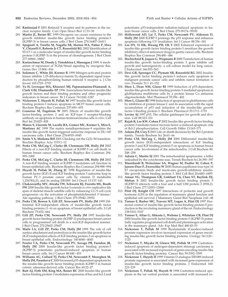
280. Rastinejad F 2001 Retinoid X receptor and its partners in the nu-clear receptor family. Curr Opin Struct Biol 11:33–38
281. Martin JL, Baxter RC 1999 Oncogenic ras causes resistance to thegrowth inhibitor insulin-like growth factor binding protein-3(IGFBP-3) in breast cancer cells. J Biol Chem 274:16407–16411
282. Spagnoli A, Torello M, Nagalla SR, Horton WA, Pattee P, HwaV, Chiarelli F, Roberts Jr CT, Rosenfeld RG 2002 Identification ofSTAT-1 as a molecular target of insulin-like growth factor bindingprotein-3 (IGFBP-3) in the process of chondrogenesis. J Biol Chem277:18860–18867
283. Kretzschmar M, Doody J, Timokhina I, Massague J 1999 A mech-anism of repression of TGF�/Smad signaling by oncogenic Ras.Genes Dev 13:804–816
284. Solomon C, White JH, Kremer R 1999 Mitogen-activated proteinkinase inhibits 1,25-dihydroxyvitamin D3-dependent signal trans-duction by phosphorylating human retinoid X receptor �. J ClinInvest 103:1729–1735
285. Yu H, Levesque MA, Khosravi MJ, Papanastasiou-Diamandi A,Clark GM, Diamandis EP 1996 Associations between insulin-likegrowth factors and their binding proteins and other prognosticindicators in breast cancer. Br J Cancer 74:1242–1247
286. Nickerson T, Huynh H, Pollak M 1997 Insulin-like growth factorbinding protein-3 induces apoptosis in MCF7 breast cancer cells.Biochem Biophys Res Commun 237:690–693
287. Granerus M, Engstrom W 2001 Effects of insulin-like growthfactor-binding protein 2 and an IGF-type I receptor-blockingantibody on apoptosis in human teratocarcinoma cells in vitro. CellBiol Int 25:825–828
288. Manes S, Llorente M, Lacalle RA, Gomez MC, Kremer L, Mira E,Martinez AC 1999 The matrix metalloproteinase-9 regulates theinsulin-like growth factor-triggered autocrine response in DU-145carcinoma cells. J Biol Chem 274:6935–6945
289. Smits VA, Medema RH 2001 Checking out the G(2)/M transition.Biochim Biophys Acta 1519:1–12
290. Perks CM, McCaig C, Clarke JB, Clemmons DR, Holly JM 2002Effects of a non-IGF binding mutant of IGFBP-5 on cell death inhuman breast cancer cells. Biochem Biophys Res Commun 294:995–1000
291. Perks CM, McCaig C, Clarke JB, Clemmons DR, Holly JM 2002A non-IGF binding mutant of IGFBP-3 modulates cell function inbreast epithelial cells. Biochem Biophys Res Commun 294:988–994
292. Huynh H, Pollak M, Zhang JC 1998 Regulation of insulin-likegrowth factor (IGF) II and IGF binding protein 3 autocrine loop inhuman PC-3 prostate cancer cells by vitamin D metabolite1,25(OH)2D3 and its analog EB1089. Int J Oncol 13:137–143
293. Chakravarthy MV, Abraha TW, Schwartz RJ, Fiorotto ML, BoothFW 2000 Insulin-like growth factor-I extends in vitro replicative lifespan of skeletal muscle satellite cells by enhancing G1/S cell cycleprogression via the activation of phosphatidylinositol 3�-kinase/Akt signaling pathway. J Biol Chem 275:35942–35952
294. Perks CM, Bowen S, Gill ZP, Newcomb PV, Holly JM 1999 Dif-ferential IGF-independent effects of insulin-like growth factorbinding proteins (1–6) on apoptosis of breast epithelial cells. J CellBiochem 75:652–664
295. Gill ZP, Perks CM, Newcomb PV, Holly JM 1997 Insulin-likegrowth factor-binding protein (IGFBP-3) predisposes breast cancercells to programmed cell death in a non-IGF-dependent manner.J Biol Chem 272:25602–25607
296. Maile LA, Gill ZP, Perks CM, Holly JM 1999 The role of cellsurface attachment and proteolysis in the insulin-like growth factor(IGF)-independent effects of IGF-binding protein-3 on apoptosis inbreast epithelial cells. Endocrinology 140:4040–4045
297. Fowler CA, Perks CM, Newcomb PV, Savage PB, Farndon JR,Holly JM 2000 Insulin-like growth factor binding protein-3(IGFBP-3) potentiates paclitaxel-induced apoptosis in humanbreast cancer cells. Int J Cancer 88:448–453
298. Williams AC, Collard TJ, Perks CM, Newcomb P, Moorghen M,Holly JM, Paraskeva C 2000 Increased p53-dependent apoptosis bythe insulin-like growth factor binding protein IGFBP-3 in humancolonic adenoma-derived cells. Cancer Res 60:22–27
299. Butt AJ, Firth SM, King MA, Baxter RC 2000 Insulin-like growthfactor-binding protein-3 modulates expression of bax and bcl-2 and
potentiates p53-independent radiation-induced apoptosis in hu-man breast cancer cells. J Biol Chem 275:39174–39181
300. Hollowood AD, Lai T, Perks CM, Newcomb PV, Alderson D,Holly JM 2000 IGFBP-3 prolongs the p53 response and enhancesapoptosis following UV irradiation. Int J Cancer 88:336–341
301. Lee DY, Yi HK, Hwang PH, Oh Y 2002 Enhanced expression ofinsulin-like growth factor binding protein-3 sensitizes the growthinhibitory effect of anticancer drugs in gastric cancer cells. BiochemBiophys Res Commun 294:480–486
302. Hochscheid R, Jaques G, Wegmann B 2000 Transfection of humaninsulin-like growth factor-binding protein 3 gene inhibits cellgrowth and tumorigenicity: a cell culture model for lung cancer.J Endocrinol 166:553–563
303. Devi GR, Sprenger CC, Plymate SR, Rosenfeld RG 2002 Insulin-like growth factor binding protein-3 induces early apoptosis inmalignant prostate cancer cells and inhibits tumor formation invivo. Prostate 51:1 41–152
304. Shen L, Dean NM, Glazer RI 1999 Induction of p53-dependent,insulin-like growth factor-binding protein-3-mediated apoptosis inglioblastoma multiforme cells by a protein kinase C� antisenseoligonucleotide. Mol Pharmacol 55:396–402
305. Shen L, Glazer RI 1998 Induction of apoptosis in glioblastoma cellsby inhibition of protein kinase C and its association with the rapidaccumulation of p53 and induction of the insulin-like growthfactor-1-binding protein-3. Biochem Pharmacol 55:1711–1719
306. Levine AJ 1997 p53, The cellular gatekeeper for growth and divi-sion. Cell 88:323–331
307. Rajah R, Lee KW, Cohen P 2002 Insulin-like growth factor bindingprotein-3 mediates tumor necrosis factor-�-induced apoptosis: roleof Bcl-2 phosphorylation. Cell Growth Differ 13:163–171
308. Adams JM, Cory S 2001 Life-or-death decisions by the Bcl-2 proteinfamily. Trends Biochem Sci 26:61–66
309. Perks CM, McCaig C, Holly JM 2000 Differential insulin-likegrowth factor (IGF)-independent interactions of IGF bindingprotein-3 and IGF binding protein-5 on apoptosis in human breastcancer cells. Involvement of the mitochondria. J Cell Biochem 80:248–258
310. Adrain C, Martin SJ 2001 The mitochondrial apoptosome: a killerunleashed by the cytochrome seas. Trends Biochem Sci 26:390–397
311. Mannhardt B, Weinzimer SA, Wagner M, Fiedler M, Cohen P,Jansen-Durr P, Zwerschke W 2000 Human papillomavirus type 16E7 oncoprotein binds and inactivates growth-inhibitory insulin-like growth factor binding protein 3. Mol Cell Biol 20:6483–6495
312. Amaar YG, Thompson GR, Linkhart TA, Chen ST, Baylink DJ,Mohan S 2002 Insulin-like growth factor binding protein 5(IGFBP-5) interacts with a four and a half LIM protein 2 (FHL2).J Biol Chem 277:12053–12060
313. Flint DJ, Knight CH 1997 Interactions of prolactin and growthhormone (GH) in the regulation of mammary gland function andepithelial cell survival. J Mammary Gland Biol Neoplasia 2:41–48
314. Tonner E, Barber MC, Travers MT, Logan A, Flint DJ 1997 Hor-monal control of insulin-like growth factor-binding protein-5 pro-duction in the involuting mammary gland of the rat. Endocrinology138:5101–5107
315. Tonner E, Allan G, Shkreta L, Webster J, Whitelaw CB, Flint DJ2000 Insulin-like growth factor binding protein-5 (IGFBP-5) poten-tially regulates programmed cell death and plasminogen activationin the mammary gland. Adv Exp Med Biol 480:45–53
316. Nickerson T, Pollak M 1999 Bicalutamide (Casodex)-inducedprostate regression involves increased expression of genes encod-ing insulin-like growth factor binding proteins. Urology 54:1120–1125
317. Nickerson T, Miyake H, Gleave ME, Pollak M 1999 Castration-induced apoptosis of androgen-dependent shionogi carcinoma isassociated with increased expression of genes encoding insulin-likegrowth factor-binding proteins. Cancer Res 59:3392–3395
318. Nickerson T, Huynh H 1999 Vitamin D analogue EB1089-inducedprostate regression is associated with increased gene expression ofinsulin-like growth factor binding proteins. J Endocrinol 160:223–229
319. Nickerson T, Pollak M, Huynh H 1998 Castration-induced apo-ptosis in the rat ventral prostate is associated with increased ex-
852 Endocrine Reviews, December 2002, 23(6):824–854 Firth and Baxter • Cellular Actions of IGFBPs
pression of genes encoding insulin-like growth factor binding pro-teins 2, 3, 4, and 5. Endocrinology 139:807–810
320. Miyake H, Pollak M, Gleave ME 2000 Castration-induced up-regulation of insulin-like growth factor binding protein-5 poten-tiates insulin-like growth factor-I activity and accelerates progres-sion to androgen independence in prostate cancer models. CancerRes 60:3058–3064
321. Stewart CE, James PL, Fant ME, Rotwein P 1996 Overexpressionof insulin-like growth factor-II induces accelerated myoblast dif-ferentiation. J Cell Physiol 169:23–32
322. Stewart CE, Rotwein P 1996 Insulin-like growth factor-II is anautocrine survival factor for differentiating myoblasts. J Biol Chem271:11330–11338
323. Meadows KA, Holly JM, Stewart CE 2000 Tumor necrosis factor-�-induced apoptosis is associated with suppression of insulin-likegrowth factor binding protein-5 secretion in differentiating murineskeletal myoblasts. J Cell Physiol 183:330–337
324. Guthrie HD, Grimes RW, Hammond JM 1995 Changes in insulin-like growth factor-binding protein-2 and -3 in follicular fluid dur-ing atresia of follicles grown after ovulation in pigs. J Reprod Fertil104:225–230
325. Wandji SA, Wood TL, Crawford J, Levison SW, Hammond JM1998 Expression of mouse ovarian insulin growth factor systemcomponents during follicular development and atresia. Endocri-nology 139:5205–5214
326. Fortune JE, Rivera GM, Evans AC, Turzillo AM 2001 Differenti-ation of dominant vs. subordinate follicles in cattle. Biol Reprod65:648–654
327. Mazerbourg S, Zapf J, Bar RS, Brigstock DR, Monget P 2000Insulin-like growth factor (IGF)-binding protein-4 proteolytic deg-radation in bovine, equine, and porcine preovulatory follicles: reg-ulation by IGFs and heparin-binding domain-containing peptides.Biol Reprod 63:390–400
328. Adashi EY, Resnick CE, Payne DW, Rosenfeld RG, MatsumotoT, Hunter MK, Gargosky SE, Zhou J, Bondy CA 1997 The mouseintraovarian insulin-like growth factor I system: departures fromthe rat paradigm. Endocrinology 138:3881–3890
329. Resnick CE, Fielder PJ, Rosenfeld RG, Adashi EY 1998 Charac-terization and hormonal regulation of a rat ovarian insulin-likegrowth factor binding protein-5 endopeptidase: an FSH-induciblegranulosa cell-derived metalloprotease. Endocrinology 139:1249–1257
330. Zhou J, Dsupin BA, Giudice LC, Bondy CA 1994 Insulin-likegrowth factor system gene expression in human endometriumduring the menstrual cycle. J Clin Endocrinol Metab 79:1723–1734
331. Besnard V, Corroyer S, Trugnan G, Chadelat K, Nabeyrat E,Cazals V, Clement A 2001 Distinct patterns of insulin-like growthfactor binding protein (IGFBP)-2 and IGFBP-3 expression in oxi-dant exposed lung epithelial cells. Biochim Biophys Acta 1538:47–58
332. Mouhieddine OB, Cazals V, Kuto E, Le Bouc Y, Clement A 1996Glucocorticoid-induced growth arrest of lung alveolar epithelialcells is associated with increased production of insulin-like growthfactor binding protein-2. Endocrinology 137:287–295
333. Mouhieddine OB, Cazals V, Maitre B, Le Bouc Y, Chadelat K,Clement A 1994 Insulin-like growth factor-II (IGF-II), type 2 IGFreceptor, and IGF-binding protein-2 gene expression in rat lungalveolar epithelial cells: relation to proliferation. Endocrinology135:83–91
334. Cazals V, Mouhieddine B, Maitre B, Le Bouc Y, Chadelat K, BrodyJS, Clement A 1994 Insulin-like growth factors, their binding pro-teins, and transforming growth factor-� 1 in oxidant-arrested lungalveolar epithelial cells. J Biol Chem 269:14111–14117
335. Sueoka N, Lee HY, Wiehle S, Cristiano RJ, Fang B, Ji L, Roth JA,Hong WK, Cohen P, Kurie JM 2000 Insulin-like growth factorbinding protein-6 activates programmed cell death in non-smallcell lung cancer cells. Oncogene 19:4432–4436
336. Schmid C, Keller C, Gosteli-Peter M, Zapf J 1999 Mitogenic andantiapoptotic effects of insulin-like growth factor binding protein-6in the human osteoblastic osteosarcoma cell line Saos-2/B-10. Bio-chem Biophys Res Commun 263:786–789
337. Wood TL, Rogler LE, Czick ME, Schuller AG, Pintar JE 2000Selective alterations in organ sizes in mice with a targeted disrup-
tion of the insulin-like growth factor binding protein-2 gene. MolEndocrinol 14:1472–1482
338. Pintar JE, Hoang B, Ning T, Schuller AG, Single and multipleknockouts of the IGFBPs. Program of the 83rd Meeting of TheEndocrine Society, Toronto, Canada, 2001, p 44 (Abstract S33-1)
339. Dai Z, Xing Y, Boney CM, Clemmons DR, D’Ercole AJ 1994Human insulin-like growth factor-binding protein-1 (hIGFBP-1) intransgenic mice: characterization and insights into the regulation ofIGFBP-1 expression. Endocrinology 135:1316–1327
340. Rajkumar K, Barron D, Lewitt MS, Murphy LJ 1995 Growthretardation and hyperglycemia in insulin-like growth factor bind-ing protein-1 transgenic mice. Endocrinology 136:4029–4034
341. Gay E, Seurin D, Babajko S, Doublier S, Cazillis M, Binoux M1997 Liver-specific expression of human insulin-like growth factorbinding protein-1 in transgenic mice: repercussions on reproduc-tion, ante- and perinatal mortality and postnatal growth. Endocri-nology 138:2937–2947
342. Crossey PA, Jones JS, Miell JP 2000 Dysregulation of the insulin/IGF binding protein-1 axis in transgenic mice is associated withhyperinsulinemia and glucose intolerance. Diabetes 49:457–465
343. Murphy LJ, Molnar P, Lu X, Huang H 1995 Expression of humaninsulin-like growth factor-binding protein-3 in transgenic mice. JMol Endocrinol 15:293–303
344. Neuenschwander S, Schwartz A, Wood TL, Roberts Jr CT, Hen-ninghausen L, LeRoith D 1996 Involution of the lactating mam-mary gland is inhibited by the IGF system in a transgenic mousemodel. J Clin Invest 97:2225–2232
345. Modric T, Silha JV, Shi Z, Gui Y, Suwanichkul A, Durham SK,Powell DR, Murphy LJ 2001 Phenotypic manifestations of insulin-like growth factor-binding protein-3 overexpression in transgenicmice. Endocrinology 142:1958–1967
346. Wang J, Niu W, Witte DP, Chernausek SD, Nikiforov YE, Cle-mens TL, Sharifi B, Strauch AR, Fagin JA 1998 Overexpression ofinsulin-like growth factor-binding protein-4 (IGFBP-4) in smoothmuscle cells of transgenic mice through a smooth muscle �-actin-IGFBP-4 fusion gene induces smooth muscle hypoplasia. Endocri-nology 139:2605–2614
347. Rajkumar K, Krsek M, Dheen ST, Murphy LJ 1996 Impairedglucose homeostasis in insulin-like growth factor binding protein-1transgenic mice. J Clin Invest 98:1818–1825
348. Yeoh SI, Baxter RC 1988 Metabolic regulation of the growth hor-mone independent insulin-like growth factor binding protein inhuman plasma. Acta Endocrinol (Copenh) 119:465–473
349. Ye P, Carson J, D’Ercole AJ 1995 In vivo actions of insulin-likegrowth factor-I (IGF-I) on brain myelination: studies of IGF-I andIGF binding protein-1 (IGFBP-1) transgenic mice. J Neurosci 15:7344–7356
350. Ni W, Rajkumar K, Nagy JI, Murphy LJ 1997 Impaired braindevelopment and reduced astrocyte response to injury in trans-genic mice expressing IGF binding protein-1. Brain Res 769:97–107
351. Doublier S, Duyckaerts C, Seurin D, Binoux M 2000 Impairedbrain development and hydrocephalus in a line of transgenic micewith liver-specific expression of human insulin-like growth factorbinding protein-1. Growth Horm IGF Res 10:267–274
352. Preece MA 1997 Making a rational diagnosis of growth-hormonedeficiency. J Pediatr 131:S61–S64
353. Marzullo P, Di Somma C, Pratt KL, Khosravi J, Diamandis A,Lombardi G, Colao A, Rosenfeld RG 2001 Usefulness of differentbiochemical markers of the insulin-like growth factor (IGF) familyin diagnosing growth hormone excess and deficiency in adults.J Clin Endocrinol Metab 86:3001–3008
354. Baxter RC 1996 The role of insulin-like growth factors and theirbinding proteins in tumor hypoglycemia. Horm Res 46:195–201
355. Lemne C, Brismar K 1998 Insulin-like growth factor bindingprotein-1 as a marker of the metabolic syndrome—a study in bor-derline hypertension. Blood Press 7:89–95
356. Hankinson SE, Willett WC, Colditz GA, Hunter DJ, Michaud DS,Deroo B, Rosner B, Speizer FE, Pollak M 1998 Circulating con-centrations of insulin-like growth factor-I and risk of breast cancer.Lancet 351:1393–1396
357. Chan JM, Stampfer MJ, Giovannucci E, Gann PH, Ma J, Wilkin-son P, Hennekens CH, Pollak M 1998 Plasma insulin-like growth
Firth and Baxter • Cellular Actions of IGFBPs Endocrine Reviews, December 2002, 23(6):824–854 853
factor-I and prostate cancer risk: a prospective study. Science 279:563–566
358. Yu H, Spitz MR, Mistry J, Gu J, Hong WK, Wu X 1999 Plasmalevels of insulin-like growth factor-I and lung cancer risk: a case-control analysis. J Natl Cancer Inst 91:151–156
359. Giovannucci E, Pollak MN, Platz EA, Willett WC, Stampfer MJ,Majeed N, Colditz GA, Speizer FE, Hankinson SE 2000 A pro-spective study of plasma insulin-like growth factor-1 and bindingprotein-3 and risk of colorectal neoplasia in women. Cancer Epi-demiol Biomarkers Prev 9:345–349
360. Kaaks R, Toniolo P, Akhmedkhanov A, Lukanova A, Biessy C,Dechaud H, Rinaldi S, Zeleniuch-Jacquotte A, Shore RE, RiboliE 2000 Serum C-peptide, insulin-like growth factor (IGF)-I, IGF-binding proteins, and colorectal cancer risk in women. J Natl Can-cer Inst 92:1592–1600
361. Li BD, Khosravi MJ, Berkel HJ, Diamandi A, Dayton MA, SmithM, Yu H 2001 Free insulin-like growth factor-I and breast cancerrisk. Int J Cancer 91:736–739
362. Lukanova A, Toniolo P, Akhmedkhanov A, Biessy C, Haley NJ,Shore RE, Riboli E, Rinaldi S, Kaaks R 2001 A prospective studyof insulin-like growth factor-I, IGF-binding proteins-1, -2 and -3and lung cancer risk in women. Int J Cancer 92:888–892
363. Wolk A, Mantzoros CS, Andersson SO, Bergstrom R, SignorelloLB, Lagiou P, Adami HO, Trichopoulos D 1998 Insulin-likegrowth factor 1 and prostate cancer risk: a population-based, case-control study. J Natl Cancer Inst 90:911–915
364. Bruning PF, Van Doorn J, Bonfrer JM, Van Noord PA, Korse CM,Linders TC, Hart AA 1995 Insulin-like growth-factor-binding pro-tein 3 is decreased in early-stage operable pre-menopausal breastcancer. Int J Cancer 62:266–270
365. Kaulsay KK, Ng EH, Ji CY, Ho GH, Aw TC, Lee KO 1999 SerumIGF-binding protein-6 and prostate specific antigen in breast can-cer. Eur J Endocrinol 140:164–168
366. Vadgama JV, Wu Y, Datta G, Khan H, Chillar R 1999 Plasmainsulin-like growth factor-I and serum IGF-binding protein 3 canbe associated with the progression of breast cancer, and predict therisk of recurrence and the probability of survival in African-Amer-ican and Hispanic women. Oncology 57:330–340
367. Holdaway IM, Mason BH, Lethaby AE, Singh V, Harman JE,MacCormick M, Civil ID 1999 Serum levels of insulin-like growthfactor binding protein-3 in benign and malignant breast disease.Aust NZ J Surg 69:495–500
368. Cohen P, Peehl DM, Stamey TA, Wilson KF, Clemmons DR,Rosenfeld RG 1993 Elevated levels of insulin-like growth factor-binding protein-2 in the serum of prostate cancer patients. J ClinEndocrinol Metab 76:1031–1035
369. Kanety H, Madjar Y, Dagan Y, Levi J, Papa MZ, Pariente C,Goldwasser B, Karasik A 1993 Serum insulin-like growth factor-binding protein-2 (IGFBP-2) is increased and IGFBP-3 is decreasedin patients with prostate cancer: correlation with serum prostate-specific antigen. J Clin Endocrinol Metab 77:229–233
370. Ho PJ, Baxter RC 1997 Insulin-like growth factor-binding protein-2in patients with prostate carcinoma and benign prostatic hyper-plasia. Clin Endocrinol (Oxf) 46:145–154
371. Flyvbjerg A, Mogensen O, Mogensen B, Nielsen OS 1997 Ele-vated serum insulin-like growth factor-binding protein 2 (IGFBP-2)and decreased IGFBP-3 in epithelial ovarian cancer: correlationwith cancer antigen 125 and tumor-associated trypsin inhibitor.J Clin Endocrinol Metab 82:2308–2313
372. Rocha RL, Hilsenbeck SG, Jackson JG, VanDenBerg CL, Weng C,Lee AV, Yee D 1997 Insulin-like growth factor binding protein-3and insulin receptor substrate-1 in breast cancer: correlation withclinical parameters and disease-free survival. Clin Cancer Res3:103–109
373. Karasik A, Menczer J, Pariente C, Kanety H 1994 Insulin-likegrowth factor-I (IGF-I) and IGF-binding protein-2 are increased incyst fluids of epithelial ovarian cancer. J Clin Endocrinol Metab78:271–276
374. Mishra L, Bass B, Ooi BS, Sidawy A, Korman L 1998 Role ofinsulin-like growth factor-I (IGF-I) receptor, IGF-I, and IGF bindingprotein-2 in human colorectal cancers. Growth Horm IGF Res8:473–479
375. Brinkman A, Kortleve DJ, Schuller AG, Zwarthoff EC, Drop SL
1991 Site-directed mutagenesis of the N-terminal region of IGFbinding protein 1; analysis of IGF binding capability. FEBS Lett291:264–268
376. Hobba GD, Lothgren A, Holmberg E, Forbes BE, Francis GL,Wallace JC 1998 Alanine screening mutagenesis establishes ty-rosine 60 of bovine insulin-like growth factor binding protein-2 asa determinant of insulin-like growth factor binding. J Biol Chem273:19691–19698
377. Vorwerk P, Yamanaka Y, Spagnoli A, Oh Y, Rosenfeld RG 1998Insulin and IGF binding by IGFBP-3 fragments derived from pro-teolysis, baculovirus expression and normal human urine. J ClinEndocrinol Metab 83:1392–1395
378. Brinkman A, Kortleve DJ, Zwarthoff EC, Drop SL 1991 Mutationsin the C-terminal part of insulin-like growth factor (IGF)-bindingprotein-1 result in dimer formation and loss of IGF binding ca-pacity. Mol Endocrinol 5:987–994
379. Booth BA, Boes M, Bar RS 1996 IGFBP-3 proteolysis by plasmin,thrombin, serum-heparin binding, IGF binding, and structure offragments. Am J Physiol 271:E465–E470
380. Lalou C, Sawamura S, Segovia B, Ogawa Y, Binoux M 1997 Pro-teolytic fragments of insulin-like growth factor binding protein-3:N-terminal sequences and relationships between structure and bi-ological activity. C R Acad Sci III 320:621–628
381. Fielder PJ, Rosenfeld RG, Graves HCB, Grandbois K, Maack CA,Sawamura S, Ogawa Y, Sommer A, Cohen P 1994 Biochemicalanalysis of prostate specific antigen-proteolyzed insulin-likegrowth factor binding protein-3. Growth Regul 1:164–172
382. Okabe E, Kajihara J, Usami Y, Hirano K 1999 The cleavage sitespecificity of human prostate specific antigen for insulin-likegrowth factor binding protein-3. FEBS Lett 447:87–90
383. Fowlkes JL, Enghild JJ, Suzuki K, Nagase H 1994 Matrix metal-loproteinases degrade insulin-like growth factor-binding protein-3in dermal fibroblast cultures. J Biol Chem 269:25742–25746
384. Zwad O, Kubler B, Roth W, Scharf JG, Saftig P, Peters C, BraulkeT 2002 Decreased intracellular degradation of insulin-like growthfactor binding protein-3 in cathepsin L-deficient fibroblasts. FEBSLett 510:211–215
385. Zheng B, Clarke JB, Busby WH, Duan C, Clemmons DR 1998Insulin-like growth factor-binding protein-5 is cleaved by physi-ological concentrations of thrombin. Endocrinology 139:1708–1714
386. Xu W, Murphy LJ, Interaction of IGFBP-3 with latent transforminggrowth factor-� binding protein-1 identified using the yeast two-hybrid system. Program of the 80th Annual Meeting of The Endo-crine Society, New Orleans, LA, 1998, p 313 (Abstract P2-293)
387. Liu B, Gibson TB, Collett-Solberg PF, Zhao H, Cerri RW, Mas-carenhas D, Cohen P, Type 1� collagen is an IGFBP-3 bindingprotein. Program of the 81st Annual Meeting of The EndocrineSociety, San Diego, CA, 1999, p 405 (Abstract P2-586)
388. Nam T, Moralez A, Clemmons D 2002 Vitronectin binding to IGFbinding protein-5 (IGFBP-5) alters IGFBP-5 modulation of IGF-Iactions. Endocrinology 143:30–36
389. Falquet L, Pagni M, Bucher P, Hulo N, Sigrist CJ, Hofmann K,Bairoch A 2002 The PROSITE database, its status in 2002. NucleicAcids Res 30:235–238
390. Hansen JE, Lund O, Tolstrup N, Gooley AA, Williams KL,Brunak S 1998 NetOglyc: prediction of mucin-type O-glycosylationsites based on sequence context and surface accessibility. Glyco-conjugate J 15:115–130
391. Martin JL, Baxter RC 1986 Insulin-like growth factor binding pro-tein from human plasma. Purification and characterization. J BiolChem 261:8754–8760
392. Bach LA, Thotakura NR, Rechler MM 1992 Human insulin-likegrowth factor binding protein-6 is O-glycosylated. Biochem Bio-phys Res Commun 186:301–307
393. Hoeck WG, Mukku VR 1994 Identification of the major sites ofphosphorylation in IGF binding protein-3. J Cell Biochem 56:262–273
394. Ceda GP, Fielder PJ, Henzel WJ, Louie A, Donovan SM, HoffmanAR, Rosenfeld RG 1991 Differential effects of insulin-like growthfactor (IGF)-I and IGF-II on the expression of IGF binding proteins(IGFBPs) in a rat neuroblastoma cell line: isolation and character-ization of two forms of IGFBP-4. Endocrinology 128:2815–2824
854 Endocrine Reviews, December 2002, 23(6):824–854 Firth and Baxter • Cellular Actions of IGFBPs