cellular and molecular synergy in as01-adjuvanted … open cellular and molecular synergy in...
TRANSCRIPT

ARTICLE OPEN
Cellular and molecular synergy in AS01-adjuvanted vaccinesresults in an early IFNγ response promoting vaccineimmunogenicityMargherita Coccia1, Catherine Collignon1, Caroline Hervé1, Aurélie Chalon1, Iain Welsby2, Sophie Detienne2, Mary J. van Helden3,Sheetij Dutta4, Christopher J. Genito4, Norman C. Waters4, Katrijn Van Deun5,6, Age K. Smilde7, Robert A. van den Berg1, David Franco1,Patricia Bourguignon1, Sandra Morel1, Nathalie Garçon1, Bart N. Lambrecht3, Stanislas Goriely2, Robbert van der Most1 andArnaud M. Didierlaurent1
Combining immunostimulants in adjuvants can improve the quality of the immune response to vaccines. Here, we report a uniquemechanism of molecular and cellular synergy between a TLR4 ligand, 3-O-desacyl-4’-monophosphoryl lipid A (MPL), and a saponin,QS-21, the constituents of the Adjuvant System AS01. AS01 is part of the malaria and herpes zoster vaccine candidates that havedemonstrated efficacy in phase III studies. Hours after injection of AS01-adjuvanted vaccine, resident cells, such as NK cells andCD8+ T cells, release IFNγ in the lymph node draining the injection site. This effect results from MPL and QS-21 synergy and iscontrolled by macrophages, IL-12 and IL-18. Depletion strategies showed that this early IFNγ production was essential for theactivation of dendritic cells and the development of Th1 immunity by AS01-adjuvanted vaccine. A similar activation was observedin the lymph node of AS01-injected macaques as well as in the blood of individuals receiving the malaria RTS,S vaccine. Thismechanism, previously described for infections, illustrates how adjuvants trigger naturally occurring pathways to improve theefficacy of vaccines.
npj Vaccines (2017) 2:25 ; doi:10.1038/s41541-017-0027-3
INTRODUCTIONAdjuvants are included in vaccines to improve humoral andcellular immune responses, particularly with poorly immunogenicsubunit vaccines. Similar to natural infections by pathogens,adjuvants activate the innate immune system to promote long-lasting adaptive immunity.1 As simultaneous activation of multipleinnate pathways is a feature of infections, adjuvants such as theAdjuvant Systems combine multiple immunostimulants to pro-mote adaptive immune responses to vaccination.1
The Adjuvant System AS01 is a liposome-based adjuvant whichcontains two immunostimulants, 3-O-desacyl-4’-monophosphoryllipid A (MPL) and QS-21.2 MPL is a non-toxic derivative of thelipopolysaccharide from Salmonella minnesota. QS-21 is a saponinfraction extracted from Quillaja saponaria Molina.2 AS01 isincluded in the recently developed malaria vaccine RTS,S(Mosquirix)3 and in other candidate vaccines in developmentagainst herpes zoster (HZ/su),4 HIV,5 and tuberculosis.6 Thepromotion of antigen-specific CD4+ T cells in addition toantigen-specific antibodies sets AS01 apart from other adjuvants.2
AS01-adjuvanted vaccines have also been effective in challengingpopulations, such as infants (with RTS,S against malaria3) and theolder adults (HZ/su against herpes zoster4).AS01 injection results in rapid and transient activation of innate
immunity in animal models. Activated MHCIIhigh dendritic cells
(DCs), which are necessary for T-cell priming, neutrophils andmonocytes are rapidly recruited to the draining lymph node(dLN).7 It is also recognized that the two components of AS01 canhave distinct functions. MPL signals via TLR4, stimulating NF-ĸBtranscriptional activity and cytokine production and directlyactivates antigen-presenting cells (APCs) both in human and inmice.8 QS-21 can activate the inflammasome in vitro9 andpromotes high antigen-specific antibody and CD8+ T-cellresponses in mice and antigen-specific antibody responses inhumans.10 However, what is less clear is how immunostimulantsfunction in combination.Here, we investigate how combining immunostimulants results
in complex patterns of innate immune activation. We presentevidence from mouse, non-human primate and clinical studiesthat the MPL-QS-21 combination in AS01 results in the stimulationof de novo pathways that were not triggered by the individualcomponents, but were essential for the promotion of a polyfunc-tional CD4+ T-cell response and Th1-antibody isotype switching.We also reveal that AS01 triggers naturally occurring defensemechanisms, including NK-cell and innate-like CD8+ T-cellactivation in the lymph node at the earliest stages of theinnate immune response. Finally, we have shown that thosemechanisms play a role in protection conferred by AS01-containing vaccine.
Received: 10 November 2016 Revised: 7 July 2017 Accepted: 11 July 2017
1GSK Vaccines, Rixensart, Belgium; 2Institute for Medical Immunology, Université libre de Bruxelles, Gosselies, Belgium; 3Vlaams Instituut voor Biotechnologie, InflammationResearch Center, Ghent University, Ghent, Belgium; 4Malaria Vaccine Branch, Military Malaria Research Program, Walter Reed Army Institute of Research, Silver Spring, Maryland,USA; 5Department of Methodology and Statistics, Tilburg University, Tilburg, The Netherlands; 6Department of Psychology, Katholieke Universiteit Leuven, Leuven, Belgium and7Biosystems data analysis, Swammerdam Institute for Life Sciences, University of Amsterdam, Amsterdam, The NetherlandsCorrespondence: Arnaud M. Didierlaurent ([email protected])
www.nature.com/npjvaccines
Published in partnership with the Sealy Center for Vaccine Development

RESULTSThe combination of MPL and QS-21 induces potent adaptiveimmune response to AS01-adjuvanted vaccinesWe initially assessed the contribution of MPL and QS-21 in theadjuvant effect of AS01 using the RTS,S vaccine. The RTS,S antigenconsists of a fusion of the central repeat and C-terminal flankingregions of Plasmodium falciparum circumsporozoite protein (CSP)with the hepatitis B surface antigen (HBs), coexpressed with HBsalone. Naive wild-type (WT) mice were immunized with RTS,Sformulated either without adjuvant, with MPL, with QS-21 (bothformulated in liposomes) or with AS01. Two immunizations wereadministered 2 weeks apart and the HBs- and CSP-specificimmune response (serum IgG and T-cell responses) was measuredafter the second dose. RTS,S/AS01-induced antigen-specific IgGresponses were higher than those induced by either RTS,S/MPL orRTS,S/QS-21 (Fig. 1a). RTS,S/AS01 also induced higher antigen-specific CD4+ and CD8+ T-cell responses than RTS,S/QS-21,whereas RTS,S/MPL induced virtually no antigen-specific T-cells(Fig. 1b, c). Notably, very low levels of CD8+ T-cell responses weregenerated against CSP (Fig. 1c), as previously reported.11
Additionally, RTS,S/AS01 induced predominantly a Th1 response,12
with a relatively high prevalence of IFNγ+IL-2+TNFα+ phenotype(Fig. 1b), as reported for other antigens.12, 13 As previous reportssuggested that triple-positive CD4+ T cells produce higher levels ofcytokines,14 we assessed this in WT mice immunized with HBs/AS01 on day 0 and day 14 (Supplementary Fig. 1). HBs is theantigen used in the Engerix vaccine and it induced cellularresponses in mice vaccinated with RTS,S/AS01(Fig. 1b, c). For eachCD4+ T-cell phenotype, the mean fluorescence intensity (MFI) of
each cytokine was measured as a proxy for the level of proteinproduction. This analysis confirmed that the triple-positiveIFNγ+IL-2+TNFα+ CD4+ T cells produced the highest levels of eachof the cytokines, in comparison with single- or double-positiveT cells (Supplementary Fig. 1). Hence, combining MPL and QS-21in AS01 results in enhanced adaptive immune responses to RTS,Sin comparison with the individual immunostimulants.
Complex interplay between MPL and QS-21 in AS01transcriptional responseTo investigate the molecular pathways induced by AS01, weanalyzed the gene expression profile in the dLN of C57BL/6 miceinjected i.m. with AS01 after 2, 4, and 6 h. Bioinformatics analysisidentified a number of genes differentially expressed over shamtreatment (differentially expressed genes = DEG; log2(fold change)> 2; p < 0.01; Supplementary Figure 2A). Pathway enrichmentanalysis of DEG showed that the response to AS01 wasconcentrated at 4 and 6 h, with few pathways enriched at 2 h(Fig. 2a). A notable exception was the pathway “Immune response– Th17 related cytokines”, which includes multiple pleiotropicinnate cytokines (Supplementary Fig. 2B), confirming that AS01rapidly activates innate immunity in the dLN.7 The processesenriched by AS01 included those related to cytokines, such asinterferons, MIF and IL-2. In particular, the interferon-signalingpathway was the most highly enriched pathway at 4 and 6 h, andcontributed the most to the variability in the microarray data set(Supplementary Fig. 2C). Moreover, at 6 h, an IL-10-driven anti-inflammatory pathway was also induced by AS01.
Fig. 1 MPL and QS-21 act synergistically to induce strong antigen-specific CD4+ T-cells and antibody responses. C57BL/6 mice wereimmunized twice 2 weeks apart with the RTS,S antigen formulated together with indicated adjuvants (RTS,S= 50 µg/ml, AS01=MPL + QS-21,MPL= 50 µg/ml, QS-21= 50 µg/ml, 2 × 50 µl/injection, i.m.). a 14 days after the last immunization, antigen-specific IgG titers were assessed byELISA (n= 30 mice/group, 1 out of 2 independent experiments is shown). b, c Spleens were obtained 7 days after the last dose of vaccine andsplenocytes were restimulated with a pool of peptide spanning the length of HBs or CSP. b Antigen-specific cytokine production by CD4+
T cells as assessed by ICS. c Antigen-specific cytokine production by CD8+ T cells as assessed by ICS (n= 5 mice/group, 1 out of 2 independentexperiments is shown). Bar charts show mean± SEM
Synergistic effects in AS01 innate immune responseM Coccia et al.
2
npj Vaccines (2018) 25 Published in partnership with the Sealy Center for Vaccine Development

To understand the contribution of MPL and QS-21 to geneexpression induced by AS01, we compared the transcriptionresponse of the three adjuvants (pathways enriched in MPL andQS-21-induced responses are described in Supplementary Fig. 2D).To go beyond the simple pairwise comparison between immu-nostimulants, we used a statistical approach initially designed forthe analysis of combination drugs.15 This approach categorizes thepotential contributive relationships that MPL and QS-21 provide inAS01 for each gene expressed (Fig. 2b, examples are shown inSupplementary Fig. 2D). As shown by the high proportion ofgenes falling into the irrelevance of MPL or irrelevance of QS-21categories, over half the genes affected by AS01 can be ascribedas MPL-specific or QS-21-specific (Fig. 2c). Altogether, those MPL-or QS-21-specific genes were generally more frequent than genesaffected in an additive fashion by MPL and QS-21. Beyond thesimple additive effect, a number of genes were affected in asynergistic fashion and interestingly, we also identified a group ofgenes which were not induced by either QS-21 or MPLimmunization alone, but which were present in AS01-immunizedanimals (Fig. 2c). We classified these genes as “emergent”.
Principal component analysis (PCA) was then used to assesswhich relationships contributed the most to the variation in geneexpression over time after injection of AS01 or PBS. The first 2principal components (PCs) accounted for 78% of the variance(PC1 = 58%, PC2 = 20%; see bivariate plot Fig. 2d). Projecting eachsample in this plot showed good separation between PBS andAS01 samples (across PC1), and between samples at different timepoints (across PC2). Further analysis showed that PC1 was enrichedin early emergent genes, together with genes driven by QS-21 (asshown by enrichment in the irrelevance of MPL category). PC2 datawere instead enriched with genes driven by antagonistic andadditive interplays, together with QS-21-driven interplays such aspotentiation by QS-21 and inhibitory by QS-21. Overall, this analysishighlights the importance of emergent interplays and QS-21-driven effects in the transcriptional response to AS01.
Innate IFNγ promotes adaptive immune response to AS01adjuvanted antigensOur analysis showed that interferon-related signals were enrichedin the transcriptional response to AS01 (Fig. 2a and
Fig. 2 Statistical analysis reveals multiple types of interplays between the components of AS01. C57BL/6 mice (n= 3/adjuvant/time point)were immunized once with HBs + AS01=MPL + QS-21, MPL = 100 µg/ml, QS-21= 100 µg/ml, 2 × 50 µl injections, i.m). Draining lymph nodes(dLNs) were isolated at indicated time points and gene expression was assessed by microarray analysis. Linear models and contrast analysiswere used to identify differentially expressed genes (DEGs, cutoffs: fold Change> 2, p-value for contrast < 0.01, adjusted for multiplecomparisons). a Functional analysis of DEGs was performed in MetaCore. The top 10 most enriched process networks for each time point arepresented. Hierarchical clustering by column was performed to highlight similarities between time points. b A taxonomy of possible interplaysbetween QS-21 and MPL in AS01 is shown. The conditions to be met for each category are represented in bold. Mi represents the geneexpression level for each gene i. Numbers in italics represent the number of AS01 DEGs belonging to each category at 2 h, 4 h, and 6 h afterimmunization, respectively. c Plots represent the distribution of AS01 DEGs among the different interplay catagories. The size of each pie isproportional to the total number of DEGs at that time point. d Principal componant (PC) analysis was used to reduce the dimensionality of theDEG list. PC1 and PC2 are represented here, together with the scores of each sample.Word Clouds represent the interplay enriched in each PC’sloadings. The size of the character is proportional to the –log(p-value) of the enrichment, while the intensity of the color is proportional to theeffect size
Synergistic effects in AS01 innate immune responseM Coccia et al.
3
Published in partnership with the Sealy Center for Vaccine Development npj Vaccines (2018) 25

Supplementary Fig. 2C). A more focused evaluation of thissignature at 4 h revealed a strong, synergistic induction of Ifngand the induction of number of genes in its signaling pathway,including Stat1, Jak2, and Ifngr2 (Fig. 3a). Noticeably, genes in thispathway fell into a number of interplay categories, highlightinghow within a same function, MPL and QS-21-mediated signalscontribute in different ways to the response to AS01.Numerous studies indicate an important role for innate
immune-derived IFNγ in the induction of protective CD4+ T-cell
responses to infections.16–19 We have previously shown that theinnate immune response to AS01, but not to AS04 or emulsion-based adjuvants, is characterized by IFNγ-related cytokineproduction in the dLN.7, 20, 21 To confirm the synergistic inductionof IFNγ by MPL and QS-21 in AS01, we analyzed the levels of thiscytokine in the dLN after immunization with HBs adjuvanted withAS01 or its components. AS01-induced the early production ofIFNγ and the IFNγ-related cytokines CXCL9 and CXCL10, with IFNγand CXCL10 concentrations peaking at 6 h (Fig. 3b). In
Synergistic effects in AS01 innate immune responseM Coccia et al.
4
npj Vaccines (2018) 25 Published in partnership with the Sealy Center for Vaccine Development

comparison, MPL and QS-21 also induced CXCL9 and CXCL10production, albeit at concentrations lower than those observedwith AS01. However, they failed to significantly induce IFNγ (Fig.3b). AS01 also induced significantly higher level of IFNγ mRNA at4 h than MPL or QS-21 alone (Fig. 3c).The requirement for IFNγ in the induction of an adaptive
immune response was first assessed in IFNγR-deficient mice. Micereceived two i.m. injections on Day 0 and Day 14 of AS01-adjuvanted or unadjuvanted vaccines containing two antigens,HBs and ovalbumin (OVA). HBs was selected because it is clinicallyrelevant, and OVA was selected because it allows Ag-specific CD8+
T cells to be detected using SIINFEKL-H2Kb pentamers. On Day 21,splenocytes were restimulated with HBs peptides and analyzed byintracellular cytokine staining (ICS) (Fig. 3d). As expected, theAS01-adjuvanted vaccine (HBs + OVA/AS01) elicited much stron-ger HBs-specific CD4+ and CD8+ T-cell responses than the non-adjuvanted vaccine (Fig. 3e–g). Immunization with HBs + OVA/AS01 induced very limited Th2 and Th17 responses, which wereunaffected by IFNγR deficiency (Supplementary Fig. 3). In IFNγR-deficient mice, HBs + OVA/AS01 elicited a much lower HBs-specificCD4+ T-cell response than in WT mice, with CD4+ T cells producingmore than one cytokine particularly affected (Fig. 3e, f). HBs +OVA/AS01 also elicited a lower (although not significant) OVA-specific CD8+ T-cell response than WT mice (interaction p-value =0.16; Fig. 3g). The overall IgG titers to HBs + OVA/AS01 wereunaffected by the IFNγR deficiency, but IFNγR−/− mice showeddecreased titers of Ag-specific IgG2c antibodies (Fig. 3h).As the IFNγR deficiency may affect the response to vaccination
beyond the activity of AS01 on the innate phase of the response,we depleted early IFNγ in the dLN by administrating a low dose ofαIFNγ antibody (XMG1.2, 100 μg i.p.22) or an isotype antibodycontrol to WT mice 2 days before vaccination with HBs + OVA/AS01. As shown in Fig. 3i, j, a single administration of αIFNγ mAbresulted in lower HBs-specific CD4+ T-cell responses in IFNγ-depleted mice than in control mice. By contrast and similar towhat was observed in the IFNγR-deficient mice, the αIFNγmAb didnot significantly lower OVA-specific CD8+ T-cell responses orhumoral responses (Fig. 3k, l). Overall, these data indicate thatIFNγ production in the early phase after vaccination is importantfor the induction of polyfunctional CD4+ T-cell response (mainly ofa Th1 phenotype) and impact the quality of the antibody responseto AS01-adjuvanted antigens.Given that IFNγ is known to modulate the function of APCs, we
addressed its role in promoting the accumulation and maturationof innate immune cells in the dLN after injection of AS01. We havepreviously shown that AS01 induces the recruitment of neutro-phils, monocytes, and DCs in the dLN, peaking at 24 h afterinjection.7 As expected, in the dLN of WT mice, HBs/AS01 elicitedthe recruitment of CD11b+ Ly6GHi neutrophils, CD11b+ Ly6CHi
monocytes and CD11c+MHCIIHi DCs compared with non-
adjuvanted HBs (Fig. 4a). In IFNγR-deficient mice, HBs/AS01elicited a similar recruitment of neutrophils and monocytes tothat in WT mice, but elicited a lower (although not significant)recruitment of DCs (Fig. 4a). Additionally, these recruited DCsexpressed lower levels of the costimulatory molecules CD40,CD86, and CD80 (Fig. 4b, c) than those DCs in WT mice. Overall,these data show that the early IFNγ signal in response to AS01 isimportant in promoting APC activation, which may explain theimpact on T-cell and antibody responses described above.
NK cells and CD8+ T cells produce IFNγ in the dLN within the first6 h of the response to AS01The source of early IFNγ production in the dLN after injection ofAS01 was investigated by flow cytometry. This showed that CD45+
cells produce IFNγ after 6 h (Fig. 4d). The CD45+ IFNγ+ populationwas composed on average of ≈51% NK cells, ≈37% TCRαβT cells (mainly CD8+ T cells), ≈1.5% NKT cells and ≈10% ofother cells (Fig. 4e). Overall, ≈33% of NK cells produced IFNγ,whereas only 0.2% of CD8+ T cells produced IFNγ. Whencompared with AS01, MPL or QS-21 induced only a minimalaccumulation of IFNγ+ cells in the dLN at 6 h (Fig. 4f), confirmingthe synergistic induction of IFNγ by AS01. Moreover, the relativeproportion of cells producing IFNγ differed in mice injected withAS01, MPL, or QS-21 (Fig. 4g). Notably, in mice injected with MPL,NK cells contributed to IFNγ production (≈41% IFNγ+CD45+ cells),whereas, in QS-21-injected mice, NK cells contributed little toIFNγ+ cells (≈6% IFNγ+CD45+ cells). The proportion of IFNγ+ CD8+
T cells differed between conditions (≈32% of IFNγ+CD45+ cellswith AS01, ≈16% with MPL and ≈20% with QS-21); and theproportions of IFNγ+ NKT cells and IFNγ+ TCRγδ+ cells weregenerally low (NKT + TCRγδ+cells combined: ≈5% of IFNγ+CD45+
cells with AS01; ≈2% with MPL and ≈3% with QS-21). These dataconfirm that QS-21 and MPL in AS01 synergistically induce earlyIFNγ production by NK cells and CD8+ T cells in the dLN.We found that AS01-induced IFNγ+NK cells were CD49b+(DX5+),
Eomes+, RORγ-, a phenotype compatible with conventionalNK cells (Fig. 5a). Murine NK-cells developmental stages can beclassified via the differential expression of the markers CD27 andCD11b.23 AS01-induced IFNγ+NK cells expressed high levels ofCD27, and included both CD11b+ and CD11b− populations,phenotypes consistent with a high cytokine-producing capacity(Fig. 5a).23 In addition, the high-expression levels of CD44, CD69,and CD25 in IFNγ+NK cells in comparison with IFNγ- NK cellssuggest that they are activated effector cells (Fig. 5a). By contrast,IFNγ+ NK cells expressed lower levels of the chemokine receptorCXCR3 than their IFNγ- counterparts (Fig. 5a). IFNγ+ CD8+ T cellswere CD49blow, Eomes+, CD122+, CXCR3+, and for the majorityCD44HiCD49dlow (Fig. 5b, c). Together with their rapid IFNγresponse to AS01, this phenotype suggests that most CD8+
T cells are unconventional virtual memory T cells. As with IFNγ+ NK
Fig. 3 IFNγ is required for the induction of adaptive immune responses after immunization with an AS01-adjuvanted vaccine. aTranscriptional analysis was performed as in Fig. 2. DEGs at 4 h post immunization in the “Inflammation interferon_signalling” process network(MetaCore) are represented in a heatmaps showing their −log2FC over PBS and their interplay classification. b, c C57BL/6 mice wereimmunized once with HBs + indicated adjuvants (AS01 =MPL + QS-21 MPL = 100 µg/ml, QS-21= 100 µg/ml, 2 × 10 µl injections, i.m.). dLNswere isolated at indicated time points and homogenized. b Cytokine concentration as assessed by Luminex (n= 5 mice/group, 1 out of 2independent experiments is shown). c Levels of IFNγ mRNA transcript in the dLN as measured by RT-PCR (n= 5 mice/group). d Timeline ofimmunization protocol. Mice were immunized with AS01 and HBs/OVA (HBs= 80 µg/ml; OVA= 20 µg/ml; AS01= 100 µg/ml MPL + 100 µg/mlQS-21, 2 × 50 µl/injection i.m.) twice at a 2 week interval. At day 21 levels of antigen-specific antibodies were titrated in the serum. Spleenswere collected and splenocytes were restimulated with a pool of peptide spanning the length of HBs or OVA and analyzed by ICS. e–g C57BL/6 or Ifngr−/− were immunized as in c. N= 6/condition, one out of two independent experiment is shown. e Frequency of total cytokine-producing antigen-specific CD4+ T cell. f Frequency of antigen-specific CD4+ cytokine-producing species. g Frequency of OVA-specificSIINFEKL-H2Kb+ CD8+ T cells. h Levels of HBs-specific IgG and ratio of HBs-specific IgG1/IgG2c in the serum. i–k WTmice were treated with anαIFNγ mAb (XMG1.2, 100 µg/mouse i.p.) and 48 h later immunized with AS01 and HBs/OVA as in d. i Frequency of total cytokine-producingantigen-specific CD4+ T cells. j Frequency of antigen-specific CD4+ T cells by cytokine expression. k Frequency of OVA-specific SIINFEKL-H2Kb+
CD8+ T cells. l HBs-specific IgG measured by ELISA in the serum. (N= 5/condition, one representative out of two independent experiments isshown. All graphs show mean± SEM
Synergistic effects in AS01 innate immune responseM Coccia et al.
5
Published in partnership with the Sealy Center for Vaccine Development npj Vaccines (2018) 25

Fig. 4 IFNγ is required for the recruitment and activation of APCs in the dLN after immunization with an AS01-adjuvanted vaccine. WT orIfngr−/− mice were immunized with AS01 + HBs (HBs = 100 µg/ml, 2 × 50 µl/injection i.m.). Twenty-four hours after immunization, dLNs werecollected and innate immune populations were evaluated by FACS. a Numbers of neutrophils (CD45+CD11b+Ly6GHi), monocytes(CD45+CD11b+Ly6CHi) and DCs (CD45+CD11c+MHCIIHi) (N= 4 for PBS injected and 5 for AS01 injected, 1 representative out of 2 independentexperiments is shown). b, c Mean Fluorescence Intensity (MFI) for indicated maturation markers on DCs. b Representative FACS histograms. cQuantification. (N= 5, 1 representative plot is shown). d–g C57BL/6 mice were immunized with AS01 + HBs as above. Six hours later, dLNswere collected and the frequency and identity of IFNγ producing cells was assessed by ICS. d Representative FACS plots of all live cellsshowing IFNγ+ CD45+ cells and their identity. One representative out of four independent experiments is shown. e Distribution of IFNγ+ cellsamong different cell population. f C57BL/6 mice were immunized with AS01 or its components (AS01=MPL + QS-21 MPL = 100 μg/ml, QS-21= 100 μg/ml, 2 × 10 μl injections, i.m.). After 6 h, dLNs were collected and IFNγ+ production assessed by ICS. g Pie charts showing thedistribution of IFNγ+ cells among different cell types depending on immunostimulant used. The area of the chart is proportional to thenumber of IFNγ+ cells for each condition (1 out of 2 independent experiments is shown). Bar graphs show mean ± SEM. Data were analyzed byone-way or two-way ANOVA followed by Bonferroni post hoc test to compare all groups or by unpaired Student's t-test, as appropriate
Synergistic effects in AS01 innate immune responseM Coccia et al.
6
npj Vaccines (2018) 25 Published in partnership with the Sealy Center for Vaccine Development

cells, IFNγ+ CD8+ T cells also expressed the activation markersCD25 and CD69 (Fig. 5b).In the dLN and after the injection of AS01, the changes in
the IFNγ+ subpopulation of NK cells differed from that of the entire
NK cell population. The size of IFNγ+ NK-cell subpopulation waslargest at 6 h, returning to baseline at 24 h, while the number oftotal NK cells remained the same within the same timeframe(Supplementary Fig. 4A). Both populations then steadily increased
Synergistic effects in AS01 innate immune responseM Coccia et al.
7
Published in partnership with the Sealy Center for Vaccine Development npj Vaccines (2018) 25

by 48 h. By contrast, the size of IFNγ+ CD8+ T-cell subpopulationsteadily increased from 3 h and paralleled that of the entire CD8+
T-cell population (Fig. 5d). Finally, the induction of IFNγ+ cells wasunaffected across a range of AS01 doses (dilutions up to 25-fold ofAS01, Supplementary Fig. 4B), excluding the possibility that thesynergistic aspect of the induction of IFNγ by AS01 was dosedependent.As NK cells were the major IFNγ-producing cells in the dLN, we
assessed the effect of NK-cell deficiency on the response to AS01-adjuvanted vaccines. For this purpose, we used a cre-lox mousemodel, in which the lethal diptheria toxin subunit was expressedunder the control of the NK-cell-specific Nkp46 promotor (NKp46-DTA mice).24, 25 As expected, NKp46-DTA mice lacked NK cells inthe dLN (Supplementary Fig. 4C). After immunization with HBs/AS01, serum IFNγ levels were lower in the NK-cell-deficient micethan in WT mice (Fig. 5e). In the dLN, although IFNγ production byNK cells was abrogated, the levels of IFNγ+ CD45+ cells weresimilar in the NK-cell-deficient mice to that in WT mice, mainly dueto compensatory production by other cells, including T cells (Fig.5f, g). After immunization with HBs + OVA/AS01 (as in Fig. 3d), NK-cell deficiency resulted in a mild but significant reduction inantigen-specific CD4+ T-cell responses in the NKp46-DTA mice incomparison with littermate controls (Fig. 5h, i), suggesting a partialrole for NK cells in promoting CD4+ T-cell response to vaccination.Antigen-specific CD8+ T cells and antibody responses were notaffected in this model (data not shown).
Early IFNγ induction by AS01 is mediated by subcapsularmacrophages via IL-18 secretion, and supported by IL-12Subcapsular sinus macrophages (SSM) promote innate IFNγresponses to infections, orchestrating both NK and CD8+ T cellsin this response.26, 27 To investigate whether SSM support earlyIFNγ production by AS01, we depleted SSM by i.m. administrationof clodronate liposomes 6 days before the injection of AS01.Clodronate liposomes have been reported to efficiently deplete LNmacrophages, without affecting other immune-cell populations atsteady state.28 The levels of NK cells and CD8+ T cells in the dLNwere lower with clodronate treatment than with the controltreatment in mice injected with PBS or AS01, but the differencewas only significant (in a post hoc test) in the mice injected withAS01 (Fig. 6a). The depletion of SSM almost entirely blocked theAS01-induced IFNγ production by lymphoid (CD45+) cells (Fig. 6b).Given that SSM are an essential reservoir of IL-18 and that this
cytokine was required for innate IFNγ responses in the dLN uponinfection with lymph-borne bacteria,27 we investigated its role inour model. Both NK and CD8+ T cells in the dLN expressed thereceptor for IL-18 (IL-18R), with higher levels observed on NK cells,and no upregulation observed after AS01 injection (Fig. 6c, top).NK cells expressed similar levels of IL-18R irrespective of IFNγproduction, whereas IFNγ+ CD8+ T cells expressed higher levels ofIL-18R than their IFNγ− counterparts (Fig. 6c, bottom). We theninvestigated whether AS01-induced innate IFNγ production wasaffected by IL-18 deficiency by using Il18−/− mice, and byantibody-mediated depletion of IL-12 (αIL-12p40 antibody i.p. 1day before vaccination), because of the known synergy between
IL-12 and IL-18 to promote IFNγ responses. We observed thatAS01-induced accumulation of IFNγ+ lymphoid cells was depen-dent on both IL-18 and IL-12 (Fig. 6d). IFNγ+ NK-cell levels werereduced in the absence of IL-18 or depletion of IL-12, and furtherreduced when both cytokines were targeted (Fig. 6e). By contrast,AS01-induced accumulation of IFNγ+ CD8+ T cells was mostlydependent on IL-18 (Fig. 6f).Next, we asked whether targeting IL-12 and IL-18 decreased
adaptive immune responses to vaccination. Mice were immunizedwith HBs + OVA/AS01 or unadjuvanted HBs + OVA as in Fig. 3d.Neither IL-12 depletion nor IL-18 deficiency significantly affectedthe HBs-specific CD4+ T-cell response to HBs + OVA/AS01 (Fig. 6g).By contrast, IL-12 depletion together with IL-18 deficiencysignificantly lowered the HBs-specific CD4+ T-cell response, mostnotably among the (multifunctional) HBs-specific CD4+ T cellsexpressing IFNγ and at least one other cytokine (SupplementaryFig. 5A). Overall, these results suggest that IL-18 and IL-12contribute to the innate IFNγ response, both individually and in asynergistic fashion. Moreover, they support the hypothesis thatthese two cytokines synergize in promoting cellular adaptiveimmune responses to vaccination with AS01-adjuvanted antigens.
Immunization with an AS01-adjuvanted vaccine is associated withinnate IFNγ response across species, and it is associated withprotection in a mouse model of malaria vaccinationTo evaluate the relevance of our findings in mice with respect tohuman vaccination, we analyzed the serum IFNγ data from aclinical trial of the candidate malaria vaccine RTS,S/AS01 (TrialNCT0007504912). Data were collected at 24 h after the first RTS,S/AS01 dose, a time point in which elevated serum IFNγ wasdetectable after vaccination in mice (Supplementary Fig. 5B). RTS,S/AS01 vaccination resulted in increased serum levels of IFNγ (Fig.6h), indicating that an AS01-adjuvanted vaccine elicits an innateIFNγ response in humans. To extend these findings to the dLN, weevaluated vaccine responses in non-human primates, a moreclinically relevant species than mice. In these experiments,macaques were vaccinated with AS01-adjuvanted VZV glycopro-tein E (VZV) (Fig. 6i, j) or HIV recombinant fusion protein F4 (Fig.6k). At 24 h, serum levels of IFNγ increased in the majority of AS01-adjuvanted vaccine recipients (Fig. 6i). In the dLN at 24 h, IFNγlevels were significantly higher in recipients of gE/AS01 thanrecipients of gE alone (Fig. 6j). After 24 h, the levels of IFNγ rapidlysubsided in both the sera and dLNs. In addition, at 24 h, the levelof CD45+ IFNγ+ cells was higher in the dLN than non-draininglymph nodes of recipients of gE/AS01, but not in the recipients ofgE/alum (Fig. 6k). Overall, these data show that injection of AS01results in the induction of IFNγ production locally in the dLN in aclinically relevant model. Hence our results suggest that elevatedserum concentrations of IFNγ (that can readily be measured invaccinees) reflect the AS01-related induction of IFNγ+ lymphoidcells in the dLN.Finally, we depleted NK and IFNγ+CD8+ T cells in the dLN (that
express asialoGM1 [AGM1]; Supplementary Fig. 5C) by adminis-tering α-AGM1 antibody before vaccination. At 6 h after vaccina-tion with RTS,S/AS01, α-AGM1 treatment resulted in the transient
Fig. 5 Characterization of IFNγ+ cells in the dLN. a–c C57BL/6 mice were immunized with AS01 + HBs (HBs = 100 μg/ml, 2 × 50 μl/injection i.m.). Six hours later, dLN were collected and the characteristics of IFNγ-producing cells were assessed by FACS. a Phenotype of IFNγ+ NK cells.b, c Phenotype of IFNγ+ CD8+ T cells. d C57BL/6 mice were treated as in a and the accumulation of IFNγ+ NK and IFNγ+ CD8+ T cells in the dLNat indicated time points was assessed (n= 4–8 from 1 of 2 independent experiments). Level of significance is indicated by * for NK cells and #for CD8+ T cells. e–g NKp46(iCre)-ROSA-stopflox-DTA (NKp46-DTA) or WT mice were immunized with AS01 + HBs/OVA (HBs 80 μg/ml, OVA 20 μg/ml, 2 × 25 μl/injection i.m.) and euthanized 6 h after immunization. e IFNγ levels in the serum. f-g dLN cell suspension was restimulated ex vivoand analyzed by ICS. f Number of IFNγ-producing cells in the dLN. g Contribution of different cell types to IFNγ+ cells. (N= 5/group). h–iNKp46-DTA or WT mice were immunized as in Fig. 3d. h Frequency of total cytokine-producing antigen-specific CD4+ T cells. i Frequency ofantigen-specific CD4+ T cells by cytokine expression (N= 9/group). Bar charts show mean± SEM. Data were analyzed by one-way or two-wayANOVA followed by Bonferroni post hoc test to compare all groups or by unpaired Student's t-test, as appropriate
Synergistic effects in AS01 innate immune responseM Coccia et al.
8
npj Vaccines (2018) 25 Published in partnership with the Sealy Center for Vaccine Development
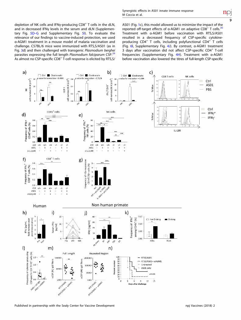
depletion of NK cells and IFNγ-producing CD8+ T cells in the dLN,and in decreased IFNγ levels in the serum and dLN (Supplemen-tary Fig. 5D–G and Supplementary Fig. 5I). To evaluate therelevance of our findings to vaccine-induced protection, we usedα-AGM1 treatment in a mouse model of malaria vaccination andchallenge. C57BL/6 mice were immunized with RTS,S/AS01 (as inFig. 3d) and then challenged with transgenic Plasmodium bergheiparasites expressing the full length Plasmodium falciparum CSP.29
As almost no CSP-specific CD8+ T-cell response is elicited by RTS,S/
AS01 (Fig. 1c), this model allowed us to minimize the impact of thereported off-target effects of α-AGM1 on adaptive CD8+ T cells.30
Treatment with α-AGM1 before vaccination with RTS,S/AS01resulted in a decreased frequency of CSP-specific cytokine-producing CD4+ T cells, including polyfunctional CD4+ T cells(Fig. 6l, Supplementary Fig. 4J). By contrast, α-AGM1 treatment3 days after vaccination did not affect CSP-specific CD4+ T-cellfrequencies (Supplementary Fig. 4H). Treatment with α-AGM1before vaccination also lowered the titres of full-length CSP-specific
Synergistic effects in AS01 innate immune responseM Coccia et al.
9
Published in partnership with the Sealy Center for Vaccine Development npj Vaccines (2018) 25
IgGs, but levels of IgGs specific for a protective repeat epitopewere not significantly affected (Fig. 6m).In response to malaria challenge, all control mice (mice that did
not receive RTS,S/AS01) developed blood-stage malaria by 4 daysafter challenge. By contrast, of the mice that received RTS,S/AS01and were not treated with α-AGM1, 30% remained protected frommalaria at 12 days after challenge (consistent with human data13).In the mice that received RTS,S/AS01 and were α-AGM1-treated,10% remained protected to day 12 post infection (Fig. 6n). Incomparison with control mice, the risk of infection was reduced byRTS/AS01 vaccination alone (hazard ratio = 0.224, 95% CI =0.077–0.654), but not by RTS/AS01 vaccination and α-AGM1-treatment (hazard ratio = 0.499, 95% CI = 0.194–1.284). Overall,these data support the hypothesis that IFNγ-producing lymphoidcells play an important role in promoting protective immunityafter vaccination with RTS,S/AS01.
DISCUSSIONCombination adjuvants promote potent antigen-specific immunityand increased protection from disease upon vaccination inhumans.1 The potency of these adjuvants is likely to rely oncomplex molecular and cellular interplays, which are still poorlyunderstood in vivo. Here, we apply a customized statisticalframework to unravel the way that MPL and QS-21 contribute tothe transcriptional response to AS01 in the dLN. We also show thatMPL and QS-21 contribute to the synergistic production of innateIFNγ at the earliest stages of vaccination. This IFNγ production iscritical for promoting polyfunctional Th1-biased antigen-specificCD4+ T-cell responses to AS01-adjuvanted antigens and alsoimpacts on the quality of the antibody response with a reductionin Th1-isotype switching. IFNγ is mainly produced by activated NKand CD8+ T cells, and the process is controlled by the SSMs and IL-12/IL-18, similar to the response to pathogens.27, 31 We alsoconfirmed the clinical relevance of these findings by showing thatIFNγ production can be detected in the serum of vaccinerecipients at 24 h after vaccination with RTS,S/AS01, and in thedLN of NHPs at 24 h after vaccination with AS01-adjuvantedantigens. Finally, we show that innate IFNγ production isimportant in mediating protection in a mouse model ofvaccination against malaria. Altogether, the study provides new
insights into how the unique design of AS01 (that is, the specificcombination of QS-21 and MPL in AS01) can account for theefficacy of AS01-containing vaccines.The rationale behind Adjuvant Systems and combination
adjuvants is that combining two immunostimulants may triggermultiple immune pathways and improve the protective responseto vaccination.1, 15 Yet, so far, most analyzes of combinationadjuvants relied on visual comparisons between components inthe absence of comprehensive statistical testing.20 Here, we useda customized taxonomy and statistical framework to understandhow MPL and QS-21 interact in AS01 at the transcriptional level. Inusing this approach, we observed complex interplays, in additionto the expected additive or specific effects related to eachimmunostimulant. Noticeably, 13–15% of differentially expressedgenes were regulated in an antagonistic fashion, suggestinginterference between MPL and QS-21 downstream processesin vivo. Moreover, 1–3% of the transcriptional response wasclassified as emergent. This highlights the complexity of theresponse to adjuvant systems, with new, not pre-existing proper-ties emerging from the combination of different immunostimu-lants. Emergent genes contributed the most to the variation ingene expression induced by AS01, suggesting an important role ofthese genes in mediating AS01’s adjuvanticity.It is well established that IFNγ plays a crucial role in the innate
immune response to infection, and that innate IFNγ can promote aTh1- polarizing environment in vivo.16, 32, 33 However, the role ofinnate IFNγ in clinically relevant adjuvant responses has not beeninvestigated. We report that the combinatorial nature of AS01promotes the production of IFNγ by lymphoid cells in the dLN inthe first few hours after vaccination. NK cells and CD8+ T cells werethe main sources of innate IFNγ. IFNγ+ NK cells were mainlyresident in the dLN, as little recruitment of NK cells was observedin the first hours after vaccination. CD8+ T cells had features ofvirtual memory CD8+ T cells, which have been shown toparticipate in innate immune responses in an antigen-independent fashion.34 Both the NK and the CD8+ T-cell responseswere dependent on SSMs, in a mechanism involving IL-18 and IL-12, and independent from monocyte recruitment in the dLN, asthe responses were preserved in Ccr2−/− mice (Kaat Fierens andBart Lambrecht, personal communication). The innate IFNγresponse to AS01 was remarkably similar to that described
Fig. 6 Innate IFNγ production is mediated by SSM-derived IL-18, in combination with IL-12. a, b C57BL/6 were treated with 20 μl of clodronateliposomes or control liposomes i.m. Six days later, mice were immunized with AS01 + HBs (HBs 100 μg/ml, 2 × 10 μl/injection i.m.). Six hoursafter immunization, dLNs were collected and analyzed by FACS. a Effect of subcapsular sinus macrophages depletion on NK (left) and CD8+
T cells (right). b Number of IFNγ+ CD45+ cells. cWTmice were immunized with AS01 + PBS or HBs (HBs 100 μg/ml, 2 × 50 μl/injection i.m.). dLNswere collected 6 h after immunization and IL-18R expression was assessed by FACS. Top panels show the expression of IL-18R on CD8+ T cellsor NK cells after immunization. The bottom panel shows IL-18R expression on IFNγ- or IFNγ+ populations. d–f C57BL/6 WT or Il18−/− mice weretreated with αIL-12p40 (C17.8, 0.5 mg, i.p.) and immunized the next day with AS01 + HBs. dLNs were collected 6 h after immunization. Theaccumulation and frequency of IFNγ-producing cells was evaluated by ICS. d Frequency of CD45+ IFNγ+ cells. e Frequency of IFNγ+ NK cells. fFrequency of IFNγ+CD8+ T cells. (N= 4–20, data from 2 pooled independent experiments are shown). g C57BL/6 WT or Il18−/− mice weretreated with αIL-12p40 (C17.8, 0.5 mg, i.p.) and immunized the next day with AS01 + HBs/OVA (HBs 80 μg/ml, OVA 20 μg/ml, 2 × 50 μl/injectioni.m.). Mice were immunized twice at 14 days intervals. On Day 21, spleens were collected and frequency of total cytokine-producing antigen-specific CD4+ T cells were anlysed by ICS (n= 8, 2 pooled experiments are shown). h Healthy individuals were immunized with RTS,S/AS01 (per0.5 ml dose: 50 μg of RTS,S, AS01= 50 μg of MPL and 50 μg of QS-21, i.m.). The concentration of IFNγ in the serum was evaluated by ELISA.Data show fold increase in the levels of IFNγ in the serum before immunization (pre) or 24 h after injection (N= 33). i–k Chinese rhesusmacaques were immunized i.m. with AS01 (2 × 250 μl) + VZV glycoprotein E (25 μg) (i, j) or HIV recombinant protein F4 (10 μg) (k). Cytokinelevels were analyzed by multiplex bead assay and ICS. i Levels of IFNγ in the serum at indicated time points. j Levels of IFNγ in the dLN atindicated time points. k Frequency of IFNγ-producing cells in draining and non-draining lymph node 24 h after immunization. For sera N= 7,for dLN N= 2–4, for ICS N= 3–6. l–n Mice were immunized with RTS,S/AS01 as in Figure 3D (1 × 50 μl, i.m.). Two days before immunization andon the day of immunization, mice were administered 100 μl of α-AGM1 Ab or control rabbit polyclonal serum. l On Day 21, spleens werecollected and splenocytes were restimulated with a pool of peptide spanning the length of CSP and frequency of total cytokine-producingantigen-specific CD4+ T cells were analyzed by ICS. m IgG titers against full length or the NANP repeated reagions of CSP. n. Two weeks afterthe last immunization, mice were infected with 3000 transgenic Plasmodium berghei sporozoites expressing Plasmodium falciparum CSP. Micewere monitored daily and considered infected after showing parasitemia by blood smear for 2 consecutive days. Kaplan–Meyer survivalcurves are shown (N= 10/group). All graphs show mean ± SEM. Data were analyzed by one-way or two-way ANOVA followed by Bonferronipost hoc test, by Student's t-test or by paired Student's t-test, as appropriate. *p<0.05, **p< 0.01, ***p< 0.001. All charts show mean± SEM
Synergistic effects in AS01 innate immune responseM Coccia et al.
10
npj Vaccines (2018) 25 Published in partnership with the Sealy Center for Vaccine Development

following infection of mice with both extracellular and intracellularpathogens.27 These results suggest that in its induction of potentadaptive responses to the vaccine antigen, AS01 targets thecomplex prepositioning of cells in the dLN that are poised to limitbacterial spread during an infection. Given the complexity ofAS01-mediated modulation of DC function and the heteroge-neous population of cells in the dLN,7 further experiments areneeded to detail the interaction between NK cells and myeloidcells in dLN. Interestingly, a recent report showed early IFNγproduction in the dLN of mice immunized with glucopyranosyllipid adjuvant-stable emulsion (GLA-SE).35 Although the levelsobserved in that study were lower than those we report for AS01,it should be noted that GLA-SE is also able to induce T-cellresponses in vivo,35 supporting the hypothesis that early IFNγproduction is important for obtaining productive T-cell responsesto adjuvanted vaccines.The adjuvanticity of AS01 is at least partially mediated by the
recruitment and activation of CD11c+ MHCIIHi DCs.7 Here, we showthat the maturation of DCs is dependent on innate IFNγ release(presumably via a cell-intrinsic mechanism36), and that IFNyRdeficiency or simultaneous impairment of both IL-12 and IL-18results in defective CD4+ cellular immune responses to vaccina-tion. In addition, mice specifically deficient in NK cells developedreduced CD4+ T-cell responses to vaccination. Overall, thisindicates that innate IFNγ is required to induce CD4+ T-cellresponses, with a Th1-bias after immunization with an AS01-adjuvanted vaccine, as further supported by the reduction inIgG2c antigen-specific antibodies in the absence of innate IFNγsignals. However, the CD8+ T-cell response and the overallmagnitude of the humoral immune response were mostlypreserved in all these models suggesting that other yetundiscovered mechanisms also contribute to AS01 adjuvanticity.Nevertheless, the depletion of IFNγ-producing cells resulted indecreased RTS,S-mediated protection in a malaria challengemodel, accompanied by a decreased CSP-specific CD4+ T-cellresponse and limited effects on protective antibodies. Thissuggests that the modulation of adaptive immunity mediatedby innate IFNγ is relevant to protection. In addition, it suggestssome role for both cellular and humoral adaptive immunity in RTS,S-mediated protection from malaria, as hinted by findings fromclinical trials.12
As QS-21 induces inflammasome activation in vitro and in vivo37
and MPL induces IL-12 secretion in vitro,8 we hypothesized thatthe combination of these two immunostimulants in AS01 isessential for early IFNγ production. Accordingly, the innate IFNγresponse to AS01 was fully dependent on both IL-18 and IL-12production in the dLN. Other members of the inflammasome-dependent cytokines, such as IL-1β, might contribute to the IFNγresponse in vivo, but are likely to be redundant with IL-18.Furthermore, the components of AS01 reach the dLN as early as30min after immunization7 (and data not shown). The liposomeformulation of AS01 is most likely to play a role in its rapiddrainage to the dLN and activation of SSM, as liposomes canimprove the targeting of their components to the lymph node38
and promote uptake by macrophages.39
We identified innate IFNγ as a major player in the innateimmune response to AS01 in the earliest stages after immuniza-tion. Although the role for adaptive, T-cell-derived IFNγ inpromoting effective immunization is well established,40 littleinformation is available regarding the role of this cytokine in theearly response to vaccination. Yet, evidence from clinical studiessuggests that major cellular and molecular changes occur in thefirst 24 hours after immunization in humans.41–43 We show thatvaccination with AS01-adjuvanted antigens results in upregulationof IFNγ levels in the serum of human subjects and in the serumand dLN of NHPs. Although caution should be used whenextending the findings from animal models to humans, this latterobservation supports the hypothesis that innate cells underlie
IFNγ production in humans who receive an AS01-adjuvantedvaccine. Indeed, CD56HiCD16– NK cells are enriched in humanlymph nodes and are more efficient at producing IFNγ thanperipheral-blood NK cells,44, 45 and CD8+ T cells with innatefeatures were recently described in human blood and cord bloodsamples.46 More investigations in appropriate models such asNHPs will be needed to understand the contribution of these celltypes to innate immune response to vaccination. Interestingly,individuals vaccinated with RTS,S/AS01 and protected frommalaria presented increased activation of an IFN-related bloodtranscriptional module at D1 post vaccination, compared withnon-protected individuals.47 However, further work will need tofully demonstrate the requirement of an innate IFNγ response forthe induction of effective vaccination in humans. It is also possiblethat such a mechanism will be amplified with successive vaccinedoses, because antigen-specific memory-T cells can promote IFNγproduction by innate immune cells.36 Indeed, a positive feedbackloop between NK cells and CD4+ T cells takes place duringvaccination, malaria and VZV infections.48–50
Overall, our study provides compelling information regardingthe mechanism of action of AS01, an adjuvant which has provensuccessful in improving immunogenicity and efficacy in vaccinesfor challenging diseases such as malaria and herpes zoster.
MATERIALS AND METHODSVaccine formulationsAll antigens (HBs, OVA, CSP, VZV glycoprotein E, all clinical-grade) and theAdjuvant System AS01 were produced at GSK. Unless otherwise stated inthe figure legends, AS01 contained 100 μg/ml 3D-MPL (GSK), 100 µg/mlQS-21 (Quillaja saponaria Molina, fraction 21, licensed by GSK fromAntigenics LLC, a wholly owned subsidiary of Agenus Inc., a Delaware, USAcorporation) and was formulated in liposomes. In experiments with RTS,S/AS01, a variant of AS01 (AS01E = 50 μg/ml 3D-MPL (GSK), 50 µg/ml QS-21)was used. Set-up experiments showed no impact of lower doses of AS01on its adjuvanticity in mice (data not shown).
Human studiesAll blood samples were obtained from the RTS,S vaccine clinical trial(ClinicalTrials.gov NCT00075049).12 The clinical trial was conducted inaccordance with all applicable regulatory requirements, including theDeclaration of Helsinki (1996). The clinical trial was approved byinstitutional review boards from the Walter Reed Army Institute ofResearch (WRAIR) Human Use Review Committee (HURC) and the UnitedStates Army Medical Research and Materiel Command (USAMRMC) HumanSubjects Research Review Board (HSRRB). Written-informed consent wasobtained from each participant prior to the performance of any study-specific procedures in accordance with relevant ICH Guidelines, US ArmyRegulations and principles of the Declaration of Helsinki. Serum IFNγ levelsat D0 and D1 after immunization were measured by ELISA (R&D Systems)in 33/35 trial participants receiving RTS,S/AS01 due to missing data in 2individuals.
Animal immunizationsAnimal husbandry and experiments were ethically reviewed and carriedout in accordance with European Directive 2010/63/EU and theGlaxoSmithKline Biologicals S.A. Policy on the Care, Welfare and Treatmentof Animals. For the malaria challenge model (conducted at WRAIR)experiments were conducted under an approved animal use protocol in anAAALACi accredited facility in compliance with the Animal Welfare Act andother federal statutes and regulations relating to animals and experimentsinvolving animals and adheres to principles stated in the Guide for theCare and Use of Laboratory Animals, NRC Publication, 2011 edition.C57Bl/6 mice were purchased from Harlan Horst, Netherlands. Ifngr−/−,
Il18−/− and ROSA-stopflox−DTA mice were purchased from JacksonLaboratories (USA). Nkp46(iCre) mice were a gift of Prof. Eric Vivier. Micewere allocated across experimental groups by random draw. Experimen-ters were not blinded during tests. The i.m. injections were performed in6–8 week-old mice, in both hind limbs, in the gastrocnemius (gcm) musclesand in a volume of 50 µl/muscle, unless otherwise stated. Iliac lymph nodes
Synergistic effects in AS01 innate immune responseM Coccia et al.
11
Published in partnership with the Sealy Center for Vaccine Development npj Vaccines (2018) 25

were identified as draining in set-up experiments, which also showedminimal immune activation in non-draining lymph nodes. Anti-IL-12p40mAb (C17.8, 0.5 mg i.p.) was purchased from BioXCell and injected i.p. theday before immunization. Anti-IFNγ mAb (XMG1.2) (BioXCell), was injectedi.p. 48 h before immunization. Clodronate liposome and control liposomes(Clodrosome) were injected i.m. (2 × 20 μl) in the gcm muscles 6 daysbefore immunization. For NHP studies, 4–6 years old Chinese rhesusmacaques were used. Animals had been previously used in unrelatedvaccination studies, but a resting period of 6 months minimum wasimposed to minimize any impact on this study. NHPs were randomizedand immunized intramuscularly in both left and right biceps with AS01(250 μl/injection) and VZV glycoprotein E or HIV recombinant protein F4.Axillary lymph nodes were used as dLNs and cervical lymph nodes as non-draining lymph nodes.
Malaria challenge modelMice were challenged i.v. on Day 0 with 3000 transgenic P. bergheisporozoites expressing P. falciparum CSP, collected from a single mosquitolot.29 Mice were monitored from Day 2–12 for parasitemia by blood smearunder microscope. Mice were considered infected and euthanized afterexhibiting parasitemia on two consecutive days. All mice were euthanizedon Day 12.
Microarray analysisTotal RNA was isolated by homogenizing pooled dLN in Tripure reagent (1ml/100mg tissue; Roche Applied Science) and then extracted withchloroform followed by RNeasy Minikit (Qiagen) according to themanufacturer’s protocol. A DNAse treatment was applied on the RNeasycolumn to avoid genomic DNA contamination. RNA was concentrated byethanol precipitation, and quantified by RiboGreen (Life Technologies). 1µg of each RNA sample was used for target preparation, using a one-cyclecDNA synthesis kit, and hybridized to GeneChip Whole Mouse Genome430 2.0 arrays (Affymetrix). Data acquisition was performed usingGeneChip Operating Software (Affymetrix). Data were quality controlledand normalized (RMA normalization) in R using Bioconductor packages.The data analysis pipeline is presented in Supplementary Fig. 6. Linearmodeling and contrast analysis were performed using the limma Rpackage, PCA plots were generated using pca3d. Enrichment analysis onPCA components were generated using the tmod and tagcloud packages.Heatmaps were generated using the pheatmap packages. For identifica-tion of interplay between AS01 components, for each gene, an effect scoreover PBS was calculated. The type of interplay for each DEG was identifiedin R using the algorithm described in ref.15 Briefly, an empirical Bayesprocedure (in limma R package) was used to estimate the contrasts thatmake up the different forms of interplay and to assess their significance.The significance of a particular form of interplay was subsequentlyobtained by applying an intersection-union hypothesis testing procedureto the proper combination of contrasts. Equivalence intervals of [−1,1] anda p-value of <0.05 were used as cutoff. Process Network enrichmentanalysis was performed in MetaCore (Thomson Reuters).
Code availabilityR code with an implementation of the procedure to identify different formsof interplay between AS01 components is included in the SupportingInformation of ref.15
Determination of antigen-specific T-cell and antibody responses tovaccinationAntigen-specific antibody concentrations in the serum (defined by internalstandards) were measured by ELISA, as previously described.7, 13 To assessantigen-specific T-cell response, 106 splenocytes from immunized micewere stimulated in vitro with peptide pools of 15-mers with 11 amino acidsoverlap (1 µg/ml). Anti-CD49d and anti-CD28 antibodies (1 µg/ml, BDBiosciences) were added to the culture and the cells were incubated 2 h at37 °C 5% CO2 in RPMI 10% FCS + additives (l-Glutamine (2mM), Pennicilin/Streptomycin (100 U/100 µg), sodium pyruvate 1mM and MEM non-essential amino acids (1 × ) (all from Invitrogen)). Brefeldin A (BFA) (1 μg/ml, BD Biosciences) was then added and cells were further culturedovernight at 37 °C 5% CO2. Cell staining was performed as describedpreviously.7, 13 Briefly, cells were washed and stained for surface markers,then fixed and permeabilized using the BD Cytofix/Cytoperm solution kit.Cells were then stained with αIL-2 FITC, anti-IFNγ-APC, and anti-TNF-α-PE
(all from BD) for 2 h at 4 °C. The samples were analyzed with the LSRII flowcytometer (BD) and FlowJo software. A complete list of antibodies used inthis study is presented in Supplementary Table 1.
Innate cell phenotypingdLNs were first treated by mechanical dissociation in 3 ml RPMI mediumcontaining DNase I (100 µg/ml, Roche), 1 % FCS and Liberase (Roche) at0.26 U/ml for 30min under agitation at room temperature. Digestion wasstopped by adding 10mM EDTA and incubating on ice. Cells were filtered(100 µM nylon cell strainer, BD Biosciences), washed and resuspended inPBS + 2mM EDTA + 2 % FCS. The analysis of innate cell phenotype wasperformed as previously described.7 Monocytes were defined as Ly6Chigh
CD11b+,Ly6G− cells and neutrophils were defined as SSChigh, CD11b+,Ly6Ghigh. After exclusion of neutrophil, monocyte, and lymphocytepopulations, DCs were gated as CD11cmidMHCIIhigh. For identification ofIFNγ producing cells in mice, dLNs were dissociated and filtered cells wereincubated for 3 h in RPMI 10% FCS + additives in the presence of BFA (1µg/ml, BD Biosciences) and Monensin (1 µg/ml, BD Biosciences) at 37 °C 5%C02. After treatment with 2.4G2 antibody for 10min to block the Fcreceptor, cells were stained for the following markers: NK1.1, TCRβ, TCRγδ,CD45, CD8, CD49b, CD44, CD27, CD69, CD25, CD4, RORγ, EOMES, CXCR3, IL-18R, and CD122 using antibodies from BD Bioscience or eBiosciences. ForNHP studies, collagenase-treated dLN cell suspensions were incubated for5 h in RPMI 10% FCS + additives in the presence of BFA (1 µg/ml, BDBiosciences) and Monensin (1 µg/ml, BD Biosciences) at 37 °C 5% C02. Cellswere then stained for the following surface markers: CD20, CD177, NKp46,CD45, CD11c, CD123, CD8, CD11b, CD3, CD19, CD16, CD14, HLADR, CD4,and TCRγδ. Cells were then fixed and permeabilized using the BD Cytofix/Cytoperm solution kit and stained with anti-IFNγ APC, FITC or PeCy7.
Analysis of cytokine levelsFor the assessment of pro-inflammatory cytokine concentration, dLNs werecollected after immunization at the indicated time points and stored at−80 °C until further analysis. Organs were homogenized and cytokineswere measured in supernatants as described previously.7 Protein levelswere measured by Cytokine Bead Analysis (BD Biosciences) or ELISA (R&Dsystem) (mouse studies) or multiplex assay (Luminex,Milliplex Kit, Millipore)(NHP studies). For qPCR analysis, RNA was isolated by homogenizing dLNin Tripure reagent (Roche Applied Science) and then extracted withchloroform followed by RNeasy Minikit (Qiagen) according to themanufacturer’s protocol, with DNAse treatment. RNA was concentratedby ethanol precipitation, and quantified by RiboGreen (Invitrogen). 500 ngof total RNA were reverse-transcribed using Superscript II RNase H(Invitrogen) as per manufacturer’s protocol. RNA expression was quantifiedby real time PCR (RT-PCR) using the ABI SYBR Green Mastermix (AppliedBiosystems) (primers sequence for IFNγ [5′-GCATTCATGAGTATTGC-CAAGTTTG-3′, 5′-CGGATGAGCTCATTGAATGCTT-3′], for HPRT [5′-GCAAACTTTGCTTTCCCTGG-3′, 5′-ACTTCGAGAGGTCCTTTTCACC-3], primerefficiency 90–110%). RT-PCR was run the ABI Prism 7900HT SequenceDetection System (Perkin Elmer). Expression levels were quantified usingthe comparative Ct method and gene expression was determined relativeto the mean value of the matched PBS-treated controls.
Statistical analyzesSample size with adequate statistical power in animal studies wasdetermined based on previous experiments. Where appropriate, datawere log-tranformed and assumed to have a normal distribution.Depending on the study design, a Student's t-test or an ANOVA (one- ortwo-way, depending on the experimental design) followed by Bonferronicorrected t-test were applied to identify differences between treatments.Only significant differences are reported on the graphs. Two-way ANOVAwas performed to assess experiments with a 2 × 2 factorial design (forinstance, when mice from 2 different strains were vaccinated with eitherAS01 or PBS + Ag). When 2- way ANOVA is performed, the interaction testaddresses the question whether the effect of treatment (for instancevaccination) is the same in both conditions tested (for instance 2 differentmouse strains). If the p-value is < 0.05, then the treatment has differenteffects in the two conditions. Statistical significance: *p < 0.05, **p < 0.01,***p < 0.001, ****p < 0.0001.
Synergistic effects in AS01 innate immune responseM Coccia et al.
12
npj Vaccines (2018) 25 Published in partnership with the Sealy Center for Vaccine Development

Data availabilityTranscriptomics data are available on GEO database with access numberGSE98322. Preclinical data that support the findings of this study areavailable from GSK Vaccines but restrictions apply to the availability ofthese data. Data are, however, available from the authors upon reasonablerequest and with permission of GSK Vaccines. The results summary for theclinical study (GSK study number 257049/027 - NCT00075049) is availableon the GSK Clinical Study Register and can be accessed at www.gsk-clinicalstudyregister.com. For interventional studies that evaluate ourmedicines, anonymized patient-level data will be made available toindependent researchers, subject to review by an independent panel, atwww.clinicalstudydatarequest.com within 6 months of publication. Toprotect the privacy of patients and individuals involved in our studies, GSKdoes not publically disclose patient-level data.
ACKNOWLEDGMENTSThe authors are grateful to Professor Eric Vivier (Université de la Méditerranée,Marseille, France) for providing Nkp46(iCre) knock-in mice. They thank CédricVanderhaegen, Virginie Adam, Isabelle Leysen, Nabila Amanchar, Hajar Larbi, JenniferDalcq, Stephanie Quique, Bernard Hoyois and all the Vaccine Research and LabAnimal Science groups (all from GSK) for laboratory support and animal work. Finally,they thank Matthew Morgan (MG Science Communications, on behalf of GSK) forimproving of the manuscript and Ulrike Krause (GSK) for publication management.This study was sponsored by GlaxoSmithKline Biologicals SA. M.C. was supported by aMarie Curie - Intra-European Fellowship (ref. “ADJSYN”). S.G. is supported by theFonds National de la Recherche Scientifique (FRS-FNRS, Belgium) and the WELBIO. S.G., S.D., and I.W. are supported by an Interuniversity Attraction Poles Program of theBelgian Federal Science Policy. Mosquirix and Engerix are trademarks of the GSKgroup of companies. The opinions or assertions contained herein are the privateviews of the author, and are not to be construed as official, or as reflecting true viewsof the Department of the Army or the Department of Defense.
AUTHOR CONTRIBUTIONSAll authors were involved in the conception and interpretation of the study. M.C., A.C., I.W., S.D., and M.J.v.H. acquired and analyzed the data. All authors were involved indrafting the manuscript or revising it critically for important intellectual content. Allauthors had full access to the data and approved the manuscript before it wassubmitted by the corresponding author.
ADDITIONAL INFORMATIONSupplementary Information accompanies the paper on the npj Vaccines website(https://doi.org/10.1038/s41541-017-0027-3).
Competing interests: All authors have declared the following interests: M.C., C.C., C.H.,A.C., P.B., S.M., N.G., R.v.d.M., D.F., R.A.v.d.B., and A.M.D. are, or were at the time of thestudy, employees of the GSK group of companies. S.M., P.B., R.v.d.M., and A.M.D. own GSKstocks. S.G.’s laboratory received a Public-Private Partnership grant from GlaxoSmithKlineBiologicals SA and the Walloon Region (ref. “SAPOVAC”). A.K.S. was partly employed bythe Academic Medical Center (AMC) of the University of Amsterdam, which has anagreement with GlaxoSmithKline Biologicals S.A. C.J.G. was supported by an appoint-ment to the Postgraduate Research Participation Program at the Walter Reed ArmyInstitute of Research administered by the Oak Ridge Institute for Science and Educationthrough an interagency agreement between the U.S. Department of Energy and MRMC.The other authors report no financial conflict of interest.
Publisher's note: Springer Nature remains neutral with regard to jurisdictional claimsin published maps and institutional affiliations.
REFERENCES1. Mutwiri, G. et al. Combination adjuvants: the next generation of adjuvants? Expert
Rev. Vaccin. 10, 95–107 (2011).2. Didierlaurent, A. M. et al. Adjuvant system AS01: helping to overcome the chal-
lenges of modern vaccines. Expert Rev. Vaccines. 16, 1–9 (2016).3. The RTSS Clinical Trials Partnership. Efficacy and safety of RTS,S/AS01 malaria vac-
cine with or without a booster dose in infants and children in Africa: final results of aphase 3, individually randomised, controlled trial. Lancet 386, 31–45 (2015).
4. Chlibek, R. et al. Long-term immunogenicity and safety of an investigationalherpes zoster subunit vaccine in older adults. Vaccine 34, 863–868 (2016).
5. Van Braeckel, E. et al. An adjuvanted polyprotein HIV-1 vaccine induces poly-functional cross-reactive CD4+T cell responses in seronegative volunteers. Clin.Infect. Dis. 52, 522–531 (2011).
6. Leroux-Roels, I. et al. Improved CD4(+) T cell responses to Mycobacterium tuber-culosis in PPD-negative adults by M72/AS01 as compared to the M72/AS02 andMtb72F/AS02 tuberculosis candidate vaccine formulations: a randomized trial.Vaccine 31, 2196–2206 (2013).
7. Didierlaurent, A. M. et al. Enhancement of adaptive immunity by the humanvaccine adjuvant AS01 depends on activated dendritic cells. J. Immunol. 193,1920–1930 (2014).
8. Casella, C. R. & Mitchell, T. C. Putting endotoxin to work for us: monophosphoryllipid A as a safe and effective vaccine adjuvant. Cell Mol. Life Sci. 65, 3231–3240(2008).
9. Marty-Roix, R. et al. Identification of QS-21 as an Inflammasome-activatingmolecular component of saponin adjuvants. J. Biol. Chem. 291, 1123–1136 (2016).
10. Ragupathi, G., Gardner, J. R., Livingston, P. O. & Gin, D. Y. Natural and syntheticsaponin adjuvant QS-21 for vaccines against cancer. Expert Rev. Vaccin. 10,463–470 (2011).
11. Mettens, P. et al. Improved T cell responses to Plasmodium falciparum circum-sporozoite protein in mice and monkeys induced by a novel formulation of RTS,Svaccine antigen. Vaccine 26, 1072–1082 (2008).
12. Kester, K. E. et al. Randomized, double-blind, phase 2a trial of falciparummalaria vaccines RTS,S/AS01B and RTS,S/AS02A in malaria-naive adults: safety,efficacy, and immunologic associates of protection. J. Infect. Dis. 200, 337–346(2009).
13. Dendouga, N., Fochesato, M., Lockman, L., Mossman, S. & Giannini, S. L. Cell-mediated immune responses to a varicella-zoster virus glycoprotein E vaccineusing both a TLR agonist and QS21 in mice. Vaccine 30, 3126–3135 (2012).
14. Darrah, P. A. et al. Multifunctional TH1 cells define a correlate of vaccine-mediated protection against Leishmania major. Nat. Med. 13, 843–850 (2007).
15. Van Deun, K., Thorrez, L., van den Berg, R. A., Smilde, A. K. & Van Mechelen, I. Notjust a sum? identifying different types of interplay between constituents incombined interventions. PLoS ONE 10, e0125334 (2015).
16. Martin-Fontecha, A. et al. Induced recruitment of NK cells to lymph nodes pro-vides IFN-gamma for T(H)1 priming. Nat. Immunol. 5, 1260–1265 (2004).
17. Goldszmid, R. S. et al. NK cell-derived interferon-gamma orchestrates cellulardynamics and the differentiation of monocytes into dendritic cells at the site ofinfection. Immunity 36, 1047–1059 (2012).
18. Ge, M. Q. et al. NK cells regulate CD8+T cell priming and dendritic cell migrationduring influenza A infection by IFN-gamma and perforin-dependent mechan-isms. J. Immunol. 189, 2099–2109 (2012).
19. Vieira, P. L., de Jong, E. C., Wierenga, E. A., Kapsenberg, M. L. & Kalinski, P.Development of Th1-inducing capacity in myeloid dendritic cells requiresenvironmental instruction. J. Immunol. 164, 4507–4512 (2000).
20. Mosca, F. et al. Molecular and cellular signatures of human vaccine adjuvants.Proc. Natl Acad. Sci. USA 105, 10501–10506 (2008).
21. Didierlaurent, A. M. et al. AS04, an aluminum salt- and TLR4 agonist-basedadjuvant system, induces a transient localized innate immune response leadingto enhanced adaptive immunity. J. Immunol. 183, 6186–6197 (2009).
22. Cherwinski, H. M., Schumacher, J. H., Brown, K. D. & Mosmann, T. R. Two types ofmouse helper T cell clone. III. Further differences in lymphokine synthesisbetween Th1 and Th2 clones revealed by RNA hybridization, functionallymonospecific bioassays, and monoclonal antibodies. J. Exp. Med. 166, 1229–1244(1987).
23. Spits, H., Bernink, J. H. & Lanier, L. NK cells and type 1 innate lymphoid cells:partners in host defense. Nat. Immunol. 17, 758–764 (2016).
24. Voehringer, D., Liang, H. E. & Locksley, R. M. Homeostasis and effector function oflymphopenia-induced “memory-like” T cells in constitutively T cell-depleted mice.J. Immunol. 180, 4742–4753 (2008).
25. Narni-Mancinelli, E. et al. Fate mapping analysis of lymphoid cells expressing theNKp46 cell surface receptor. Proc. Natl Acad. Sci. USA 108, 18324–18329 (2011).
26. Kuka, M. & Iannacone, M. The role of lymph node sinus macrophages in hostdefense. Ann. N. Y. Acad. Sci. 1319, 38–46 (2014).
27. Kastenmüller, W., Torabi-Parizi, P., Subramanian, N., Lammermann, T. & Germain,R. N. A spatially-organized multicellular innate immune response in lymph nodeslimits systemic pathogen spread. Cell 150, 1235–1248 (2012).
28. Detienne, S. et al. Central Role of CD169+Lymph node resident macrophagesin the adjuvanticity of the QS-21 component of AS01. Sci. Rep. 6, 39475 (2016).
29. Porter, M. D. et al. Transgenic parasites stably expressing full-length Plasmodiumfalciparum circumsporozoite protein as a model for vaccine down-selection inmice using sterile protection as an endpoint. Clin. Vaccin. Immunol. 20, 803–810(2013).
30. Slifka, M. K., Pagarigan, R. R. & Whitton, J. L. NK markers are expressed on a highpercentage of virus-specific CD8+ and CD4+ T cells. J. Immunol. 164, 2009–2015(2000).
31. Kupz, A., Curtiss, R. 3rd, Bedoui, S. & Strugnell, R. A. In vivo IFN-gamma secretionby NK cells in response to Salmonella typhimurium requires NLRC4 inflamma-somes. PLoS ONE 9, e97418 (2014).
Synergistic effects in AS01 innate immune responseM Coccia et al.
13
Published in partnership with the Sealy Center for Vaccine Development npj Vaccines (2018) 25
32. Huang, S. et al. Immune response in mice that lack the interferon-gammareceptor. Science 259, 1742–1745 (1993).
33. Das, G., Sheridan, S. & Janeway, C. A. Jr. The source of early IFN-gamma that playsa role in Th1 priming. J. Immunol. 167, 2004–2010 (2001).
34. Kastenmüller, W. et al. Peripheral prepositioning and local CXCL9 chemokine-mediated guidance orchestrate rapid memory CD8+T cell responses in the lymphnode. Immunity 38, 502–513 (2013).
35. Cauwelaert, N. D. et al. The TLR4 agonist vaccine adjuvant, GLA-SE, requirescanonical and atypical mechanisms of action for TH1 induction. PLoS ONE 11,e0146372 (2016).
36. Soudja, S. M. et al. Memory-T-cell-derived interferon-gamma instructs potent innatecell activation for protective immunity. Immunity 40, 974–988 (2014).
37. Marty-Roix, R. et al. Identification of QS-21 as an inflammasome-activating molecular component of saponin adjuvants. J. Biol. Chem. 291,1123–1136 (2015).
38. Xie, Y., Bagby, T. R., Cohen, M. S. & Forrest, M. L. Drug delivery to the lymphaticsystem: importance in future cancer diagnosis and therapies. Expert Opin. DrugDeliv. 6, 785–792 (2009).
39. van Rooijen, N. & van Kesteren-Hendrikx, E. “In vivo” depletion of macrophagesby liposome-mediated “suicide”. Methods Enzymol. 373, 3–16 (2003).
40. Seder, R. A., Darrah, P. A. & Roederer, M. T-cell quality in memory and protection:implications for vaccine design. Nat. Rev. Immunol. 8, 247–258 (2008).
41. Obermoser, G. et al. Systems scale interactive exploration reveals quantitativeand qualitative differences in response to influenza and pneumococcal vaccines.Immunity 38, 831–844 (2013).
42. Bucasas, K. L. et al. Early patterns of gene expression correlate with the humoralimmune response to influenza vaccination in humans. J. Infect. Dis. 203, 921–929(2011).
43. Nakaya, H. I. et al. Systems analysis of immunity to influenza vaccination acrossmultiple years and in diverse populations reveals shared molecular signatures.Immunity 43, 1186–1198 (2015).
44. Fehniger, T. A. et al. CD56 bright natural killer cells are present in human lymphnodes and are activated by T cell-derived IL-2: a potential new link betweenadaptive and innate immunity. Blood 101, 3052–3057 (2003).
45. Morandi, B., Bougras, G., Muller, W. A., Ferlazzo, G. & Munz, C. NK cells of humansecondary lymphoid tissues enhance T cell polarization via IFN-gamma secretion.Eur. J. Immunol. 36, 2394–2400 (2006).
46. Jacomet, F. et al. Evidence for eomesodermin-expressing innate-like CD8 KIR/NKG2A T cells in human adults and cord blood samples. Eur. J. Immunol. 45,1926–1933 (2015).
47. Rinchai, D., Presnell, S. & Chaussabel, D. Blood interferon signatures putativelylink lack of protection conferred by the RTS,S recombinant malaria vaccine to anantigen-specific IgE response [version 1; referees: 1 approved, 1 approved withreservations]. F1000Research 4, 919 (2015).
48. Horowitz, A., Behrens, R. H., Okell, L., Fooks, A. R. & Riley, E. M. NK cells as effectorsof acquired immune responses: effector CD4+T cell-dependent activation of NKcells following vaccination. J. Immunol. 185, 2808–2818 (2010).
49. Horowitz, A. et al. Cross-talk between T cells and NK cells generates rapid effectorresponses to Plasmodium falciparum-infected erythrocytes. J. Immunol. 184,6043–6052 (2010).
50. Steain, M. et al. Analysis of T cell responses during active varicella-zoster virusreactivation in human ganglia. J. Virol. 88, 2704–2716 (2014).
Open Access This article is licensed under a Creative CommonsAttribution 4.0 International License, which permits use, sharing,
adaptation, distribution and reproduction in anymedium or format, as long as you giveappropriate credit to the original author(s) and the source, provide a link to the CreativeCommons license, and indicate if changes were made. The images or other third partymaterial in this article are included in the article’s Creative Commons license, unlessindicated otherwise in a credit line to the material. If material is not included in thearticle’s Creative Commons license and your intended use is not permitted by statutoryregulation or exceeds the permitted use, you will need to obtain permission directlyfrom the copyright holder. To view a copy of this license, visit http://creativecommons.org/licenses/by/4.0/.
© The Author(s) 2018
Synergistic effects in AS01 innate immune responseM Coccia et al.
14
npj Vaccines (2018) 25 Published in partnership with the Sealy Center for Vaccine Development