cellular micropatterns on biocompatible materials
TRANSCRIPT

Cellular Micropatterns on Biocompatible Materials
Albert Folch and Mehmet Toner*
Center for Engineering in Medicine and Surgical Services, Massachusetts General Hospital, Shriners BurnsHospital, and Harvard Medical School, Boston, Massachusetts 02114
We present a method to produce micropatterns of cells on tissue culture substrates.A network of deep elastomeric microchannels defining the desired pattern is sealedonto the surface of interest, and a protein template is created by injecting sub-milliliterquantities of protein solution into the microchannels. Protein adsorbs only on theareas that were exposed to the microflow. After the channels are flushed and theelastomer is removed, cells attach only on the protein template. Micropatterns ofcollagen or fibronectin were used to selectively adhere cells on various biomedicalpolymers and on heterogeneous or microtextured substrates. Since the bare substrateareas remain apt for seeding other, more adhesive cell types such as fibroblasts, wewere able to create micropatterned co-cultures. Our method allows for inexpensivepatterning of a rich assortment of biomolecules, cells, and surfaces under physiologicalconditions.
Introduction
Tissue function is modulated by the spatial organiza-tion of cells on a sub-millimeter scale. For this reason,artificial replication of cellular microstructures is impor-tant in understanding, measuring, and simulating theirin vivo functions in the laboratory. Biotechnologicalapplications of synthetic cellular patterns range fromtissue engineering and wound healing to single-cellassays and biosensors for toxic compounds. However,geometrically selective attachment of cells on surfacesis an ongoing technological challenge. Early studiesutilized spider webs (Harrison, 1914), grooved surfaces(Weiss, 1959; Rovensky et al., 1971), shadow-evaporatedmetallic lines (Carter, 1965), or scratched extracellularmatrix (ECM) protein patterns (Rovasio et al., 1983) toguide cell migration, attachment, and/or spreading.These studies could not address the structural dimen-sions and precise repeatability over large areas found inliving tissue. Recently, silicon microfabrication tech-niques (Ristic, 1994) and advances in surface chemistryhave allowed for micropatterning of self-assembled mono-layers (SAMs) (Laibinis et al., 1991; Ulman, 1991; Kumarand Whitesides, 1993; Kumar et al., 1994) of organicmolecules which promote or inhibit cell adhesion onsubstrates otherwise homogeneously adherent to cells(Kleinfeld et al., 1988; Clark et al., 1992; Stenger et al.,1992; Singhvi et al., 1994; Bhatia et al., 1997; Chen etal., 1997). Unfortunately, the specialized SAM chemis-tries, e.g. functionalized alkylsiloxanes on silicon oxideor alkanethiols on gold (Ulman, 1991), are not easilycombined with most tissue culture or implantable (“bio-compatible”) materials (Allara, 1995; Ratner, 1995) suchas polymers or heterogeneous surfaces and, most impor-tantly, can result in undesirable biointeraction (Schaffneret al., 1995; Tidwell et al., 1997).
Other schemes for cell micropatterning based on cy-toscribing (Klebe, 1988), thermoresponsive polymers(Takezawa et al., 1990; Ito et al., 1997), or irradiation ofproteins (Letourneau, 1975) or polymers (Matsuda et al.,1990; Rohr et al., 1991; Lee et al., 1993, 1994) have beenproposed as well. We present an alternative strategybased on physically blocking protein adsorption on se-lected areas of a substrate by means of a removable andreusable elastomeric mask. As a result, the techniquepresented here uniquely allows for patterning, withoutchemical alteration of the substrate, polystyrene petridishes and other biocompatible polymers, as well asheterogeneous surfaces containing a metallic or biomo-lecular micropattern.
Materials and Methods
Microfabrication. Photoresist (1-µm-thick) patternson 0.5-µm-thick SiO2 on Si were defined by conventionalphotolithography (Ristic, 1994) and transferred to theSiO2 layer by HF etch (5 min). The SiO2 pattern wasused to mask a 20-30-µm-deep Si plasma etch (plasmaparameters: 250 W, 350 mTorr, 100 sccm SF6, 10 sccmCCl4, 10 sccm He, 1 cm gap). The Si master was coatedwith ∼50 Å of Cr and ∼500 Å of Au to prevent adhesionof the elastomer. The poly(dimethylsiloxane) (PDMS)precursor and curing agent (Sylgard 184, Dow Corning)were mixed with a thinning fluid (Dow Corning 200) (10:1:1), poured over the master in a container (forming a∼7-mm-thick layer), put under low vacuum (∼12 Torr)to evacuate the bubbles from the microtrenches, andcured at 65 °C for 12 h (8-h overcure to avoid toxicbleaching). The cured PDMS replica, constituting amicrofluidic network of microchannels (µChs), was gentlypeeled off the master and the container by hand.Substrate Preparation. We used human plasma
fibronectin (Sigma) in sodium carbonate buffer (pH ) 9.6)and collagen type I from Lewis rat tail tendons in HCl0.1 mM (pH ) 4 to prevent gel formation inside theµChs). Dunn et al. described the collagen and rathepatocytes isolations (Dunn et al., 1991). The µChs
* Address correspondence to this author at Shriners ResearchCenter Bldg., 1400 West, One Kendall Square, Cambridge, MA02139. Telephone: (617)374-5611. Fax: (617)374-5665. E-mail:[email protected].
388 Biotechnol. Prog. 1998, 14, 388−392
S8756-7938(98)00037-X CCC: $15.00 © 1998 American Chemical Society and American Institute of Chemical EngineersPublished on Web 05/20/1998

were rinsed with acetone and ethanol and blow-driedwith N2 between each use. We either perfused the µChsin sterile conditions or sterilized the protein-patternedsubstrates with 70% ethanol or with overnight UV lightexposure. We found the latter the most practical ster-ilization procedure.Immunofluorescence. Collagen-patterned substrates
were incubated in 1% bovine serum albumin (BSA) inphosphate-buffered saline (PBS) (blocking solution) for30 min, then in rabbit anti-rat collagen antibody (BioDe-sign, 100:1 dilution in blocking solution) for 1 h; afterbeing rinsed thoroughly with PBS, they were incubatedin CY3-labeled goat anti-rabbit IgG (Jackson Immunore-search, 500:1 dilution in blocking solution) for 1 h.Cell Culture. Prior to cell seeding, the surfaces were
rinsed with deionized water or PBS and incubated at 37°C or room temperature for 30 min in 0.05% BSA toincrease attachment selectivity (Bhatia et al., 1997). Inaddition to the polymers polystyrene (PS) (Falcon, eitherplasma-treated #3002 or untreated #1007), polycarbonate(PC) (Nalgene), poly(methyl methacrylate) (PMMA) (Glas-flex), and PDMS, we also studied poly(methylpentene)Petri dishes (Nalgene), poly(vinyl chloride) sheets (Fisher),and UV-cured polyurethane films (Summers Optical), towhich cells did not adhere. These polymers may needpretreatment such as surface hydroxylation by O2 plasma(Curtis et al., 1986) to promote protein and/or celladhesion. To demonstrate selective cell seeding, hepa-tocytes were allowed to attach for 1 h in serum-freemedium (shaking every 15 min virtually eliminatesbackground attachment); then the substrates were rinsedtwice with serum-free medium to remove nonadheredhepatocytes. The same seeding/rinse sequence wasrepeated twice. Finally, the hepatocytes were culturedin medium containing 10% fetal bovine serum (Bhatiaet al., 1997). For micropatterned co-cultures, 3T3-J2fibroblasts were added 24 h after hepatocyte seeding andallowed to attach for another 24 h as described by Bhatiaet al. (1997, 1998).
Results and Discussion
Elastomeric Microchannels. In our method, anetwork of deep (∼20-30 µm) elastomeric µChs sealedonto the surface of interest is perfused with sub-milliliterquantities of an ECM protein solution. Thus proteinadsorbs on the areas exposed to the microflow and isblocked in the areas where the elastomer contacts thesurface. Essentially, we implement, in a microfluidicenvironment, noncovalent procedures for protein adsorp-tion. These procedures are simple, they do not compro-mise the specific cell functions associated with the richchemical complexity of polymeric surfaces (Tidwell et al.,1997), and they have been used in cell culture for decadeson a wide variety of materials.Recently, others have filled shallow (∼1.5 µm) µChs
by capillary action to pattern curable polymers (Kim etal., 1995, 1996) and immunoglobulins (Delamarche et al.,1997). However, capillarity patterning is limited tolengths on the order of a few millimeters and, moreimportantly, it results in protein gradients due to deple-tion of protein adsorbing on the µCh walls (Delamarcheet al., 1997). Deeper µChs yielding smaller pressuredrops (Potter and Foss, 1982) are needed to pattern largerareas and reach steady-flow, nondepleting conditions.Our method is schematized in Figure 1. We used ananisotropic SF6/CCl4-based plasma etch (Ristic, 1994) tofabricate 20-30-mm-deep vertical-sidewall trenches ofvarious shapes, widths, and lengths on a Si wafer (Figure
1a). We followed a simple replication procedure (Kumarand Whitesides, 1993; Kumar et al., 1994) to mold theµChs in PDMS from the Au-coated Si master (Figure 1b).A post glued to the center of the Si pattern resulted in avia hole in the PDMS (Figure 1c) for liquid injection orsuction. The molding procedure leaves the Si masterintact; therefore, it can be used repeatedly. Upon drycontact with a smooth surface, the µChs self-seal revers-ibly (Kim et al., 1995, 1996; Delamarche et al., 1997) andcan be perfused with various solutions over large areas(up to tens of square centimeters). The elastomer, byvirtue of its inert nature, does not appear to leave debrisor toxic remains on the substrate or to degrade over time.Thus it can be reused virtually indefinitely.Micropatterning. The minimum achievable line
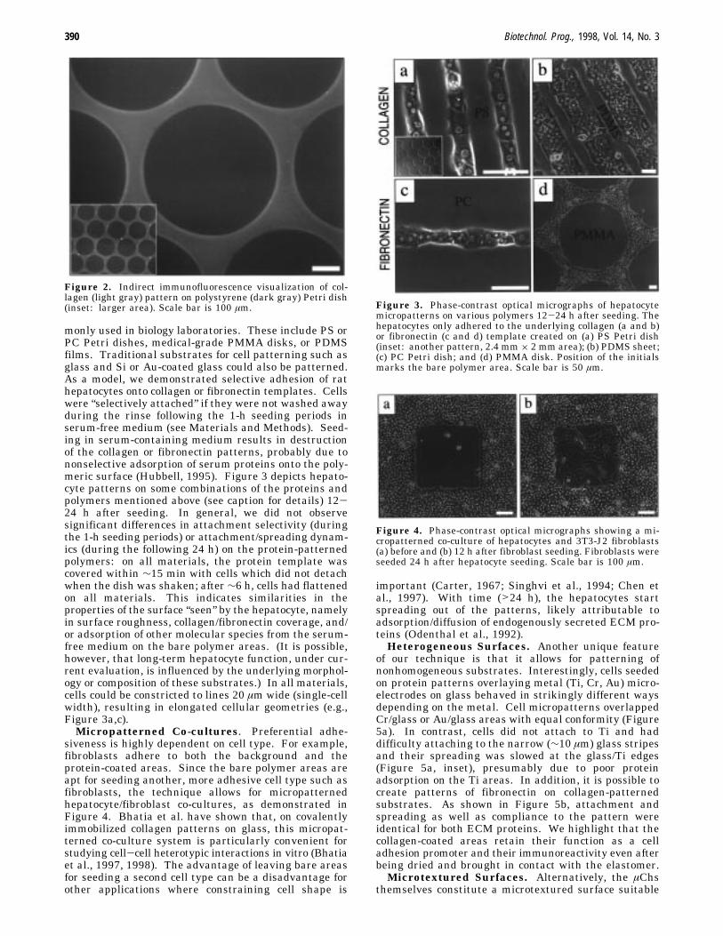
width, the maximum patternable area, and the maximuminjection pressure depend on the sealing pressure andµCh geometry and pattern and must, therefore, bedetermined empirically for each application. As shownbelow, we straightforwardly achieved single-cell linewidths. We typically inject <1 mL (∼0.1 mL/min) ofcollagen (0.2 mg/mL) or fibronectin (10 mg/mL) solutionfor ∼5 min to create the ECM protein template (Figure1d). To prevent protein solution from spreading ontoadjacent areas, the µChs are flushed with deionized wateror saline solution before being separated from the surface.Cells adhere preferentially to the protein template (Fig-ure 1e). Note that this method is compatible with non-denaturing as well as sterile conditions. The fact thatthe µChs are continuously perfused (as opposed to filledby capillary action (Delamarche et al., 1997)) preventsdepletion of protein from the solution and ensures thatthe protein coverage is uniform throughout the perfusedarea, as demonstrated by indirect immunofluorescencestaining (Figure 2). The exact protein coverage (pres-ently undergoing characterization in our laboratory) isnot crucial to the current study, but it is known to affectadhesion strength (Truskey and Pirone, 1990; Garcia etal., 1997), growth, and differentiation (Mooney et al.,1992) and could, in principle, be adjusted by varying thesurface hydroxylation (Curtis et al., 1986) and the timeof exposure to the protein solution as well as its temper-ature, pH, ionic strength, and concentration (Lee andRuckenstein, 1988). Slight local variations in proteincoverage may result from parabolic flow velocity vectordistribution (typical of channel fluidics) (Potter and Foss,1982), from anomalies in the microflow around edges,particles, or trapped bubbles or (in the case of collagen)from gel nucleation.Biocompatible Surfaces. Our method is readily
compatible with off-the-shelf polymeric substrates com-
Figure 1. Cross-section schematics of our cell patterningmethod: (a) Si master fabrication; (b and c) replica molding ofPDMS from the Au-coated master (Au layer not depicted); (d)protein patterning by injecting protein solution into the µChs;(e) cell seeding after elastomer removal, resulting in selectivecell attachment only on the protein template.
Biotechnol. Prog., 1998, Vol. 14, No. 3 389
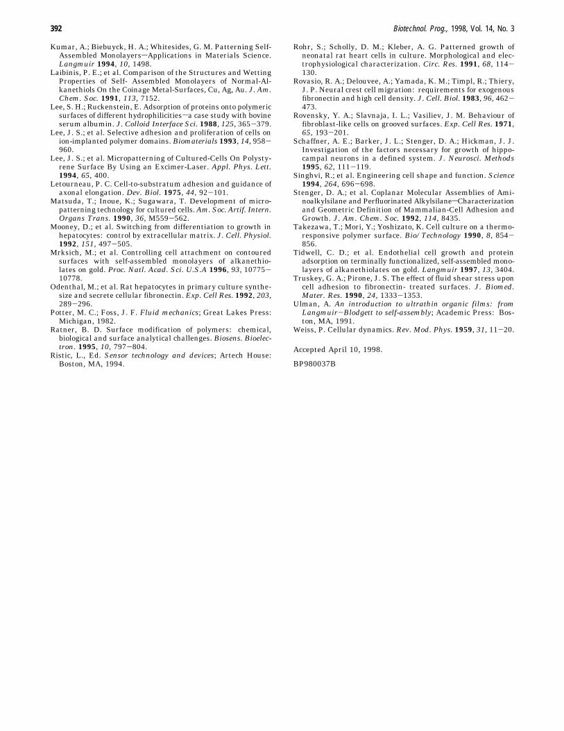
monly used in biology laboratories. These include PS orPC Petri dishes, medical-grade PMMA disks, or PDMSfilms. Traditional substrates for cell patterning such asglass and Si or Au-coated glass could also be patterned.As a model, we demonstrated selective adhesion of rathepatocytes onto collagen or fibronectin templates. Cellswere “selectively attached” if they were not washed awayduring the rinse following the 1-h seeding periods inserum-free medium (see Materials and Methods). Seed-ing in serum-containing medium results in destructionof the collagen or fibronectin patterns, probably due tononselective adsorption of serum proteins onto the poly-meric surface (Hubbell, 1995). Figure 3 depicts hepato-cyte patterns on some combinations of the proteins andpolymers mentioned above (see caption for details) 12-24 h after seeding. In general, we did not observesignificant differences in attachment selectivity (duringthe 1-h seeding periods) or attachment/spreading dynam-ics (during the following 24 h) on the protein-patternedpolymers: on all materials, the protein template wascovered within ∼15 min with cells which did not detachwhen the dish was shaken; after∼6 h, cells had flattenedon all materials. This indicates similarities in theproperties of the surface “seen” by the hepatocyte, namelyin surface roughness, collagen/fibronectin coverage, and/or adsorption of other molecular species from the serum-free medium on the bare polymer areas. (It is possible,however, that long-term hepatocyte function, under cur-rent evaluation, is influenced by the underlying morphol-ogy or composition of these substrates.) In all materials,cells could be constricted to lines 20 µm wide (single-cellwidth), resulting in elongated cellular geometries (e.g.,Figure 3a,c).Micropatterned Co-cultures. Preferential adhe-
siveness is highly dependent on cell type. For example,fibroblasts adhere to both the background and theprotein-coated areas. Since the bare polymer areas areapt for seeding another, more adhesive cell type such asfibroblasts, the technique allows for micropatternedhepatocyte/fibroblast co-cultures, as demonstrated inFigure 4. Bhatia et al. have shown that, on covalentlyimmobilized collagen patterns on glass, this micropat-terned co-culture system is particularly convenient forstudying cell-cell heterotypic interactions in vitro (Bhatiaet al., 1997, 1998). The advantage of leaving bare areasfor seeding a second cell type can be a disadvantage forother applications where constraining cell shape is
important (Carter, 1967; Singhvi et al., 1994; Chen etal., 1997). With time (>24 h), the hepatocytes startspreading out of the patterns, likely attributable toadsorption/diffusion of endogenously secreted ECM pro-teins (Odenthal et al., 1992).Heterogeneous Surfaces. Another unique feature
of our technique is that it allows for patterning ofnonhomogeneous substrates. Interestingly, cells seededon protein patterns overlaying metal (Ti, Cr, Au) micro-electrodes on glass behaved in strikingly different waysdepending on the metal. Cell micropatterns overlappedCr/glass or Au/glass areas with equal conformity (Figure5a). In contrast, cells did not attach to Ti and haddifficulty attaching to the narrow (∼10 µm) glass stripesand their spreading was slowed at the glass/Ti edges(Figure 5a, inset), presumably due to poor proteinadsorption on the Ti areas. In addition, it is possible tocreate patterns of fibronectin on collagen-patternedsubstrates. As shown in Figure 5b, attachment andspreading as well as compliance to the pattern wereidentical for both ECM proteins. We highlight that thecollagen-coated areas retain their function as a celladhesion promoter and their immunoreactivity even afterbeing dried and brought in contact with the elastomer.Microtextured Surfaces. Alternatively, the µChs
themselves constitute a microtextured surface suitable
Figure 2. Indirect immunofluorescence visualization of col-lagen (light gray) pattern on polystyrene (dark gray) Petri dish(inset: larger area). Scale bar is 100 µm. Figure 3. Phase-contrast optical micrographs of hepatocyte
micropatterns on various polymers 12-24 h after seeding. Thehepatocytes only adhered to the underlying collagen (a and b)or fibronectin (c and d) template created on (a) PS Petri dish(inset: another pattern, 2.4 mm× 2 mm area); (b) PDMS sheet;(c) PC Petri dish; and (d) PMMA disk. Position of the initialsmarks the bare polymer area. Scale bar is 50 µm.
Figure 4. Phase-contrast optical micrographs showing a mi-cropatterned co-culture of hepatocytes and 3T3-J2 fibroblasts(a) before and (b) 12 h after fibroblast seeding. Fibroblasts wereseeded 24 h after hepatocyte seeding. Scale bar is 100 µm.
390 Biotechnol. Prog., 1998, Vol. 14, No. 3

for selective cell seeding (Mrksich et al., 1996). Indeed,all three walls of a µCh are rendered adhesive to cellswhen perfused with protein solution. When hepatocyteswere seeded on PDMS µChs which had been perfusedwith a protein solution, cells attached selectively to thebottom and sidewalls of the trenches and not on theelevated bare PDMS areas (Figure 5c). Cells do notattach to PDMS (and that is valid for the inside of thetrenches as well as the elevated areas) unless it is coatedwith protein (see Figure 3b). This constitutes a versatileapproach for cellular patterning in the vertical dimen-sion. We stress that the ability to produce micropatternsof cells on moldable polymers is of paramount importancein the artificial simulation of complex three-dimensional(3D) cellular microstructures, such as liver sinusoids orblood capillaries.Cellular “Islands”. Note that, although our method
only allows for connected patterns, it can generatedisconnected patterns when used in combination with aheterogeneous surface capable of differential adhesionsuch as Ti/glass. Cellular “islands” can also be createdby a variation of our technique which uses agarose as acell adhesion repellent. Hot liquid agarose (1% w/w inwater at ∼80 °C) injected into the µChs solidifies withinthe µChs when cooled to room temperature; when sepa-rated from the sealing surface, the µChs constitute aPDMS surface with agarose-filled trenches. After beingincubated (37 °C) in protein solution for 30 min, cellsattach to the elevated PDMS areas surrounded byagarose (Figure 5d). Note how hepatocyte spreadingconforms to the shape of the island.In conclusion, we have been able to make micropat-
terned cell cultures using ECM protein templates on avariety of tissue culture materials. Our protein pattern-ing method is compatible with nondenaturing and physi-ological conditions, does not present toxic side effects, andcan be extended to other biomolecules that spontaneouslyadsorb on surfaces from aqueous solution. Furthermore,it does not require routine access to microfabricationfacilities. Due to its low cost, versatility, and 3D capa-
bilities, the method has strong implications in basic cellbiology and biotechnology, encompassing tissue engineer-ing, electrophysiology, bioartificial devices or implants,and biosensors.
Acknowledgment
We are thankful to U. Balis, S. Bhatia, O. Hurtado, L.Horowitz, J. Voldman, and M. Yarmush for insightfulcomments and to W. Jastromb, C. Pligavko, and S.Beshad for hepatocyte and collagen isolations and mediapreparation. This study was partially funded by theShriners Hospitals for Children and NIH. We microfab-ricated the Si masters at the Microsystems TechnologyLaboratory (MIT).
References and Notes
Allara, D. L. Critical Issues in Applications of Self-AssembledMonolayers. Biosens. Bioelectron. 1995, 10, 771.
Bhatia, S. N.; Yarmush, M. L.; Toner, M. Controlling cellinteractions by micropatterning in co-cultures: hepatocytesand 3T3 fibroblasts. J. Biomed. Mater. Res. 1997, 34, 189-199.
Bhatia, S. N.; Balis, U. J.; Yarmush, M. L.; Toner, M. Micro-fabrication of Hapatocyte/Fibroblast Co-cultures: Role ofHomotype Cell Interactions. Biotechnol. Prog. 1998, 14, 378-387.
Carter, S. B. Principles of cell motility: the direction of cellmovement and cancer invasion. Nature 1965, 208, 1183-1187.
Carter, S. B. Haptotactic islands: a method of confining singlecells to study individual cell reactions and clone formation.Exp. Cell. Res. 1967, 48, 189-193.
Chen, C. S.; Mrksich, M.; Huang, S.; Whitesides, G. M.; Ingber,D. E. Geometric control of cell life and death. Science 1997,276, 1425-1428.
Clark, P.; Connolly, P.; Moores, G. R. Cell guidance by micro-patterned adhesiveness in vitro. J. Cell Sci. 1992, 103, 287-292.
Curtis, A. S. G.; Forrester, J. V.; Clark, P. Substrate hydroxyl-ation and cell adhesion. J. Cell Sci. 1986, 86, 9-24.
Delamarche, E.; Bernard, A.; Schmid, H.; Michel, B.; Biebuyck,H. Patterned delivery of immunoglobulins to surfaces usingmicrofluidic networks. Science 1997, 276, 779-781.
Dunn, J. C.; Tompkins, R. G.; Yarmush, M. L. Long-term in vitrofunction of adult hepatocytes in a collagen sandwich config-uration. Biotechnol. Prog. 1991, 7, 237-245.
Garcia, A. J.; Ducheyne, P.; Boettiger, D. Cell adhesion strengthincreases linearly with adsorbed fibronectin density. TissueEng. 1997, 3, 197-206.
Harrison, R. G. The reaction of embryonic cells to solid struc-tures. J. Exp. Zool. 1914, 17, 521-544.
Hubbell, J. A. Biomaterials in tissue engineering. Bio/Technol-ogy 1995, 13, 565-576.
Ito, Y.; Chen, G. P.; Guan, Y. Q.; Imanishi, Y. Patternedimmobilization of thermoresponsive polymer. Langmuir 1997,13, 2756.
Kim, E.; Xia, Y. N.; Whitesides, G. M. Polymer MicrostructuresFormed By Molding in Capillaries. Nature 1995, 376, 581.
Kim, E.; Xia, Y. N.; Whitesides, G. M. Micromolding in capil-laries: Applications in materials science. J. Am. Chem. Soc.1996, 118, 5722.
Klebe, R. J. Cytoscribing: a method for micropositioning cellsand the construction of two- and three-dimensional synthetictissues. Exp. Cell Res. 1988, 179, 362-373.
Kleinfeld, D.; Kahler, K. H.; Hockberger, P. E. Controlledoutgrowth of dissociated neurons on patterned substrates. J.Neurosci. 1988, 8, 4098-4120.
Kumar, A.; Whitesides, G. M. Features of Gold Having Mi-crometer to Centimeter Dimensions Can Be Formed Througha Combination of Stamping With an Elastomeric Stamp andan Alkanethiol Ink Followed By Chemical Etching. Appl.Phys. Lett. 1993, 63, 2002.
Figure 5. Hepatocyte micropatterns on nonhomogeneous sur-faces: (a) collagen patterns on Au/glass and (inset) Ti/glassmicroelectrodes (glass areas appear brighter); (b) 20-mm-widefibronectin lines crossing a mosaic of collagen circles (samepattern as in Figure 3d) on PS; (c) inside collagen-coated PDMStrenches; (d) fibronectin-coated PDMS islands surrounded byagarose-filled PDMS trenches (inset: larger area). Scale bar is50 µm.
Biotechnol. Prog., 1998, Vol. 14, No. 3 391
Kumar, A.; Biebuyck, H. A.; Whitesides, G. M. Patterning Self-Assembled MonolayerssApplications in Materials Science.Langmuir 1994, 10, 1498.
Laibinis, P. E.; et al. Comparison of the Structures and WettingProperties of Self- Assembled Monolayers of Normal-Al-kanethiols On the Coinage Metal-Surfaces, Cu, Ag, Au. J. Am.Chem. Soc. 1991, 113, 7152.
Lee, S. H.; Ruckenstein, E. Adsorption of proteins onto polymericsurfaces of different hydrophilicitiessa case study with bovineserum albumin. J. Colloid Interface Sci. 1988, 125, 365-379.
Lee, J. S.; et al. Selective adhesion and proliferation of cells onion-implanted polymer domains. Biomaterials 1993, 14, 958-960.
Lee, J. S.; et al. Micropatterning of Cultured-Cells On Polysty-rene Surface By Using an Excimer-Laser. Appl. Phys. Lett.1994, 65, 400.
Letourneau, P. C. Cell-to-substratum adhesion and guidance ofaxonal elongation. Dev. Biol. 1975, 44, 92-101.
Matsuda, T.; Inoue, K.; Sugawara, T. Development of micro-patterning technology for cultured cells.Am. Soc. Artif. Intern.Organs Trans. 1990, 36, M559-562.
Mooney, D.; et al. Switching from differentiation to growth inhepatocytes: control by extracellular matrix. J. Cell. Physiol.1992, 151, 497-505.
Mrksich, M.; et al. Controlling cell attachment on contouredsurfaces with self-assembled monolayers of alkanethio-lates on gold. Proc. Natl. Acad. Sci. U.S.A 1996, 93, 10775-10778.
Odenthal, M.; et al. Rat hepatocytes in primary culture synthe-size and secrete cellular fibronectin. Exp. Cell Res. 1992, 203,289-296.
Potter, M. C.; Foss, J. F. Fluid mechanics; Great Lakes Press:Michigan, 1982.
Ratner, B. D. Surface modification of polymers: chemical,biological and surface analytical challenges. Biosens. Bioelec-tron. 1995, 10, 797-804.
Ristic, L., Ed. Sensor technology and devices; Artech House:Boston, MA, 1994.
Rohr, S.; Scholly, D. M.; Kleber, A. G. Patterned growth ofneonatal rat heart cells in culture. Morphological and elec-trophysiological characterization. Circ. Res. 1991, 68, 114-130.
Rovasio, R. A.; Delouvee, A.; Yamada, K. M.; Timpl, R.; Thiery,J. P. Neural crest cell migration: requirements for exogenousfibronectin and high cell density. J. Cell. Biol. 1983, 96, 462-473.
Rovensky, Y. A.; Slavnaja, I. L.; Vasiliev, J. M. Behaviour offibroblast-like cells on grooved surfaces. Exp. Cell Res. 1971,65, 193-201.
Schaffner, A. E.; Barker, J. L.; Stenger, D. A.; Hickman, J. J.Investigation of the factors necessary for growth of hippo-campal neurons in a defined system. J. Neurosci. Methods1995, 62, 111-119.
Singhvi, R.; et al. Engineering cell shape and function. Science1994, 264, 696-698.
Stenger, D. A.; et al. Coplanar Molecular Assemblies of Ami-noalkylsilane and Perfluorinated AlkylsilanesCharacterizationand Geometric Definition of Mammalian-Cell Adhesion andGrowth. J. Am. Chem. Soc. 1992, 114, 8435.
Takezawa, T.; Mori, Y.; Yoshizato, K. Cell culture on a thermo-responsive polymer surface. Bio/Technology 1990, 8, 854-856.
Tidwell, C. D.; et al. Endothelial cell growth and proteinadsorption on terminally functionalized, self-assembled mono-layers of alkanethiolates on gold. Langmuir 1997, 13, 3404.
Truskey, G. A.; Pirone, J. S. The effect of fluid shear stress uponcell adhesion to fibronectin- treated surfaces. J. Biomed.Mater. Res. 1990, 24, 1333-1353.
Ulman, A. An introduction to ultrathin organic films: fromLangmuir-Blodgett to self-assembly; Academic Press: Bos-ton, MA, 1991.
Weiss, P. Cellular dynamics. Rev. Mod. Phys. 1959, 31, 11-20.
Accepted April 10, 1998.
BP980037B
392 Biotechnol. Prog., 1998, Vol. 14, No. 3