cellular rna binding proteins ns1-bp and hnrnp k … rna binding proteins ns1-bp and hnrnp k...
TRANSCRIPT

Cellular RNA Binding Proteins NS1-BP and hnRNP KRegulate Influenza A Virus RNA SplicingPei-Ling Tsai1, Ni-Ting Chiou2, Sharon Kuss1, Adolfo Garcı́a-Sastre3, Kristen W. Lynch2,
Beatriz M. A. Fontoura1*
1 Department of Cell Biology, University of Texas Southwestern Medical Center, Dallas, Texas, United States of America, 2 Department of Biochemistry and Biophysics,
University of Pennsylvania School of Medicine, Philadelphia, Pennsylvania, United States of America, 3 Departments of Microbiology, Medicine, Infectious Diseases, Global
Health and Emerging Pathogens Institute, Mount Sinai School of Medicine, New York, New York, United States of America
Abstract
Influenza A virus is a major human pathogen with a genome comprised of eight single-strand, negative-sense, RNAsegments. Two viral RNA segments, NS1 and M, undergo alternative splicing and yield several proteins including NS1, NS2,M1 and M2 proteins. However, the mechanisms or players involved in splicing of these viral RNA segments have not beenfully studied. Here, by investigating the interacting partners and function of the cellular protein NS1-binding protein (NS1-BP), we revealed novel players in the splicing of the M1 segment. Using a proteomics approach, we identified a complex ofRNA binding proteins containing NS1-BP and heterogeneous nuclear ribonucleoproteins (hnRNPs), among which arehnRNPs involved in host pre-mRNA splicing. We found that low levels of NS1-BP specifically impaired proper alternativesplicing of the viral M1 mRNA segment to yield the M2 mRNA without affecting splicing of mRNA3, M4, or the NS mRNAsegments. Further biochemical analysis by formaldehyde and UV cross-linking demonstrated that NS1-BP did not interactdirectly with viral M1 mRNA but its interacting partners, hnRNPs A1, K, L, and M, directly bound M1 mRNA. Among thesehnRNPs, we identified hnRNP K as a major mediator of M1 mRNA splicing. The M1 mRNA segment generates the matrixprotein M1 and the M2 ion channel, which are essential proteins involved in viral trafficking, release into the cytoplasm, andbudding. Thus, reduction of NS1-BP and/or hnRNP K levels altered M2/M1 mRNA and protein ratios, decreasing M2 levelsand inhibiting virus replication. Thus, NS1-BP-hnRNPK complex is a key mediator of influenza A virus gene expression.
Citation: Tsai P-L, Chiou N-T, Kuss S, Garcı́a-Sastre A, Lynch KW, et al. (2013) Cellular RNA Binding Proteins NS1-BP and hnRNP K Regulate Influenza A Virus RNASplicing. PLoS Pathog 9(6): e1003460. doi:10.1371/journal.ppat.1003460
Editor: Andrew Pekosz, Johns Hopkins University - Bloomberg School of Public Health, United States of America
Received March 1, 2013; Accepted May 12, 2013; Published June 27, 2013
Copyright: � 2013 Tsai et al. This is an open-access article distributed under the terms of the Creative Commons Attribution License, which permits unrestricteduse, distribution, and reproduction in any medium, provided the original author and source are credited.
Funding: This work was supported by the following grants from The National Institutes of Health and The Cancer Prevention Research Institute of Texas (CPRIT):NIH R01 GM07159; NIH R01 AI079110; NIH R01AI089539; NIH R01AI046954, NIH P01 AI058113; NIH U19 AI083025; NIH T32AI070116-06; CPRIT RP121003-RP120718-P2. The funders had no role in study design, data collection and analysis, decision to publish, or preparation of the manuscript.
Competing Interests: The authors have declared that no competing interests exist.
* E-mail: [email protected]
Introduction
Influenza A virus belongs to the Orthomyxoviridae family of RNA
viruses and infects mammals and birds. Pathogenic strains of
influenza A virus cause high mortality in humans, which usually
results in ,250,000 to 500,000 deaths/year worldwide [1]. In
pandemic years, influenza infection can lead to even higher
mortality rates, as in 1918, when at least 20 million deaths
occurred worldwide [2]. Influenza A virus is an enveloped virus
with a genome comprised of eight single-strand, negative-sense
RNA segments that encode an increasing list of proteins [3].
Projecting from the viral surface are two glycoproteins, hemag-
glutinin (HA) and neuraminidase (NA), which determine the
subtypes of influenza A viruses. Underneath the lipid bilayer, there
are viral ribonucleoprotein complexes (vRNPs) comprised of
RNAs and viral polymerase complex (PB1, PB2, and PA). Each
viral RNA segment is wrapped with viral nucleoprotein (NP), and
the viral polymerase complex binds to a panhandle/fork/
corkscrew structure formed by the complementary base pairing
at the 59 and 39 ends of the untranslated regions of viral RNAs [4].
In addition, N40 is a newly identified viral protein derived from
the PB1 segment whose function is unclear [5], and some viruses
also encode the PB1-F2 protein, which is expressed from a
different open reading frame within the PB1 segment [6,7,8].
Recently, a ribosomal frameshift product derived from the RNA
segment that encodes PA has been identified and termed PA-X
[9]. This protein was then shown to modulate host response [9].
Furthermore, additional products from the PA segment have been
reported including PA-N155 and PA-N182, which may also
function in virus replication [10].
Two of the influenza virus RNA segments generate spliced
products: NS segment encodes the non-structural protein (NS1)
and nuclear export protein (NEP/NS2); M segment encodes the
matrix protein (M1) and ion channel (M2). More recently, a
splicing variant of M2 has been identified [11]. In addition,
another splice product of the NS segment termed NS3 has been
identified by adaptation of a human virus in a mouse host but its
function remains unknown [12].
Influenza A virus initiates infection by attaching to sialic acids
on the host cell surface via its HA protein, and then enters the cell
through endocytosis. The low pH environment of the endosome
triggers membrane fusion between the viral envelope and
endosomal membrane, which is induced by HA. The viral core
is acidified by the opening of the M2 ion channel, resulting in the
PLOS Pathogens | www.plospathogens.org 1 June 2013 | Volume 9 | Issue 6 | e1003460

release of the viral genome into the cytoplasm. Influenza virus
replication depends on cellular factors present in the nucleus, so
the released cytoplasmic vRNPs are imported into the nucleus and
generate two positive-sense RNAs, mRNA and cRNA. Viral
mRNA is then exported to the cytoplasm for translation. On the
other hand, cRNP is used as template for producing progeny
vRNA. The newly synthesized vRNPs are exported to the
cytoplasm where new viral particles are assembled and then exit
the host cell by budding [13].
The influenza A virus non-structural protein (NS1) down-
regulates host pathways to evade the host innate immune system.
In the cytoplasm, NS1 represses interferon a/b production by
inhibiting signaling pathways [14]. In the nucleus, NS1 interacts
with constituents of the mRNA processing and export machiner-
ies, including CPSF and poly(A) binding protein II [15], and the
mRNA export complex containing the NXF1/TAP-p15/NXT
export receptors [16]. These nuclear interactions result in blockage
of host mRNA nuclear export [16,17], which requires pyrimidines
and prevents expression of mRNAs that encode antiviral factors
[17]. In addition, NS1 was found to interact with a host protein
termed NS1 binding protein (NS1-BP), which was previously
shown to inhibit pre-mRNA splicing of a reporter gene in vitro [18].
Upon infection, NS1-BP was dispersed from speckles and re-
distributed throughout the nucleus [18]. In this regard, NS1 was
also found to alter the subcellular localization of splicing factors
[19]. However, the role of NS1-BP during influenza A virus
infection remained unknown. Here, we used biochemical and
functional approaches to investigate NS1-BP function in the
context of host cell-viral interaction. We found that NS1-BP
interacted with the heterogenous nuclear ribonucleoprotein
(hnRNP) K to promote splicing of M1 mRNA, which yields the
viral M2 mRNA segment. Thus, NS1-BP and hnRNP K were
revealed as key mediators of influenza A viral gene expression and
replication.
Results
NS1-BP interacts with RNA binding proteinsTo investigate the function of NS1-BP, we first took a systematic
biochemical approach to identify its interacting partners. We
developed polyclonal antibodies that specifically recognized NS1-
BP and immunoprecipitated NS1-BP from cell extracts (Figure 1A,
top panel). Knockdown of NS1-BP with siRNA oligos followed by
immunoblot analysis further demonstrate antibody specificity
(Figure 1A, bottom panel). As also shown in Figure 1A and Table
S1, we identified NS1-BP interacting partners by mass spectrom-
etry, which included RNA-binding proteins. These proteins can be
classified into the following categories: heterogeneous nuclear
ribonucleoprotein and splicing factors (hnRNP family), RNA
helicase (DEAD box polypeptide family), antiviral proteins
[nuclear factor associated with dsRNA (NFAR-2), zinc finger
antiviral protein], poly(A) binding protein, nuclear cap binding
protein, chaperones (Hsp70 and Hsp90), and DNA damage repair
protein [poly (ADP-ribose) polymerase, PARP]. Next, we exam-
ined whether these interactions were RNA-dependent or inde-
pendent. NS1-BP was then immunoprecipitated in the absence or
presence of RNase A. While NS1-BP interacted with RNA-
binding proteins in the absence of RNase, most of these
interactions were lost in the presence of RNase A. However,
hnRNPs K and M remained associated with NS1-BP to a lesser
extent upon RNase treatment (Figure 1B). These results suggested
that NS1-BP is likely an RNP-binding protein, which could be
involved in gene expression.
By comparing our proteomics results with other interactomes,
we noticed that certain proteins that bound NS1-BP were found
associated with the influenza A virus ribonucleoprotein (vRNP) or
the heterotrimeric polymerase complex including PARP-1,
NFAR-2, poly(A)-binding protein, hnRNP M, hnRNP A1,
DDX17, Hsp70, and Hsp90 [20,21,22]. In addition, host-
influenza polymerase interactomes also identified the largest
subunit of cellular RNA polymerase II (Pol II CTD), which has
been shown to bind the viral polymerase complex [23,24]. Since
NS1-BP shared some of the binding partners of the host-influenza
polymerase interactome, we investigated the sedimentation profile
of hnRNP M, viral and host polymerases, viral NP protein, which
binds viral polymerase [25], and NS1-BP. Cell extracts were
subjected to gel filtration and fractions were processed for
immunoblot analysis with antibodies against NS1-BP, hnRNP
M, PB1, NP, and host RNA polymerase II. We showed that a pool
of NS1-BP, hnRNP M, PB1, NP, and a pool of the host
polymerase II sedimented with high molecular weight complexes
(Figure 1C). Interestingly, PB1 and NP are only found co-
sedimenting with high molecular weight complexes. We then
tested whether NS1-BP binds the cellular and viral RNA
polymerases. Indeed, as shown in figure 1D, immunoprecipitation
of NS1-BP revealed its interaction with both viral and host RNA
polymerase II. Taking together all the results above, these findings
indicated that NS1-BP physically interacts with hnRNPs and viral
and host polymerases to likely regulate gene expression.
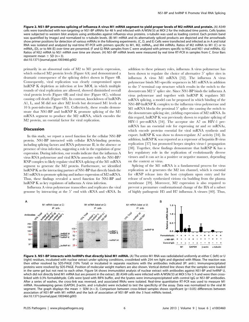
NS1-BP and its interacting partner hnRNP K regulatesplicing of influenza A virus M1 mRNA
To investigate the function of NS1-BP in the influenza virus life
cycle, we knocked down NS1-BP in cells and infected them with
A/WSN/33. We first assessed whether NS1-BP depletion affected
viral entry or vRNP nuclear import by examining the localization
of viral nucleoprotein (NP) in NS1-BP-depleted cells after infection
and in the presence of the translation inhibitor cycloheximide.
Compared to control, NS1-BP knockdown did not decrease
nuclear NP staining (Figure S1), indicating that reduction of viral
RNAs in NS1-BP depleted cells did not alter viral entry or vRNP
nuclear import. Then, viral protein levels were assessed over time
by immunoblot analysis (Figure 2A). While the levels of most viral
proteins were not significantly altered, we observed an abnormal
M2/M1 protein ratio in NS1-BP depleted cells suggesting a role
Author Summary
Influenza A virus is a major human pathogen, which causesapproximately 500,000 deaths/year worldwide. In pan-demic years, influenza infection can lead to even highermortality rates, as in 1918, when ,30–50 million deathsoccurred worldwide. In this manuscript, we identified anovel function for the cellular protein termed NS1-BP as aregulator of the influenza A virus life cycle. We found thatNS1-BP, together with other host factors, mediates theexpression of a key viral protein termed M2. NS1-BP and itsinteracting partner hnRNP K specifically regulate alterna-tive splicing of the viral M1 mRNA segment, whichgenerates the M2 mRNA that is translated into theessential viral M2 protein. The M2 protein is key for viraluncoating and entry into the host cell cytoplasm.Altogether, inhibition of NS1-BP and hnRNP K functionsregulate influenza A virus gene expression and replication.In sum, these studies revealed new functions for thecellular proteins NS1-BP and hnRNP K during viral RNAexpression, which facilitate the influenza A virus life cycle.
NS1-BP and hnRNP K Promote Viral RNA Splicing
PLOS Pathogens | www.plospathogens.org 2 June 2013 | Volume 9 | Issue 6 | e1003460
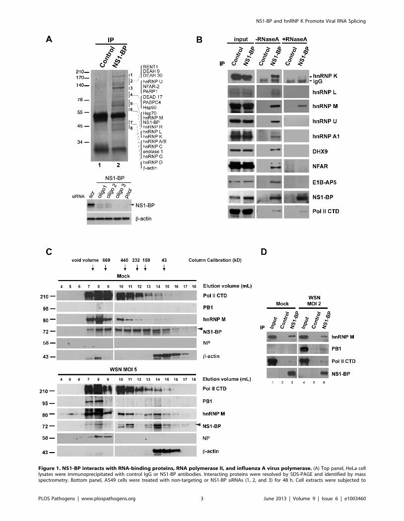
Figure 1. NS1-BP interacts with RNA-binding proteins, RNA polymerase II, and influenza A virus polymerase. (A) Top panel, HeLa celllysates were immunoprecipitated with control IgG or NS1-BP antibodies. Interacting proteins were resolved by SDS-PAGE and identified by massspectrometry. Bottom panel, A549 cells were treated with non-targeting or NS1-BP siRNAs (1, 2, and 3) for 48 h. Cell extracts were subjected to
NS1-BP and hnRNP K Promote Viral RNA Splicing
PLOS Pathogens | www.plospathogens.org 3 June 2013 | Volume 9 | Issue 6 | e1003460

for NS1-BP in regulating splicing of a specific viral mRNA, the M1
mRNA segment.
To investigate this potential effect on splicing, mRNA levels of
M2 and M1 were measured by RT-qPCR. Indeed, the ratio of the
M2 over M1 mRNAs decreased in cells depleted of NS1-BP and
infected with A/WSN/33, indicating a splicing defect (Figure 2B
and 2C). The dynamics of M2 and M1 expression in control cells
was similar to what has been previously reported [26]. Different
dynamics of M2 and M1 expression has also been reported [27],
which may be due to differences in cell types. M2 mRNA is
derived from alternative splicing of the M1 mRNA segment. In
addition, the M1 mRNA segment can generate mRNA3 and, in
certain strains such as A/WSN/33, mRNA4. However, it is
unknown whether mRNA3 encodes a functional peptide
[28,29,30,31]. Recently, mRNA4 was shown to express an isoform
of the M2 ion channel [11]. The 59 splice site that yields mRNA3 is
strong and has to be inhibited by influenza A virus polymerase to
allow usage of the downstream weak splice site that generates the
M2 mRNA, which happens later in infection. Therefore, we also
examined mRNA3 and M4 levels relative to M1 mRNA over time
and found no difference between control and NS1-BP knockdown
cells (Figure 2D and 2E). Together, these findings reveal that
sequences surrounding the M2-specific 59 splice site and/or factors
bound to this region are required for NS1-BP activity on splicing.
We have also tested another viral segment, NS, which undergoes
alternative splicing and generates the NS1 and NS2 mRNAs. In
this case, no differences in ratio were observed between control
and NS1-BP depleted cells (Figure 2F and 2G). Upon NS1-BP
knockdown under these conditions, NS1-BP mRNA levels
remained down-regulated throughout these mRNA measurements
as compared to control (Figure 2H). Thus, these findings show that
NS1-BP specifically regulates splicing of the M1 mRNA segment.
NS1-BP effect on M1 mRNA splicing is consistent with our
findings of NS1-BP interaction with the influenza virus polymer-
ase, which was previously reported to regulate splicing of the M1
mRNA [32]. In addition, our proteomics analysis showed NS1-BP
interaction with hnRNPs previously known to be involved in
splicing such as hnRNP M [33] and hnRNP K [34]. Thus, we
tested whether NS1-BP and the hnRNPs that interacted with NS1-
BP (Figure 1A and 1B) bound directly to M1 RNA. Capped and32P-labeled M1 RNA was incubated with nuclear extracts and
direct protein-RNA interactions were crosslinked with UV,
followed by RNA digestion and immunoprecipitation with
antibodies specific to various individual hnRNPs. The hnRNPs
bound to a 32P-labeled nucleotide (C or U) were then detected. As
shown in Figure 3A, hnRNP U and NS1-BP did not directly bind
M1 RNA whereas hnRNPs A1, M, L, and K directly interacted
with M1 RNA. By contrast, we detect no binding of hnRNP K to
a control RNA derived from the CD45 pre-mRNA (Figure S2)
[35,36]. However, NS1-BP bound the viral M1 segment upon
formaldehyde cross-linking (Figure 3B). In this case, A549 cells
were infected with influenza A/WSN/33 virus at MOI 5 for 5 h,
crosslinked with formaldehyde, and then lysed. Cell lysates were
subjected to immunoprecipitation, and the associated viral RNAs
were isolated and quantified by RT-qPCR. Each experiment
included three conditions as following: immunoprecipitations were
performed with NS1-BP antibodies using lysates with or without
formaldehyde crosslinking, and control IgG was incubated with
lysates subjected to formaldehyde crosslinking. The results indicate
that formaldehyde cross-linked M1 mRNA to NS1-BP as opposed
to three host mRNAs encoding GAPDH, b-actin, and a-tubulin,
which were not cross-linked to M1 mRNA (Figure 3B). Since NS1-
BP interacted with the M1 segment upon formaldehyde treatment
but not upon UV crosslinking, these results indicate that NS1-BP
likely requires adaptor proteins, such as hnRNPs, to interact with
the M1 segment.
The hnRNPs that directly bound the M1 mRNA were then
tested for regulation of M1 mRNA splicing in virus-infected cells.
Each hnRNP was knocked down individually and the ratios of the
M2 over M1 mRNAs were assessed over time. As shown in
Figures 4A and 4B, only depletion of hnRNP K showed a striking
decrease in M1 mRNA splicing, reducing the levels of M2 mRNA.
Furthermore, simultaneous knockdown of NS1-BP and hnRNP K
prevented M2 expression (Figure 4C), indicating that hnRNP K
depletion was sufficient to inhibit M1 mRNA splicing. Taken
together, a model could be proposed in which NS1-BP regulates
M1 mRNA splicing via hnRNP K, resulting in M2 mRNA
expression.
NS1-BP and hnRNP K are required for optimal expressionof viral proteins and viral replication
To determine whether the effect of NS1-BP or hnRNP K on
viral gene expression would result in alteration of viral protein
levels and replication, we examined both parameters in cells
depleted of NS1-BP or hnRNP K versus control cells. As shown in
Figure 2A, the ratio of M2 to M1 at the protein level follows the
effect of NS1-BP depletion on the ratio of M2 and M1 mRNAs
(Figure 2C). Regulation of M2 expression by NS1-BP is an
important step in the viral life cycle as M2 is the ion channel that
allows entry of hydrogen ions from the endosome into the viral
particle resulting in uncoating of the virus and release of viral
content into the cytoplasm. Consequently, the observed effects of
NS1-BP depletion on viral gene expression resulted in down-
regulation of influenza A virus replication in A549 cells (Figure 5A
and 5B). siRNA oligo 1 was more efficient in down-regulating
virus replication (,21 fold at 24 h and ,12 fold at 36 h) than
oligos 2 and 3 (,4–6 fold). While all oligos down-regulated NS1-
BP levels efficiently (Figure 5F), the range of viral titers is likely due
to differences in the kinetics of knockdown for each siRNA oligo.
ATP levels were also measured and the results did not show
significant difference in cell death between control and NS1-BP
depletion during the experimental period (Figure 5C and 5D),
indicating that the decrease of virus replication in NS1-BP
depleted cells was not due to cell death but likely due to NS1-
BP effects on viral gene expression. We have also analyzed virus
replication in human bronchial epithelial cells (HBEC) and
observed a decrease in influenza A virus replication between
,3–9 fold, depending on the siRNA oligo and depicted time
points (Figure 5E and 5F).
Furthermore, depletion of hnRNP K followed by influenza virus
infection at high MOI, single round of viral replication, resulted
immunoblot analysis, which shows NS1-BP knockdown. b-actin served as loading control. (B) Immunoprecipitation was performed with control IgG oranti-NS1-BP antibodies, in the absence or presence of RNase A. Western blots were then performed with the depicted antibodies, selected based onthe proteins identified in A. (C) A549 cells were mock-infected or infected with influenza virus at MOI 5 for 5 h. Cells were lysed and subjected to sizeexclusion chromatography. The fractions were concentrated by TCA precipitation and analyzed by western blot with the indicated antibodies. (D)A549 cells were mock-infected or infected with influenza virus (A/WSN/33) at MOI 2 for 16 h. Cell lysates were immunoprecipitated with control IgGor anti-NS1-BP antibodies. Western blots were performed with the depicted antibodies.doi:10.1371/journal.ppat.1003460.g001
NS1-BP and hnRNP K Promote Viral RNA Splicing
PLOS Pathogens | www.plospathogens.org 4 June 2013 | Volume 9 | Issue 6 | e1003460

NS1-BP and hnRNP K Promote Viral RNA Splicing
PLOS Pathogens | www.plospathogens.org 5 June 2013 | Volume 9 | Issue 6 | e1003460
primarily in an abnormal ratio of M2 to M1 protein expression,
which reduced M2 protein levels (Figure 6A) and demonstrated a
dramatic consequence of the splicing defect shown in Figure 4B.
Consequently, viral replication was clearly compromised upon
hnRNP K depletion as infection at low MOI, in which multiple
rounds of viral replication are allowed, showed diminished overall
viral protein levels (Figure 6B) and viral titer (Figure 6C), without
causing cell death (Figure 6D). In contrast, knockdown of hnRNPs
A1, L, and M did not alter M2 levels but decreased M1 levels at
10 h post-infection (Figure S3). Collectively, these results demon-
strate that NS1-BP and hnRNP K promote splicing of the M1
mRNA segment to produce the M2 mRNA, which encodes the
M2 protein, an essential factor for viral replication.
Discussion
In this study, we report a novel function for the cellular NS1-BP
protein. NS1-BP interacted with cellular RNA-binding proteins,
including splicing factors and RNA polymerase II, in the absence or
presence of virus infection, suggesting a role in the regulation of gene
expression. During infection, our results indicate that the influenza A
virus RNA polymerase and viral RNAs associate with the NS1-BP/
RNP complex to likely regulate viral RNA splicing of the M1 mRNA
segment to generate the M2 protein. Furthermore, we identified
hnRNP K as the interacting partner of NS1-BP that directly binds the
M1 mRNA to promote splicing and induce expression of M2 mRNA.
Thus, these findings revealed a novel function for NS1-BP and
hnRNP K as key regulators of influenza A virus infection.
Influenza A virus polymerase transcribes and replicates the viral
genome by interacting at the 59 end with cRNA and vRNA. In
addition to these primary roles, influenza A virus polymerase has
been shown to regulate the choice of alternative 59 splice sites in
influenza A virus M1 mRNA [32]. The influenza A virus
polymerase binds M1-specific sequences in M1 mRNA in addition
to the 59-terminal cap structure which results in the switch to the
downstream M2 59 splice site. Since NS1-BP binds the influenza A
virus polymerase and together with hnRNP K regulates M1
mRNA splicing, a model can be proposed in which binding of the
NS1-BP-hnRNP K complex to the influenza virus polymerase and
M1 mRNA blocks the proximal 59 splice site causing the switch to
the downstream splicing site, yielding expression of M2 mRNA. In
this regard, hnRNP K was previously shown to regulate splicing of
HIV-1 pre-mRNA [34]. The acceptor site A7 on HIV-1 pre-
mRNA has an essential role for expressing tat and rev mRNAs,
which encode proteins essential for viral mRNA synthesis and
export. hnRNP K was show to down-regulate A7 activity [34]. In
addition, hnRNP K was reported as a repressor of hepatitis B virus
replication [37] but promoted herpes simplex virus-1 propagation
[38]. Together, these findings demonstrate that hnRNP K has a
key regulatory role in the replication of evolutionarily diverse
viruses and it can act in a positive or negative manner, depending
on the context or virus.
Splicing of the M1 mRNA is a fundamental process for virus
replication as it generates the M2 ion channel, which is essential
for vRNP release into the host cytoplasm upon entry and for
release of newly synthesized virions via budding from the plasma
membrane [39]. Moreover, M2 expression is also required to
prevent a premature conformational change of the HA of a subset
of highly pathogenic H5 and H7 influenza A viruses [40]. Thus,
Figure 2. NS1-BP promotes splicing of influenza A virus M1 mRNA segment to yield proper levels of M2 mRNA and protein. (A) A549cells were transfected with non-targeting or NS1-BP siRNAs for 48 h and infected with A/WSN/33 at MOI 2 for the indicated time points. Cells lysateswere subjected to western blot analysis using antibodies against influenza virus proteins. a-tubulin was used as loading control. Each protein bandwas quantified by ImageJ and normalized to a-tubulin levels. (B) M1 mRNA and its alternatively spliced products are depicted and the arrowheadsshow primer positions for detection of various mRNAs in the following experiments. (C, D, and E) Cells were transfected and infected as in A and totalRNA was isolated and analyzed by real-time RT-PCR with primers specific to M1, M2, mRNA3, and M4 mRNAs. Ratios of M2 mRNA to M1 (C) or tomRNA3 (D), or to M4 (E) over time are presented. (F and G) RNA samples from C were analyzed with primers specific to NS2 and NS1 viral mRNAs. (G)Ratios of NS2 mRNA to NS1 mRNA over time are shown. (H) NS1-BP mRNA levels were measured by real time RT-PCR in samples from C. Error Barsrepresent mean 6 SD (n = 3).doi:10.1371/journal.ppat.1003460.g002
Figure 3. NS1-BP interacts with hnRNPs that directly bind M1 mRNA. (A) The entire M1 RNA was radiolabeled uniformly at either C (left) or U(right) residues, incubated with nuclear extract under splicing conditions, crosslinked with 254 nm light and digested with RNase. The reaction wasthen either resolved by SDS-PAGE (10% Total) or incubated in separate reactions with the antibodies indicated (IP: anti-). Immunoprecipitatedproteins were resolved by SDS-PAGE. Position of molecular weight markers are also shown. Vertical dotted line shows that the samples were loadedin the same gel but not next to each other. Figure S4 shows immunoblot analysis of nuclear extract with antibodies against NS1-BP and hnRNP U,which did not directly bind M1 mRNA but are present in the extract. (B) A549 cells were infected with A/WSN/33 at MOI 5 for 5 h and were then cross-linked with 0.3% formaldehyde. Cells were lysed with RIPA buffer, and the lysates were immunoprecipitated with control IgG or NS1-BP antibodies.After a series of washes, the cross-link was reversed, and associated RNAs were isolated. Real-time quantitative RT-PCR was used to measure M1mRNA. Housekeeping genes (GAPDH, b-actin, and a-tubulin) were included to test the specificity of the assay. Data was normalized to the viral Msegment. The graph displays the mean 6 SEM (n = 3). Comparison between cross-linked samples shows significant (p,0.05) differences betweenassociation of NS1-BP with M1 mRNA and the lack of association of NS1-BP with the 3 host mRNAs tested.doi:10.1371/journal.ppat.1003460.g003
NS1-BP and hnRNP K Promote Viral RNA Splicing
PLOS Pathogens | www.plospathogens.org 6 June 2013 | Volume 9 | Issue 6 | e1003460

depletion of NS1-BP or hnRNP K is likely to have more impact on
these highly pathogenic avian influenza viruses.
The influenza A virus NS1 protein was recently shown to
regulate the M1 mRNA splicing [27] and the involvement of the
influenza A virus polymerase is not clear in this case. However,
NS1 was previously shown to interact with the influenza A virus
polymerase [41] and temperature-sensitive viruses expressing C-
terminal truncations of NS1 presented defective RNA replication
and late gene expression [42]. Therefore, future studies are
necessary to determine whether NS1 regulates influenza A virus
polymerase function in splicing and if this mechanism is related to
NS1-BP function. One possibility is that influenza A virus may use
NS1-dependent and independent mechanisms to regulate splicing
of the M1 mRNA segment, which might occur at different stages
of infection. In fact, host splicing can be differentially regulated
depending on various stimuli, resulting in diverse effects such as
differential protein-protein interactions, nucleo-cytoplasmic traf-
ficking and transcription elongation [43]. Further experimentation
using influenza A virus lacking the NS1 gene, which replicates in
interferon-deficient systems [44], will be necessary to understand
the impact of NS1 on NS1-BP function. Interestingly, NS1-BP
levels did not affect splicing of the NS mRNA, indicating again
differential splicing or factors involved in the splicing regulation of
the M1 and NS1 mRNAs.
In sum, our findings reveal a novel role for NS1-BP and hnRNP
K as host regulators of influenza A virus RNA biogenesis. In future
studies, these findings may serve as paradigm to uncover a
potential role for NS1-BP in splicing of specific classes of host pre-
mRNAs. Since specific sequences surrounding the M2 59 splice
site are required for NS1-BP-hnRNP K activity on splicing, one
can envisage NS1-BP regulating splicing, via hnRNP K, of a
subset of host pre-mRNAs containing similar sequences. It would
be interesting if host mRNAs regulated by NS1-BP and hnRNP K
encode proteins involved in immune response during viral
infection, which is an interesting topic for future investigation. In
sum, the study presented here exposed novel host-viral interactions
that regulate splicing of influenza A virus RNA, which play a
significant role in the virus life cycle.
Materials and Methods
Cells and virusHuman lung adenocarcinoma epithelial cells (A549) were
cultured in RPMI 1640 media supplemented with 10% heat-
inactivated fetal bovine serum (Invitrogen), 100 units/mL penicil-
lin, and 100 mg/mL streptomycin. HeLa and MDCK cells were
cultured in DMEM media containing the same amount of serum
and antibiotics described above. Human bronchial epithelial cells
(HBEC) were cultured in Keratinocyte-SFM supplied with human
recombinant epidermal growth factor, bovine pituitary extract
(Invitrogen), 100 units/mL penicillin, and 100 mg/mL streptomy-
cin. All cells were maintained at 37uC with 5% CO2.
Influenza virus (A/WSN/33) was propagated in MDCK cells.
In brief, MDCK cells (26107) were seeded into a 15 cm dish. At
the second day, cells were infected with viruses at multiplicity of
infection (MOI) of 0.001 in 2.5 mL of infection media [EMEM,
10 mM HEPES, 0.2% BSA, 100 units/mL penicillin, 100 mg/mL
streptomycin, and 0.5 mg/mL TPCK-treated trypsin]. After 1 h,
cells were washed with PBS, overlaid with 10 mL infection media,
and incubated until viral cytopathic effect was observed (,36 h).
Supernatants were harvested, centrifuged at 10006g for 10 min,
aliquotted, and stored at 280uC. Virus titer was determined by
plaque assay (see below). Infection of A549 cells was performed in
the same manner as above, except that the infection media did not
contain TPCK-treated trypsin.
PlasmidsFull-length cDNA clone of human NS1-BP (NM_006469.4) was
purchased from OriGene Technologies, Inc. (Rockville, MD) and
subcloned into Sal I and Not I sites of pGEX-4T-2 (GE
Healthcare). The M2 was cloned from infected A549 cells by
RT-PCR and inserted into the pGEM-T vector.
AntibodiesGST-tagged full-length NS1-BP was used as antigen to develop
polyclonal antibodies in rabbits. The specific antiserum was
purified by affinity chromatography [45] with some modifications.
In brief, GST and full-length GST-NS1-BP were expressed in
BL21(DE3) cells and purified with glutathione-Sepharose 4B
beads (GE Healthcare). Without elution, protein GST and GST-
NS1-BP were covalently linked to glutathione-Sepharose beads by
using the cross-linker dimethyl pimelimidate-HCl (Sigma). Crude
serum was loaded onto the GST column to remove the non-
specific antibodies and the flow through was collected and loaded
onto the GST-NS1-BP column. After washes, the antibodies were
eluted with 0.1 M Glycine (pH 2.5) and immediately neutralized
to pH 7.5 by 3M Tris-HCl (pH 8.8). The eluted fractions were
collected and concentrated in a Centricon (Millipore). The
concentration of antibodies was adjusted to 1 mg/mL and used
for western blotting at a dilution of 1:1000.
hnRNP M monoclonal antibody (a gift from Dr. Maurice S.
Swanson, University of Florida, FL) was used for western blot
Figure 4. hnRNP K regulates M1 mRNA splicing. (A) Cell extracts from A549 cells transfected with non-targeting siRNAs or siRNAs that targetvarious indicated hnRNPs for 48 h were subjected to immunoblot analysis with antibodies specific to each hnRNP. (B and C) A549 cells weretransfected as in B and then infected with A/WSN/33 at MOI 2 for the indicated time points. In C, NS1-BP and hnRNP K were simultaneously knockeddown. Total RNA was isolated at each depicted time point and analyzed by real-time RT-PCR with primers specific to M1 and M2 mRNAs. M2/M1mRNA ratios are shown. Error bars represent mean 6 SD (n = 3). *p,0.05, **p,0.01, ***p,0.001.doi:10.1371/journal.ppat.1003460.g004
NS1-BP and hnRNP K Promote Viral RNA Splicing
PLOS Pathogens | www.plospathogens.org 7 June 2013 | Volume 9 | Issue 6 | e1003460

analysis at a 1:4000 dilution. hnRNP A1 monoclonal antibody (a
gift from Dr. Michael Matunis, Johns Hopkins School of Public
Health, MD) was used at a dilution 1:1000. NFAR polyclonal
antibody (a gift from Dr. Glen Barber, University of Miami Miller
School of Medicine, FL) was used at a dilution 1:2000. hnRNP U
monoclonal antibody (ImmuQuest) was used at a 1:1000 dilution.
hnRNPs K and L antibodies (abcam) were used at dilutions 1:3000
and 1:2000, respectively. E1B-AP5 polyclonal antibody (Protein-
tech Group Inc) was used at a 1:2000 dilution. DHX9 monoclonal
antibody (Abnova) was used at a 1:500 dilution. RNA polymerase
II monoclonal antibody, clone CTD4H8, recognized both
phosphorylated and non-phosphorylated forms (Millipore), was
Figure 5. NS1-BP is required for optimal influenza A virus replication. (A) and (B) A549 cells were transfected with non-targeting or NS1-BPsiRNAs for 48 h and infected with A/WSN/33 at MOI 0.001. Supernatants were harvested at 24 h and 36 h post-infection, and virus titer wasdetermined by plaque assay. (C) and (D) After removal of supernatants from plaque assays in A and B, the remaining cells were used to measure ATPlevels, at 24 h and 36 h post-infection, to determine cell viability. (E) Human bronchial epithelial cells (HBEC) were transfected and infected as in Aexcept that different NS1-BP siRNAs were used. Supernatants were subjected to plaque assay. (F) Cell extracts from A549 cells transfected with siRNAsas in A and HBEC transfected with siRNAs as in E were subjected to immunoblot analysis with anti-NS1-BP antibodies. * denotes a cross-reacting bandthat serves as loading control. *p,0.05, **p,0.01.doi:10.1371/journal.ppat.1003460.g005
NS1-BP and hnRNP K Promote Viral RNA Splicing
PLOS Pathogens | www.plospathogens.org 8 June 2013 | Volume 9 | Issue 6 | e1003460

used at a 1:5000 dilution. Influenza A PB1 polyclonal antibody
(Santa Cruz Biotechnology) was used at a 1:5000 dilution. Anti-
NS1 polyclonal antibody [46] was used at a 1:30,000 dilution, and
anti-M1/M2 monoclonal antibody [47] was used at a 1:10,000
dilution. Anti-NP monoclonal antibody (ab20343, Abcam) was
used at a 1:3200 dilution for immunostaining. Anti-influenza A
virion polyclonal antibodies (Meridian Life Science, Inc.) was used
for NP western blot analysis at a 1:10,000 dilution. b-actin
monoclonal antibody (Sigma) was used at a 1:20,000 dilution. a-
tubulin monoclonal antibody (Sigma) was used at a 1:20,000
dilution.
Immunoprecipitation and mass spectrometryHeLa cells were lysed with a buffer containing: 50 mM Tris,
pH 7.5, 150 mM NaCl, 1% IGEPAL CA-630 (Sigma), 0.1 mM
Na3VO4, 1 mM NaF, 1 mM DTT, 1 mM EDTA, 1 mM PMSF,
16 complete protease inhibitor cocktail (Roche), and 15%
glycerol, for 20 min on ice. Cell lysates were centrifuged at
13,0006g for 15 min to pellet cellular debris. For experiments
treated with RNase A, the enzyme was added to the lysates at a
final concentration of 1 mg/mL. Five mg of cell lysates were pre-
cleared with protein A beads (GE Healthcare) for 1 h; the pre-
cleared lysates were incubated with new protein A beads and 6 mg
of control rabbit IgG (Santa Cruz Biotechnology) or purified anti-
NS1-BP antibody at 4uC for 2 h. Beads were washed with lysis
buffer 46, mixed with sample buffer and loaded onto a SDS-
PAGE. After electrophoresis, the gel was stained with Colloidal
blue (Invitrogen). Each lane was excised into 8 fragments
containing proteins ranging from 250 kDa to 30 kDa. Gel slices
were subjected to in-gel digestions followed by LC/MS/MS
analysis that were performed at the Protein Identification core
facility at University of Texas Southwestern Medical Center at
Dallas. Acquired results were searched against the NCBI-nr
protein database with Mascot software (Matrix Science).
Size exclusion chromatographyA549 cells were grown to 90–100% confluency and mock-
infected or infected with virus at MOI 5 for 5 h. Cells were lysed,
Figure 6. hnRNP K is required for optimal M2 protein production and influenza A virus replication. A549 cells were transfected withnon-targeting or hnRNP K siRNAs for 48 h prior to infection. siRNA transfected cells were infected with A/WSN/33 at (A) MOI 2 or (B) MOI 0.001. Cellswere harvested at the indicated hours post-infection, and viral protein accumulation was assessed by immunoblot analysis. Each protein band in (A)and (B) was quantified by ImageJ and normalized to a-tubulin levels. (C) Control or hnRNP K siRNA transfected cells were infected with A/WSN/33 atMOI 0.001. At 36 hours post-infection, cell supernatants were collected and subjected to viral titer analysis (n = 3) or (D) ATP level analysis todetermine cell viability (n = 3, representative experiment). Error bars denote mean + SEM. *p,0.05.doi:10.1371/journal.ppat.1003460.g006
NS1-BP and hnRNP K Promote Viral RNA Splicing
PLOS Pathogens | www.plospathogens.org 9 June 2013 | Volume 9 | Issue 6 | e1003460

as described in the previous section, and passed through
QIAshredder columns (Qiagen). Lysates (8 mg) were centrifuged
twice at 150006g for 20 min and subjected to a Superdex 200
column (GE Healthcare). One-milliliter fractions were collected
from number 1 to 25, and 200 mL of each fraction were
precipitated with 2% trichloroacetic acid with 8 volume of cold
acetone. The solution was incubated at 220uC overnight, and
protein pellets were precipitated at 150006g for 20 min and
washed with cold acetone. The pellets were air dried, resuspended
in sample buffer, and resolved by SDS-PAGE.
RNA interference and transfectionsiRNA oligos (siGENOME SMARTpool) designed for silencing
human IVNS1ABP and hnRNPs were purchased from Dharma-
con (Thermo Fisher Scientific). Non-targeting siRNAs, AUGAAC-
GUGAAUUGCUCAAdTdT together with siGENOME non-
targeting siRNA #3 (Thermo Fisher Scientific) were used as
controls. The siRNAs were transfected with RNAiMAX (Invitro-
gen), 1.5 mL of RNAiMax was used per 25 pmol siRNA,
according to manufacture’s instruction. A549 cells were seeded
into 12-well plates at a density 16105 the day before transfection,
and cells were transfected with 50 nM of non-targeting siRNAs or
siRNAs specific for NS1-BP or hnRNPs. Knockdown efficiency
was detected by western blot 48 h after transfection.
Isolation of RNAs associated with NS1-BPRNA associated with NS1-BP were isolated as described [48].
In brief, A549 cells were grown to 90–100% confluency
(,7.56106 cells) and infected with influenza virus at MOI 5 for
5 h. To induce crosslinking, cells were incubated with 0.3%
formaldehyde/PBS (Electron Microscopy Sciences) at room
temperature for 10 min. Non-crosslink control was incubated
with 10 mL PBS. Crosslinking reaction was quenched by adding
1.25 mL of 2 M glycine (pH 7.0) and incubated at room
temperature for 5 min. Cells were lysed with RIPA buffer and
then centrifuged at 16,000 g for 10 min at 4uC. Five percent of
lysates were saved as input. Immunoprecipitation was performed
as previously described with 6 mg of anti-NS1-BP antibodies or
purified IgG from pre-immune rabbit serum. After washes, 100%
beads and the 5% of input were mixed with reverse buffer [10 mM
Tris (pH 6.8), 5 mM EDTA, 10 mM DTT, and 1% SDS] and
incubated at 70uC for 45 min to reverse the cross-link. Samples
were then mixed with Proteinase K at 37uC for 30 min, and RNA
were extracted with Phenol:Chloroform:Isoamyl Alcohol (Invitro-
gen). After precipitation, RNA was subjected to DNase I digestion
followed by phenol:chloroform extraction. RNA was reversed
transcribed into cDNA and analyzed by qPCR. The amount of
RNA was calculated as 22Ct, and the RNA associated with NS1-
BP was normalized to the input amount and expressed as
percentage of IP [%IP = (22Ct Bead/22Ct input)65%]. To show
the relative amount of different RNAs, we set the %IP of pull-
down M segment to 1, and the %IP of each mRNA was
normalized to the M segment.
RNA purification and RT-qPCRTotal RNA was isolated from A549 cells with RNA Isolation
Kit (Roche) and 0.5 mg of total RNA was reverse transcribed into
cDNA by SuperScript II reverse transcriptase (Invitrogen).
Reactions were set up according to the manufacturer’s protocol
in the presence of 1 mM of specific primers as following: oligo-
(dT)15 for mRNA and random hexamers (Roche) for host 18S
rRNA. RT was carried on 42uC for 1 h and inactivated by heating
at 80uC for 20 min. RT reactions were then diluted with water at
a ratio 1:5, and 1.25 mL of cDNA mixture was subjected to
quantitative real-time PCR (qPCR) using SYBR green I Master
combined LightCycler 480 System (Roche). Real-time PCR was
carried out by initial denaturation at 95uC for 3 min, 40 cycles of
95uC for 15 sec, 60uC for 15 sec, 72uC for 20 s, and followed by a
melting curve cycle from 65uC to 95uC for quality assurance.
Gene-specific primers were designed by D-LUX program
(Invitrogen) and validated by analysis of template titration and
dissociation curves. Primer Sequences used in this study are listed
as follows (59R39):
18S rRNA (Forward: ACCGCAGCTAGGAATAATGGA,
Reverse: GCCTCAGTTCCGAA AACCA); RPL32 (Forward:
CGGCGTGCAACAAATCTTACTGTGCCG, Reverse: CCAG
TTGGGCAGCTCTTTCC); GAPDH (Forward: CGACCG-
GAGTCAACGGATTTGGTCG, Reverse: CCAGTTGGGCA-
GCTCTTTCC); b-actin (Forward: CCGCGAGAAGATGACCC
AGAT, Reverse: CGTTGGCACAGCCTGGATAGCAACG); a-
tubulin (Forward: CACTCT GATTGTGCCTTCATGG, Re-
verse: CGAGCTTAGTGTAGGTTGGGCGCTCG); NS1-BP
(Forward: CGCTGGTAATCAACTGGGTGCAGCG, Reverse:
ACCTCTTCCATCAGCTC TTCCA); NS (Forward: CAGGA-
CATACTGATGAGGATG, Reverse: GTTTCAGAGACTC-
GAACTGTG) NS1 (Forward: TGGAAAGCAAATA GTG-
GAGCG, Reverse: GTAGCGCGATGCAGGTACAGAG); NS2
(Forward: CAAGCTT TCAGGACATACTGATGAG, Reverse:
CTTCTCCAAGCGAATCTCTGTAGA); M1 (Forward: AT-
CAGACATGAGAACAGAATGG, Reverse: TGCCTGGCCT-
GACTAGCAA TATC); M2 (Forward: CGAGGTCGAA-
ACGCCTATCAGAAAC, Reverse: CCAATGATA TTTGCTG-
CAATGACGAG); mRNA3 (Forward: GCAAAAGCAGGGCC-
TATCAGAAAC, Reverse: CCAATGATA TTTGCTGCAAT-
GACGAG); M4 (Forward: ACCGATCTTGAGGCCTATCA-
GAAAC, Reverse: CCAATGATA TTTGCTGCAATGACG-
AG).
UV crosslinking and RNA-protein interactionThe full length M1 RNA (1070 nt) was transcribed from a PCR
template by T7 polymerase using the standard GpppG cap
analogue to cap the 59 end and the indicated 32P-NTPs to 32P-
labeled the RNA throughout it’s length. Labeled M1 RNA
(10 nM) was incubated with 60% JSL1 nuclear extract in a total
volume of 12.5 ml under splicing condition, which contains (final
concentration): 12 mM Tris-HCl, pH 7.5, 3.2 mM MgCl2, 1 mM
ATP, 20 mM CP, 1 mM DTT, 0.125 U RNasin (Promega),
60 mM KCl and 12% glycerol. Reactions were incubated at 30uCfor 20 min, crosslinked using UV light (254 nm) for 20 min on ice,
and digested with RNaseT1 and RNase A for 20 min at 37uC.
Reactions were resuspended in 26SDS loading buffer, denatured
for 5 min at 95uC, analyzed under denaturing conditions on an
SDS-PAGE gel (Acrylamide/Bis 37.5:1, BioRad), and detected by
autoradiography. Immunoprecipitation after crosslinking was
carried out as described previously [35,36], with the antibodies
against hnRNP L (4D11, abcam), hnRNP K (3C2, abcam),
hnRNP A1 (4B10, abcam) hnRNP M (2A6, abcam), hnRNP U
(3G6, abcam), and NS1BP.
Plaque assayOne day before the assay, MDCK cells were seeded into 6-well
plates at a density of 56105 cells/well. Ten-fold serial dilutions of
each supernatant (from 1021 to 1027) were prepared in 400 mL
PBS containing 100 units/mL penicillin, 100 mg/mL streptomy-
cin, 0.2% BSA, 0.9 mM CaCl2, and 1.05 mM MgCl2. Cells were
infected with 300 mL of each dilution. After 1 h of adsorption,
inoculums were removed, and cells were overlaid with 2 mL of
agar mixture [16 EMEM, 0.6% agar 100 units/mL penicillin,
NS1-BP and hnRNP K Promote Viral RNA Splicing
PLOS Pathogens | www.plospathogens.org 10 June 2013 | Volume 9 | Issue 6 | e1003460

100 mg/mL streptomycin, 2 mM L-glutamine, 0.2% bovine serum
albumin (BSA), 10 mM HEPES, 0.22% sodium bicarbonate,
0.01% DEAE-Dextran, and 0.5 mg/mL TPCK-treated trypsin].
Plates were incubated at 37uC with 5% CO2 for 48 h. After 2
days, cells were fixed by adding 2 mL of 2% formaldehyde on the
top of the agar and incubated at room temperature for 1 h. After
removal of the agar, monolayers were stained with 0.1% crystal
violet prepared in 0.25% methanol, and the plaques were counted.
SDS-PAGE and western blot analysisGel electrophoresis was performed using the Tricine-SDS
PAGE system as described [49]. Acrylamide gels used in this
study were 7%, acrylamide solution was 40%, and acrylamide/bis-
acrylamide ratio was 5%. Running gel buffer contained 1.5 M
Tris-HCl, pH 8.45; stacking gel buffer contained 0.3 M Tris-HCl
and 0.4% SDS, pH 7.8. Cathode buffer contained 0.1 M Tris,
0.1 M Tricine, and 0.1% SDS, pH 8.25. Anode buffer was 0.2 M
Tris-HCl, pH 8.9. Thirty ml of the running gel mixture was
prepared for 4 gels, which includes 5.25 mL of acrylamide
solution, 10 mL of running gel buffer, 4 g glycerol, 10.64 mL
water, 100 mL ammonium persulfate (10%), and 10 mL of
TEMED (N,N,N9,N9-tetramethylethane-1,2-diamine). The stack-
ing gel mixture contained 1.23 mL of acrylamide solution, 3 mL
of stacking gel buffer, 7.77 mL of water, 100 mL of ammonium
persulfate (10%), and 10 mL of TEMED. Samples were mixed
with equal volume of 26 sample buffer [100 mM Tris-HCl, 8%
SDS, 24% glycerol, 200 mM DTT, and 0.02% bromophenol
blue, pH 7.8], heated at 95uC for 10 min, and loaded onto the
wells. The electrophoresis was performed at a constant voltage of
120 V until the tracking dye reached the edge of the gel.
After electrophoresis, proteins were transferred to a PVDF
membrane (Millipore) using a mini transfer electrophoretic cell
with transfer buffer [20 mM Tris, 192 mM Glycine, and 10%
methanol] at a constant voltage of 30 V overnight. The PVDF
membrane was then blocked with 5% skim milk in PBS for 1 h,
followed by two PBS-T (0.05% Tween 20 in PBS) washes, and
incubated with primary antibody (prepared in 10 mg/mL BSA/
PBS) at room temperature for 1 h. After incubation, the
membrane was washed 5 times with PBS-T buffer for 5 min
each, and then incubated with HRP-conjugated secondary
antibodies for another hour. Finally, the PVDF membrane was
washed 4 times with PBS-T buffer for 10 min each, submerged in
ECL reagent (34096, Pierce, Thermo Scientific) for 5 min, and
exposed to a film in the dark room for 20 s to 5 min until the
signal was visible.
Expression and purification of GST-NS1-BPThe plasmid pGEX-NS1BP was transformed into BL21(DE3)
cells. A single colony was inoculated into 2.5 ml of LB with
100 mg/ml of ampicillin and incubated at 37uC in a shaker at
225 rpm overnight. Next day, the overnight culture was added
into 250 ml of 2-YT broth (Invitrogen) and grown to OD 0.6 at
37uC. Protein expression was induced by 0.2 mM isopropyl-beta-
D-thiogalactopyranoside (IPTG) at 20uC for 6 h. Bacteria were
pelleted and re-suspended in 25 ml of cold lysis buffer [20 mM
HEPES, pH 7.5, 1 mM EDTA, 300 mM NaCl, and 16protease
inhibitor cocktail (Roche)]. Cells were lysed by five passages
through an EmulsiFlex-C5 homogenizer (Avestin, Canada) at
8000 psi, and lysates were centrifuged at 13,0006g for 15 min.
The supernatant was incubated with 2 ml (50% slurry) glutathione
Sepharose beads at 4uC for 1 h, and beads were washed 10 times
with cold lysis buffer. GST-NS1-BP was eluted with 50 mM
glutathione solution (pH 7.5).
Immunostaining and fluorescence microscopyA549 cells were seeded in 12-well plates at a density of 16105
cells/well the day before siRNA transfection. At the second day,
cells were treated with 50 nM of non-targeting or NS1-BP
siRNAs. After 24 h, cells in each well were trypsinized and split
into 4 wells of 24-well plate including a coverslip per well, and
incubated for another 24 hours. To synchronize the infection, cells
were pre-chilled on ice for 10 min before infection, washed with
cold PBS, and infected with influenza virus at MOI 50. After
45 min adsorption on ice, cells were washed with PBS and
overlaid with 0.5 ml of warm infection media including 1 mM
cycloheximide. At the indicated time points, cells on coverslips
were fixed with 3% formaldehyde/PBS at room temperature for
15 min, washed 36with PBS, and then permeabilized with 0.5%
TritonX-100/PBS at room temperature for 5 min. Then,
coverslips were incubated with NP antibody (ab20343, Abcam)
prepared in BSA solution (1 mg/ml) at a dilution of 1:3200 at
room temperature for 1 h. After washed 36 in PBS, coverslips
were incubated with Alexa Fluor 488 donkey anti-mouse IgG
(Invitrogen) at a dilution of 1:500 in BSA for another 1 h, followed
by three washes in PBS. Coverslips were further incubated with
Hoechst (0.5 mg/ml) (Invitrogen) at room temperature for 5 min
and washed in PBS. After the final PBS wash, coverslips were
mounted onto glass microscope slides with mounting media (Dako,
Denmark). Images were taken and processed in the Zeiss Axioplan
2E microscope, Hamamatsu monochrome digital camera, and
OpenLab software.
Accession numbersNS1-BP (NM_006469); hnRNP M (EAW68921) hnRNP A1
(P09651); hnRNP U (Q00839); DDX17 (CAG30318); Hsp 70
(NP_006588); Hsp90 (AAA36025); NFAR-2 (AAD51099); PARP-
1 (EAW69785); polyadenylate-binding protein 4 (NP_003810); Pol
II CTD (NM_000937), Influenza virus (A/WSN/1933(H1N1))
strain: PB1 (CY034138); PA (CY034137); M1 (L25818); NP
(ACF54602); M2 (AAA91324); NS1 (K01076); NS2 (ABF47961).
Supporting Information
Figure S1 NS1-BP depletion does not affect virus entryand vRNP nuclear import. A549 cells were grown on
coverslips, transfected with non-targeting or NS1-BP siRNAs for
48 h, and infected with A/WSN/33 at MOI 50 on ice to
synchronize infection. After virus adsorption, cells were incubated
with warm media containing 1 mM cycloheximide and cells were
then fixed with 3% formaldehyde at each indicated time point. (A)
Cells were stained with Hoechst (top panels) and antibody against
influenza A virus nucleoprotein (NP) (bottom panels). Scale bar:
10 mm. (B) The ratio of NP nuclear localization/cell number at
3.5 h post-infection was determined in control and NS1-BP-
depleted cells. The results represent the average ratio from three
independent experiments and ,150 cells were counted in each
experiment. Error bars are mean6SD (n = 3). (C) and (D) Control
for cycloheximide treatment showing low levels of p53, which is
short-lived. In parallel wells, cells were transfected with non-
targeting or NS1-BP siRNAs for 48 h and used as a control to
demonstrate knockdown efficiency.
(TIFF)
Figure S2 hnRNP L but not hnRNP K bind to CD45 exon4. A 100 nt, capped and 32P-labeled RNA corresponding to exon
4 of the human CD45 gene was incubated in nuclear extract and
crosslinked with UV light to analyze protein-RNA complexes.
Antibodies specific to hnRNP L efficiently immunoprecipitated a
protein-RNA complex while no bound protein was observed with
NS1-BP and hnRNP K Promote Viral RNA Splicing
PLOS Pathogens | www.plospathogens.org 11 June 2013 | Volume 9 | Issue 6 | e1003460

antibody to hnRNP K or an IgG control. Molecular weight
markers and a total crosslink reaction are shown.
(TIFF)
Figure S3 hnRNPs A1, L, and M do not alter M2expression. A549 cells were transfected with non-targeting or
hnRNPA A1, L, and M siRNAs for 48 h prior to infection. siRNA
transfected cells were infected with A/WSN/33 at MOI 2. Cells
were harvested at the indicated hours post-infection, and viral
protein accumulation was assessed by immunoblot analysis. Each
protein band was quantified by ImageJ and normalized to a-
tubulin levels.
(TIFF)
Figure S4 Detection of NS1-BP and hnRNP U in JSL1nuclear extract. JSL1 nuclear extract was subjected to
immunoblot analysis with antibodies specific to hnRNP U (A)
and NS1-BP (B), detected using different amounts of extract. MW,
molecular weight markers.
(TIFF)
Table S1 Identification of NS1-BP binding proteins bymass spectrometry. HeLa cell lysates were immunoprecipitat-
ed with control IgG or NS1-BP antibodies in the absence of
RNase. Interacting proteins were resolved by SDS-PAGE and
identified by mass spectrometry. The gel lane was divided into
eight segments from molecular weight 250 kDa to 35 kDa, and
the top hits of each segment are listed in the table.
(DOCX)
Acknowledgments
We thank Nicholas Conrad for scientific advice and Ramanavelan
Sakthivel for assistance.
Author Contributions
Conceived and designed the experiments: BMAF KWL AGS PLT.
Performed the experiments: PLT NTC SK. Analyzed the data: BMAF
PLT SK NTC AGS KWL. Contributed reagents/materials/analysis tools:
BMAF AGS KWL. Wrote the paper: PLT BMAF AGS.
References
1. WHO (April 2009) http://www.who.int/mediacentre/factsheets/fs211/en/
index.html. Fact sheet Number 211.
2. Webster RG (1999) 1918 Spanish influenza: the secrets remain elusive. Proc Natl
Acad Sci U S A 96: 1164–1166.
3. Palese P, Shaw ML (2007) Orthomyxoviridae: The virus and their replication.
In: Knipe, D.M, et al. Eds. Fields Virology. 5th edition. Philadelphia, USA:
Lippincott Williams & Wilkins. 1647–1689.
4. Palese P, Shaw ML (2007) Orthomyxoviridae: The virus and their replication.
In: Knipe, D.M, et al. Eds. Fields Virology. 5th edition. Lippincott Williams &
Wilkins, Philadelphia, USA.
5. Wise HM, Foeglein A, Sun J, Dalton RM, Patel S, et al. (2009) A complicated
message: Identification of a novel PB1-related protein translated from influenza
A virus segment 2 mRNA. J Virol 83: 8021–8031.
6. Chen W, Calvo PA, Malide D, Gibbs J, Schubert U, et al. (2001) A novel
influenza A virus mitochondrial protein that induces cell death. Nat Med 7:
1306–1312.
7. Gibbs JS, Malide D, Hornung F, Bennink JR, Yewdell JW (2003) The influenza
A virus PB1-F2 protein targets the inner mitochondrial membrane via a
predicted basic amphipathic helix that disrupts mitochondrial function. J Virol
77: 7214–7224.
8. Zamarin D, Garcia-Sastre A, Xiao X, Wang R, Palese P (2005) Influenza virus
PB1-F2 protein induces cell death through mitochondrial ANT3 and VDAC1.
PLoS Pathog 1: e4.
9. Jagger BW, Wise HM, Kash JC, Walters KA, Wills NM, et al. (2012) An
overlapping protein-coding region in influenza A virus segment 3 modulates the
host response. Science 337: 199–204.
10. Muramoto Y, Noda T, Kawakami E, Akkina R, Kawaoka Y (2013)
Identification of novel influenza A virus proteins translated from PA mRNA.
J Virol 87: 2455–2462.
11. Wise HM, Hutchinson EC, Jagger BW, Stuart AD, Kang ZH, et al. (2012)
Identification of a novel splice variant form of the influenza A virus M2 ion
channel with an antigenically distinct ectodomain. PLoS pathogens 8:
e1002998.
12. Selman M, Dankar SK, Forbes NE, Jia J-J, Brown EG (2012) Adaptive mutation
in influenza A virus non-structural gene is linked to host switching and induces a
novel protein by alternative splicing. . Emerg Microbes Infect 1: e42.
13. Medina RA, Garcia-Sastre A (2011) Influenza A viruses: new research
developments. Nature reviews Microbiology 9: 590–603.
14. Versteeg GA, Garcia-Sastre A (2010) Viral tricks to grid-lock the type I
interferon system. Curr Opin Microbiol 13: 508–516.
15. Nemeroff ME, Barabino SM, Li Y, Keller W, Krug RM (1998) Influenza virus
NS1 protein interacts with the cellular 30 kDa subunit of CPSF and inhibits 39
end formation of cellular pre-mRNAs. Mol Cell 1: 991–1000.
16. Satterly N, Tsai PL, van Deursen J, Nussenzveig DR, Wang Y, et al. (2007)
Influenza virus targets the mRNA export machinery and the nuclear pore
complex. Proc Natl Acad Sci U S A 104: 1853–1858.
17. Zhang L, Das P, Schmolke M, Manicassamy B, Wang Y, et al. (2012) Inhibition
of Pyrimidine Synthesis Reverses Viral Virulence Factor-Mediated Block of
mRNA Nuclear Export. J Cell Biol 196: 315–326.
18. Woff T, O’Neil RE, Palese P (1998) NS1-Binding protein (NS1-BP): a novel
human protein that interacts with the influenza A virus nonstructural NS1
protein is relocalized in the nuclei of infected cells. J Virol 72: 7170–7180.
19. Fortes P, Lamond AI, Ortin J (1995) Influenza virus NS1 protein alters the
subnuclear localization of cellular splicing components. J Gen Virol 76: 1001–
1007.
20. Bortz E, Westera L, Maamary J, Steel J, Albrecht RA, et al. (2011) Host- and
strain-specific regulation of influenza virus polymerase activity by interacting
cellular proteins. mBio 2: e00151–00111.
21. Jorba N, Juarez S, Torreira E, Gastaminza P, Zamarreno N, et al. (2008)
Analysis of the interaction of influenza virus polymerase complex with human
cell factors. Proteomics 8: 2077–2088.
22. Mayer D, Molawi K, Martinez-Sobrido L, Ghanem A, Thomas S, et al. (2007)
Identification of cellular interaction partners of the influenza virus ribonucleo-
protein complex and polymerase complex using proteomic-based approaches.
Journal of proteome research 6: 672–682.
23. Engelhardt OG, Smith M, Fodor E (2005) Association of the influenza A virus
RNA-dependent RNA polymerase with cellular RNA polymerase II. J Virol 79:
5812–5818.
24. Engelhardt OG, Fodor E (2006) Functional association between viral and
cellular transcription during influenza virus infection. Rev Med Virol 16: 329–
345.
25. Biswas SK, Boutz PL, Nayak DP (1998) Influenza virus nucleoprotein interacts
with influenza virus polymerase proteins. J Virol 72: 5493–5501.
26. Valcarcel J, Portela A, Ortin J (1991) Regulated M1 mRNA splicing in influenza
virus-infected cells. J Gen Virol 72 (Pt 6): 1301–1308.
27. Robb NC, Fodor E (2012) The accumulation of influenza A virus segment 7
spliced mRNAs is regulated by the NS1 protein. J Gen Virol 93: 113–118.
28. Lamb RA, Choppin PW (1981) Identification of a second protein (M2) encoded
by RNA segment 7 of influenza virus. Virology 112: 729–737.
29. Lamb RA, Lai CJ, Choppin PW (1981) Sequences of mRNAs derived from
genome RNA segment 7 of influenza virus: colinear and interrupted mRNAs
code for overlapping proteins. Proc Natl Acad Sci U S A 78: 4170–4174.
30. Shih SR, Suen PC, Chen YS, Chang SC (1998) A novel spliced transcript of
influenza A/WSN/33 virus. Virus genes 17: 179–183.
31. Winter G, Fields S (1980) Cloning of influenza cDNA ino M13: the sequence of
the RNA segment encoding the A/PR/8/34 matrix protein. Nucleic acids
research 8: 1965–1974.
32. Shih SR, Nemeroff ME, Krug RM (1995) The choice of alternative 59 splice sites
in influenza virus M1 mRNA is regulated by the viral polymerase complex. Proc
Natl Acad Sci U S A 92: 6324–6328.
33. Hovhannisyan RH, Carstens RP (2007) Heterogeneous ribonucleoprotein m is a
splicing regulatory protein that can enhance or silence splicing of alternatively
spliced exons. J Biol Chem 282: 36265–36274.
34. Marchand V, Santerre M, Aigueperse C, Fouillen L, Saliou JM, et al. (2011)
Identification of protein partners of the human immunodeficiency virus 1 tat/rev
exon 3 leads to the discovery of a new HIV-1 splicing regulator, protein hnRNP
K. RNA biology 8: 325–342.
35. Lynch KW, Maniatis T (1996) Assembly of specific SR protein complexes on
distinct regulatory elements of the Drosophila doublesex splicing enhancer.
Genes & development 10: 2089–2101.
36. Rothrock CR, House AE, Lynch KW (2005) HnRNP L represses exon splicing
via a regulated exonic splicing silencer. The EMBO journal 24: 2792–2802.
37. Ng LF, Chan M, Chan SH, Cheng PC, Leung EH, et al. (2005) Host
heterogeneous ribonucleoprotein K (hnRNP K) as a potential target to suppress
hepatitis B virus replication. PLoS medicine 2: e163.
38. Schmidt T, Striebinger H, Haas J, Bailer SM (2010) The heterogeneous nuclear
ribonucleoprotein K is important for Herpes simplex virus-1 propagation. FEBS
Lett 584: 4361–4365.
39. Rossman JS, Jing X, Leser GP, Lamb RA (2010) Influenza virus M2 protein
mediates ESCRT-independent membrane scission. Cell 142: 902–913.
NS1-BP and hnRNP K Promote Viral RNA Splicing
PLOS Pathogens | www.plospathogens.org 12 June 2013 | Volume 9 | Issue 6 | e1003460
40. Ciampor F, Bayley PM, Nermut MV, Hirst EM, Sugrue RJ, et al. (1992)
Evidence that the amantadine-induced, M2-mediated conversion of influenza Avirus hemagglutinin to the low pH conformation occurs in an acidic trans Golgi
compartment. Virology 188: 14–24.
41. Marion RM, Zurcher T, de la Luna S, Ortin J (1997) Influenza virus NS1protein interacts with viral transcription-replication complexes in vivo. J Gen
Virol 78 (Pt 10): 2447–2451.42. Falcon AM, Marion RM, Zurcher T, Gomez P, Portela A, et al. (2004) Defective
RNA replication and late gene expression in temperature-sensitive influenza
viruses expressing deleted forms of the NS1 protein. J Virol 78: 3880–3888.43. Heyd F, Lynch KW (2011) Degrade, move, regroup: signaling control of splicing
proteins. Trends in biochemical sciences 36: 397–404.44. Garcia-Sastre A, Egorov A, Matassov D, Brandt S, Levy DE, et al. (1998)
Influenza A virus lacking the NS1 gene replicates in interferon-deficient systems.Virology 252: 324–330.
45. Bar-Peled M, Raikhel NV (1996) A method for isolation and purification of
specific antibodies to a protein fused to the GST. Anal Biochem 241: 140–142.
46. Solorzano A, Webby RJ, Lager KM, Janke BH, Garcia-Sastre A, et al. (2005)
Mutations in the NS1 protein of swine influenza virus impair anti-interferon
activity and confer attenuation in pigs. J Virol 79: 7535–7543.
47. Bourmakina SV, Garcia-Sastre A (2005) The morphology and composition of
influenza A virus particles are not affected by low levels of M1 and M2 proteins
in infected cells. J Virol 79: 7926–7932.
48. Conrad NK (2008) Chapter 15. Co-immunoprecipitation techniques for
assessing RNA-protein interactions in vivo. Methods Enzymol 449: 317–342.
49. Schagger H, von Jagow G (1987) Tricine-sodium dodecyl sulfate-polyacrylamide
gel electrophoresis for the separation of proteins in the range from 1 to 100 kDa.
Anal Biochem 166: 368–379.
NS1-BP and hnRNP K Promote Viral RNA Splicing
PLOS Pathogens | www.plospathogens.org 13 June 2013 | Volume 9 | Issue 6 | e1003460