cellular to el-deleted - pnas · be activated with the inducible cellular factor e2f (27)....
TRANSCRIPT
Proc. Natl. Acad. Sci. USAVol. 91, pp 4407-4411, May 1994Medical Sciences
Cellular immunity to viral antigens limits El-deleted adenovirusesfor gene therapy
(liver/cytotoxic T lymphocytes/genetic diseases)
YIPING YANG*t, FREDERICK A. NUNES*t, KLARA BERENCSIt, EMMA E. FURTH§, EVA G6NCZ6L0,AND JAMES M. WILSON*t#1*Institute for Human Gene Therapy and Departments of tMolecular and Cellular Engineering, and IPathology and Laboratory Medicine, University ofPennsylvania Medical Center, and OThe Wistar Institute of Anatomy and Biology, Philadelphia, PA 19104
Communicated by Francis S. Collins, January 4, 1994 (receivedfor review November 3, 1993)
ABSTRACT An important limitation that has emerged inthe use of adenoviruses for gene therapy has been loss ofrecombinant gene expression that occurs concurrent with thedevelopment of pathology In the organ exp g the trans-gene. We have used liver-dircted approaches to gene therapyin mice to study mecanims that underlie the problems withtransient expression and pathology that have characterized invivo applications of first-generation recombinant adenoviruses(i.e., those deleted of Ela and Elb). Our data are consistentwith the following hypothesis. Cells harboring the recombinantviral genome express the transgene as desired; however, low-level expression of viral genes also occurs. A virus-specificcellular immune response is stimulated that leads to destructionof the genetically modified hepatocytes, massive hepatitis, andrepopulation of the liver with nontransgene-containing hepa-tocytes. These findigs suggest approaches for improving re-combinant adenoviruses that are based on further crippling thevirus to limit expression of nondeleted viral genes.
Human adenoviruses have emerged as a promising technol-ogy for in vivo gene therapy. The 36-kb double-strandedDNA genome of these viruses contains 25 a series of earlygenes encoding regulatory products and late genes encodingstructural products. An important step in the design ofrecombinant adenoviruses for gene therapy was the isolationof a packaging cell line that provides in trans the essentialregulatory genes Ela and Elb (1). By using this cell line, it ispossible to produce recombinants containing a transgene inwhich the virus was rendered replication-defective by delet-ing El sequences.Recombinant adenoviruses have many of the requisite fea-
tures for a useful in vivo gene delivery vehicle in that they canbe purified and grown in large quantities, and they very effi-ciently transduce genes into a wide spectrum of nondividingcells in vivo (2-8). Application ofadenoviral technology for thetreatment of chronic disorders such as genetic diseases willrequire very prolonged, if not stable, expression of the recom-binant gene or repeated administration of the vehicle withoutthe development of destructive or inhibiting responses.
Critical evaluation of this technology for in vivo genetherapy has revealed several important limitations. In thebest characterized systems (i.e., gene transfer to lung andliver), expression of the transgene has been transient andassociated with the development of pathology in the targetorgan (9-13). In addition, it appears that repeated adminis-tration of the recombinant virus is associated with confound-ing immune responses. In this report, we have used mousemodels to evaluate the role of the immune system in the
pathology and transient recombinant gene expression thathas characterized El-deleted viruses for in vivo gene therapy.
MATERIALS AND METHODSIn Vivo Delivery ofRecombinant AdenovisestoMouse Liver.
Ad.CBlacZ, an El-deleted replication-deficient recombinantadenovirus carrying the lacZ minigene driven by a cytomega-lovirus-enhanced (3-actin promoter, was constructed (14). It isbased on human adenovirus type 5 (AdS) with a small deletionof the E3 region. None of the viral stocks used in the experi-ments contained detectable replication-competent virus.Ad5dlE3, a replication-competent AdS with deletion of E3region, was described (15). Virus (1 x 10¹¹ plaque-formingunits/ml) in 0.1 ml of phosphate-buffered saline (PBS, pH 7.4)was infused retrograde into the biliary tract offemale CBA andnu/nu mice as described (16). All animals that received recom-binant virus survived to necropsy.DNA and RNA Analyses. Total cellular DNA and RNA
were prepared from liver tissues and analyzed as described(17, 18). Probes included a 2.0-kb Escherichia coli lacZDNAfragment (Cla I-EcoRI) for DNA hybridization and a 1.6-kbAdS hexon DNA fragment for RNA hybridization.
Morphological Analyses. S-Bromo-4-chloro-3-indolyl (-D-galactoside (X-Gal) histochemistry. Frozen sections (6 ,um)were fixed in 0.5% glutaraldehyde and stained for 3-galac-tosidase activity as described-(16).
Histopathology. Paraffin sections (5 pm) were stained withhematoxylin/eosin. Random sections were examined forhistopathology in a blinded fashion.Immunohistochemical detection of viral proteins. Frozen
sections (6 ian) were fixed in 2% (wt/vol) paraformaldehydeand permeabilized as described (19). Indirect immunofluo-rescence was performed with a hybridoma supernatant toAd5 DNA binding protein (an E2a gene product) (20), andAdS hexon protein was detected with fluorescein isothiocy-anate-labeled polyclonal antibody (Ab1O56F, Chemicon).BrdUrd labeling and detection of mitotically active cells.
Animals were labeled for 4 h with BrdUrd at 100 mg/kgbefore sacrifice at each time point. Frozen sections werefixed in methanol and analyzed by immunohistochemicalmethod using a monoclonal antibody to BrdUrd as described(21). The percentage of BrdUrd labeling was calculated bycounting 2000 cells from 10 random fields for each animal.
Cytotoxic T-Lymphocyte (CTL) Assay. CBA mice infusedwith Ad.CBlacZ were sacrificed at day 14 and the spleenswere aseptically removed for preparation of cell suspensions.Cells were suspended at 2.5 x 10⁶ viable cells per ml andrestimulated in vitro using Ad5dlE3 with multiplicity of
Abbreviations: AdS, adenovirus type 5; X-Gal, 5-bromo4chloro-3-indolyl P-D-galactoside; CTL, cytotoxic T lymphocyte; MOI, mul-tiplicity of infection; MHC, major histocompatibility complex.ITo whom reprint requests should be addressed.
4407
The publication costs of this article were defrayed in part by page chargepayment. This article must therefore be hereby marked "advertisement"in accordance with 18 U.S.C. §1734 solely to indicate this fact.
4408 Medical Sciences: Yang et al.
infection (MOI) of 20 for 5 days in 24-well plates. Cytolyticactivity of nonadherent spleen cells (effectors) was tested ina chromium release assay.
RESULTS AND DISCUSSIONThe most extensive evaluation of adenoviral technology forin vivo gene therapy has been performed in the lung and liver(9-13). In these organs, expression of the recombinant virus
Proc. Natl. Acad. Sci. USA 91 (1994)
has been efficient but transient and, when carefully exam-ined, associated with inflammation. Transient expression ofthe transgene has also been associated with adenoviral-mediated gene transfer to other organs (5, 8). Exceptions tothis include studies that evaluated adenovirus-mediated genetransfer in (i) newborn animals (2, 22), (ii) immunoprivilegedorgans (i.e., retina) (7), and (iii) human xenografts grown inimmunodeficient animals (23). In each case, transgene ex-
*h )\ n'-YJ~~~~~~
Iw ~v -
44*
j~~~~~~~~~~E U,; *,~ *Q i , aa-*
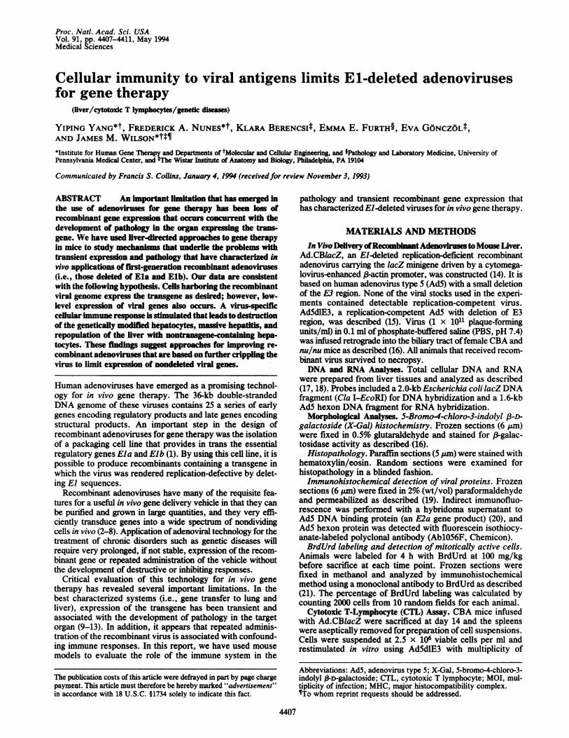
mlofPBS) and_euhnzd and.lie tissus wereealuate fo Z exrsso usin h X-al hitceia stiig"dC~aZi
nulnu~~~~Xmic at 3pday (A) 7 dy(C, 2days (Ead6 days (G1) an Ad.C~aZif e CA mic at 3,days (, 7 as hi) 21 das~~~s&and35 day (H are #vshown .\ ;§;X160.r
*, S. Q
_¢s .Z gb< *,° e t1 .r
and~~~~~~~~~~~~~ #35day()aeshwn(x10.
Proc. Natl. Acad. Sci. USA 91 (1994) 4409
CBA Nude
0 2 7 14 21 0 2 7 14 21B 1234
8.4-
28 S-4.8-3.6-
2 3-
18 S
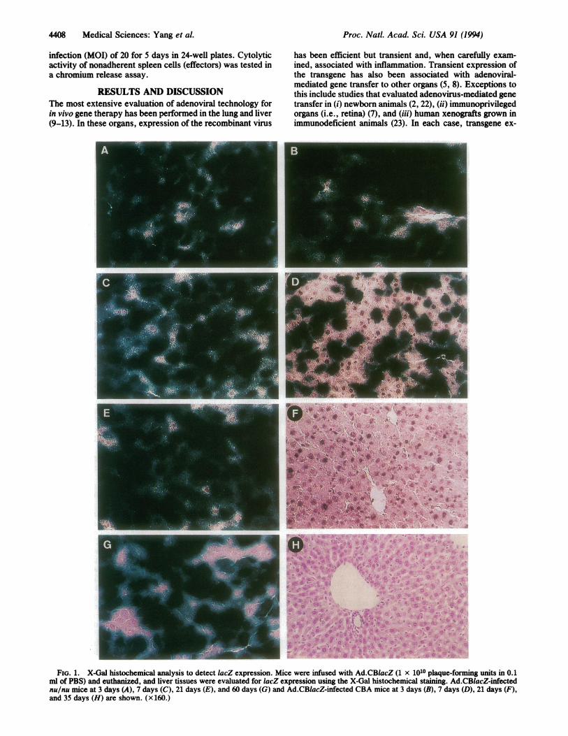
FIG. 2. Analysis of liver for viral DNA and RNA. (A) Totalcellular DNA (10 jig) was prepared from liver tissues of CBA (lanesCBA) and nu/nu (lanes Nude) mice, digested with BamHI, andanalyzed on a Southern blot using an E. coli lacZ probe. Numbersindicate the day after gene transfer on which tissue was harvested.DNA (10 pg) from uninfected mice plus 7.5 pg (lane 1 copy), 75 pg(lane 10 copies), and 750 pg (lane 100 copies) ofpAd.CBlacZ plasmidDNA was used as copy number controls. Molecular size standardsare indicated to the left. (B) Total cellular RNA (10 ug) fromuninfected (lane 3), Ad.CBlacZ-infected (lanes 1 and 2), andAd5dlE3-infected (lane 4) liver tissues ofCBA mice were analyzed.The intensity of the ribosomal markers was identical in each lane,indicating equivalent quantities of electrophoresed RNA.
pression was stable for prolonged periods of time withoutsignificant pathology. These informative exceptions suggest
that the immune system may play a role in mediating themajor limitations of adenovirus technology.We propose a hypothesis to explain the mechanisms that
underlie the development of pathology and loss of geneexpression that have limited the utility of El-deleted adeno-viruses for in vivo gene therapy. We suggest that cellsharboring the recombinant genome express adenoviral pro-teins despite the absence of Ela and Elb. This could occurthrough transactivation by cellular El-like factors or basalexpression of adenoviral promoters (24, 25). Presentation ofthe newly synthesized viral proteins in the context of histo-compatibility antigens would lead to the generation of spe-cific cellular immune responses to, and destruction of, thegenetically modified cells. The end result is a pathologicalinflammatory response and loss of gene expression as thetissue is replaced with cells that do not contain the transgene.Adenovirus-mediated gene transfer to mouse liver in vivo
was used as a model to evaluate this hypothesis. The mouseis an attractive species for studying immunological mecha-nisms because of the availability of congenic strains andextensive panels of reagents for in vitro immunologic assays.In addition, previous studies have shown that wild-typehuman AdS is capable of lytic infection and replication inmouse hepatocytes that, when administered in vivo, leads todose-dependent hepatitis (26).The role of the immune system in the stability of recom-
binant adenoviral transgene expression was demonstrated inexperiments in which livers from both immunocompetent(CBA) and genetically athymic (nu/nu) strains of mice wereexposed to Ad.CBlacZ by instillation of virus retrograde intothe biliary tract. lacZ expression was demonstrated by X-Gal
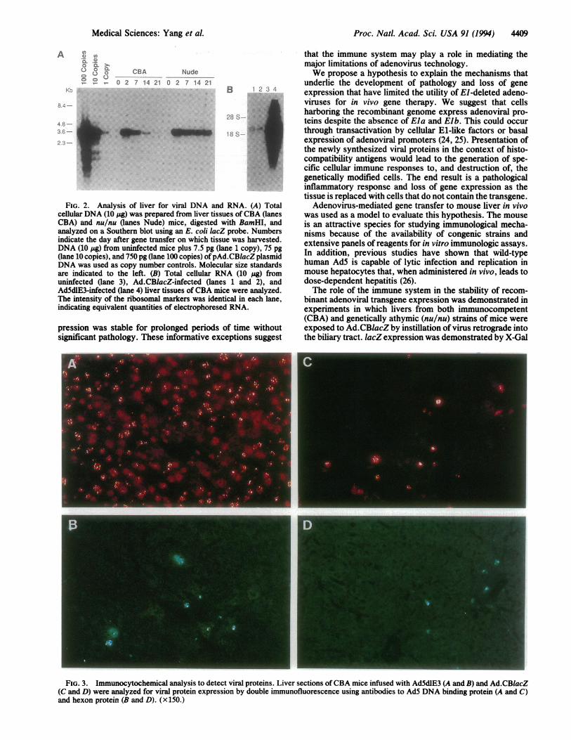
FIG. 3. Immunocytochemical analysis to detect viral proteins. Liver sections of CBA mice infused with Ad5dlE3 (A and B) and Ad.CBlacZ(C and D) were analyzed for viral protein expression by double immunofluorescence using antibodies to AdS DNA binding protein (A and C)and hexon protein (B and D). (x 150.)
A 0 Cn
Kbl
Medical Sciences: Yang et al.
4410 Medical Sciences: Yang et al.
Table 1. Adenovirus-specific MHC class I-restricted lysis of target cells by spleen cells fromAd.CBlacZ-infected CBA mice
Target E/T Specific lysis of infected target cellsMice cells ratio Mock Ad.CBlacZ Ad5dlE3
Ad.CBlacZ-infected L-929 100:1 12.9 ± 0.3 25.0 ± 0.8 26.8 ± 0.650:1 9.6 ± 1.2 18.8 ± 1.2 20.1 ± 1.5
MC57 50:1 4.7 ± 0.6 5.2 ± 0.9 7.8 ± 3.2Uninfected L-929 100:1 10.3 ± 0.9 8.8 ± 0.7 11.7 ± 0.2
50:1 3.0 ± 0.8 5.6 ± 0.8 8.5 ± 2.1MC57 50:1 3.1 ± 0.2 3.0 ± 1.3 7.6 ± 5.6
CBA(H-2k) mice were infected with Ad.CBlacZ. Two weeks later spleen cells of two mice from eachgroup were pooled and restimulated in vitro with Ad5dlE3 for 5 days. MHC-matched L-929 (H-2k) andMHC-mismatched MC57 (H-2b) target cells were mock-infected or infected with Ad.CBlacZ (MOI =100-200) for 72 h orwith Ad5dlE3 (MOI = 50-100) for 48 h. Target cell killing is expressed as a percentage(mean ± SD) of CTL-induced release of incorporated 51Cr from target cells in a 4-h cytolysis assay.
histochemistry in >80% of hepatocytes of both strains ofmice when analyzed 2 days after gene transfer (Fig. 1 A andB). In CBA mice, expression diminished to undetectablelevels by day 21 (Fig. 1F), whereas no diminution in expres-sion was detected in nu/nu mice during the longest intervalstudied, which was 60 days in the initial studies (Fig. 1G).Southern blot analysis of total cellular DNA from liver ofrecipient animals was performed to further delineate themechanism for the selective loss of gene expression inimmunocompetent animals (Fig. 2A). ViralDNA remained athigh levels (%5 copies per cell) for the duration of theexperiment in nu/nu mice, whereas the viral DNA progres-sively diminished from -5 copies per cell to <0.2 copy percell over 21 days in CBA mice. This indicates that loss of theviral genome, rather than extinction of expression, is theprimary mechanism responsible for loss of lacZ activity.CBA mice were exposed to either recombinant adenovirus
and liver tissue was analyzed for presence of viral proteins byimmunocytochemistry and viralRNAs byRNA hybridization.Expression of the early gene E2a, which encodes a DNAbinding protein essential forDNA replication, was detected inthe nuclei of the majority of hepatocytes in animals treatedwith Ad5dlE3 (Fig. 3A) and in a subpopulation of hepatocytesin animals treated with Ad.CBlacZ (Fig. 3C). Expression ofthis gene in the absence ofEla was not surprising since it canbe activated with the inducible cellular factor E2F (27).Expression of the prototypic late gene product hexon wasdemonstrated in a smaller population ofperiportal hepatocytesin Ad.CBlacZ-infected animals (Fig. 3D). Hexon-containingtranscripts were also detected in these tissues, at levels-200-fold lower than that achieved with Ad5dlE3 (Fig. 2B,compare lanes 1 and 2 to lane 4). The requirement of Ela foractivation of late transcription would suggest that little hexonshould be present in Ad.CBlacZ-infected animals. The findingof small but detectable hexon RNA and protein suggestslow-level transactivation of the major late promoter by Ela-independent mechanisms.Animals were analyzed for the development of CTLs to
adenoviral proteins in splenocytes harvested 2 weeks afterexposure to Ad.CBlacZ (Table 1). Significant lysis (P < 0.01)was demonstrated to major histocompatibility complex(MHC)-matched target cells (L-929) infected with eitherAd5dlE3 or Ad.CBlacZ, compared to mock-infected L-929cells. Specificity of the assay was confirmed by absence ofsignificant lysis with infected MHC-mismatched target cells(MC57). A CTL response was not detected in splenocytesfrom naive animals.
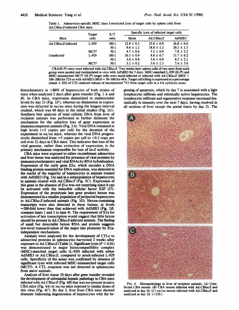
Analysis of liver tissue 10 days after gene transfer revealedthe development of substantial hepatic pathology in CBA miceinfected with Ad.CBlacZ (Fig. 4B) that was not present in naiveCBA mice (Fig. 4A) or nu/nu mice exposed to similar doses ofthis virus (Fig. 4C). By day 2, liver from CBA mice showeddramatic ballooning degeneration of hepatocytes with the be-
ginning of apoptosis, which by day 7 is associated with a lightlymphocytic infiltrate and mitotically active hepatocytes. Thelymphocytic infiltrate and regenerative response increased dra-matically in intensity over the next 7 days, having resolved inall sections of liver except the portal tracts by day 21. The
-- .. .. -.- V
FIG. 4. Histopathology in liver of recipient animals. (A) Unin-fected CBA mouse. (B) CBA mouse infected with Ad.CBlacZ andanalyzed at day 10. (C) nu/nu mouse infected with Ad.CBlacZ andanalyzed at day 10. (x130.)
Proc. Natl. Acad Sci. USA 91 (1994)

Proc. Natl. Acad. Sci. USA 91 (1994) 4411
L-~~~~~~~1oD 0)
0.P
OO0 10
1'
Day
FIG. 5. Labeling of hepatocytes with BrdUrd after exposure tovirus. Animals were labeled with BrdUrd (100 mg/kg) and analyzedfor its incorporation into DNA by immunofluorescent staining usinga monoclonal antibody to BrdUrd. The percentage of cells positivefor BrdUrd labeling over time with the solid line and the dash lineindicating CBA and nu/nu mice, respectively.
extensive pathology noted in liver of Ad.CBlacZ-infected ani-mals is not due to direct toxicity of the virus because it was notpresent when UV-inactivated virus was introduced into theseanimals (data not shown). The nu/nu mice developed similarabnormalities in hepatocyte morphology 2 days after exposureto virus but they did not progress to develop lymphocyticinfiltrates and hepatocyte regeneration. The level ofhepatocyteregeneration was quantitated by labeling hepatocytes with thenucleoside analog BrdUrd whose incorporation into DNA canbe detected by immunocytochemistry. On selected days ani-mals were infused with abolus ofBrdUrd and sacrificed 4 h laterfor analysis. Less than 0.01% of nuclei were labeled in unin-fected control animals, whereas at its peak (day 2 after instil-lation of virus), 7% of hepatocytes were labeled in nu/nuanimals and 15-20%o ofhepatocytes were labeled from the CBAmice (Fig. 5). Nuclei labeling was primarily periportal and itquickly diminished to baseline levels in nu/nu animals,whereas, in CBA mice, the labeling was distributed throughoutthe hepatic lobule and it persisted at very high levels for severalweeks (data not shown). The massive regeneration observed inCBA mice is consistent with that which would be required torepopulate this organ with nontransgene-containing cells.Our data are consistent with at least two mechanisms of
injury to the liver in the context of gene transfer withEl-deleted recombinant adenoviruses. One mechanism illus-trated in the nu/nu mouse is characterized by low level andtransient hepatocyte degeneration with compensatory regen-eration in the areas of the liver in which expression of lacZwas the highest (i.e., periportal). These are areas of liverexposed to the highest MOI of virus consistent with directtoxicity of the virus as possibly mediating this effect; thedevelopment of pathology and regeneration within days afterinfusion of virus is consistent with this hypothesis. The majormechanism that underlies loss of recombinant gene expres-sion with the development of pathology is a cellular immuneresponse to de novo-synthesized viral antigens. This effect isindependent of MOI (data not shown) and occurs severaldays later. Our findings are consistent with results of Gins-berg and coworkers (28, 29) who studied the mechanisms ofhuman adenovirus pneumonia in cotton rat and mouse mod-els. They describe a biphasic inflammatory response thelatter of which is mediated by CTLs to viral antigens and isabsent in nu/nu mice.The hypothesis that emerges from this study suggests
mechanisms to explain the major limitations of first genera-tion adenoviruses. The data presented in this report are frommouse liver; however, similar results have been obtained inmouse and cotton rat lung, suggesting this may be a generalparadigm (Y.Y., J.M.W., and J. Engelhardt, unpublished
results). These studies provide a basis for rationally improv-ing this technology. One approach would be to manipulatethe immune system of the recipient to abrogate the cellularimmune response. This could be accomplished by initiallytolerizing the recipient to viral antigens or subjecting therecipient to chronic immunosuppression. A more attractiveapproach would be to further cripple the virus to diminish orablate viral protein expression.
We thank Kim Alston for technical support and the Cell Morphol-ogy Core and Vector Core of the Institute for Human Gene Therapy.This work was supported by the Cystic Fibrosis Foundation andNational Institutes of Health Grants P30 DK 47757 and RO1 HL49040.
1. Graham, F. L., Smiley, J., Russell, W. L. & Nairn, R. (1977) J. Gen.Virol. 36, 59-72.
2. Stratford-Perncaudet, L. D., Levrero, M., Chasse, J. F., Perricaudet,M. & Briand, P. (1990) Hum. Gene Ther. 1, 241-256.
3. Rosenfeld, M. A., Siegfried, W., Yoshimura, K., Yoneyama, K.,Fukayama, M., Steir, L. E., Pfak6, P. K., Gilardi, P., Stratford-Perri-caudet, L. D., Perricaudet, M., Jalla, S., Pavirani, A., Lecocq, J.-P. &Crystal, R. G. (1991) Science 252, 431-434.
4. Quantin, B., Perricaudet, L. D., Tajbakhsh, S. & Mandel, J.-L. (1992)Proc. Nat!. Acad. Sci. USA 89, 2581-2584.
5. Lemarchand, P., Ari Jaffe, H., Danel, C., Cid, M. C., Kleinman, H. K.,Stratford-Perricaudet, L. D., Perricaudet, M., Pavirani, A., Lecocq,J.-P. & Crystal, R. G. (1992) Proc. Natl. Acad. Sci. USA 89,6482-6486.
6. Le Gal La Salle, G., Robert, J. J., Berrard, S., Ridoux, V., Stratford-Perricaudet, L. D., Perricaudet, M. & Mallet, J. (1993) Science 259,988-990.
7. Bennett, J., Wilson, J., Sun, D., Forbes, B. & Maguire, A. (1993) Invest.Ophthalmol. Visual Sci., in press.
8. Roessler, B. J., Allen, E. D., Wilson, J. M., Hartman, J. W. & David-son, B. L. (1993) J. Clin. Invest. 92, 1085-1092.
9. Jaffe, H. A., Danel, C., Longenecker, G., Metzger, M., Setoguchi, Y.,Rosenfeld, M. A., Gant, T. W., Thorgeirsson, S. S., Stratford-Perricau-det, L. D., Perricaudet, M., Pavirani, A., Lecocq, J.-P. & Crystal, R. G.(1992) Nat. Genet. 1, 372-378.
10. Li, Q., Kay, M. A., Finegold, M., Stratford-Perricaudet, L. D. & Woo,S. L. C. (1993) Hum. Gene Ther. 4, 403-409.
11. Ishibashi, S., Brown, M. S., Goldstein, J. L., Gerard, R. D., Hammer,R. E. & Herz, J. (1993) J. Clin. Invest. 92, 883-893.
12. Engelhardt, J. F., Simon, R., Yang, Y., Zepeda, M., Weber-Pendelton,S., Doranz, B., Grossman, M. & Wilson, J. M., (1993) Hum. Gene Ther.4, 759-769.
13. Simon, R., Engelhardt, J. F., Yang, Y., Zepeda, M., Weber-Pendelton,S., Grossman, M. & Wilson, J. M. (1993) Hum. Gene Ther. 4, 771-780.
14. Kozarsky, K., Grossman, M. & Wilson, J. M. (1993) Somatic Cell Mol.Genet. 19, 449-458.
15. Berencsi, K., Rando, R. F., deTaisne, C., Paoletti, E., Plotkin, S. A. &Gonczol, E. (1994) J. Gen. Virol. 74, in press.
16. Yang, Y., Raper, S. E., Cohn, J. A., Engelhardt, J. F. & Wilson, J. M.(1993) Proc. Nat!. Acad. Sci. USA 90, 4601-4605.
17. Wilson, J. M., Johnston, D. E., Jefferson, D. M. & Mulligan, R. C.(1988) Proc. Nat!. Acad. Sci. USA 85, 4421-4425.
18. Chomczynski, P. & Sacchi, N. (1987) Anal. Biochem. 162, 156-159.19. Yang, Y., Devor, D. C., Engelhardt, J. F., Ernst, S. A., Strong, T. V.,
Collins, F. S., Cohn, J. A., Frizzell, R. A. & Wilson, J. M. (1993) Hum.Mol. Genet. 2, 1253-1261.
20. Reich, N. C., Sarnow, P., Durpey, E. & Levine, A. (1983) Virology 128,480-484.
21. Engelhardt, J. F., Yankaskas, J. R. & Wilson, J. M. (1992) J. Clin.Invest. 90, 2598-2607.
22. Ragot, T., Vincent, N., Chafey, P., Vigne, E., Gilgenkrantz, H., Couton,D., Cartaud, J., Briand, P., Kaplan, J. C., Perricaudet, M. & Kahn, A.(1993) Nature (London) 361, 647-650.
23. Engelhardt, J. F., Yang, Y., Stratford-Perricaudet, L. D., Allen, E. D.,Kozarsky, K., Perricaudet, M., Yankaskas, J. R. & Wilson, J. M. (1993)Nat. Genet. 4, 27-34.
24. Spergel, J. M., Hsu, W., Akira, S., Thimmappaya, B., Kishimoto, T. &Chen-Kiang, S. (1992) J. Virol. 66, 1021-1030.
25. Imperiale, M. J., Kao, H.-T., Feldman, L. T., Nevins, J. R. & Strick-land, S. (1984) Mol. Cell. Biol. 4, 867-874.
26. Duncan, S. J., Gordon, F. C. A., Gregory, D. W., McPhie, J. L.,Postlethwaite, R., White, R. & Willcox, H. N. A. (1978) J. Gen. Virol.40, 45-61.
27. Reichel, R., Kovesdi, I. & Nevins, J. R. (1988) Proc. Natl. Acad. Sci.USA 85, 387-390.
28. Ginsberg, H. S., Moldawer, L. L., Sehgal, P. B., Redington, M., Kilian,P. L., Chanock, R. M. & Prince, G. A. (1991) Proc. Natl. Acad. Sci.USA 88, 11651-11655.
29. Prince, G. A., Porter, D. D., Jenson, A. B., Horswood, R. L., Chanock,R. M. & Ginsberg, H. S. (1993) J. Virol. 67, 101-111.
Medical Sciences: Yang et al.