cellular uptake of coumarin-6 as a model drug … · journal of physiologyand pharmacology 2011,...
TRANSCRIPT

INTRODUCTIONThe epithelial surface of the lungs is the largest surface area
of the human body in direct contact with the environment and,furthermore, its thickness, in the so called thin part, is in therange 0.1-0.3 µm, reaching about 3 µm in the thick part thatincludes the fibrous components. Accordingly, the alveolarsurface represents an important pathway for inhalation drugdelivery being routinely used to treat allergic, genetic, infectiveor chronic diseases of the respiratory system (1). Only recently,though, the delivery to the lung has been considered foradministering drugs not only for local pathology, but also forsystemic diseases. The pulmonary route, in fact, is attractive forseveral reasons. It is non-invasive, it might allow drugabsorption from a large, highly vascularized surface area, theflow of blood perfusing the lungs is ~5 L/min at rest and drugsthat reach the circulation avoid the first-pass metabolism,actually the main disadvantage of orally administration route (2).
The advent of nanotechnology has introduced a great varietyof nanoparticles (NPs), defined as “objects” with at least onedimension in the nanometer range. This has provoked greatexpectations for new inhalation-targeted drug delivery strategiesincluding the application of therapeutics, vaccines anddiagnostics. On the other hand, the different physico-chemicalproperties of nano-sized particles have raised substantialconcerns about the safety of organic and inorganic nano-sizedmaterial (3). Nevertheless, the application of nanoparticle-based
drug delivery in respiratory diseases has been somewhat limitedby the ability of nanoparticles to reach the alveolarcompartment; in fact, the delivery to the most peripheral lungportions, requires particle size within a very narrow range, about1 µm for Poly(Lactic-co-Glycolic Acid) composites (PLGA) (4,5), and in the nanometer range, less than 250 nm, forbiodegradable particles (6).
Some reports highlight the efficacy of nanoparticles deliveryto the alveolar compartment using either aerosol inhalation (7)and an air-liquid interface exposure system (8-10). Of course,the ideal approach of a therapeutic carrier that targets a potentdrug specifically at site of pathology is still quite far. Thisapproach requires, in fact, the recognition by NPs of a highlyspecific molecular epitope on the plasma-membrane surface, inorder to deliver the drug to the target site minimizing any effectelsewhere. The identification of the epitope is still a majorunsolved problem preventing, so far, the specific targeting ofdiseased or cancer cells. The case of drug delivery system ininflammatory disease, as for tuberculosis (11), is in principleeasier as it resides in phagocytosis by macrophages.
A further problem in alveolar delivery is due to overcomingthe surfactant layer. There is a report indicating that metal NPsalter the surface properties of semi-synthetic surfactant andinterfere with cyclic changes in surface tension oncompression/expansion of the surface film (12). Another reportconfirms an alteration in fatty distribution within the surfactantlayer in presence of poly(amidoamine) dendrimers (13).
JOURNAL OF PHYSIOLOGY AND PHARMACOLOGY 2011, 62, 1, 45-53www.jpp.krakow.pl
I. RIVOLTA1, A. PANARITI1, B. LETTIERO1, S. SESANA, P. GASCO2, M.R. GASCO2, M. MASSERINI1, G. MISEROCCHI1
CELLULAR UPTAKE OF COUMARIN-6 AS A MODEL DRUG LOADED IN SOLID LIPID NANOPARTICLES
1Department of Experimental Medicine, University of Milano Bicocca, Monza, Italy; 2Nanovector, Turin, Italy
The aim of present work was to elucidate the interaction of solid lipid nanoparticles (SLNs) with cellular plasma-membraneto gain insight of intracellular drug delivery. To this aim we followed the uptake of coumarin-6 (a drug model) either freein the extracellular medium or loaded on SLN (c-SLN). Alveolar epithelial cells were exposed to a biocompatibleconcentration of c-SLN (0.01 mg/ml of tripalmitin) prepared by warm microemulsion whose lipid matrix was constitutedby low melting point molecules (fatty acids, triglycerides). Intracellular fluorescence and preferential accumulation in theperinuclear region were increased by 54.8% on comparing c-SLN to the same amount of free coumarin-6 in the medium.Lowering temperature from 37° to 4°C decreased the intracellular signal intensity by about 48% equally for the free as wellas for loaded drug, thus suggesting the inhibition of a similar non-endocytotic entrance pathway. No specific co-localizationof the fluorescence with intracellular organelles was found. The c-SLN calorimetric profile obtained with differentialscanning calorimetry (DSC), revealing transition within the range 58-62°C, altered remarkably upon incubation with cells,suggesting a change in SLN structure after association with cells membranes. We propose that the uptake of the model drugloaded on SLN is only partly related to the endocytotic pathway; it occurs despite the loss of integrity of the original SLNstructure and it appears to be more efficient when the drug is vehicled rather than being free in the culture medium.
K e y w o r d s : coumarin-6, differential scanning calorimetry, effect of temperature on uptake, nanoparticles uptake, solid lipidnanoparticles, reactive oxigen species

A further criticism concerns the toxicity of nanomaterials. Itis known that NPs inhaled from the ambient or at the work placemay trigger inflammatory reactions: NPs translocation maydecrease the immune defences, cause pulmonary endothelialdisfunction and general systemic effects (3). In this context it isof interest to recall the attempt to use carbon nanotubes,extremely toxic by nature (14, 15), as biocompatible drug carrierafter coating them with a naturally derived surfactant (16).
Solid lipid nanoparticles (SLN) represent an interesting drugdelivery system as an alternative to liposomes and polymericnanoparticles. SLN are constituted of biocompatible componentssimilar to plasma membrane as well as to the lipidic part ofalveolar surfactant and can be prepared by several methods (highpressure homogenization, warm microemulsions, solventemulsification-evaporation-diffusion, high speed stirring, and/orsonication) (17).
Our interest is based on previous results indicating therelatively easy uptake of SLN by alveolar cells (18). Therefore,the aim of present work is to elucidate the interaction of SLNwith plasma membrane of alveolar epithelial cells to gain insightconcerning the process of intracellular drug delivery. To this aimwe followed the uptake of coumarin-6 used as a lipophilic drugmodel either free in the extracellular medium or loaded on SLN.
MATERIALS AND METHODS
Preparation and characterizationSolid lipid nanoparticles (SLN) were produced by
NANOVECTOR, in the terms of the CE Contract STREP NLSHB-CT-2006-037639-BONSAI (Bio-imaging with SmartFunctional Nanoparticles) by choosing tripalmitin as lipidicmatrix. SLN have been loaded with coumarin-6 (3-(2’-benzothiazolyl)-7-diethylaminocoumarin -MW 350,4 Da), c-SLN, to allow their visualization by means of fluorescentmicroscope (λexc 450 nm, λem 505 nm). c-SLN were prepared bydispersing in cold water (2°C) a warm microemulsion consistingof tripalmitin (Sigma), Epikuron 200 (Cargill), sodiumtaurocholate (PCA-Italy) and ultrapure water (Millipore-Milliq).Coumarin-6 (Acros) was added to the lipid phase ofmicroemulsion. 2-phenylethanol (Fluka) was present aspreservant. c-SLN dispersion has been washed four times bytangential flow filtration (Sartorius, Vivaflow 50 Cassette, RCMembrane, Cut Off 100 kDa), followed by heat sterilization andfinal storage overtime at 4°C until use.
The concentration of coumarin-6 in c-SLN dispersion wasdetermined by fluorimetry (Jasco FP2020 Plus Fluorimeter, λexc450 nm and λem 505 nm) after dissolving SLN dispersion inchloroform/methanol (6/4).
In experiments where coumarin-6 was used as free drug, weprepared a stock solution at a concentration of 20 µg/ml indimethylsulfoxide (DMSO) to be diluted at the working solutiondesiderated.
The hydrodynamic diameter (Zave), polydispersity index (PI)and Zeta Potential (Zpot) of SLN dispersion was characterized byphoton correlation spectroscopy (PCS) using a Malvern Zetasizer3000 HSA instrument at a fixed angle of 90° and a temperature of25°C (Laser λ 633 nm). Each value was the average of tenmeasuraments. To evaluate the stability of c-SLN in physiologicalmedium, NPs were incubated up to 24 hours in the cell culturemedium supplemented with Pen/Strep and L-glutamine.
Cell cultureHuman embrionic kidney (HEK), African green monkey
SV40-transfected kidney fibroblast cell line (COS-7) and A30
cells were grown on Petri dishes in Dulbecco’s modified eagle’smedium (DMEM) supplied with 10% fetal bovine serum (FBS),1% of L-glutamine and 1% of penicillin/streptomycin(Pen/Strep) and incubated in a controlled environment at 37°Cwith 5% CO2. During all the experiments cells were incubatedwith medium supplemented with 1% FBS to prevent seruminterference in the toxicity assay. A30 cells (19) represent acontinuous alveolar cell line obtained from lung specimens frompatients who underwent lobectomy in the Thoracic Surgery Unitof S. Gerardo Hospital, Monza. These cells were isolated fromthe apparently healthy portion of the excised lung lobe.
ToxicityThe evaluation of biocompatibility of c-SLN on A30 was
analyzed by lactate dehydrogenase (LDH) release assay and byMTT ((3-(4,5-dimethylthiazol-2-yl)-2,5-diphenyltetrazoliumbromide) test according to the direction of the manufacturer(Clonetech and Sigma Aldrich). A dose-response curve, from 0.9ng/ml up to 9 mg/ml, was performed to determine the celldamage.
Time course of fluorescence intracellular distributionThe experimental set up consisted in a wide field
fluorescence microscope NIKON Eclipse FN1, equipped withmotorized table PRIOR and Metamorph software for imagingacquisition and analysis. In order to obtain the images of the c-SLN we used a Fluorescein Isothiocyanate (FITC) filter (λmax488 nm). The kinetic of the internalization up to 30 minutes, wasstudied on cells kept under the microscope at 32°C. During theexperiment c-SLNs were added at a concentration of 9 µg/ml, ina way to guarantee no toxicity for the cells for the time of theexposure. The magnification of the objective was 63x andallowed a field view of at least 5 cells. The acquisition time was400 ms. Intracellular fluorescence was expressed as percentageof a maximum value detected or as arbitrary units (a.u.), asdescribed in figure legends.
Effect of temperature on fluorescence intracellular distributionCells were exposed for 45 minutes to c-SLN 9 µg/ml either
at 37°C, or at 4°C. In this latter case cells were pre-cooled at 4°Cfor 30 min before the incubation. Images were acquired alongthe Z axis at 0.5 µm distance starting from top of the cell; on theaverage we acquired images over 40 Z planes. We considered theoverall fluorescence acquired in wide field as well as, through adeconvolution process (Autodeblur), the fluorescence estimatedon a Z plane corresponding to the mid cell height.
ImmunocytochemistryCells were plated on glass coverslips and, at confluence,
they were incubated with or without c-SLN 9 µg/ml. After 45minutes, cells were fixed with paraformaldehyde 4% in PBS atroom temperature for 20 min. Cells were washed three times inphosphate buffered saline (PBS), LS (low salt PBS) and in HS(high salt PBS) respectively and then permeabilized withdigitonin 0.01% in GDB for 30 min. Fixed and permeabilizedcells were incubated with primary antibody diluted in GDB atroom temperature (RT) for 2 hours. After washing with HS forthree times, cells were incubated with Alexa conjugatedsecondary antibody (Alexa fluor 594, Invitrogen), diluted ingelatin detergent buffer (GDB) (1:100). Actin filaments werevisualized with Texas red-conjugated phalloidin (Invitrogen).Cells were then washed with HS and LS three times respectively.DAPI (4’-6-diamidino-2-phenylindole) was used to stain cell
46

nuclei at a concentration of 1 µM in PBS for 5 min. Coverslipswere mounted with glycerol.
Differential scanning calorimetry (DSC) experimentsMeasurements were performed with a VP-DSC high
sensitivity differential scanning calorimeter (Microcal, Amherst,MA), equipped with twin 0.6 ml cells, interfaced to a personalcomputer for automatic data collection and analysis. All samplesand buffers were degassed (10 min; 20°C) in a MicroCalThermovac immediately before loading. The calorimetric scanswere performed at a rate of 20°C/h starting from 1°C to 80°C.Analysis of the resulting thermograms was performed usingMicroCal Origin software. c-SLN were suspended in PBS bufferat a concentration of 9 µg/ml tripalmitin and then submitted toDSC. DSC experiments were also carried out with A30 cells. Forthis purpose, the cell cultures were incubated o/n with or withoutc-SLN 9 µg/ml diluted in medium 1%FBS. After incubation,cells were washed twice with PBS, collected and centrifugatedfor 10 min at 1000 rpm at RT. The pellet was resuspended in 500µl of PBS and sonicated with a vibrating probe (Vibra cell,Sonics and Materials) for a total duration of 2 min at 40 kHz.Homogenates, corresponded to 23E6 cells, were then submittedto DSC.
Statistical analysisStatistical analysis was carried out by t-test and significance
level was set at p<0.001 (in the figures indicated with *) Valuesare reported as means±S.D.
RESULTS
Solid lipid nanoparticlesIn c-SLN, the concentration of tripalmitin, chosen as the
most representative lipid, was 9 mg/ml of dispersion, while theconcentration of coumarin-6 in was 3.42 µM (corresponding to50 µg/mL dispersion of coumarin-6). The molar ratio 6-coumarin:tripalmitin was about 79. Zave of heat sterilised c-SLN dispersion was 116.1±15 nm, the polydispersity index was0.31±0.05, while Zpot was -24.5 mV. The averagehydrodynamic diameter of the particles dispersed in bufferremained essentially stable (130 nm) for the three timedeterminations (0, 6 and 24 hrs) on varying their concentrationby two order of magnitude. Similar results were obtained byadding 1% FBS to the medium.
c-solid lipid nanoparticles and cells penetrationc-SLN may represent a good vehicle for drugs because of
their lipophilic nature. In fact they penetrate differentmammalian cell lines (Fig. 1), showing the same pattern offluorescence distribution: a labelling of the plasma membranewhose intensity may depend on the composition of the lipidbilayer and an involvement of the cytoplasm while the nucleusappeared never labelled.
We focused our attention on A30 cell line since they arecontinuous cells from native tissue revealing greater sensitivityto stress, such as hypoxia (19), compared to the commercialA549 cells. Moreover A30 show a higher mobility of lipidmicrodomains with respect to A549 (19) that has been proposedas a mechanism to promote a rapid receptor response as well asturnover in the cellular signalling in response to exogenousagents. In our opinion they represent a sensitive system to studydrug delivery to the lungs.
BiocompatibilityThe biocompatibility of c-SLN was estimated by assessing
the integrity of the plasma membrane and the metabolic activityof the cells following the incubation to nanoparticles for 24hours. The LDH assay is based on the consideration that when themembrane is damaged, this cytosolic enzyme is released atincreased amount in the medium. MTT assay evaluates theamount of cellular MTT that mirrors the activity of mitochondrialdehydrogenase, whose production decreases with increasingcellular distress. We therefore assume that the increase in LDH inthe medium and a decrease of MTT represent cytotoxicitymarkers. In Fig. 2, open circles refer to LDH release assay, whilefilled circles show the percentage decrease in MTT productionassay: it can be appreciated that above a tripalmitin concentration
47
Fig.1. c-SLN cells penetration. A30, HEK and COS-7 wereincubated with c-SLN 9 µg/ml for 45 minutes at 37°C.Fluorescent images were taken using FITC filter.

of 0.9 µg/ml, the MTT test appears more sensitive than the LDHto unravel cellular distress. Indeed, for a 5 order of magnitudeincrease in c-SLN concentration (90 µg/ml), LDH increased byless than 10%, while the MTT production decreased by as much
as 45±4% At a concentration of 4.5 mg/ml, both LDH and MTTprovided the same percentage of cellular distress.
SLN are loaded with coumarin-6, in a molar lipids tocoumarin-6 ratio of about 1/182. We therefore evaluated thetoxicity of the fluorophore itself considering this ratio. No toxicitywas found for the concentration of coumarin-6 corresponding to0.9 mg/ml of SLN (data not shown). Thus, we conclude that thefluorescent dye is not the cause of distress for the cells.
Based on the above toxicity data, we performed the study usingSLN concentration of 9 µg/ml that is in the range where we maysuppose no lesional effects on the plasma membrane (1.56 ±1.11%of LDH release) and a minor reduction in MTT (31±2.5%).
Intracellular coumarin-6 uptake and distributionWe performed a dose-response curve incubating A30 cells to
different bath concentration of coumarin-6 and verified that themeasure of intracellular fluorescence emission increases withthe increase of the extracellular concentration of the fluorophorechosen (Table 1). We thus considered the cytoplasmicfluorescence as a function of the cellular uptake of coumarin-6.Intracellular fluorescence was evaluated based on a generalpattern of distribution, namely a progressive increase inemission from plasma membrane towards the nucleus.Perinuclear accumulation was not homogenous, but occurredpreferentially at one location where the distance from plasmamembrane to nuclear membrane was the greatest.
In order to analyze the intracellular distribution we selecteda line on the acquired images, indicated as emission line (EL)running from the extracellular space (just outside of the plasmamembrane, indicated as 0% of the overall distance to theperinuclear region), up to the nuclear membrane (100% of thedistance) (Fig. 3 inset). We selected four cytoplasmic regions ofinterest (ROI) along EL at 0%, 25%, 50% and 75% of the totallength. We considered the averaged emission of each ROI,whose length averaged 10 pixels, acquired at 3, 9, 15 and 30minutes from the beginning of exposure to NPs.
Starting from the third minute of incubation, fluorescentsignal increased over time in the cytoplasm showing a maximalfluorescence in the perinuclear region. After the 9th minute ofexposure to SLN, the fluorescence of the cytoplasm wasincreased so as to allow a clear distinction between theintracellular and the extracellular compartment. Within the timeframe of observation, the plasma membrane also labelled whilethis was never observed for the nucleus (Fig. 1 and 3).
Effect of temperatureTo gain insight about the mechanism responsible for the
transmembrane transport and uptake of the fluorophore, weevaluated how intracellular fluorescence distribution was affectedby changing the incubation temperature. Cells were exposed eitherto c-SLN or to free coumarin-6 for 45 minutes at 37° or at 4°C, atemperature at which endocytosis is strongly inhibited (20). In thiscase a single acquisition was obtained along the EL line at the endof the exposure time. Data in Fig. 4 show that the decrease intemperature significantly decreased, but did not abolish, thecellular uptake, thus suggesting that the uptake process is notentirely mediated by endocytosis. Interestingly, coumarin-6 intakewas decreased by 44% either when the fluorophore was free in themedium or loaded on SLN (Fig. 4B). Since the community’sinterest in SLN is mainly driven by the possibility to use them asdrug delivery systems, we evaluated the cellular uptake ofcoumarin-6 when loaded on SLN or when free in the medium. Tothis aim, we compared the average cytoplasmic emission of A30cells after 30 minutes of exposure to either c-SLN or freecoumarine-6 at the same fluorophore concentration. The absolute
48
100 101 102 103 104 105 106 107-20
0
20
40
60
80
100
120
Cyt
otox
icity
c-SLN concentration (tripalmitin, ng/ml)
Fig. 2. Biocompatibility of c-SLN. Dose-response curve for A30cells incubated with progressively increasing c-SLNconcentration from 0.9 ng/ml up to 9 mg/ml for 24 hrs (n=6).Open circles refer to LDH release assay, filled circles show thepercentage decrease in MTT production assay.
Fig. 3. Fluorescence analysis of intracellular distribution. Theplot shows the cytoplasmic emission in A30 cells exposed withc-SLN for 9 (empty circles) and 30 (filled circles) minutes. Thefluorescence is expressed as percentage of the maximum valuemeasured at 30 min and it is calculated from the ROIs placed at0, 25, 50, 75% of EL (see inset), averaged from 6 cells. In theinset a typical image of A30 cells incubated with c-SLN.
Coumarin-6 (µg/ml) Fluorescence (a.u.) SD 25 11.4 1.8 50 27.6 5.3
250 61.8 6.1
Table 1. Dose-response relation between the concentration of thefluorophore in the bath and the fluorescence cytoplasmicemission, measured in arbitrary units (a.u.) in A30 cells.

value of the fluorescence signal detected in the perinuclear regionwhen cells were exposed to c-SLN was 1.5 fold higher comparedto the one obtained with free coumarin-6 (Fig. 5A). This resultmay suggest that the fluorophore uptake was favoured when it waslinked to SLN as a carrier. Moreover, when we focused ourattention to a single region of interest, for example 50% of the EL,we found and the slope of the fluorescence intensity versus timeprofile, in the time frame 9-30 minutes, was doubled for loaded,compared to free coumarin-6 (Fig. 5B). This result suggests thatthe rate of intracellular accumulation was increased when the drugmodel was loaded in SLN.
In view of the marked perinuclear and cytoplasmic typicalgranulation of the emission signal, we investigated whether theemission could co-localize with known intracellular organellesor cytoplasmic protein. Fig. 6 shows that no specific co-localization of fluorescence was found with known markers forGolgi apparatus, endosomes, lysosomes and peroxisomes. We
also labelled the c-SLN incubated cells with antibodies against acytosolic structural protein such as actin, and a cytosolic solubleglyceraldehyde-3-phosphate dehydrogenase (GAPDH). In thefirst case we observed no co-localization, while in the latter, dueto widespread cytosolic distribution of GAPDH, a partial signalsoverlap was found in the perinuclear region.
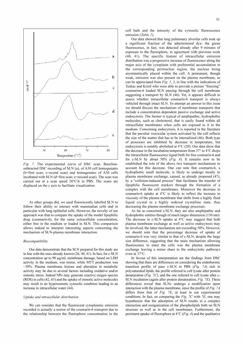
Differential scanning calorimetryThe DSC scan of c-SLN resuspended in PBS showed a
series of endothermic transitions (Fig. 7A) within the range 58-62°C, likely corresponding to the phase transition of the SLNlipidic component (21). A30 cell homogenates, submitted toDSC, showed the presence of endothermic transitions with amain prominent peak centered at a temperature of 61°C (Fig.7B). The overall transition observed by DSC appears to beirreversible, since no significant transition was detected during a
49
0 25 50 750
20
40
60
80
100
120
140
% o
f flu
ores
cenc
e
% of EL
37°C4°C
*
6-cum
0 25 50 750
20
40
60
80
100
120
140
% o
f flu
ores
cenc
e
% of EL
37°C 4°C
*
c-SLN
A B
Fig. 4. Effect of temperature on uptake and intracellular distribution of c-SLN and coumarin-6. The two plots illustrate the fluorescentdistribution of c-SLN (panel A) or coumarin-6 (panel B) after 45 minutes of incubation at 32°C (filled symbols) and 4°C (opensymbols). The fluorescence is expressed as percentage of the maximum value measured at 45 min and at 75% of EL. For allexperiments, n=6 (p<0.001, t-student test).
0 25 50 750
20
40
60
80
100
120
140
*
Inte
nsity
(a.u
.)
% of EL
c-SLN cum-6 *
A
10 15 20 25 30
0
10
20
30
40
50
60
70
Inte
nsity
(a.u
.)
Time (min)
c-SLN cum-6
*
*
B
Fig. 5. Comparison between the intensity and the rate of accumulation of the fluorescence signal in cells incubated with c-SLN (filledcircles) or with coumarin-6 (open circles). Panel A represents the average of fluorescent intensity expressed as absolute value obtainedafter 30 minutes of incubation in the two conditions respectively (113.27+16.6 a.u. for c-SLN vs. 73.17+11 a.u. for cum-6, p<0.001,n=8). Panel B show that the kinetics of accumulation analyzed at 50% of EL is faster in the presence of SLN as vector of the fluorophore:the slope calculated for perfusion interval between 9 and 30 minutes is 1.24 for coumarin-6 and 2.47 for c-SLN (p<0.001, n=8).

second heating cycle (Fig. 7C), likely reflecting the thermaldenaturation of cell proteins (22). Also, the first heating DSCscan of homogenates of A30 cells, previously incubated with c-SLN, showed the presence of an endothermic transition centeredat about 61°C (Fig. 7D). Contrary to untreated cell homogenates,the second heating scan showed the presence of a series of smallendothermic transitions in the range from 57 to 62°C (Fig. 7E).These transitions were reversible, as they were detected also insuccessive scans (not shown). Since these transitions were notpresent in untreated A30 cells, and showed up only after cellincubation with c-SLN, they likely correspond to nanoparticleslipids present within the cell homogenate or to the association ofc-SLN to the cells. It is noteworthy to say that the plasmamembrane phospholipids lack of phase transition in DSC, is dueto the high proportion of very low melting (poly)unsaturatedlipids and cholesterol in cell membranes (23).
DISCUSSIONA drug delivery system can be defined as any method used
to incorporate drugs to improve their pharmacokineticsparameters (22). SLN might represent a good delivery system
because of their lipophilic nature. They show high specificsurface area that facilitates the contact with the plasmamembrane. As far as in vivo application is concerned, SLN havebeen used to improve skin/dermal uptake of several drugs, thusthey can be employed as the carrier for the topical delivery (24-27). Moreover they have been used to improve thebioavailability of drugs via duodenal or oral administration inrats or mice (28-32). Furthermore, they have been administeredintravenously in rats for anticancer drug delivery (33). Finally,they have been used as carriers for topical ocular delivery oftobramycin and administered by ocular route as eye-drops inrabbits (34).
At cellular level SLN have already been used as non viralsystem for DNA (35) or for anticancer drugs delivery (36-38).
In the present study we consider SLN, prepared by warmmicroemulsions, whose lipidic matrix is made of biocompatiblemolecules, with medium-low melting point (fatty acids,triglycerides). Other components of microemulsions arephospholipids as surfactant, water, biliar salts or short chain fattyacids as co-surfactant. SLN obtained from warm microemulsionscan incorporate hydrophilic and lipophilic drugs; they can befreeze dried and sterilized by filtration or by heat according to theproperties of the molecule incorporated. (39).
50
Fig. 6. Evaluation of co-localizationof c-SLN with subcellular organellesand cityplasmic proteins. A30 cellsincubated with c-SLN, afterimmunocytochemistry protocol withantibodies against selected markersof Golgi apparatus (GM130),endosomes (EEA1), lysosomes(Lysotracker), peroxisomes(PMP70), actin (phalloidin) andGAPDH. C-SLN cytoplasmiclabelling (in green) is shown in thefirst column, the antibodies labelling(in red) in the central column and themerged images in the third one. In allthe images, nuclei are stained in blue.
As other groups did, we used fluorescently labelled SLN tofollow their ability to interact with mammalian cells and inparticular with lung epithelial cells. However, the novelty of ourapproach was that to compare the uptake of the model lipophilicdrug (coumarin-6), for the same extracellular concentration,either free in the medium or loaded in SLN. This comparisonallows indeed to interpret interesting aspects concerning themechanism of SLN-plasma membrane interaction.
BiocompatibilityOur data demonstrate that the SLN prepared for this study are
in line with the ones already known (26, 40, 41). In fact, for c-SLNconcentration up to 90 µg/ml, membrane damage, based on LDHactivity in the medium, was minor, while MTT production was~30%. Plasma membrane lesions and alteration in metabolicactivity may be due to several factors including oxidative and/orosmotic stress. Indeed NPs may generate reactive oxigen species(ROS) in cells (42, 43) and the uptake of osmotic active moleculesmay result in an hyperosmotic cytosolic condition leading to anincrease in intracellular water (44).
Uptake and intracellular distributionWe can consider that the fluorescent cytoplasmic emission
recorded is actually a mirror of the coumarin-6 transport due tothe relationship between the fluorophore concentration in the
cell bath and the intensity of the cytosolic fluorescenceemission (Table 1).
Our data showed that lung pulmonary alveolar cells took upa significant fraction of the administered dye: the greenfluorescence, in fact, was detected already after 9 minutes ofexposure to the fluorophore, in agreement with previous work(40, 41). The specific feature of intracellular emissiondistribution was a progressive increase of fluorescence along themajor axis of the cytoplasm with preferential accumulation inthe corresponding perinuclear region, the nucleus beingasymmetrically placed within the cell. A permanent, thoughweak, emission was also present on the plasma membrane, ascan be appreciated from Fig. 1, 3, in line with the indications ofTeskac and Kristl who were able to provide a picture “freezing”coumarine-6 loaded SLN passing through the cell membranesuggesting a transport by SLN (46). Yet, it appears difficult toassess whether intracellular coumarin-6 transport is alwaysvehicled through intact SLN. To attempt an answer to this issuewe should discuss the mechanism of membrane transports thatinclude a concentration dependent passive exchange and activeendocytosis. The former is typical of amphipathic, hydrophobicmolecules, such as cholesterol, that is easily found within allintracellular membranes when cells are exposed to it in themedium. Concerning endocytosis, it is reported in the literaturethat the peculiar vescicular system activated by the cell reflectsthe size of the matter that has to be internalized (46). Both typeof processes are inhibited by decrease in temperature, butendocytosis is notably abolished at 4°C (20). Our data show thatthe decrease in the incubation temperature from 37° to 4°C dropsthe intracellular fluorescence signal both for free coumarin-6 andfor c-SLN by about 50% (Fig. 4). It remains now to beestablished the role of the above two transport mechanisms toaccount for this decrease. One can note that coumarin-6, ahydrophobic small molecule, is likely to undergo mostly toplasma membrane exchange, caused, as already proposed (47),by a “collision-induced process” that facilitates the transfer oflipophilic fluorescent markers through the formation of acomplex with the cell membranes. Moreover the decrease incoumarin-6 uptake at 4°C is likely to reflect the increase inviscosity of the plasma membrane that shifts from a highly fluidliquid crystal to a highly ordered crystalline state, thusdecreasing the plasma membrane exchange processes.
As far as concerned c-SLN, they are also amphipathic andhydrophobic entities though of much larger dimension (130 nm).The decrease in c-SLN uptake at 4°C may suggest that bothplasma membrane exchange as well as active endocytosis maybe involved, the latter mechanism not exceeding 50%. However,we should note that the percentage decrease of uptake ofcoumarin-6 was very similar to that of c-SLN, despite the largesize difference, suggesting that the main mechanism allowingfluorescence to enter the cells was the plasma membraneexchange leaving a minor share to the endocytotic pathwayseven at 37°C.
In favour of this interpretation are the findings from DSCshowing that there are differences on considering the endothermictransition profile of pure c-SLN in PBS (Fig. 7A) rich inpolysaturated lipids, the profile referred to cell lysate after proteindenaturation (Fig. 7C), and the one referred to cell lysate after c-SLN incubation (again after protein denaturation, Fig. 7E). Thesedifferences reveal that SLNs undergo a modification uponinteraction with the plasma membrane, since the profile of Fig. 7Adiffers from that of Fig. 7E, at least in our experimentalconditions. In fact, on comparing the Fig. 7C with 7E, one mayhypothesize that the adsorption of SLN results in a complexinteraction and reorganization of the phospholipids both on SLNstructure as well as in the cell membranes. Furthermore, thepersistent uptake of fluorophore at 4°C (Fig. 4) and the qualitative
51
Fig. 7. The experimental curve of DSC scan. Baseline-subtracted DSC recording of SLN (a), of A30 cell homogenates(b=first scan; c=econd scan) and homogenates of A30 cellsincubated with SLN (d=first scan; e=second scan). The scan wascarried out at a scan speed 20°C/h in PBS. The scans aredisplaced on the y axis to facilitate visualization.

non specific co-localization with intracellular organelles (Fig. 6)suggest a minor role of direct incorporation of SLN through anendocytotic pathway. Our interpretation is in keeping with datafrom scanning electron microscopy revealing that SLN interactingwith epidermal cells (pig skin), spread over the surface and loosetheir primary shape (26). This suggests that a major change occursto the particle structure during the chemico-physical interactionprocess (27) and such modification may account for a “functionalburst” of SLN and subsequent release of the loaded drug rapidlylabelling the cytosol. Our data support a direct cytoplasmicdelivery into the cells of the model drug, sharing an idea alreadydeveloped by Partlow et al. (48). One can indeed envisage a directmixing or exchange of phospholipids between the target cellplasma membrane and particle. In fact, since SLN have a strongchemical and structural similarity with the plasma membrane, itsstructural lipids may merge with cell membranes and facilitatedrug delivery into the interior of the cell.
The other interesting point emerging from our study is that, forthe same amount of the model drug coumarin-6 present in themedium, its uptake was higher and faster when loaded on SLN,compared to when it was free in the medium (Fig 5A, B). Thus wemay suggest that, although SLN do not necessarily reach the cellcytoplasm through an endocytotic pathway, yet, they represent acapacity reservoir for the loaded drug as they add efficiency toendocellular drug delivery through a quantum-like release process.
Acknowledgements: A special thanks to Prof P. Palestini,Prof R. Perego and Dr C. Bianchi (DIMS, University of MilanoBicocca) for providing A30 and COS-7 cell lines. This work wassupported by CE Contract STREP N° LSHB-CT-2006-037639-BONSAI (Bio-imaging with Smart Functional Nanoparticles).
Conflict of interests: None declared.
REFERENCES1. Pison U, Welte T, Giersig M, Groneberg DA.
Nanomedicine for respiratory diseases. Eur J Pharmacol2006; 533: 341-350.
2. Siekmeier R, Scheuch G. Systemic treatment by inhalationof macromolecules- principles, problems and examples. JPhysiol Pharmacol 2008; 59: 53-79.
3. Muhlfeld C, Rothen-Rutishauser B, Blank F, Vanhecke D,Ochs M, Gehr P. Interaction of nanoparticles withpulmonary structures and cellular responses. Am J Lung CellMol Physiol 2008; 294: L817-L829.
4. Kraft KS, Grant M. Preparation of macromolecule-containing dry powders for pulmonary delivery. Meth MolBiol 2009; 480: 165-174.
5. Tomoda K, Ohkoshi T, Nakajima T, Makino K. Preparationand properties of inhalable nanocomposite particles: effectsof the size, weight ratio of the primary nanoparticles innanocomposite particles and temperature at a spray-dryerinlet upon properties of nanocomposite particles. ColloidsSurf B Biointerfaces 2008; 64: 70-76.
6. Dailey LA, Jekel N, Fink L, et al. Investigation of the pro-inflammatory potential of biodegradable nanoparticle drugdelivery system in the lung. Toxicol Appl Pharmacol 2006;215: 100-108.
7. Siekmeier R, Scheuch G. Treatment of systemic diseases byinhalation of biomolecules aerosol. J Physiol Pharmacol2009; 60: 15-26.
8. Brandenberger C, Muhlfeld C, Ali Z, et al. Quantitativeevaluation of cellular uptake and trafficking of plain andpolyethylene glycol-coated gold nanoparticles. Small 2010;6: 1669-1678.
9. Lenz AG, Karg E, Lentner B, et al. A dose-controlled systemfor air-liquid interface cell exposure and application to zincoxide nanoparticles. Part Fibre Toxicol 2009; 6: 32.
10. Aufderheide M, Knebel JW, Ritter D. Novel approaches forstudying pulmonary toxicity in vitro. Toxicol Lett 2003; 140-141: 205-211.
11. Suarez S, O’Hara P, Kazantseva M, et al. Respirable PLGAmicrospheres containing rifampicin for the treatment oftuberculosis: screening in an infectious disease model.Pharm Res 2001; 18: 1315-1319.
12. Bakshi MS, Zhao L, Smith R, Possmayer F, Peterse NO.Metal nanoparticle pollutants interfere with pulmonarysurfactant function in vitro..Biophys J 2008; 94: 855-868.
13. Erickson B, DiMaggio S, Mulle DG, et al. Interaction ofpoly(amidoamine) dendrimers with survanta lung surfactant:the importance of lipid domains. Langmuir 2008; 24: 11003-11008.
14. Zhiqing L, Zhuge X, Fuhuan C, et al. ICAM-1 and VCAM-1 expression in rat aortic endothelial cells after single-walledcarbon nanotube exposure. J Nanosci Nanotechnol 2010; 10:8562-8574.
15. Wang X, Zang JJ, Wang H, et al. Pulmonary toxicity in miceexposed to low and medium doses of water-soluble multi-walled carbon nanotubes. J Nanosci Nanotechnol 2010; 10:8516-8526.
16. Gasser M, Rothen-Rutishauser B, Krug HF, et al. Theadsorption of biomolecules to multi-walled carbon nanotubes(MWCNTs) is influenced by both pulmonary surfactant lipidsand surface chemistry. J Nanobiotechnol 2010; 8: 31.
17. Muller RH, Kader K, Gohla S. Solid lipid nanoparticles(SLN) for controlled drug delivery. A review of the state ofthe art. Eur J Pharm Biopharm 2000; 50: 161-177.
18. Panariti A, Rivolta I, Lettiero B, Chirico G, Gasco P,Miserocchi G. SLN as vehicle for a model drug: abiophysical study. AIP Conference Proceedings 2010; 1275:115-117.
19. Botto L, Beretta E, Bulbarelli A, et al. Hypoxia-inducedmodifications in plasma membranes and lipid microdomainsin A549 cells and primary human alveolar cells. J CellBiochem 2008; 105: 503-513.
20. Steinman RM, Mellman IS, Muller WA, Cohn ZA.Endocytosis and the recycling of plasma membrane. J CellBiol 1983; 96: 1-27.
21. Muller RH, Runge SA, Ravelli V, Thunemann AF, MehnertW, Souto EB. Cyclosporine-loaded solid lipid nanoparticles(SLN): drug-lipid physicochemical interactions andcharacterization of drug incorporation. Eur J PharmBiopharm 2008; 68: 535-544.
22. Lepock JR, Frey HE, Ritchie KP. Protein denaturation inintact hepatocytes and isolated cellular organelles duringheat shock. J Cell Biol 1993; 122: 1267-1276.
23. Masserini M, Pitto M, Raimondo F, Cazzaniga E, Sesana S,Bellini T. Methyl-b-cyclodextrin treatment affects thethermotropic behaviour of membranes and detergent-resistant membrane fractions of cultured A431 cells. BiolPharm Bull 2005; 28: 2185-2188.
24. Lv Q, Yu A, Xi Y, et al. Development and evaluation ofpenciclovir-loaded solid lipid nanoparticles for topicaldelivery. Int J Pharm 2009; 372: 191-198.
25. Priano L, Esposti D, Esposti R, et al. Solid lipidnanoparticles incorporating melatonin as new model forsustained oral and transdermal delivery system. J NanosciNanotechnol 2007; 7: 3596-3601.
26. Kuchler S, Radowski MR, Blaschke T, et al. Nanoparticlesfor skin penetration enhancement – a comparison of adendritic core-multishell-nanotransporter and solid lipidnanoparticles. Eur J Pharm Biopharm 2009; 71: 243-250.
52

27. Kuchler S, Herrmann W, Panek-Minkin G, et al. SLN fortopical application in skin diseases – characterization ofdrug-carrier and carrier-target interaction. Int J Pharm 2010;390: 225-233.
28. Suresh G, Manjunath K, Venkateswarlu V, Satyanarayana V.Preparation, characterization, and in vitro and in vivoevaluation of lovastatin solid lipid nanoparticles. AAPSPharmSciTech 2007; 8: E1-E9.
29. Yang S, Zhu J, Lu Y, Liang B, Yang C. Body distribution ofcamptothecin solid lipid nanoparticles after oraladministration. Pharm Res 1999; 16: 751-757.
30. Holpuc AS, Hummel GJ, Tong M, et al. Nanoparticles forlocal drug delivery to the oral mucosa: proof of principlestudies. Pharm Res 2010; 27: 1224-1236.
31. Hauss DJ, Fogal SE, Ficorilli JV, Ahuss DJ, et al. Lipid-based delivery systems for improving the bioavailability andlymphatic transport of a poorly water-soluble LTB4inhibitor. J Pharm Sci 1998; 87: 164-169.
32. Cavalli R, Bargoni A, Podio V, Muntoni E, Zara GP, GascoMR. Duodenal administration of solid lipid nanoparticlesloaded with different percentages of tobramycin. J PharmSci 2003; 92: 1085-1094.
33. Zara GP, Cavalli R, Bargoni A, Fundarn A, Vighetto D,Gasco MR. Intravenous administration to rabbits of non-stealth and stealth doxorubicin loaded solid lipidnanoparticles at increasing concentrations of stealth agent:pharmacokinetics and distribution of doxorubicin in brainand in other tissues J Drug Target 2002; 10: 327-335.
34. Cavalli R, Gasco MR, Chetoni P, Burgalassi S, Saettone MF.Solid lipid nanoparticles (SLN) as ocular delivery system fortobramycin. Int J Pharm 2002; 238: 241-245.
35. del Pozo-Rodriguez A, Delgado D, Solinis MA, Gascon AR,Pedraz JL. Solid lipid nanoparticles for retinal gene therapy:transfection and intracellular trafficking in RPE cells. Int JPharm 2008; 360: 177-183.
36. Serpe L, Guido M, Canaparo R, et al. Intracellularaccumulation and cytotoxicity of doxorubicin with differentpharmaceutical formulations in human cancer cells. JNanosci Nanotecnol 2006; 6: 3062-3069.
37. Dianzani C, Cavalli R, Zara GP, et al. Cholestryl butyratesolid lipid nanoparticles inhibit adhesion of humanneutrophils to endothelial cells. Br J Pharmacol 2006; 148:648-656.
38. Brioschi A, Zenga F, Zara GP, Gasco MR, Ducati A, MauroA. Solid lipid nanoparticles: could they help to improve the
efficacy of pharmacologic treatments for brain tumors?Neurol Res 2007; 29: 324-330.
39. Cavalli R, Caputo O, Carlotti ME, Trotta M, Scarnecchia C,Gasco MR. Sterilization and freeze-drying of drug-free anddrug-loaded solid lipid nanoparticles. Int J Pharm 1997; 148:47-54.
40. Cryan S. Carrier-based strategies for targeting protein andpeptide drugs to the lungs. AAPS J 2005; 7: E20-E41.
41. Nassimi M, Schleh C, Lauenstein HD, et al. A toxicologicalevaluation of inhaled solid lipid nanoparticles used as apotential drug delivery system for the lung. Eur J PharmBiopharm 2010; 75: 107-116.
42. Kovacic P, Somanathan R. Biomechanisms of nanoparticles(toxicants, antioxidants and therapeutics): electron transferand reactive oxygen species. J Nanosci Nanotechnol 2010;10: 7919-7930.
43. Potter TM, Neun BW, Stern ST. Assay to detect lipidperoxidation upon exposure to nanoparticles. Meth Mol Biol2011; 697: 181-189.
44. Nielsen MB, Christensen ST, Hoffmann EK. Effects ofosmotic stress on the activity of MAPKs and PDGFR-beta-mediated signal transduction in NIH-3T3 fibroblasts. Am JPhysiol Cell Physiol 2008; 294: C1046-C1055.
45. Teskac K, Kristl J. The evidence for solid lipid nanoparticlesmediated cell uptake of resveratol. Int J Pharm 2010; 390:61-69.
46. Rejman J, Oberle V, Zuhorn IS, Hoekstra D. Size-dependentinternalization of particles via the pathways of clathrin andcaveolae-mediated endocytosis. Biochem J 2004; 377: 159-179.
47. Pietzonka P, Rothen-Rutishauser B, Langguth P, Wunderli-Allenspach H, Walter E, Merklel HP. Transfer of lipophilicmarkers from PLGA and polystyrene nanoparticles to Caco-2 monolayers mimics particle uptake. Pharm Res 2002; 19:595-561.
48. Partlow KC, Lanza GM, Wickline SA. Exploiting lipid rafttransport with membrane targeted nanoparticles: a strategy forcytosolic drug delivery. Biomaterials 2008; 23: 3367-3375.R e c e i v e d : July 23, 2010A c c e p t e d : January 20, 2011Author’s address: Dr. Ilaria Rivolta, Department of
Experimental Medicine, University of Milano-Bicocca, ViaCadore, 48, 20052 Monza, Italy; E-mail: [email protected]
53